


Languages
Pages
Legal

Review
Understanding the evolution of restriction-modification sys-
tems: Clues from sequence and structure comparisons�
Janusz M. Bujnicki�
Bioinformatics Laboratory, International Institute of Molecular and Cell Biology, ks. Trojdena 4,
02-109 Warszawa, Poland; BioInfoBank Institute, Limanowskiego 24A, 60-744 Poznañ, Poland
Received: 24 September, 2001; accepted: 3 December, 2001
Key words: methyltransferases, endonucleases, protein structure, molecular evolution, bioinformatics
Restriction-modification (RM) systems comprise two opposing enzymatic activities:
a restriction endonuclease, that targets specific DNA sequences and performs
endonucleolytic cleavage, and a modification methyltransferase that renders these se-
quences resistant to cleavage. Studies on molecular genetics and biochemistry of RM
systems have been carried out over the past four decades, laying foundations for mod-
ern molecular biology and providing important models for mechanisms of highly spe-
cific protein–DNA interactions. Although the number of known, relevant sequences
3D structures of RM proteins is growing steadily, we do not fully understand their
functional diversities from an evolutionary perspective and we are not yet able to en-
gineer new sequence specificities based on rational approaches. Recent findings on
the evolution of RM systems and on their structures and mechanisms of action have
led to a picture in which conserved modules with defined function are shared between
different RM proteins and other enzymes involved in nucleic acid biochemistry. On
the other hand, it has been realized that some of the modules have been replaced in the
evolution by unrelated domains exerting similar function. The aim of this review is to
give a survey on the recent progress in the field of structural phylogeny of RM en-
zymes with special emphasis on studies of sequence–structure–function relationships
and emerging potential applications in biotechnology.
Vol. 48 No. 4/2001
935–967
QUARTERLY
�The author’s research on RM enzymes is supported by the State Committee for Scientific Research
(KBN, Poland) grant 6P04 B00519.�
tel: (48 22) 668 5384; fax: (48 22) 668 5288; e-mail [email protected]
Abbreviations: RM, restriction-modification; MTase, methyltransferase; ENase, endonuclease;
M/HsdM, R/HsdR, S/HsdS, protein subunits responsible for: modification, restriction and sequence
recognition; TRD, target recognition domain; m6A, N
6-methyladenine; m
4C, N4-methylcytosine; m
5C,
C5-methylcytosine; hm5C, C5-hydroxymethylcytosine; AdoMet, S-adenosyl-L-methionine.
Restriction-modification (RM) systems oc-
cur exclusively in unicellular organisms and
their viruses. They comprise opposing intra-
cellular enzyme activities: DNA endo-
deoxyribonuclease (ENase), that recognizes
and cleaves its target site, and a DNA
methyltransferase (MTase), that transfers
methyl group from S-adenosyl-L-methionine
(AdoMet) onto specific nucleobases within the
target, thereby protecting it from the action of
the ENase. Methylation occurs either at ade-
nine or cytosine, yielding N6-methyladenine
(m6A), N4-methylcytosine (m4C) or C5-me-
thylcytosine (m5C). In symmetrical se-
quences, the same base is methylated on both
strands. The methyl groups lie in the major
groove of the DNA helix, in positions that do
not interfere with base-pairing, but that
change the “epigenetic” information content
of DNA. For instance, methylation of only one
strand of the target (hemimethylation) is usu-
ally sufficient to prevent cleavage by sterically
hindering binding of the ENase to the target.
This guarantees that after DNA replication
hemimethylated daughter duplexes eventu-
ally become fully re-methylated rather than
being cleaved [1].
RM systems were originally suggested to
evolve as a defense mechanism against phage
infection and other types of DNA invasion [2],
and serve evolutionary purposes by producing
gene-size fragments of foreign DNA to be inte-
grated into the host chromosome via recombi-
nation [3]. There are also cases of seemingly
quite typical RM proteins known, which are
involved in quite sophisticated physiological
processes, such as regulating competence for
DNA uptake [4]. Moreover, some DNA repair
systems in Eubacteria can be regarded as de-
scendants of RM systems or vice versa. Pres-
ently there is a large body of evidence that
many RM systems are highly mobile elements
involved in various genome rearrangements,
and that many of them exhibit ”selfish” behav-
ior, regardless of potential benefits for the
host they may confer (reviewed in ref. [5]).
With the abundance of literature it is beyond
the scope of this review to fully cover all re-
search articles on the biochemistry and genet-
ics of RM systems; instead I will focus on the
recent studies of their sequences and struc-
tures and rather recommend several excellent
reviews that can provide a complementary
viewpoint to that presented in this article
[6–10].
CLASSIFICATION OF RM SYSTEMS
RM systems were subdivided into three ba-
sic types (I, II, and III) based on the number
and organization of subunits, regulation of
their expression, cofactor requirements, enzy-
matic mechanism, and sequence specificity
[1]. However, further types and subtypes have
been proposed as new, distinct RM systems
have been discovered. The biochemical prop-
erties of the “novel” systems are intermediate
to those of the “old” ones, and their most strik-
ing feature is that they seem to combine pro-
tein domains originated from the “old” sys-
tems in unprecedented structural contexts.
Recently, a novel nomenclature for the “sub-
types” has been proposed at the “DNA En-
zymes: Structures & Mechanisms” conference
in Bangalore (December 2000) [11].
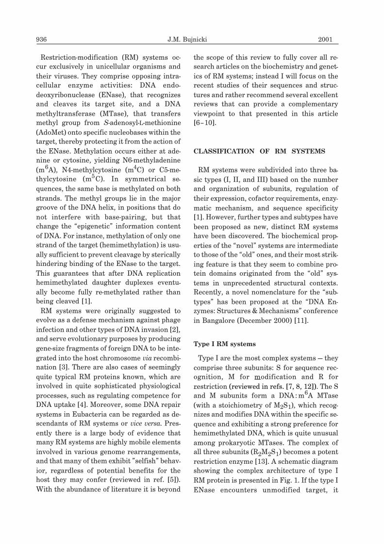
Type I RM systems
Type I are the most complex systems — they
comprise three subunits: S for sequence rec-
ognition, M for modification and R for
restriction (reviewed in refs. [7, 8, 12]). The S
and M subunits form a DNA:m6A MTase
(with a stoichiometry of M2S1), which recog-
nizes and modifies DNA within the specific se-
quence and exhibiting a strong preference for
hemimethylated DNA, which is quite unusual
among prokaryotic MTases. The complex of
all three subunits (R2M2S1) becomes a potent
restriction enzyme [13]. A schematic diagram
showing the complex architecture of type I
RM protein is presented in Fig. 1. If the type I
ENase encounters unmodified target, it
936 J.M. Bujnicki 2001
dimerizes rapidly [14] and initiates an
ATP-dependent translocation of DNA towards
itself simultaneously from both directions
[15]. This process causes the extrusion or con-
traction of DNA loops and results in extensive
supercoiling of DNA. Cleavage is elicited at
variable distance from the recognition se-
quence once translocation stalls [16]. Since
type I systems cleave DNA nonspecifically at
considerable distances from the unmethyl-
ated target sequences, they have so far failed
to provide useful analytical reagents for mod-
ern molecular biology.
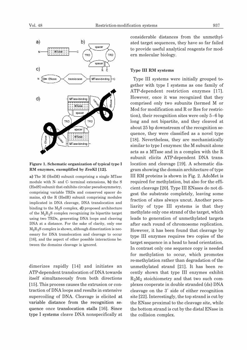
Type III RM systems
Type III systems were initially grouped to-
gether with type I systems as one family of
ATP-dependent restriction enzymes [17].
However, once it was recognized that they
comprised only two subunits (termed M or
Mod for modification and R or Res for restric-
tion), their recognition sites were only 5–6 bp
long and not bipartite, and they cleaved at
about 25 bp downstream of the recognition se-
quence, they were classified as a novel type
[18]. Nevertheless, they are mechanistically
similar to type I enzymes: the M subunit alone
acts as a MTase and in a complex with the R
subunit elicits ATP-dependent DNA trans-
location and cleavage [19]. A schematic dia-
gram showing the domain architecture of type
III RM proteins is shown in Fig. 2. AdoMet is
required for methylation, but also for the effi-
cient cleavage [20]. Type III ENases do not di-
gest the substrate completely, leaving some
fraction of sites always uncut. Another pecu-
liarity of type III systems is that they
methylate only one strand of the target, which
leads to generation of unmethylated targets
after each round of chromosome replication.
However, it has been found that cleavage by
type III enzymes requires two copies of the
target sequence in a head to head orientation.
In contrast only one sequence copy is needed
for methylation to occur, which promotes
re-methylation rather than degradation of the
unmethylated strand [21]. It has been re-
cently shown that type III enzymes exhibit
R2M2 stoichiometry and that two such com-
plexes cooperate in double stranded (ds) DNA
cleavage on the 3� side of either recognition
site [22]. Interestingly, the top strand is cut by
the ENase proximal to the cleavage site, while
the bottom strand is cut by the distal ENase in
the collision complex.
Vol. 48 Restriction-modification systems 937
Figure 1. Schematic organization of typical type I
RM enzymes, exemplified by EcoKI [12].
a) The M (HsdM) subunit comprising a single MTase
module with N- and C- terminal extensions, b) the S
(HsdS) subunit that exhibits circular pseudosymmetry,
comprising variable TRDs and conserved spacer do-
mains, c) the R (HsdR) subunit comprising modules
implicated in DNA cleavage, DNA translocation and
binding to the M2S complex, d) proposed architecture
of the M2R2S complex recognizing its bipartite target
using two TRDs, generating DNA loops and cleaving
DNA at a distance. For the sake of clarity, only one
M2R2S complex is shown, although dimerization is nec-
essary for DNA translocation and cleavage to occur
[16], and the aspect of other possible interactions be-
tween the domains cleavage is ignored.
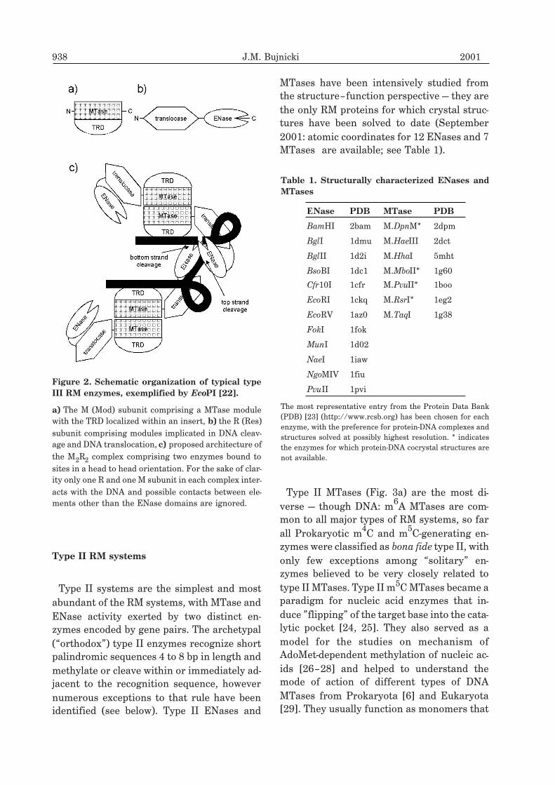
Type II RM systems
Type II systems are the simplest and most
abundant of the RM systems, with MTase and
ENase activity exerted by two distinct en-
zymes encoded by gene pairs. The archetypal
(“orthodox”) type II enzymes recognize short
palindromic sequences 4 to 8 bp in length and
methylate or cleave within or immediately ad-
jacent to the recognition sequence, however
numerous exceptions to that rule have been
identified (see below). Type II ENases and
MTases have been intensively studied from
the structure–function perspective — they are
the only RM proteins for which crystal struc-
tures have been solved to date (September
2001: atomic coordinates for 12 ENases and 7
MTases are available; see Table 1).
Type II MTases (Fig. 3a) are the most di-
verse — though DNA: m6A MTases are com-
mon to all major types of RM systems, so far
all Prokaryotic m4C and m
5C-generating en-
zymes were classified as bona fide type II, with
only few exceptions among “solitary” en-
zymes believed to be very closely related to
type II MTases. Type II m5C MTases became a
paradigm for nucleic acid enzymes that in-
duce ”flipping” of the target base into the cata-
lytic pocket [24, 25]. They also served as a
model for the studies on mechanism of
AdoMet-dependent methylation of nucleic ac-
ids [26–28] and helped to understand the
mode of action of different types of DNA
MTases from Prokaryota [6] and Eukaryota
[29]. They usually function as monomers that
938 J.M. Bujnicki 2001
Figure 2. Schematic organization of typical type
III RM enzymes, exemplified by EcoPI [22].
a) The M (Mod) subunit comprising a MTase module
with the TRD localized within an insert, b) the R (Res)
subunit comprising modules implicated in DNA cleav-
age and DNA translocation, c) proposed architecture of
the M2R2 complex comprising two enzymes bound to
sites in a head to head orientation. For the sake of clar-
ity only one R and one M subunit in each complex inter-
acts with the DNA and possible contacts between ele-
ments other than the ENase domains are ignored.
Table 1. Structurally characterized ENases and
MTases
ENase PDB MTase PDB
BamHI 2bam M.DpnM* 2dpm
BglI 1dmu M.HaeIII 2dct
BglII 1d2i M.HhaI 5mht
BsoBI 1dc1 M.MboII* 1g60
Cfr10I 1cfr M.PvuII* 1boo
EcoRI 1ckq M.RsrI* 1eg2
EcoRV 1az0 M.TaqI 1g38
FokI 1fok
MunI 1d02
NaeI 1iaw
NgoMIV 1fiu
PvuII 1pvi
The most representative entry from the Protein Data Bank
(PDB) [23] (http://www.rcsb.org) has been chosen for each
enzyme, with the preference for protein-DNA complexes and
structures solved at possibly highest resolution. * indicates
the enzymes for which protein-DNA cocrystal structures are
not available.
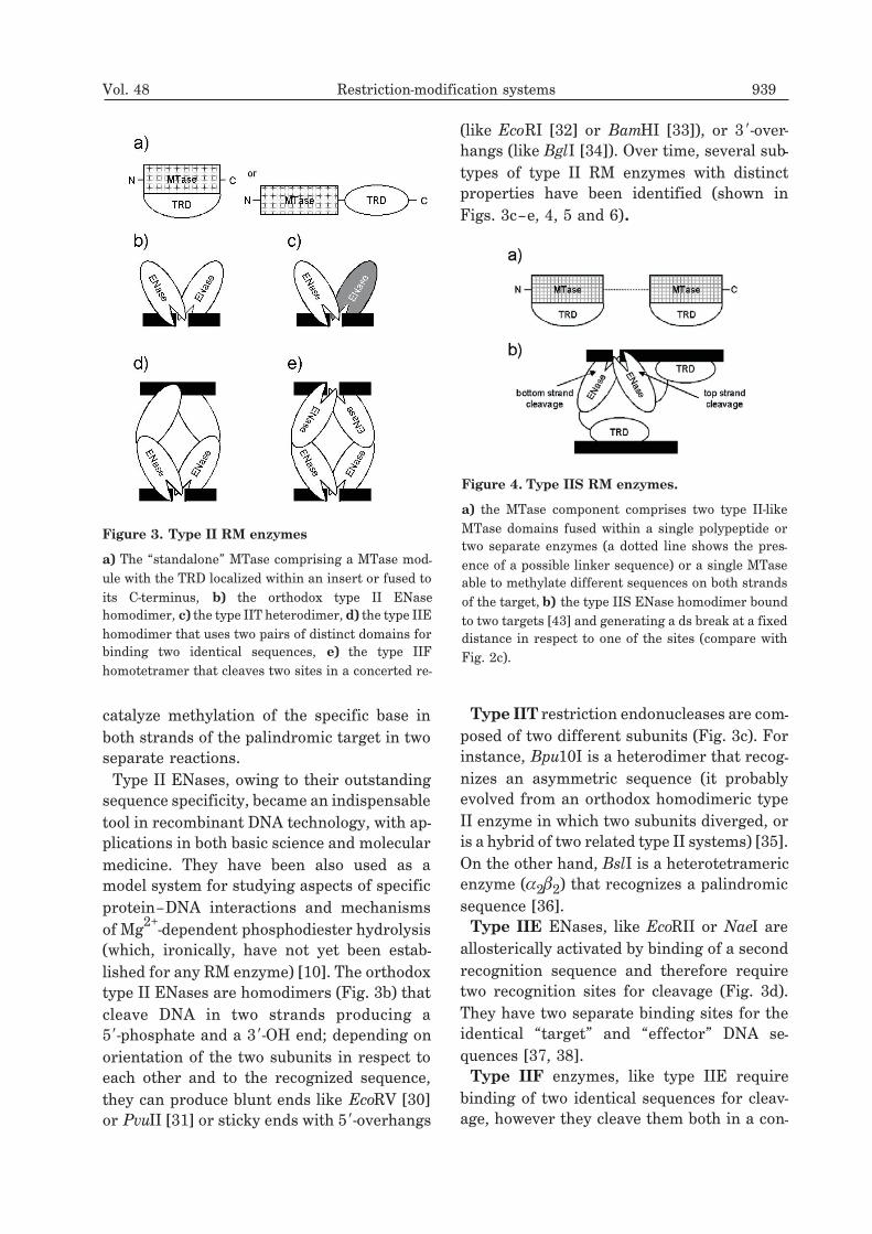
catalyze methylation of the specific base in
both strands of the palindromic target in two
separate reactions.
Type II ENases, owing to their outstanding
sequence specificity, became an indispensable
tool in recombinant DNA technology, with ap-
plications in both basic science and molecular
medicine. They have been also used as a
model system for studying aspects of specific
protein–DNA interactions and mechanisms
of Mg2+
-dependent phosphodiester hydrolysis
(which, ironically, have not yet been estab-
lished for any RM enzyme) [10]. The orthodox
type II ENases are homodimers (Fig. 3b) that
cleave DNA in two strands producing a
5�-phosphate and a 3�-OH end; depending on
orientation of the two subunits in respect to
each other and to the recognized sequence,
they can produce blunt ends like EcoRV [30]
or PvuII [31] or sticky ends with 5�-overhangs
(like EcoRI [32] or BamHI [33]), or 3�-over-
hangs (like BglI [34]). Over time, several sub-
types of type II RM enzymes with distinct
properties have been identified (shown in
Figs. 3c–e, 4, 5 and 6).
Type IIT restriction endonucleases are com-
posed of two different subunits (Fig. 3c). For
instance, Bpu10I is a heterodimer that recog-
nizes an asymmetric sequence (it probably
evolved from an orthodox homodimeric type
II enzyme in which two subunits diverged, or
is a hybrid of two related type II systems) [35].
On the other hand, BslI is a heterotetrameric
enzyme (�2�2) that recognizes a palindromic
sequence [36].
Type IIE ENases, like EcoRII or NaeI are
allosterically activated by binding of a second
recognition sequence and therefore require
two recognition sites for cleavage (Fig. 3d).
They have two separate binding sites for the
identical “target” and “effector” DNA se-
quences [37, 38].
Type IIF enzymes, like type IIE require
binding of two identical sequences for cleav-
age, however they cleave them both in a con-
Vol. 48 Restriction-modification systems 939
Figure 3. Type II RM enzymes
a) The “standalone” MTase comprising a MTase mod-
ule with the TRD localized within an insert or fused to
its C-terminus, b) the orthodox type II ENase
homodimer, c) the type IIT heterodimer, d) the type IIE
homodimer that uses two pairs of distinct domains for
binding two identical sequences, e) the type IIF
homotetramer that cleaves two sites in a concerted re-
Figure 4. Type IIS RM enzymes.
a) the MTase component comprises two type II-like
MTase domains fused within a single polypeptide or
two separate enzymes (a dotted line shows the pres-
ence of a possible linker sequence) or a single MTase
able to methylate different sequences on both strands
of the target, b) the type IIS ENase homodimer bound
to two targets [43] and generating a ds break at a fixed
distance in respect to one of the sites (compare with
Fig. 2c).

certed reaction (Fig. 3e). Those proteins char-
acterized to date are tetrameric, for example
NgoMIV [39] and SfiI [40].
Type IIS ENases cut at a fixed distance near
their short, asymmetric target site [41]. This
makes them similar to type III enzymes, but
type IIS ENases do not require ATP or
AdoMet or the presence of the MTase subunit
for cleavage. They exist as monomers, with
the DNA recognition and cleavage functions
located on distinct domains (Fig. 4); however
a dimerization of cleavage domains from two
DNA-bound complexes is obligatory for ds
DNA cleavage, as demonstrated for FokI [42,
43]. Since the TRDs of type IIS ENases effec-
tively interact with two sites, of which only
one is cut is a single catalytic event, they can
be regarded as a subclass of type IIE enzymes.
Because of the unusual bipartite structure,
type IIS ENases have proven particularly use-
ful in creating chimeric enzymes by attaching
the nonspecific cleavage domain to the
DNA-binding domain of transcription factors
[44–46].
The enzyme N.BstNBI related to type IIS
ENases has been characterized as a “nicking”
ENase, which cleaves only on the top strand 4
bp away from its recognition sequence [47].
Interestingly, it has been shown that its close
homologs, MlyI and PleI introduce nicks prior
to ds cleavage, which presumably occurs only
after the ENases dimerize [48]. Hence, it has
been suggested that the peculiar limited bot-
tom strand cleavage activity of N.BstNBI re-
sults from the inability of its cleavage domain
to dimerize. These results suggest that type
IIS enzymes exert ds DNA cleavage in a simi-
lar manner to type III enzymes, i.e. the top
strand is cut by the ENase bound to the target
sequence proximal to the cleavage site, while
the bottom strand is cut by the distal ENase
(Figs. 3, 4).
Type IIS MTases must methylate an asym-
metric target, hence this kind of RM systems
comprises two MTases specific for each
strand, which may methylate different bases,
like adenine (GGTGA) and cytosine (TCACC)
in the case of NgoBVIII [49], or one fusion pro-
tein with two MTase domains with distinct
specificities, like in the case of FokI (GGATG
and CATCC), [50]. Another possibility is to
employ a MTase, which recognizes a degener-
ated sequence and is able to methylate both
strands, like it has been suggested for
GASTC-specific (S=G or C) M.BstNBI (unpub-
lished data cited in ref. [48]) or for the hypo-
thetical SSATSS-specific ancestor of the
C-terminal MTase domain of M.FokI [50].
Type IIG (formerly type IV) RM systems
are composed of two MTases, of which one
modifies both strands of the asymmetric sub-
strate, while the other modifies only one
strand, but in addition exhibits also the ENase
activity (Fig. 5), cutting the target 16/14 bp in
3� direction from the recognition site [51].
Some type IIG enzymes exhibit peculiar bio-
chemical properties that make them similar to
type III enzymes (see below): For instance
Eco57I cleaves the substrate only partially
and is stimulated by AdoMet [51], while for
BseMII AdoMet is essential for cleavage [52].
On the other hand, cleavage at a fixed dis-
tance from the target resembles both type IIS
and type III enzymes. Hence, type IIG en-
zymes were suggested to be the evolutionary
link between type III and type IIS systems,
however this hypothesis has never been sup-
ported by a genuine phylogenetic study [53].
940 J.M. Bujnicki 2001
Figure 5. Schematic organization of type IIG RM
enzymes.
a) the type II-like MTase, b) the ENase/MTase subunit,
whose mechanism of interaction with the target or the
possible multimerization mode is unknown, but may be
related to that of type III and type IIS ENases (Figs. 2c
and 4b)
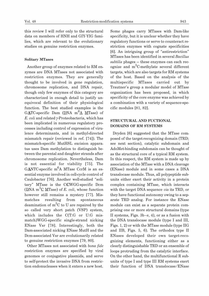
Type IIB (formerly type V or “BcgI-like”)
RM systems encode both ENase and MTase
activities within one polypeptide chain, simi-
larly to the type IIG bifunctional
ENase/MTase, but with the ability to modify
both strands of the symmetric, bipartite tar-
get sequence [54]. The pattern of cleavage,
which makes them distinct from other types,
results from unprecedented combination of
previously known features: all type IIB en-
zymes cleave DNA on both sides of their bind-
ing site (like type I ENases) at a fixed distance
(like type IIs, IIG and III), resulting in exci-
sion of a short DNA fragment (Fig. 6). Some
of them, like BcgI, require a separate subunit
(S) to bind to DNA and recognize the target,
but others, like CjeI [55] and HaeIV [56] seem
to exert all three functions with one chain.
The S subunit of the BcgI RM system is re-
lated to the type I S subunits, while in CjeI the
S subunit is fused to the C-terminus of the
ENase/MTase subunit. In the HaeIV RM sys-
tem, no region homologous to the typical S
subunits has been identified to date, but it is
likely that its TRD maps to the C-terminus
[56].
Generally, many type IIB enzymes exhibit
various peculiarities, which may be or may be
not specific to other proteins of this class. For
instance, HaeIV was shown to release an
asymmetric fragment after cleavage [56] and
BcgI requires two bipartite target sites for
cleavage [57] similarly to the enzymes of types
I, IIE, IIF, and III. It is tempting to speculate
that type IIB enzymes are a compact variant
of type I enzymes that lack the DNA trans-
locase module, but may show the same mecha-
nism of DNA binding and cleavage on both
sides of the target (compare Figs. 1 and 6).
To my best knowledge, interactions between
a pair of the ENase domains, each cleaving
one strand of the double strand target, has
been shown only for the orthodox type II and
related “standalone” ENases (types IIT, IIE
and IIF) and for the ENase modules of type
IIS and type III RM enzymes. It is tempting to
speculate that other RM enzymes, including
type I, type IIG and type IIB ENases also re-
quire a dimer of ENase domains to exert cleav-
age as opposed to a single domain that would
introduce two nicks in both strands of the tar-
get, thereby making a ds break. If this hypoth-
esis is corroborated by experiment, it would
be interesting to learn if in those complex en-
zymes that possess two ENase domains, the
catalytically competent dimers are formed in
cis (i.e. by the ENase domains of a multi-
protein complex bound to the same target) or
in trans (i.e. by the ENase domains that belong
to different proteins, as in the case of type IIS
enzymes). Remarkably, different in trans con-
figurations can be envisaged for proteins with
more than two ENase domains in the catalytic
unit [22].
Some type II RM enzymes recognize
lengthy, discontinuous sites, such as SfiI
(GGCCNNNNNGGCC), BglI (GCCNNNNNG-
GC) or XcmI (CCANNNNNNNNNTGG), but
most likely they acquired this functional pecu-
liarity independently in the evolution [58] and
they have not been classified as a separate
Vol. 48 Restriction-modification systems 941
Figure 6. Schematic organization of an archetypal
type IIB RM enzyme BcgI [54].
a) The ENase/MTase subunit, b) the S subunit, c) pro-
posed architecture of the (MR)2S complex of the BcgI
RM system that cleaves DNA at a limited distance at
both sides of its bipartite type-I like target (compare
with Fig. 1d). The aspect of dimerization required for
the bilateral cleavage is ignored for clarity and because
it is unclear if and how the four ENase domains of the
[(MR)2S]2 complex cooperate during the cleavage.
type or subtype. There have been several ex-
cellent reviews articles in the last decade fo-
cusing on various aspects of type II ENases [9,
10, 59–61], however only recently experimen-
tal and computational studies on their se-
quences and structures provided new data
and interpretations, considerably broadening
our view on these enzymes and their relation-
ship to other protein families (see the para-
graph devoted to the ENase domain within
the subsequent section of this paper).
RM systems of other types
There are also some RM systems that do not
fit into any of these classes — they likely repre-
sent genuine hybrids of ”regular” types, which
arose by fusions of their separated compo-
nents, but so far no robust phylogenetic study
has been undertaken to infer the pathways of
their evolution. For example it has been also
suggested that type II ENases may couple
with type I MTases with a cognate sequence
specificity, giving rise to the chimerical “type
I&1/2” systems (G.G. Wilson, cited as per-
sonal communication in ref. [6]). On the other
hand, the LlaI system consists of four pro-
teins, one of which is a fusion of two type
II-like m6A MTases, a typical IIS MTase simi-
lar to FokI (see above) [62] and the other three
are remotely related to the McrBC nuclease
(see below). There are also RM systems com-
prised of multiple ENases and MTases; in sev-
eral such cases, like DpnII [63] or BcnI [64],
one of the two MTases of the same specificity
may also methylate single stranded DNA.
Solitary ENases
Paradoxically, the first restriction enzymes
described were McrA (RglA) and McrBC
(RglB) from E. coli, which do not form a part
of a RM system since they do not associate
functionally with any particular MTase and
their ENase activity is not inhibited by
methylation of the target. Conversely, they
specifically recognize and cleave sequences
containing methylated or hydroxymethylated
cytosine (m4C, m5C or hm5C, respectively),
unless it is glucosylated as in wild type T-even
coliphages [65, 66]. Together with the E. coli
Mrr enzyme, which targets modified adenine
or cytosine in a poorly defined sequence con-
text [67] and Streptococcus pneumoniae DpnI
ENases [68] they make up a separate type of
modification-directed restriction (MDR) en-
zymes. Another unusual enzyme of this class
is PvuRts1I, which restricts DNA containing
hm5C, even when it is glucosylated. A
MTase-like gene has been found near
PvuRts1I, but neither its activity as a modifi-
cation MTase nor influence on the
PvuRts1I-mediated restriction could be dem-
onstrated [69]. The MDR enzymes can be
thought of as free-standing predecessors of
RM system components or as nucleases that
abandoned RM systems (for instance follow-
ing the ”death” of their cognate MTase) to be-
come “ENases on the loose”. Alternatively,
the MDR systems may be seen as products of
the “arms race” between bacteria developing
new defensive weapons against T-even phages
and the viruses protecting their DNA using in-
creasingly more complex modifications (re-
viewed in ref. [70]).
Another class of sequence-specific nu-
cleases, whose relationships with restriction
enzymes were not known until very recently,
are the so called “homing” ENases (reviewed
in refs. [71, 72]). A large number of these en-
zymes has been identified in Eukaryotic nu-
clear and organellar genes, but there are also
a few, which have been found in Prokaryota
and their phages. They function in dissemina-
tion of certain mobile introns and inteins by
cleavage of long, asymmetric, and degenerate
sequences. Creation of recombinogenic ends
promotes gene conversion, which leads to du-
plication of the intron. Homing ENases and
some freestanding intergenic ENases, which
share functional properties and sequence sim-
ilarities, can be grouped into three families of
presumably independent evolutionary origin
(LAGLIDADG, HNH, and GIY-YIG) [73]. In
942 J.M. Bujnicki 2001
this review I will refer only to the structural
data on members of HNH and GIY-YIG fami-
lies, which are relevant to the evolutionary
studies on genuine restriction enzymes.
Solitary MTases
Another group of enzymes related to RM en-
zymes are DNA MTases not associated with
restriction enzymes. They are generally
thought to be involved in gene regulation,
chromosome replication, and DNA repair,
though only few enzymes of this category are
characterized in enough detail to justify un-
equivocal definition of their physiological
function. The best studied examples is the
GATC-specific Dam (DNA m6A MTase) of
E. coli and related �-Proteobacteria, which has
been implicated in numerous regulatory pro-
cesses including control of expression of viru-
lence determinants, and in methyl-directed
mismatch repair (reviewed in ref. [74]). The
mismatch-specific MutHSL excision appara-
tus uses Dam methylation to distinguish be-
tween the parental and daughter strands after
chromosome replication. Nevertheless, Dam
is not essential for viability [75]. The
GANTC-specific m6A MTase CcrM is an es-
sential enzyme involved in cell-cycle control of
Caulobacter [76]. Another well-studied “soli-
tary” MTase is the CCWGG-specific Dcm
(DNA m5C MTase) of E. coli, whose function
however still remains a mystery [77]. Mis-
matches resulting from spontaneous
deamination of m5C to U are repaired by the
so called very short patch (VSP) system,
which includes the C(T:G or U:G mis-
match)WGG-specific single-strand nicking
ENase Vsr [78]. Interestingly, both the
Dam-associated nicking ENase MutH and the
Dcm-associated Vsr are evolutionarily related
to genuine restriction enzymes [79, 80].
Other MTases not associated with bona fide
restriction enzymes are specified by viral
genomes or conjugative plasmids, and serve
to self-protect the invasive DNA from restric-
tion endonucleases when it enters a new host.
Some phages carry MTases with Dam-like
specificity, but it is unclear whether they have
regulatory functions or serve to counteract re-
striction enzymes with cognate specificities
[6]. An intriguing group of “antirestriction”
MTases has been identified in several Bacillus
subtilis phages — these enzymes can each rec-
ognize and m5C-methylate several different
targets, which are also targets for RM systems
of the host. Based on the analysis of the
multispecific MTases carried out by
Trautner’s group a modular model of MTase
organization has been proposed, in which
specificity of the core enzyme was achieved by
a combination with a variety of sequence-spe-
cific modules [81, 82].
STRUCTURAL AND FUCTIONAL
DOMAINS OF RM SYSTEMS
Dryden [6] suggested that the MTase com-
posed of the target-recognizing domain (TRD;
see next section), catalytic subdomain and
AdoMet-binding subdomain can be thought of
as the structural core of a typical RM system.
In this respect, the RM system is made up by
association of the MTase with a DNA cleavage
(ENase) module and in some cases a DNA
translocase module. Thus, all polypeptide sub-
units either exert their activity in a protein
complex containing MTase, which interacts
with the target DNA sequence via its TRD, or
they have functional autonomy owing to a sep-
arate TRD analog. For instance the ENase
module can exist as a separate protein com-
prising one or more structural domains (type
II systems, Figs. 3b–e, 4), or as a fusion with
the DNA translocase module (type I and III,
Figs. 1, 2) or with the MTase module (type IIG
and IIB, Figs. 5, 6). The orthodox type II
ENases developed their own target-reco-
gnizing elements, functioning either as a
clearly distinguishable TRD or an ensemble of
loops protruding from the catalytic interface.
On the other hand, the multifunctional R sub-
units of type I and type III RM systems exert
their function of DNA translocase/ENase
Vol. 48 Restriction-modification systems 943
only when complexed with the MTase. In type
I R subunits a special domain responsible for
establishing protein–protein contacts has
been identified in the C-terminus [12] (Fig. 1);
to my knowledge, such domain has not been
delineated to date in primary structures of
type III R subunits. The apparent modular ar-
chitecture of all enzyme types suggested that
shuffling of a quite limited repertoire of mod-
ules and domains conferring particular func-
tions is the main force driving their functional
diversification (Figs. 1–6).
The target recognition domain (TRD)
Target recognition domains have been oper-
ationally defined as regions responsible for se-
quence-specific binding of RM proteins to the
target DNA. They have been initially (and
most clearly) defined for mono- and
multi-specific m5C MTases [81] and the S sub-
units of type I RM systems [83], in which they
are long, variable sequences, surrounded by
well conserved motifs. In the multi-specific
m5C MTases from several bacteriophages of
Bacillus subtilis, certain mutations in the vari-
able region can abolish one target specificity
while leaving the others intact. By mapping
the mutations and studying the specificity of
chimeric proteins, Trautner and coworkers
determined that each target sequence is rec-
ognized by its own TRD and defined its mini-
mal size as approximately 40 amino acids.
Nevertheless, they failed to generate enzymes
with novel specificities by shuffling of gene
fragments except for instances where entire
TRDs were exchanged [81, 84–87]. TRD swap-
ping has also been successfully applied to al-
ter the DNA sequence specificity of mono-
specific m5C MTases from Bacteria and
Eukaryota [88, 89], in agreement with the con-
clusion of a recent phylogenetic study focused
on the m5C MTase family ([90], J.M. Bujnicki,
unpublished).
In type I RM systems, which recognize two
short defined regions separated by a non-spe-
cific spacer of fixed length, each of these re-
gions is recognized by an independent TRD
(reviewed in ref. [91]). Most of the S subunits
carry two separable TRDs, each approxi-
mately 150 aa in length, within a single poly-
peptide. It has been proposed that the TRDs
and the “conserved” domains in the S sub-
units have a circular organization (Fig. 1) pro-
viding the symmetry for their interaction with
the other subunits and with the bipartite,
asymmetric DNA target [92]. However, a nat-
urally or artificially truncated S subunit com-
prising a single TRD and a set of conserved
motifs can function as a dimer, specifying the
bipartite, symmetric DNA target, suggesting
that the present day S subunits are the result
of a gene duplication [93]. The conserved re-
gions can be thought of as a scaffold upon
which TRDs are mounted, allowing them to be
swapped among type I RM systems to gener-
ate new specificities. Indeed, natural combi-
natorial variation of the S subunits and the
half-subunits in certain type I RM systems
have been reported [91, 94–96].
By analogy, the large variable regions found
in most m4C and m6A MTases were also pre-
dicted to function as TRDs [97]. X-Ray crystal-
lographic studies of the m5C MTases M.HhaI
[98] and M.HaeIII [99], m6A MTases M.TaqI
[100], M.DpnM [101] and M.RsrI [102], and
m4C MTase M.PvuII [28] demonstrated that
the TRDs of all these proteins (excepting the
pair of m5C MTases) are structurally dissimi-
lar (Fig. 7). It is not clear if these similar TRDs
result from independent gene fusion events or
evolutionary convergence. Based on structure
prediction and random mutagenesis, Dryden
and coworkers suggested that the TRDs of
type I enzymes may be similar to the TRDs of
m5C MTases [103, 104]. Nevertheless, it is un-
clear to what degree the “alternative” TRDs
are conserved in individual MTase sub-
families and if there are novel types of TRD
yet to be discovered. For instance, sequence
analysis demonstrated that certain mono-
specific MTases possess several variable re-
gions, which may share the function of a spa-
tially-discontinuous TRD [97, 105]. Some
944 J.M. Bujnicki 2001
small MTases seem TRD-less, and it has been
suggested that their specificity determinants
reside within the short loops protruding from
the catalytic face of the catalytic domain [106,
107]. Moreover, even the typical TRD-con-
taining enzyme M.EcoRV (and presumably its
numerous homologs) has recruited residues
from at least two loops in the catalytic domain
to make specific protein–DNA contacts [108].
In addition, it is not known how the series of
TRDs are arranged in the multispecific m5C
MTases, or how these complex enzymes inter-
act with their multiple targets.
ENases also have to achieve sequence speci-
ficity. In the type I systems, the ENase speci-
ficity is provided by the same S subunit that is
used by the MTase. Type II ENases, which in-
teract with their DNA targets independently
from their cognate MTases, may recognize
target sequences using either an autonomous
TRD fused to the catalytic domain, an ensem-
ble of elongated loops projected from the cata-
lytic domain or combination of both (reviewed
in ref. [10]). Generally, the first strategy is
characteristic for type IIS enzymes that cleave
at a distance and the latter two strategies for
most other type II enzymes. For instance,
X-ray crystallography demonstrated that type
IIS FokI endonuclease comprises a non-spe-
cific cleavage domain and a large, compact
TRD composed of three subdomains resem-
bling helix-turn-helix domains [111, 112]. Sim-
ilar bipartite architecture, albeit comprising
structurally dissimilar TRDs and catalytic do-
mains, has been predicted from computa-
tional sequence analysis for the type IIS en-
Vol. 48 Restriction-modification systems 945
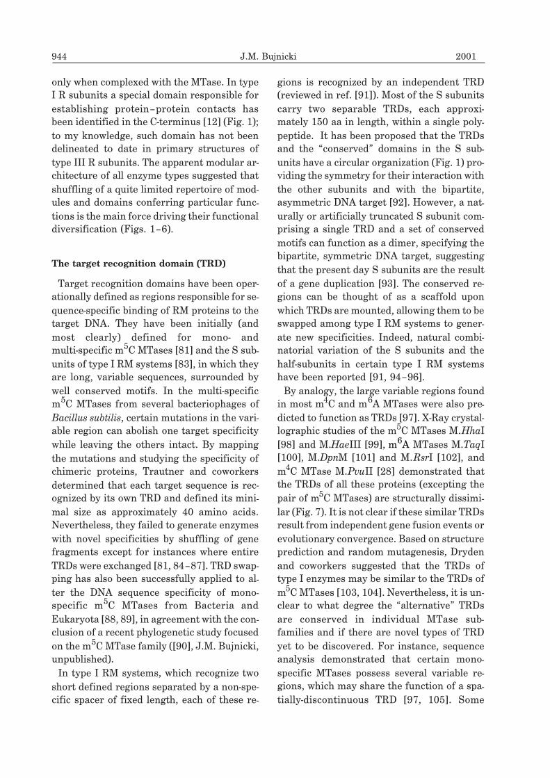
Figure 7. Cartoon diagrams of four structurally characterized DNA MTases depicting similarities be-
tween their catalytic domains and differences between their TRDs.
The core of the consensus MTase fold, recognizable by the 7-stranded �-sheet, is in the same relative orientation in
all four images. a) The m5C MTase M.HhaI co-crystalized with its target DNA (PDB coordinate file 5mht [109]), the
TRD is “behind” the DNA, b) the �-m6A MTase M.TaqI (1g38 [110]) co-crystalized with its target DNA, the
C-terminal TRD is on the left hand side, c) the �-m6A MTase M.DpnM (2dpm [101]) manually docked to its target,
the TRD (localized within an insert in the catalytic domain) is on the right hand side, d) the �-m4C MTase M.PvuII
(1boo [28]) manually docked to its target DNA, the proposed TRD (localized within an insert in the catalytic domain
that maps to the upper left hand side of the image) is disordered in the crystal of the DNA-free form and therefore
not shown.
zymes BfiI [113] and MboII [114], and for
homing nucleases from the GIY-YIG super-
family [115]. It should be stressed that identi-
fication of potential TRDs in sequences of re-
striction enzymes is particularly difficult,
since unlike in MTases the catalytic domains
of ENases contain no obviously conserved se-
quence motifs, which renders the simplistic
criterion of sequence variability inadequate.
Moreover, the key functions of type II restric-
tion enzymes, i.e. multimerization, se-
quence-specific DNA binding and cleavage are
interwoven such that some regions and resi-
dues are crucial for more than one aspect of
the ENase function [10].
The MTase domain
The MTase domain, which transfers the
methyl group from AdoMet onto the target
base, is the only truly conserved domain
among RM systems; that is, representatives of
only one of several unrelated protein families
known to catalyze this kind of reaction have
been identified in the context of RM systems
(reviewed in ref. [116]). Other enzymes, which
generate different modifications to inhibit re-
striction, are evolutionarily unrelated and
structurally dissimilar, including the only en-
zyme that generates a chemically similar
product, the tetrahydrofolate-dependent cyto-
sine-C5 hydroxymetyltransferase of T-even
coliphages [117]. The conserved ”MTase fold”
is characterized by an ��� domain with a cen-
tral seven-stranded �-sheet sandwiched be-
tween two layers of �-helices (Figs. 7, 8a). It
strongly resembles the architecture of the du-
plicated Rossmann-fold, with the only excep-
tion of a characteristic �-hairpin, involving
strands 6 and 7, which is absent from
Rossmann-fold proteins [118]. All DNA
MTase structures exhibit very similar fold,
with only minor variations of orientation and
number of peripheral secondary structural el-
ements. The approximate two-fold pseudo
symmetry reflects the structural similarity of
the AdoMet binding site to the target nucleo-
tide-binding active site. This observation has
led to the suggestion that the ancestral MTase
arose after gene duplication converted an
AdoMet-binding protein into a protein that
bound two molecules of AdoMet and that the
two halves then diverged [119]. An alternative
hypothesis has been put forward that various
MTases could have originated independently
from Rossmann-fold proteins [101]. Sup-
porting this view, a subsequent phylogenetic
study using both atomic coordinates and cor-
responding amino-acid sequences suggested
that MTases exhibiting the “typical fold” origi-
946 J.M. Bujnicki 2001
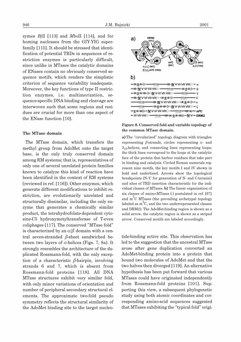
Figure 8.Conserved fold and variable topology of
the common MTase domain.
a) The “circularized” topology diagram with triangles
representing �-strands, circles representing �- and
310-helices, and connecting lines representing loops;
the thick lines correspond to the loops at the catalytic
face of the protein that harbor residues that take part
in binding and catalysis. Circled Roman numerals rep-
resent nine motifs, the key motifs I and IV shown in
bold and underlined. Arrows show the topological
breakpoints (N/C for generation of N- and C-termini)
and sites of TRD insertion characteristic for the indi-
vidual classes of MTases. b) The linear organization of
six classes of amino-MTases (-) postulated in ref. [97]
and m5C MTases (the prevailing archetypal topology
labeled as m5C, and the two underrepresented classes
and DRM2). The AdoMet-binding region is shown as a
solid arrow, the catalytic region is shown as a striped
arrow. Conserved motifs are labeled accordingly.
nated from one common Rossmann-fold an-
cestor [118].
Based on the methylated nucleotide that is
generated, DNA MTases can be divided into
three different groups: m6A, m4C, and m5C
MTases. m6A and m4C MTases methylate the
exocyclic amino group of the nucleobase and
are collectively termed “amino-MTases”,
while m5C MTases methylate the C-5 atom of
cytosine. It has been suggested that m4C and
m6A MTases are more closely related to each
other than to m5C MTases [97]. Remarkably,
certain m6A MTases display cryptic m4C ac-
tivity on mismatched cytosines [120] and
some m4C MTases may methylate mis-
matched adenine [121]. Moreover, experimen-
tal and bioinformatics studies suggested that
m4C-specific enzymes may have evolved inde-
pendently multiple times from m6A MTases,
although no consensus has been reached re-
garding the evolutionary pathways leading to
the present-day distribution of specificities
[105, 106, 120, 122]. Recently, it has been
shown that a change of the target base speci-
ficity from m6A to m4C is possible with only a
few amino acid substitutions. In an elegant ex-
periment Roth and Jeltsch reduced the size of
the target base binding pocket of M.EcoRV by
site-directed mutagenesis, generating an en-
zyme variant that no longer methylated ade-
nine and whose activity towards mismatched
cytosine was reduced only 17-fold [108, 123].
Nevertheless, such variant was not able to
methylate cytosine if it was base-paired with
guanine, suggesting that additional mutations
are needed to change the base flipping mecha-
nism of amino-MTase.
Amino-acid sequence alignments of MTases
revealed 9 relatively weakly conserved motifs
and a variable region, localized differently in
distinct families [124, 125] (Fig. 8b). Based on
the results of X-ray crystallography of m5C
MTase HhaI [98] and on structure-based mul-
tiple sequence alignment, motifs IV–VIII
were assigned to the active-site subdomain,
motifs X and I-III to the AdoMet-binding
subdomain, and the variable region with the
adjacent motif IX (present only in m5C
MTases) was recognized as the TRD, sug-
gested to be acting as an autonomous struc-
tural and functional domain [6, 97, 126]. That
alignment has been validated and its details
refined by comparison with crystal structures
of m6A MTases TaqI [100], DpnM [101], and
RsrI [102] and m4C MTase PvuII [28].
According to the possible linear arrange-
ments of the AdoMet-binding subdomain, the
active site subdomain, and the variable region
assumed to function as a TRD, the
amino-MTases were subdivided into 6 classes:
�� �� �� �� � and [97] (Fig. 8). The majority of
known DNA amino-MTases fall into the �� ��
and � classes, with no bona fide �-m4C MTases
discovered yet. M.NgoMXV and its homolog
M.LmoA118I are the only experimentally
characterized m4C MTases relatively closely
similar to �-m6A MTases, however they lack a
well-defined TRD [106, 127]. Similarly, se-
quence analysis and structure prediction for a
small group of viral �-like Dam MTases indi-
cated that due to the lack of TRD they cannot
be put into any of the proposed classes [107,
128]. Besides, we have identified two families
of enzymes closely related to DNA amino-
MTases, namely 16S rRNA: guanine-N2
MTases and the HemK family of putative nu-
cleic acid MTases that possess a large variable
region at the N-terminus, and therefore
should be classified as putative members of
the class [129, 130]. It has been also found
that the m4C MTase M.MwoI exhibits the � ar-
chitecture [131], rather than previously pro-
posed � [97]. Nearly all m5C MTases differ
from the group � MTases only in the position
of motif X, corresponding to a helix packing
against the central beta-sheet next to motif I:
in m5C MTases it is as the C-terminus, while
in � MTases it is in N-terminus. Nevertheless,
two exceptions to this rule have been identi-
fied: the M.BssHII MTase, which is a typical
member of the class with the TRD at the
N-terminus followed by the conserved motifs
IX, X, I–VIII [132], and a family of putative de
novo DNA MTases from Arabidopsis and
Vol. 48 Restriction-modification systems 947
maize (DRM2), that contain a MTase module
with a unique arrangement of motifs:
VI–VIII–TRD–IX–X, I–V [133] (Fig. 8).
Based on careful sequence analysis and molec-
ular modeling it has been proposed that the
atypical architecture of M.BssHII is not a re-
sult of a simple gene permutation event, but
rather a series of recombination events be-
tween of fragments of genes coding for up to
three different m5C MTases [134].
Lately, models of circular permutation dur-
ing evolution of m4C [105] and m6A MTases
[135] have been proposed. Jeltsch argued that
the domain permutation process needs dupli-
cation of a MTase gene, producing one en-
zyme with two catalytic domains. For in-
stance, after formation of new start and stop
codons in a hypothetical tandem ��-class
MTase, a - or �-like permutant would arise.
This model corresponds to the widely ac-
cepted concept that a permuted protein may
arise naturally from tandem repeats by ex-
traction of the C-terminal portion of one re-
peat together with the N-terminal portion of
the subsequent repeat, if the protein’s N and
C termini are in close spatial proximity [136].
Although the idea itself offers a plausible ex-
planation for the origin of permutants within
many protein families, the only duplicated
m6A MTases known to date are the type IIS
enzymes of the ��-class, whose permutation
would eventually produce enzymes of the � or
� classes that have not been identified to date.
M.MwoI, the only plausible candidate for the
� class known to date, is closely related to �MTases, and its putative TRD seems to have
“jumped” from the position in the middle of
the protein to the C-terminus without convinc-
ing evidence for duplication of the entire
MTase gene (Ref. [131] and J.M. Bujnicki and
M. Radlinska, unpublished data). In my opin-
ion, simple interconversions of topologies
from �� to � or from �� to � are rather implau-
sible, since the TRDs of known MTases from �
and � classes are unrelated [100, 102]. More-
over, the N- and C-termini of M.TaqI, the only
�-m6A MTase whose 3D structure is known,
are quite distant in space [100]. Still, this
scheme may be valid for enzymes, which have
not been identified yet, or whose sequences
have not been studied in enough detail. None-
theless, I believe that in most cases permuta-
tion of m4C and m6A MTases occurred via
intragenic relocations of gene segments (i.e.
“domain shuffling” [137]), which left no evi-
dent intermediates or fusions and rearrange-
ments of gene fragments [105], rather than
solely according to the “duplicate and get rid
of redundant termini” scheme. However, to
my knowledge, no systematic study has been
published, which would infer the evolutionary
history of shuffled fragments of MTase do-
mains in enzymes other than M.BssHII [134].
ENase domain
ENase exerts the second key activity of the
RM system and therefore could be predicted
to exhibit the degree of conservation at least
similar to that of the MTase counterpart.
However, among numerous ENase sequences
known there are only a few that exhibit statis-
tically significant similarity. The lack of se-
quence conservation has led to speculation
that despite common features, such as a re-
quirement for Mg2+
and outstanding se-
quence specificity, most ENases may be unre-
lated to one another [138]. Initially, the only
similarities were detected between type II
izoschizomers, enzymes with identical cleav-
age specificity, which may be regarded as di-
rect descendants of one ancestor, transferred
horizontally to different hosts [59, 139]. Nev-
ertheless, X-ray crystallographic studies of 13
seemingly dissimilar type II ENases demon-
strated unequivocally that they share a com-
mon structural core and metal-binding/cata-
lytic site, arguing for extreme divergence
rather than independent evolution of a similar
fair-sized domain (for the most recent reviews
see [10, 38, 61, 140]). This domain, termed
”PD-(D/E)XK” for a very weakly conserved
signature of the active site, turned out to be
common to other nucleases, including phage
948 J.M. Bujnicki 2001

exonuclease [141], two Archaeal Holliday
junction resolvases Hjc [142, 143], phage T7
Endonuclease I [144], transposase TnsA [145]
and two enzymes exerting ssDNA nicking in
the context of methyl-directed and very short
patch DNA repair: MutH [79] and Vsr [80]. It
is particularly interesting that MutH and Vsr
are genetically linked with DNA MTases Dam
and Dcm, respectively. Since the sequences of
structurally characterized PD-(D/E)XK cleav-
age domains seemed too divergent for ”regu-
lar” phylogenetic analysis, a structure-based
treeing has been carried out in a similar man-
ner to that performed for MTase domains
[140]. From this and other structure-based
comparative studies it can be concluded that
the PD-(D/E)XK superfamily can be divided
into two lineages, roughly corresponding to
“5� four-base overhang cutters” like EcoRI or
BamHI that interacts with the target DNA
predominantly via an �-helix and a loop and
the “blunt end cutters” like PvuII and EcoRV
that use a �-strand for DNA recognition [38].
A hypothetical evolutionary scenario of evolu-
tion of the two main ENase lineages based on
comparison of publicly available crystal struc-
tures is shown in Fig. 9.
Recently, despite limitations resulting from
extreme divergence of the PD-(D/E)XK do-
main, state-of-the-art algorithms for sequence
comparisons and structure prediction allowed
to identify it in a variety of other genuine and
putative nucleases, including the (m6A or
m5C)-specific restriction enzyme Mrr and its
homologs, the McrC subunit of the (m4C, m
5C
or hm5C)-specific restriction enzyme McrBC,
the hm5C-specific restriction enzyme
PvuRts1I, herpesvirus alkaline exonucleases,
Archaeal-type Holliday junction resolvases
Hjc, various proteins containing the NTPase
module like the RecB and DNA2 nuclease fam-
ilies or other enzymes involved in DNA recom-
bination and repair [146–151]. It has been
also found out that the catalytic domain of
tRNA splicing endonuclease EndA bears strik-
ing resemblance to the minimal core of the
PD-(D/E)XK fold [152], although it developed
the RNase A-like active site in a distinct loca-
tion [153]. It is tempting to speculate that
EndA may be related to a ”common ancestor”
of the PD-(D/E)XK superfamily (Fig. 9), how-
ever this hypothesis must await a thorough
structure-based phylogenetic study with
atomic coordinates of more ancient nucleases
available.
Ironically, following the series of crystallo-
graphic studies suggesting common origin of
all ENase domains in restriction enzymes and
related DNA repair and recombination en-
zymes, bioinformatics studies provided evi-
dence that some bona fide type II ENases are
in fact diverged members of other well-studied
nuclease superfamilies, unrelated to the
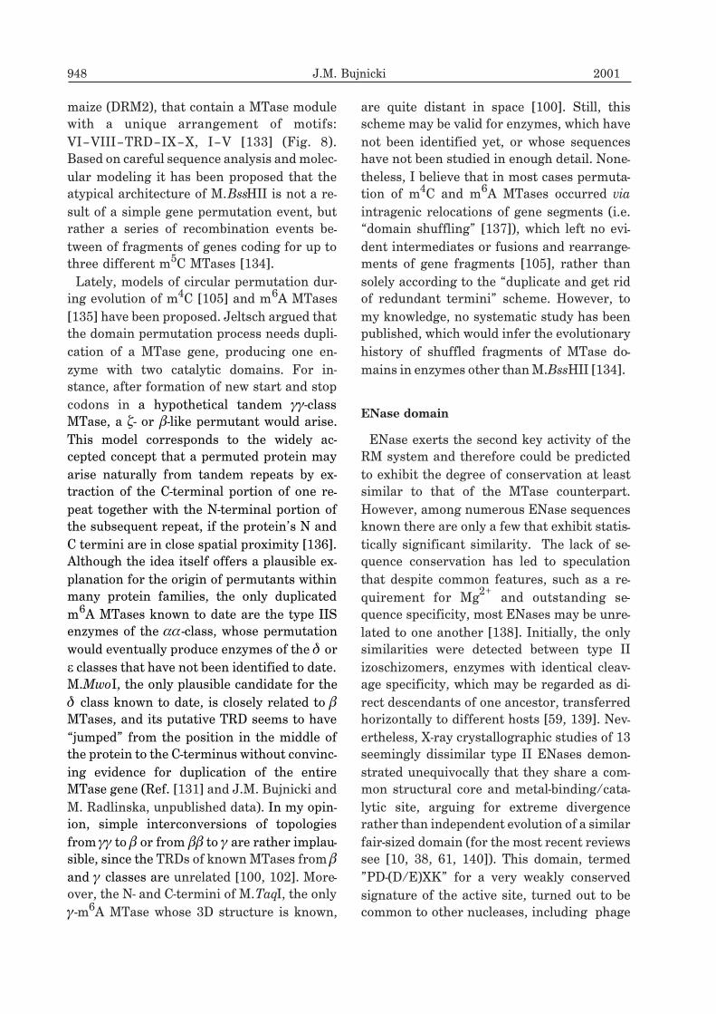
PD-(D/E)XK enzymes (Fig. 10). It has been
found that the N-terminal part of the type IIS
restriction enzyme exhibits low sequence sim-
ilarity to an EDTA-resistant nuclease (Nuc) of
Salmonella typhimurium, and the relationship
of these nuclease domains has been confirmed
experimentally [113]. We have also identified
the Nuc-like domain in type II restriction en-
zymes NgoFVII, NgoAVII, and CglI (J.M.
Bujnicki, M. Radliñska, V. Siksnys, unpub-
lished data). Another evolutionarily unrelated
nuclease domain, similar to the catalytic do-
main of nucleases from the HNH superfamily,
has been identified in the m5C-specific restric-
tion enzyme McrA, type II restriction en-
zymes HpyI, NlaIII, SphI, SapI, NspHI, NspI
and KpnI, and in type IIS enzyme MboII and
its homologs from Helicobacter pylori by our
group [114, 154] and by Eugene Koonin’s
group [147]. We have also found that type II
enzymes Eco29kI, NgoMIII, NgoAIII, and
MraI are homologous to the GIY-YIG endo-
nuclease domain present in certain homing
endonucleases and DNA repair and recombi-
nation enzymes [114] and that the HgiDII en-
zyme is related to the DNA repair enzyme
MutL, which also possesses a distinct fold
(J.M. Bujnicki, unpublished, and P. Friedhoff,
cited as personal communication in ref. [10]).
Presently, most of these predictions await ex-
perimental confirmation, however even in the
Vol. 48 Restriction-modification systems 949

absence of crystal structures of ENases with
any of the three “alternative” folds it became
clear that restriction enzymes have evolved on
multiple occasions. Moreover, analysis of the
various combinations of structural modules
present in homing endonucleases and type IIS
950 J.M. Bujnicki 2001
Figure 9. Proposed scheme of evolution of the PD-(D/E)XK family of proteins that depicts radiation and
divergence of the � and � subfamilies of restriction enzymes [38, 140].
Secondary structural elements in the topological diagrams are coded as described in Fig. 7a. Evolutionary steps (ac-
quisition and loss of structural elements) are indicated by arrows, elements that are conserved in a given step and in
a given sub-lineage are shaded, novel elements are shown in white. The major features that allow distinction be-
tween the two lineages are depicted by dotted circles: i) the directionality of the 5th
�-strand (parallel in the �-lineage
and antiparallel in the �-lineage) and ii) the appearance of an additional small �-sheet that participates in target rec-
ognition in the �-lineage. The additional �-sheet of -exo and other �-enzymes is a topologically different and hence
independently acquired feature. Other peculiarities are the unusual left-handed �-�-� element at the C-teriminal
edge of the �-strand in Vsr [80], as opposed to the typical right-handed structure in other proteins, and the fact that
the core of T7 Endo I is made of fragments of two polypeptide chains forming a swapped dimer [144].
and certain multimodular type II restriction
enzymes suggests that the “remote cutters”
arose independently multiple times from vari-
ous combinations of “cleavage domains” and
TRDs with alternative folds and therefore rep-
resent an interesting example of convergent
evolution.
DNA translocase (helicase-like) domain
All type I and III restriction enzymes, to-
gether with the modification-dependent en-
zyme McrBC, require two recognition sites in
linear DNA and nucleotide triphosphate
(NTP) hydrolysis before DNA cleavage can oc-
cur [70]. Type I and III restriction enzymes re-
quire ATP for activity (reviewed in ref. [8]),
while McrBC requires GTP [157]. Type I en-
zymes and McrBC exhibit a similar mecha-
nism: they translocate along DNA from their
recognition sites in a reaction powered by
NTP hydrolysis until they encounter a block
to translocation, which stimulates DNA cleav-
age [158, 159]. The block is normally another
enzyme molecule translocating from another
site or a topological barrier resulting from
supercoiling of the loop between the two en-
zymes, explaining the dependence of reaction
on two sites. However, other non-specific
blocks to translocation, such as a bound
repressor or a Holliday junction also stimu-
late cleavage. One peculiarity of type I en-
zymes is that they do not turn over in the
cleavage reaction, but they hydrolyze ATP
long after DNA cleavage has stopped [160]. In
contrast, type III enzymes, require a specific
contact between the two translocating enzyme
molecules and non-specific blocks are inhibi-
tory [19]. Bickle and coworkers demonstrated
that cooperation between two enzymes is nec-
essary for ds DNA cleavage, since each
translocating enzyme complex cuts only one
strand of DNA [22].
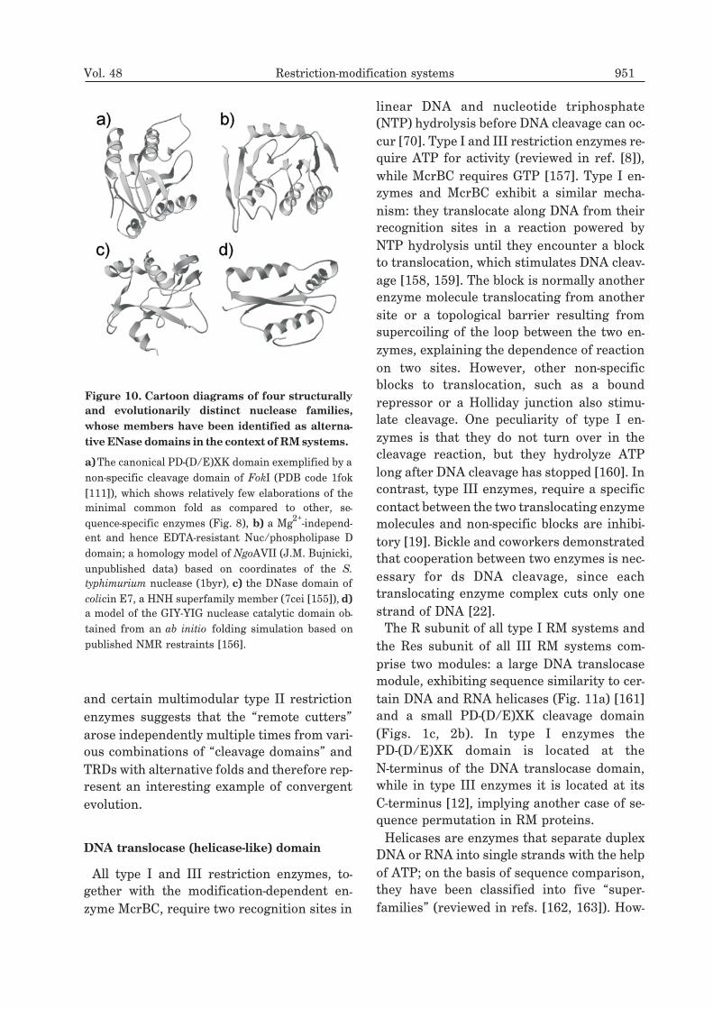
The R subunit of all type I RM systems and
the Res subunit of all III RM systems com-
prise two modules: a large DNA translocase
module, exhibiting sequence similarity to cer-
tain DNA and RNA helicases (Fig. 11a) [161]
and a small PD-(D/E)XK cleavage domain
(Figs. 1c, 2b). In type I enzymes the
PD-(D/E)XK domain is located at the
N-terminus of the DNA translocase domain,
while in type III enzymes it is located at its
C-terminus [12], implying another case of se-
quence permutation in RM proteins.
Helicases are enzymes that separate duplex
DNA or RNA into single strands with the help
of ATP; on the basis of sequence comparison,
they have been classified into five “super-
families” (reviewed in refs. [162, 163]). How-
Vol. 48 Restriction-modification systems 951
Figure 10. Cartoon diagrams of four structurally
and evolutionarily distinct nuclease families,
whose members have been identified as alterna-
tive ENase domains in the context of RM systems.
a)The canonical PD-(D/E)XK domain exemplified by a
non-specific cleavage domain of FokI (PDB code 1fok
[111]), which shows relatively few elaborations of the
minimal common fold as compared to other, se-
quence-specific enzymes (Fig. 8), b) a Mg2+
-independ-
ent and hence EDTA-resistant Nuc/phospholipase D
domain; a homology model of NgoAVII (J.M. Bujnicki,
unpublished data) based on coordinates of the S.
typhimurium nuclease (1byr), c) the DNase domain of
colicin E7, a HNH superfamily member (7cei [155]), d)
a model of the GIY-YIG nuclease catalytic domain ob-
tained from an ab initio folding simulation based on
published NMR restraints [156].
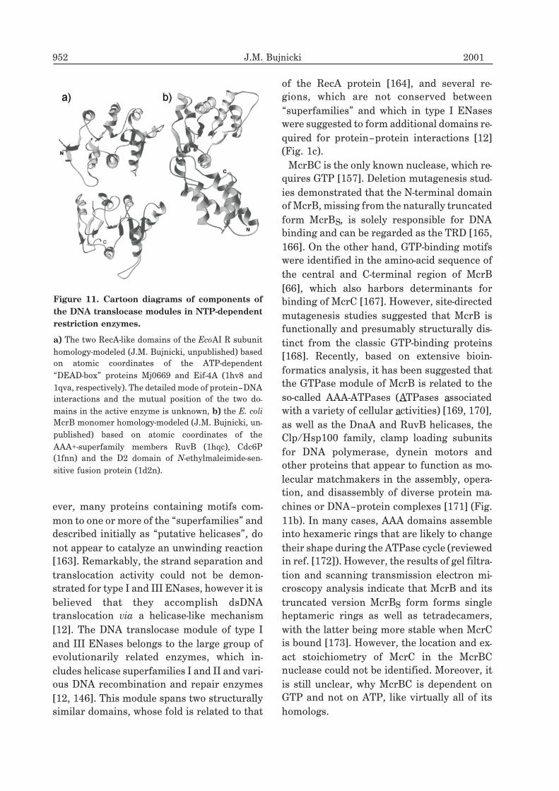
ever, many proteins containing motifs com-
mon to one or more of the “superfamilies” and
described initially as “putative helicases”, do
not appear to catalyze an unwinding reaction
[163]. Remarkably, the strand separation and
translocation activity could not be demon-
strated for type I and III ENases, however it is
believed that they accomplish dsDNA
translocation via a helicase-like mechanism
[12]. The DNA translocase module of type I
and III ENases belongs to the large group of
evolutionarily related enzymes, which in-
cludes helicase superfamilies I and II and vari-
ous DNA recombination and repair enzymes
[12, 146]. This module spans two structurally
similar domains, whose fold is related to that
of the RecA protein [164], and several re-
gions, which are not conserved between
“superfamilies” and which in type I ENases
were suggested to form additional domains re-
quired for protein–protein interactions [12]
(Fig. 1c).
McrBC is the only known nuclease, which re-
quires GTP [157]. Deletion mutagenesis stud-
ies demonstrated that the N-terminal domain
of McrB, missing from the naturally truncated
form McrBS, is solely responsible for DNA
binding and can be regarded as the TRD [165,
166]. On the other hand, GTP-binding motifs
were identified in the amino-acid sequence of
the central and C-terminal region of McrB
[66], which also harbors determinants for
binding of McrC [167]. However, site-directed
mutagenesis studies suggested that McrB is
functionally and presumably structurally dis-
tinct from the classic GTP-binding proteins
[168]. Recently, based on extensive bioin-
formatics analysis, it has been suggested that
the GTPase module of McrB is related to the
so-called AAA-ATPases (ATPases associated
with a variety of cellular activities) [169, 170],
as well as the DnaA and RuvB helicases, the
Clp/Hsp100 family, clamp loading subunits
for DNA polymerase, dynein motors and
other proteins that appear to function as mo-
lecular matchmakers in the assembly, opera-
tion, and disassembly of diverse protein ma-
chines or DNA–protein complexes [171] (Fig.
11b). In many cases, AAA domains assemble
into hexameric rings that are likely to change
their shape during the ATPase cycle (reviewed
in ref. [172]). However, the results of gel filtra-
tion and scanning transmission electron mi-
croscopy analysis indicate that McrB and its
truncated version McrBS form forms single
heptameric rings as well as tetradecamers,
with the latter being more stable when McrC
is bound [173]. However, the location and ex-
act stoichiometry of McrC in the McrBC
nuclease could not be identified. Moreover, it
is still unclear, why McrBC is dependent on
GTP and not on ATP, like virtually all of its
homologs.
952 J.M. Bujnicki 2001
Figure 11. Cartoon diagrams of components of
the DNA translocase modules in NTP-dependent
restriction enzymes.
a) The two RecA-like domains of the EcoAI R subunit
homology-modeled (J.M. Bujnicki, unpublished) based
on atomic coordinates of the ATP-dependent
“DEAD-box” proteins Mj0669 and Eif-4A (1hv8 and
1qva, respectively). The detailed mode of protein–DNA
interactions and the mutual position of the two do-
mains in the active enzyme is unknown, b) the E. coli
McrB monomer homology-modeled (J.M. Bujnicki, un-
published) based on atomic coordinates of the
AAA+-superfamily members RuvB (1hqc), Cdc6P
(1fnn) and the D2 domain of N-ethylmaleimide-sen-
sitive fusion protein (1d2n).
Regulatory proteins
The characterization of type II RM systems
has shown that some systems contain other
components in addition to the requisite
endonuclease and methyltransferase. One of
these is the C (controller) protein, which has
been proposed to allow establishment of RM
systems in new hosts by delaying the appear-
ance of restriction activity; its gene generally
precedes and in some cases partially overlaps
the ENase gene [174]. C proteins have not yet
been structurally characterized, but their
amino-acid sequences reveal that they are
probably helix-turn-helix proteins similar to
numerous known activators and repressors of
gene expression (reviewed in ref. [175]). Se-
quence comparisons have identified a con-
served DNA sequence element termed a
“C box” immediately upstream of most
C genes [176]. It has been shown that C.PvuII
and C.BamHI are DNA-binding proteins that
bind to the C box and by autogenous activa-
tion of the polycistronic pvuIICR or bamHICR
promoter contribute to the temporal activa-
tion of the ENase gene expression (ref. [177]
and A. Sohail, I. Ghosh, R.M. Fuentes, and
J.E. Brooks, unpublished results cited
therein). It has been also demonstrated that
there is some cross-complementation between
the C genes from different RM systems [178].
Kobayashi and coworkers reported that
some type II RM systems on plasmids resist
displacement by a plasmid bearing RM sys-
tems with ENase and MTase of distinct speci-
ficity but the C protein of the same specific-
ity. An apparent cell suicide results from
chromosome cleavage at unmodified sites by
prematurely expressed ENase from an in-
coming RM system [179]. In general, C genes
were found to play important roles in the
maintenance, establishment, and mutual ex-
clusion of RM systems. These roles are remi-
niscent of the strategies of temperate bacte-
riophages [180] and are in accord with the
“selfish gene” hypothesis for the spread and
maintenance of RM gene complexes [5, 181]
(see also below).
The regulatory protein from the unusual
LlaI RM system [62] was shown to enhance
expression of LlaI restriction at a post-trans-
criptional level rather than to function as a
transcriptional activator, despite its sequence
similarity to HTH proteins [182]. Similarly,
regulation of the ENase activity by inhibiting
intracellular subunit association was reported
for the PvuII enzyme and a 28-amino-acid pep-
tide, designated W.PvuII [183].
Other elements associated with RM systems
There have been several reports of the close
association between enzymes involved in
DNA mobility and RM systems. Genes and
partial genes encoding phage-like integrases
and other proteins from the tyrosine
recombinase (Int) superfamily occur next to
the sinIR [184], accIM [185], ecoHK31IM, and
eaeIM genes [186]. Genes for putative pro-
teins similar to DNA invertases and
resolvases are found near the PaeR7I [187],
BglII [188], and ApaLI [189] RM systems. A
complete copy of the IS982 element with a
DDE-superfamily transposase-encoding gene
was identified between the llaKR2IR and
llaKR2IM genes [190]; a putative transposase
was also found in the intergenic area between
the eco47IR and eco47IIM genes [191]. These
proteins may facilitate the transfer of RM
genes among different bacterial strains.
Genomic context of evolution, structure,
and function of RM systems
Currently hundreds of sequences of func-
tionally characterized DNA MTases and
ENases are available in public databases
[192]. Although this number is still growing,
we are also faced with a virtual explosion in
the number of sequences of putative RM pro-
tein deduced from data produced by numer-
ous Prokaryotic genome-sequencing projects.
75% of completely sequences genomes appear
to contain multiple RM systems (up to two
dozens in the case of Helicobacter pylori J99),
Vol. 48 Restriction-modification systems 953
most of which have never been assayed bio-
chemically. However, as emphasized based on
the recent results of genome-wide analyses
carried out for putative RM systems of H.
pylori J99 [193], H. pylori 26695 [194] and
Cyanobacterium Anabaena strain PCC7120
[122], many of the candidate genes are in fact
pseudogenes in various states of decomposi-
tion. Nevertheless, as demonstrated for the
Hpy99I system, which has been identified
based on sequence analysis and subsequently
characterized biochemically, the remaining
active RM genes, may be a rich source of novel
specificities [193]. Evidently, the genome-
based screening method has several impor-
tant advantages over conventional methods
employing testing the crude cell extracts for
their restriction activity: it can save the fer-
mentation of large amount of microbes, which
may pathogenic or very difficult to grow; and
allows cloning and expression of RM systems,
whose activity is not detectable in cell ex-
tracts.
Genome-wide comparisons carried out for
pairs of related strains of: �-Proteobacteria H.
pylori [195] and Archaea Pyrococcus abyssii
and P. horikoshii [196] suggested that the
presence of RM systems is often associated
with various types of genome polymorphisms.
It has been noted, that certain chromosomal
loci in different strains of related bacteria
may be associated with unrelated or very re-
motely related RM systems that exhibit differ-
ent specificities [197]. This suggests that the
representational difference analysis may be
used for isolation of novel RM systems based
on genomic sequence analysis even if the se-
quence of the genome of the strain of interest
is not available.
From the data generated by combined theo-
retical and experimental genomic approaches
many more surprises can be expected, not
only as a result of enzymes with new speci-
ficities or new “types” combining old domains
in unprecedented manner, but also because
some RM systems may comprise novel do-
mains, not related to those described in this
article. For instance, it seems plausible that
some restriction enzymes comprise cleavage
domains homologous to the LAGLIDADG,
AP, RusA, RuvC/RNase H or other nuclease
superfamilies [147, 198], rather than the
PD-(D/E)XK, HNH, GIY-YIG and Nuc
superfamilies described to date. On the other
hand, the numerous ongoing structural
genomics programs will undoubtedly provide
more insight into cases like the yeast RPB5
subunit of RNA polymerase from Saccharo-
myces cerevisiae, which comprises a
PD-(D/E)XK-like domain without the nuclease
active site [199] or the EndA enzyme, whose
PD-(D/E)XK-like domain acquired the RNase
A-like active site on an opposite face of the
protein [153]. The latter case is especially in-
teresting, since it suggests that additional
binding or catalytic sites could be engineered
in structures of restriction ENases from the
PD-(D/E)XK superfamily [152].
The existence of specific relationships be-
tween certain restriction enzymes and other
evolutionarily conserved nucleases inferred
from structural studies and sequence compar-
isons on a genome scale suggests that they
have arisen on multiple occasions from differ-
ent nuclease lineages [147]. It is tempting to
speculate that most of restriction ENases
evolved as self-propagating, “selfish” ele-
ments from DNA repair enzymes or other cel-
lular nucleases, however the available data do
not allow to draw definite conclusions. None-
theless, in the course of comparative analysis
of sequences and structures of various nu-
cleases carried out by our group and by others
it became clear that the major families of se-
quence-specific restriction enzymes are re-
lated to either structure-specific or nonspe-
cific nucleases [114, 140, 146, 147, 149, 150,
154, 198]. It suggests that evolutionary path-
ways leading from non-specific nucleases to
highly sequence-specific restriction enzymes
or vice versa can be inferred, provided suffi-
cient number of sequences and structures cor-
responding to “evolutionary intermediates”.
Even though many putative RM genes are in-
954 J.M. Bujnicki 2001
active, their sequences may aid in generation
of multiple sequence alignments and phylo-
genetic trees. The use of “intermediate se-
quences” is also helpful in molecular model-
ing, where one attempts to predict the
three-dimensional structure of a protein of in-
terest based on sequence alignment to a ho-
mologous protein of known structure [200,
201].
Such information could guide mutagenesis
experiments aiming at rational engineering of
restriction enzymes with new specificities. To
date, attempts to change the specificity of
type II restriction enzymes using site-directed
or random mutagenesis were rather unsuc-
cessful [202, 203]. It has been concluded that
even for the very well characterized restric-
tion enzymes, like EcoRV, properties that de-
termine specificity and selectivity are difficult
to model on the basis of the available struc-
tural information [204]. However, with the
broad range of enzymes with different
specificities in hand one can systematically
analyze the structure–function relationships
and follow the evolutionary history of selected
families of RM proteins. Since MTases show
much greater sequence similarity than
ENases, several projects have been launched
aiming at engineering of their specificity
based on phylogenetic analysis and identifica-
tion of mutations correlated with functional
modifications. To date, there has been no
spectacular success; it has been concluded
that the evolutionary pathway for specificity
change leads through a stage of relaxed speci-
ficity (ref. [108], S. Klimasauskas, personal
communication, J.M. Bujnicki and M.
Radlinska, unpublished). It suggests that best
targets for specificity engineerning would be
not the highly specific enzymes studied pres-
ently, but the “sloppy” ones [205], which make
only a few key protein–DNA contacts to recog-
nize their target (or rather a broad range of
targets). A similar approach seems applicable
for engineering of ENases with novel speci-
ficities. In my opinion, engineering specificity
into polypeptide loops of inherently non-spe-
cific cleavage domains that are able to bind to
DNA on their own seems more promising
than modifying the highly elaborated
DNA-binding surface of enzymes like EcoRV.
Unfortunately, only a few crystal structures
are available for the non-specific nucleases
[112, 206] and none for the “sloppy” MTases.
Our unpublished results suggest that the
three-dimensional structure of certain
ENases can be predicted based on results of
sequence-structure threading even in the ab-
sence of significant sequence similarity, how-
ever it remains to be verified experimentally if
such models are of sufficient resolution to
guide knowledge-based redesign of DNA-bin-
ding determinants. Nevertheless, it is obvious
that good insight into evolutionary plasticity
of functionally important elements in RM pro-
teins can be obtained in the course of compar-
ative analysis carried out using advanced
computational methods. In my opinion the
elusive goal of creating MTases and ENases
with novel specificities will be achieved only if
the large-scale bioinformatics and experimen-
tal approaches are combined.
CONCLUSIONS
This review covers recent results on the
structure and evolution of RM enzymes. One
immediately obvious fact is the rapid accelera-
tion in the production of new data in this field.
This has allowed the demonstration of phylo-
genetic and mechanistic links between RM en-
zymes and other proteins that often possess
similar biochemical or enzymatic properties.
The wealth of new data becoming available
should help to answer many open questions
concerning the structure–function relation-
ships of RM proteins. No doubt the approach
of functional genomics will play a significant
role in identifying genes coding for novel
ENases and MTases, and the newly developed
computational tools will guide their experi-
mental characterization and engineering, sug-
gesting that a new era in research on the
Vol. 48 Restriction-modification systems 955
structure and function of RM systems has just
begun.
I would like to thank Drs. Ashok Bhagwat,
Thomas Bickle, Robert Blumenthal, Xiaodong
Cheng, David Dryden, Jeff Elhai, Alan Fried-
man, Peter Friedhoff, Arvydas Janulaitis, Al-
bert Jeltsch, Antal Kiss, Saulius
Klimasauskas, Ichizo Kobayashi, Daniel
Panne, Andrzej Piekarowicz, Alfred Pingoud,
Monika Radlinska, Virgis Siksnys, and
Geoffrey Wilson for stimulating discussions
and kind provision of unpublished data. I
thank Drs. Xiaodong Cheng, Alan Friedman,
Richard Gumport, Sanford A. Lacks, Michael
Topal, and Simon E. Phillips for sending me
coordinates of crystal structures of RM en-
zymes before they were made publicly avail-
able. I also thank numerous colleagues for
sending reprints and preprints of their arti-
cles and apologize for not being able to cite all
references due to space limitations. Finally, I
am indebted to Drs. Robert Blumenthal and
Monika Radlinska for critical reading of the
manuscript and to Leszek Rychlewski for his
constant support.
R E F E R E N C E S
1. Wilson, G.G. & Murray, N.E. (1991) Restric-
tion and modification systems. Annu. Rev.
Genet. 25, 585–627.
2. Arber, W. & Dussoix, D. (1962) Host specific-
ity of DNA produced by Escherichia coli. Host
controlled modification of bacteriophage �. J.
Mol. Biol. 5, 18–36.
3. Arber, W. (1979) Promotion and limitation of
genetic exchange. Science 205 , 361–365.
4. Lacks, S.A., Ayalew, S., de la Campa, A.G. &
Greenberg, B. (2000) Regulation of compe-
tence for genetic transformation in Streptococ-
cus pneumoniae: Expression of dpnA, a late
competence gene encoding a DNA methyl-
transferase of the DpnII restriction system.
Mol. Microbiol. 35, 1089–1098.
5. Kobayashi, I., Nobusato, A., Kobayashi-
Takahashi, N. & Uchiyama, I. (1999) Shaping
the genome-restriction-modification systems
as mobile genetic elements. Curr. Opin. Genet.
Dev. 9, 649–656.
6. Dryden, D.T. (1999) Bacterial DNA
methyltranferases; in: S-Adenosylmethio-
nine-dependent Methyltransferases: Structures
and Functions (Cheng, X. et al., eds.) pp. 283–
340, World Scientific Inc., Singapore.
7. Murray, N.E. (2000) Type I restriction sys-
tems: Sophisticated molecular machines
(a legacy of Bertani and Weigle). Microbiol.
Mol. Biol. Rev. 64, 412–434.
8. Rao, D.N., Saha, S. & Krishnamurthy, V.
(2000) ATP-dependent restriction enzymes.
Prog. Nucleic Acid Res. Mol. Biol. 64, 1–63.
9. Pingoud, A. and Jeltsch, A. (1997) Recognition
and cleavage of DNA by type-II restriction
endonucleases. Eur. J. Biochem. 246, 1–22.
10.Pingoud, A. & Jeltsch, A. (2001) Structure and
function of type II restriction endonucleases.
Nucleic Acids Res. 29, 3705–3727.
11. Pingoud, A., Jeltsch, A., Maxwell, A. &
Sherratt, D. (2001) Enzymes that keep DNA
under control: Meeting: DNA enzymes: struc-
tures and mechanisms. EMBO Rep. 2,
271–276.
12.Davies, G.P., Martin, I., Sturrock, S.S.,
Cronshaw, A., Murray, N.E. & Dryden, D.T.
(1999) On the structure and operation of type I
DNA restriction enzymes. J. Mol. Biol. 290,
565–579.
13.Dryden, D.T., Cooper, L.P., Thorpe, P.H. & By-
ron, O. (1997) The in vitro assembly of the
EcoKI type I DNA restriction/modification en-
zyme and its in vivo implications. Biochemis-
try, 36, 1065–1076.
14. Ellis, D.J., Dryden, D.T., Berge, T.,
Edwardson, J.M. & Henderson, R.M. (1999)
Direct observation of DNA translocation and
cleavage by the EcoKI endonuclease using
956 J.M. Bujnicki 2001
atomic force microscopy. Nat. Struct. Biol. 6,
15–17.
15.Yuan, R., Hamilton, D.L. & Burckhardt, J.
(1980) DNA translocation by the restriction
enzyme from E. coli K. Cell 20, 237–244.
16.Berge, T., Ellis, D.J., Dryden, D.T.,
Edwardson, J.M. & Henderson, R.M. (2000)
Translocation-independent dimerization of
the EcoKI endonuclease visualized by atomic
force microscopy. Biophys. J. 79, 479–484.
17. Boyer, H.W. (1971) DNA restriction and mod-
ification mechanisms in bacteria. Annu. Rev.
Microbiol. 25, 153–176.
18.Kauc, L. & Piekarowicz, A. (1978) Purification
and properties of a new restriction
endonuclease from Haemophilus influenzae Rf.
Eur. J. Biochem. 92, 417–426.
19.Meisel, A., Mackeldanz, P., Bickle, T.A.,
Kruger, D.H. & Schroeder, C. (1995) Type III
restriction endonucleases translocate DNA in
a reaction driven by recognition site-specific
ATP hydrolysis. EMBO J. 14, 2958–2966.
20.Bist, P., Sistla, S., Krishnamurthy, V.,
Acharya, A., Chandrakala, B. & Rao, D.N.
(2001) S-Adenosyl-L-methionine is required
for DNA cleavage by type III restriction en-
zymes. J Mol. Biol . 310, 93–109.
21. Meisel, A., Bickle, T.A., Kruger, D.H. &
Schroeder, C. (1992) Type III restriction en-
zymes need two inversely oriented recognition
sites for DNA cleavage. Nature 355, 467–469.
22.Janscak, P., Sandmeier, U., Szczelkun, M.D. &
Bickle, T.A. (2001) Subunit assembly and
mode of DNA cleavage of the type III restric-
tion endonucleases EcoP1I and EcoP15I. J.
Mol. Biol. 306, 417–431.
23.Berman, H.M., Westbrook, J., Feng, Z.,
Gilliland, G., Bhat, T.N., Weissig, H.,
Shindyalov, I.N. & Bourne, P.E. (2000) The
Protein Data Bank. Nucleic Acids Res. 28,
235–242.
24.Roberts, R.J. & Cheng, X. (1998) Base flip-
ping. Annu. Rev. Biochem. 67, 181–198.
25.Hornby, D.P. & Ford, G.C. (1998) Pro-
tein-mediated base flipping. Curr. Opin.
Biotechnol. 9, 354–358.
26.Ho, D.K., Wu, J.C., Santi, D.V. & Floss, H.G.
(1991) Stereochemical studies of the
C-methylation of deoxycytidine catalyzed by
HhaI methylase and the N-methylation of
deoxyadenosine catalyzed by EcoRI
methylase. Arch. Biochem. Biophys. 284,
264–269.
27. Ahmad, I. & Rao, D.N. (1996) Chemistry and
biology of DNA methyltransferases. Crit. Rev.
Biochem. Mol. Biol. 31, 361–380.
28.Gong, W., O’Gara, M., Blumenthal, R.M. &
Cheng, X. (1997) Structure of PvuII DNA-(cy-
tosine N4) methyltransferase, an example of
domain permutation and protein fold assign-
ment. Nucleic Acids Res. 25, 2702–2715.
29.Vertino, P.M. (1999) Eukaryotic DNA
methyltransferases; in: S-Adenosylmethionine-
dependent Methyltransferases: Structures and
Functions (Cheng, X. et al., eds.) pp. 341–372,
World Scientific Inc., Singapore.
30.Winkler, F.K., Banner, D.W., Oefner, C.,
Tsernoglou, D., Brown, R.S., Heathman, S.P.,
Bryan, R.K., Martin, P.D., Petratos, K. & Wil-
son, K.S. (1993) The crystal structure of
EcoRV endonuclease and of its complexes with
cognate and non-cognate DNA fragments.
EMBO J. 12, 1781–1795.
31. Cheng, X., Balendiran, K., Schildkraut, I. &
Anderson, J.E. (1994) Structure of PvuII
endonuclease with cognate DNA. EMBO J.
13, 3927–3935.
32.Kim, Y., Grable, J.C., Love, R., Green, P.J. &
Rosenberg, J.M. (1990) Refinement of EcoRI
endonuclease crystal structure: A revised pro-
tein chain tracing. Science 249, 1307–1309.
33.Newman, M., Strzelecka, T., Dorner, L.F.,
Schildkraut, I. & Aggarwal, A.K. (1995) Struc-
Vol. 48 Restriction-modification systems 957
ture of BamHI endonuclease bound to DNA:
Partial folding and unfolding on DNA binding.
Science 269, 656–663.
34.Newman, M., Lunnen, K., Wilson, G., Greci,
J., Schildkraut, I. & Phillips, S.E. (1998) Crys-
tal structure of restriction endonuclease BglI
bound to its interrupted DNA recognition se-
quence. EMBO J. 17, 5466–5476.
35.Stankevicius, K., Lubys, A., Timinskas, A.,
Vaitkevicius, D. & Janulaitis, A. (1998) Clon-
ing and analysis of the four genes coding for
Bpu10I restriction-modification enzymes. Nu-
cleic Acids Res. 26, 1084–1091.
36.Hsieh, P.C., Xiao, J.P., O’loane, D. & Xu, S.Y.
(2000) Cloning, expression and purification of
a thermostable nonhomodimeric restriction
enzyme, BslI. J Bacteriol. 182, 949–955.
37. Kruger, D.H., Barcak, G.J., Reuter, M. &
Smith, H.O. (1988) EcoRII can be activated to
cleave refractory DNA recognition sites. Nu-
cleic Acids Res. 16, 3997–4008.
38.Huai, Q., Colandene, J.D., Chen, Y., Luo, F.,
Zhao, Y., Topal, M.D. & Ke, H. (2000) Crystal
structure of NaeI-an evolutionary bridge be-
tween DNA endonuclease and topoisomerase.
EMBO J. 19, 3110–3118.
39.Deibert, M., Grazulis, S., Sasnauskas, G.,
Siksnys, V. & Huber, R. (2000) Structure of
the tetrameric restriction endonuclease
NgoMIV in complex with cleaved DNA. Nat.
Struct. Biol. 7, 792–799.
40.Bilcock, D.T. & Halford, S.E. (1999) DNA re-
striction dependent on two recognition sites:
Activities of the SfiI restriction-modification
system in Escherichia coli. Mol. Microbiol. 31,
1243–1254.
41. Szybalski, W., Kim, S.C., Hasan, N. &
Podhajska, A.J. (1991) Class-IIS restriction en-
zymes — a review. Gene 100, 13–26.
42.Bitinaite, J., Wah, D.A., Aggarwal, A.K. &
Schildkraut, I. (1998) FokI dimerization is re-
quired for DNA cleavage. Proc. Natl. Acad. Sci
U.S.A. 95, 10570–10575.
43.Vanamee, E.S., Santagata, S. & Aggarwal,
A.K. (2001) FokI requires two specific DNA
sites for cleavage. J. Mol. Biol. 309, 69–78.
44.Kim, Y.G. & Chandrasegaran, S. (1994) Chi-
meric restriction endonuclease. Proc. Natl.
Acad. Sci U.S.A. 91, 883–887.
45.Kim, Y.G., Smith, J., Durgesha, M. &
Chandrasegaran, S. (1998) Chimeric restric-
tion enzyme: Gal4 fusion to FokI cleavage do-
main. Biol. Chem. 379, 489–495.
46.Chandrasegaran, S. & Smith, J. (1999) Chime-
ric restriction enzymes: what is next? Biol.
Chem. 380, 841–848.
47. Morgan, R.D., Calvet, C., Demeter, M., Agra,
R. & Kong, H. (2000) Characterization of the
specific DNA nicking activity of restriction
endonuclease N.BstNBI. Biol. Chem. 381,
1123–1125.
48.Higgins, L.S., Besnier, C. & Kong, H. (2001)
The nicking endonuclease N.BstNBI is closely
related to Type IIs restriction endonucleases
MlyI and PleI. Nucleic Acids Res. 29,
2492–2501.
49.Gunn, J.S. & Stein, D.C. (1997) The Neisseria
gonorrhoeae S.NgoVIII restriction/modifica-
tion system: A type IIs system homologous to
the Haemophilus parahaemolyticus HphI re-
striction/modification system. Nucleic Acids
Res. 25, 4147–4152.
50.Friedrich, T., Fatemi, M., Gowhar, H.,
Leismann, O. & Jeltsch, A. (2000) Specificity
of DNA binding and methylation by the
M.FokI DNA methyltransferase. Biochim.
Biophys. Acta 1480, 145–159.
51. Janulaitis, A., Petrusyte, M., Maneliene, Z.,
Klimasauskas, S. & Butkus, V. (1992) Purifica-
tion and properties of the Eco57I restriction
endonuclease and methylase-prototypes of a
new class (type IV). Nucleic Acids Res. 20,
6043–6049.
958 J.M. Bujnicki 2001
52.Jurenaite-Urbanaviciene, S., Kazlauskiene,
R., Urbelyte, V., Maneliene, Z., Petrusyte, M.,
Lubys, A. & Janulaitis, A. (2001) Characteriza-
tion of BseMII, a new type IV restriction-mo-
dification system, which recognizes the penta-
nucleotide sequence 5�-CTCAG(N)(10/8). Nu-
cleic Acids Res. 29, 895–903.
53.Janulaitis, A., Vaisvila, R., Timinskas, A.,
Klimasauskas, S. & Butkus, V. (1992) Cloning
and sequence analysis of the genes coding for
Eco57I type IV restriction-modification en-
zymes. Nucleic Acids Res. 20, 6051–6056.
54.Kong, H. (1998) Analyzing the functional orga-
nization of a novel restriction modification
system, the BcgI system. J. Mol. Biol. 279,
823–832.
55.Vitor, J.M. & Morgan, R.D. (1995) Two novel
restriction endonucleases from Campylobacter
jejuni. Gene 157, 109–110.
56.Piekarowicz, A., Golaszewska, M., Sunday,
A.O., Siwinska, M. & Stein, D.C. (1999) The
HaeIV restriction modification system of
Haemophilus aegyptius is encoded by a single
polypeptide. J Mol. Biol. 293, 1055–1065.
57. Kong, H. & Smith, C.L. (1897) Does BcgI, a
unique restriction endonuclease, require two
recognition sites for cleavage? Biol. Chem.
379, 605–609.
58.Gormley, N.A., Bath, A.J. & Halford, S.E.
(2000) Reactions of BglI and other type II re-
striction endonucleases with discontinuous
recognition sites. J Biol. Chem. 275,
6928–6936.
59.Jeltsch, A. & Pingoud, A. (1996) Horizontal
gene transfer contributes to the wide distribu-
tion and evolution of type II restric-
tion-modification systems. J. Mol. Evol. 42,
91–96.
60.Jeltsch, A., Wenz, C., Wende, W., Selent, U. &
Pingoud, A. (1996) Engineering novel restric-
tion endonucleases: Principles and applica-
tions. Trends Biotechnol. 14, 235–238.
61. Kovall, R.A. & Matthews, B.W. (1999) Type II
restriction endonucleases: Structural, func-
tional and evolutionary relationships. Curr.
Opin. Chem. Biol. 3, 578–583.
62.O’Sullivan, D.J., Zagula, K. & Klaenhammer,
T.R. (1995) In vivo restriction by LlaI is en-
coded by three genes, arranged in an operon
with llaIM, on the conjugative Lactococcus
plasmid pTR2030. J. Bacteriol. 177, 134–143.
63.de la Campa, A.G., Kale, P., Springhorn, S.S.
& Lacks, S.A. (1987) Proteins encoded by the
DpnII restriction gene cassette. Two
methylases and an endonuclease. J. Mol. Biol.
196, 457–469.
64.Merkiene, E., Vilkaitis, G. & Klimasauskas, S.
(1998) A pair of single-strand and dou-
ble-strand DNA cytosine-N4 methyltrans-
ferases from Bacillus centrosporus. Biol. Chem.
379, 569–571.
65.Revel, H.R. (1967) Restriction of non-
glucosylated T-even bacteriophage: Properties
of permissive mutants of Escherichia coli B
and K12. Virology 31, 688–701.
66.Dila, D., Sutherland, E., Moran, L., Slatko, B.
& Raleigh, E.A. (1990) Genetic and sequence
organization of the mcrBC locus of Escherichia
coli K-12. J. Bacteriol. 172, 4888–4900.
67. Waite-Rees, P.A., Keating, C.J., Moran, L.S.,
Slatko, B.E., Hornstra, L.J. & Benner, J.S.
(1991) Characterization and expression of the
Escherichia coli Mrr restriction system. J
Bacteriol. 173 , 5207–5219.
68.Lacks, S. & Greenberg, B. (1977) Complemen-
tary specificity of restriction endonucleases of
Diplococcus pneumoniae with respect to DNA
methylation. J. Mol. Biol. 114, 153–168.
69.Janosi, L., Yonemitsu, H., Hong, H. & Kaji, A.
(1994) Molecular cloning and expression of a
novel hydroxymethylcytosine-specific restric-
tion enzyme (PvuRts1I) modulated by
glucosylation of DNA. J. Mol. Biol. 242,
45–61.
Vol. 48 Restriction-modification systems 959
70.Bickle, T.A. & Kruger, D.H. (1993) Biology of
DNA restriction. Microbiol. Rev.57, 434–450.
71. Jurica, M.S. & Stoddard, B.L. (1999) Homing
endonucleases: Structure, function and evolu-
tion. Cell Mol. Life Sci. 55, 1304–1326.
72.Gimble, F.S. (2000) Invasion of a multitude of
genetic niches by mobile endonuclease genes.
FEMS Microbiol. Lett. 185, 99–107.
73.Belfort, M. & Perlman, P.S. (1995) Mecha-
nisms of intron mobility. J. Biol. Chem. 270,
30237–30240.
74. Marinus, M.G. (1996) Methylation of DNA; in
Escherichia coli and Salmonella typhimurium
(Neidhardt, F.C., ed.) pp. 782–791, ASM
Press, Washington DC.
75.Peterson, K.R., Wertman, K.F., Mount, D.W.
& Marinus, M.G. (1985) Viability of Esche-
richia coli K-12 DNA adenine methylase (dam)
mutants requires increased expression of spe-
cific genes in the SOS regulon. Mol. Gen.
Genet. 201, 14–19.
76.Wright, R., Stephens, C. & Shapiro, L. (1997)
The CcrM DNA methyltransferase is wide-
spread in the alpha subdivision of
proteobacteria and its essential functions are
conserved in Rhizobium meliloti and
Caulobacter crescentus. J. Bacteriol. 179,
5869–5877.
77. Gomez-Eichelmann, M.C. & Ramirez- Santos,
J. (1993) Methylated cytosine at Dcm
(CCATGG) sites in Escherichia coli: Possible
function and evolutionary implications. J.
Mol. Evol. 37, 11–24.
78.Lieb, M. & Bhagwat, A.S. (1996) Very short
patch repair: Reducing the cost of cytosine
methylation. Mol. Microbiol. 20, 467–473.
79.Ban, C. & Yang, W. (1998) Structural basis for
MutH activation in E.coli mismatch repair and
relationship of MutH to restriction endo-
nucleases. EMBO J. 17, 1526–1534.
80.Tsutakawa, S.E., Jingami, H. & Morikawa, K.
(1999) Recognition of a TG mismatch: The
crystal structure of very short patch repair
endonuclease in complex with a DNA duplex.
Cell 99, 615–623.
81. Behrens, B., Noyer-Weidner, M., Pawlek, B.,
Lauster, R., Balganesh, T.S. & Trautner, T.A.
(1987) Organization of multispecific DNA
methyltransferases encoded by temperate Ba-
cillus subtilisphages. EMBO J. 6, 1137–1142.
82.Tran-Betcke, A., Behrens, B., Noyer-Weidner,
M. & Trautner, T.A. (1986) DNA methyl-
transferase genes of Bacillus subtilis phages:
Comparison of their nucleotide sequences.
Gene 42, 89–96.
83.Fuller-Pace, F.V. & Murray, N.E. (1986) Two
DNA recognition domains of the specificity
polypeptides of a family of type I restriction
enzymes. Proc. Natl. Acad. Sci. U.S.A. 83,
9368–9372.
84.Wilke, K., Rauhut, E., Noyer-Weidner, M.,
Lauster, R., Pawlek, B., Behrens, B. &
Trautner, T.A. (1988) Sequential order of tar-
get-recognizing domains in multispecific
DNA-methyltransferases. EMBO J. 7,
2601–2609.
85.Lange, C., Jugel, A., Walter, J., Noyer-
Weidner, M. & Trautner, T.A. (1991) ‘Pseudo’
domains in phage-encoded DNA methyl-
transferases. Nature 352, 645–648.
86.Lange, C., Wild, C. & Trautner, T.A. (1996)
Identification of a subdomain within DNA-(cy-
tosine-C5)-methyltransferases responsible for
the recognition of the 5� part of their DNA tar-
get. EMBO J. 15, 1443–1450.
87. Trautner, T.A., Pawlek, B., Behrens, B. &
Willert, J. (1996) Exact size and organization
of DNA target-recognizing domains of multi-
specific DNA-(cytosine-C5)-methyltransfera-
ses. EMBO J. 15, 1434–1442.
88.Mi, S. & Roberts, R.J. (1992) How M.MspI and
M.HpaII decide which base to methylate. Nu-
cleic Acids Res. 20, 4811–4816.
960 J.M. Bujnicki 2001
89.Pradhan, S. & Roberts, R.J. (2000) Hybrid
mouse-prokaryotic DNA (cytosine-5) methyl-
transferases retain the specificity of the paren-
tal C-terminal domain. EMBO J. 19,
2103–2114.
90.Bujnicki, J.M. & Radlinska, M. (1999) Molecu-
lar phylogenetics of DNA 5mC-methyl-
transferases. Acta Microbiol. Pol. 48, 19–33.
91. Gann, A.A., Campbell, A.J., Collins, J.F.,
Coulson, A.F. & Murray, N.E. (1987)
Reassortment of DNA recognition domains
and the evolution of new specificities. Mol.
Microbiol. 1, 13–22.
92.Kneale, G.G. (1994) A symmetrical model for
the domain structure of type I DNA
methyltransferases. J. Mol. Biol. 243 , 1–5.
93.MacWilliams, M.P. & Bickle, T.A. (1996) Gen-
eration of new DNA binding specificity by
truncation of the type IC EcoDXXI hsdS gene.
EMBO J. 15, 4775–4783.
94.Thorpe, P.H., Ternent, D. & Murray, N.E.
(1997) The specificity of StySKI, a type I re-
striction enzyme, implies a structure with ro-
tational symmetry. Nucleic Acids Res. 25,
1694–1700.
95.Dybvig, K., Sitaraman, R. & French, C.T.
(1998) A family of phase-variable restriction
enzymes with differing specificities generated
by high-frequency gene rearrangements. Proc.
Natl. Acad. Sci. U.S.A. 95, 13923–13928.
96.Schouler, C., Gautier, M., Ehrlich, S.D. & Cho-
pin, M.C. (1998) Combinational variation of
restriction modification specificities in Lacto-
coccus lactis. Mol. Microbiol. 28, 169–178.
97. Malone, T., Blumenthal, R.M. & Cheng, X.
(1995) Structure-guided analysis reveals nine
sequence motifs conserved among DNA
amino-methyltransferases and suggests a cat-
alytic mechanism for these enzymes. J. Mol.
Biol. 253, 618–632.
98.Cheng, X., Kumar, S., Posfai, J., Pflugrath,
J.W. & Roberts, R.J. (1993) Crystal structure
of the HhaI DNA methyltransferase com-
plexed with S-adenosyl-L-methionine. Cell 74,
299–307.
99.Reinisch, K.M., Chen, L., Verdine, G.L. &
Lipscomb, W.N. (1995) The crystal structure
of HaeIII methyltransferase convalently
complexed to DNA: An extrahelical cytosine
and rearranged base pairing. Cell 82, 143–
153.
100. Labahn, J., Granzin, J., Schluckebier, G.,
Robinson, D.P., Jack, W.E., Schildkraut, I. &
Saenger, W. (1994) Three-dimensional struc-
ture of the adenine-specific DNA methyl-
transferase M.TaqI in complex with the co-
factor S- adenosylmethionine. Proc. Natl.
Acad. Sci. U.S.A. 91, 10957–10961.
101. Tran, P.H., Korszun, Z.R., Cerritelli, S.,
Springhorn, S.S. & Lacks, S.A. (1998) Crys-
tal structure of the DpnM DNA adenine
methyltransferase from the DpnII restriction
system of Streptococcus pneumoniaebound to
S-adenosylmethionine. Structure 6, 1563–
1575.
102. Scavetta, R.D., Thomas, C.B., Walsh, M.A.,
Szegedi, S., Joachimiak, A., Gumport, R.I. &
Churchill, M.E. (2000) Structure of RsrI
methyltransferase, a member of the N6-ade-
nine beta class of DNA methyltransferases.
Nucleic Acids Res. 28, 3950–3961.
103. Dryden, D.T., Sturrock, S.S. & Winter, M.
(1995) Structural modelling of a type I DNA
methyltransferase. Nat. Struct. Biol. 2, 632–
635.
104. O’Neill, M., Dryden, D.T. & Murray, N.E.
(1998) Localization of a protein-DNA inter-
face by random mutagenesis. EMBO J. 17,
7118–7127.
105. Bujnicki, J.M. & Radlinska, M. (1999) Molec-
ular evolution of DNA-(cytosine-N4) methyl-
transferases: Evidence for their polyphyletic
origin. Nucleic Acids Res. 27, 4501–4509.
106. Radlinska, M., Bujnicki, J.M. & Piekarowicz,
A. (1999) Structural characterization of two
tandemly arranged DNA methyltransferase
genes from Neisseria gonorrhoeae MS11:
N4-cytosine specific M.NgoMXV and non-
Vol. 48 Restriction-modification systems 961

functional 5-cytosine-type M.NgoMorf2P.
Proteins 37, 717–728.
107. Radlinska, M. & Bujnicki, J.M. (2001) Clon-
ing of enterohemorrhagic Escherichia coli
phage VT-2 Dam methyltransferase. Acta
Microbiol. Pol. 50, 151–156.
108. Beck, C., Cranz, S., Solmaz, M., Roth, M. &
Jeltsch, A. (2001) How does a DNA interact-
ing enzyme change its specificity during mo-
lecular evolution? A site directed mutagene-
sis study at the DNA binding site of the
DNA-(adenine-N6)-methyltransferase EcoRV.
Biochemistry 40, 10956–10965.
109. Klimasauskas, S., Kumar, S., Roberts, R.J. &
Cheng, X. (1994) HhaI methyltransferase
flips its target base out of the DNA helix. Cell
76, 357–369.
110. Goedecke, K., Pignot, M., Goody, R.S.,
Scheidig, A.J. & Weinhold, E. (2001) Struc-
ture of the N6-adenine DNA methyl-
transferase M.TaqI in complex with DNA and
a cofactor analog. Nat. Struct. Biol. 8, 121–
125.
111. Wah, D.A., Hirsch, J.A., Dorner, L.F.,
Schildkraut, I. & Aggarwal, A.K. (1997)
Structure of the multimodular endonuclease
FokI bound to DNA. Nature 388, 97–100.
112. Wah, D.A., Bitinaite, J., Schildkraut, I. &
Aggarwal, A.K. (1998) Structure of FokI has
implications for DNA cleavage. Proc. Natl.
Acad. Sci U.S.A. 95, 10564–10569.
113. Sapranauskas, R., Sasnauskas, G., Laguna-
vicius, A., Vilkaitis, G., Lubys, A. & Siksnys,
V. (2000) Novel subtype of type IIs restric-
tion enzymes. J Biol. Chem. 275, 30878–
30885.
114. Bujnicki, J.M., Radlinska, M. & Rychlewski,
L. (2001) Polyphyletic evolution of type II re-
striction enzymes revisited: Two independ-
ent sources of second-hand folds revealed.
Trends Biochem. Sci. 26, 9–11.
115. Kowalski, J.C., Belfort, M., Stapleton, M.A.,
Holpert, M., Dansereau, J.T., Pietrokovski,
S., Baxter, S.M. & Derbyshire, V. (1999) Con-
figuration of the catalytic GIY-YIG domain of
intron endonuclease I-TevI: Coincidence of
computational and molecular findings. Nu-
cleic Acids Res. 27, 2115–2125.
116. Dixon, M., Fauman, E.B. & Ludwig, M.L.
(1999) The black sheep of the family:
AdoMet-dependent methyltransferases that
do not fit the consensus structural fold; in:
S-Adenosylmethionine-dependent Methyltrans-
ferases: Structures and Functions (Cheng, X.,
et al., eds.) pp. 39–54, World Scientific Inc.,
Singapore.
117. Song, H.K., Sohn, S.H. & Suh, S.W. (1999)
Crystal structure of deoxycytidylate
hydroxymethylase from bacteriophage T4, a
component of the deoxyribonucleoside
triphosphate-synthesizing complex. EMBO
J. 18, 1104–1113.
118. Bujnicki, J.M. (1999) Comparison of protein
structures reveals monophyletic origin of the
AdoMet-dependent methyltransferase family
and mechanistic convergence rather than re-
cent differentiation of N4-cytosine and
N6-adenine DNA methylation. In Silico Biol.
1, 1–8,
http://www.bioinfo.de/isb/1999–01/ 0016/.
119. Lauster, R. (1989) Evolution of type II DNA
methyltransferases. A gene duplication
model. J. Mol. Biol. 206 , 313–321.
120. Jeltsch, A., Christ, F., Fatemi, M. & Roth, M.
(1999) On the substrate specificity of DNA
methyltransferases. Adenine-N6 DNA
methyltransferases also modify cytosine res-
idues at position N4. J. Biol. Chem. 274,
19538–19544.
121. Jeltsch, A. (2001) The cytosine
N4-methyltransferase M.PvuII also modifies
adenine residues. J. Biol. Chem. 382,
707–710.
122. Matveyev, A.V., Young, K.T., Meng, A. &
Elhai, J. (2001) DNA methyltransferases of
the Cyanobacterium Anabaena PCC7120.
Nucleic Acids Res. 29, 1491–1506.
123. Roth, M. & Jeltsch, A. (2001) Changing the
target base specificity of the EcoRV DNA
methyltransferase by rational de novo pro-
tein-design. Nucleic Acids Res. 29, 1–8.
962 J.M. Bujnicki 2001
124. Posfai, J., Bhagwat, A.S., Posfai, G. & Rob-
erts, R.J. (1989) Predictive motifs derived
from cytosine methyltransferases. Nucleic
Acids Res. 17, 2421–2435.
125. Klimasauskas, S., Timinskas, A., Menke-
vicius, S., Butkiene, D., Butkus, V. &
Janulaitis, A. (1989) Sequence motifs charac-
teristic of DNA[cytosine-N4]methyltransfe-
rases: Similarity to adenine and cytosine-C5
DNA-methylases. Nucleic Acids Res. 17,
9823–9832.
126. Kumar, S., Cheng, X., Klimasauskas, S., Mi,
S., Posfai, J., Roberts, R.J. & Wilson, G.G.
(1994) The DNA (cytosine-5) methyltrans-
ferases. Nucleic Acids Res. 22, 1–10.
127. Bujnicki, J.M. & Radlinska, M. (2001) Clon-
ing and characterization of M.LmoA118I, a
novel DNA:m4C methyltransferase from the
Listeria monocytogenes phage A118, a close
homolog of M.NgoMXV. Acta Microbiol. Pol.
50, 151–156.
128. Piekarowicz, A. & Bujnicki, J.M. (1999) Clon-
ing of the Dam methyltransferase gene from
Haemophilus influenzae bacteriophage HP1.
Acta Microbiol. Pol. 48, 123–129.
129. Bujnicki, J.M. & Radlinska, M. (1999) Is the
HemK family of putative S-adenosylmethio-
nine-dependent methyltransferases a “miss-
ing” zeta subfamily of adenine methyl-
transferases? A hypothesis. IUBMB Life 48,
247–250.
130. Bujnicki, J.M. (2000) Phylogenomic analysis
of 16S rRNA:(guanine -N2)
methyltransferases suggests new family
members and reveals highly conserved mo-
tifs and a domain structure similar to other
nucleic acid amino-methyltransferases.
FASEB J. 14, 2365–2368.
131. Bujnicki, J.M. & Rychlewski, L. (2000) Diver-
gence and retroconvergence in the evolution
of sequence specificity and reaction mecha-
nism of DNA methyltransferases and their
relatives; in: Proceedings of the IUBMB Sym-
posium “DNA Enzymes: Structures and Mech-
anisms”, Anonymous pp. 61, Bangalore, In-
dia.
132. Xu, S.Y., Xiao, J.P., Posfai, J., Maunus, R.E.
& Benner, J.S. (1997) Cloning of the BssHII
restriction-modification system in Esche-
richia coli: BssHII methyltransferase con-
tains circularly permuted cytosine-5
methyltransferase motifs. Nucleic Acids Res.
25, 3991–3994.
133. Cao, X., Springer, N.M., Muszynski, M.G.,
Phillips, R.L., Kaeppler, S. & Jacobsen, S.E.
(2000) Conserved plant genes with similarity
to mammalian de novo DNA methyltrans-
ferases. Proc. Natl. Acad. Sci. U.S.A. 97,
4979–4984.
134. Bujnicki, J.M. (2000) Homology modelling of
the DNA 5mC methyltransferase M.BssHII.
is permutation of functional subdomains
common to all subfamilies of DNA methyl-
transferases? Int. J. Biol. Macromol. 27,
195–204.
135. Jeltsch, A. (1999) Circular permutations in
the molecular evolut ion of DNA
methyltransferases. J. Mol. Evol. 49,
161–164.
136. Heinemann, U. & Hahn, M. (1995) Circular
permutation of polypeptide chains: Implica-
tions for protein folding and stability. Prog.
Biophys. Mol. Biol. 64, 121–143.
137. Heringa, J. & Taylor, W.R. (1997) Three-di-
mensional domain duplication, swapping
and stealing. Curr. Opin. Struct. Biol. 7,
416–421.
138. Heitman, J. (1993) On the origins, structures
and functions of restriction-modification en-
zymes. Genet. Eng. N.Y. 15, 57–108.
139. Jeltsch, A., Kroger, M. & Pingoud, A. (1995)
Evidence for an evolutionary relationship
among type-II restriction endonucleases.
Gene 160, 7–16.
140. Bujnicki, J.M. (2000) Phylogeny of the re-
striction endonuclease-like superfamily in-
ferred from comparison of protein struc-
tures. J. Mol. Evol. 50, 39–44.
141. Kovall, R.A. & Matthews, B.W. (1998) Struc-
tural, functional and evolutionary relation-
ships between lambda-exonuclease and the
Vol. 48 Restriction-modification systems 963
type II restriction endonucleases. Proc. Natl.
Acad. Sci. U.S.A. 95, 7893–7897.
142. Bond, C.S., Kvaratskhelia, M., Richard, D.,
White, M.F. & Hunter, W.N. (2001) Struc-
ture of Hjc, a Holliday junction resolvase,
from Sulfolobus solfataricus. Proc. Natl.
Acad. Sci U.S.A. 98, 5509–5514.
143. Nishino, T., Komori, K., Tsuchiya, D., Ishino,
Y. & Morikawa, K. (2001) Crystal structure
of the Archaeal Holliday junction resolvase
Hjc and implications for DNA recognition.
Structure 9, 197–204.
144. Hadden, J.M., Convery, M.A., Declais, A.C.,
Lilley, D.M. & Phillips, S.E. (2001) Crystal
structure of the Holliday junction resolving
enzyme T7 endonuclease I. Nat. Struct. Biol.
8, 62–67.
145. Hickman, A.B., Li, Y., Mathew, S.V., May,
E.W., Craig, N.L. & Dyda, F. (2000) Unex-
pected structural diversity in DNA recombi-
nation: The restriction endonuclease connec-
tion. Mol. Cell. 5, 1025–1034.
146. Aravind, L., Walker, D.R. & Koonin, E.V.
(1999) Conserved domains in DNA repair
proteins and evolution of repair systems. Nu-
cleic Acids Res. 27, 1223–1242.
147. Aravind, L., Makarova, K.S. & Koonin, E.V.
(2000) Holliday junction resolvases and re-
lated nucleases: Identification of new fami-
lies, phyletic distribution and evolutionary
trajectories. Nucleic Acids Res. 28,
3417–3432.
148. Kvaratskhelia, M., Wardleworth, B.N., Nor-
man, D.G. & White, M.F. (2000) A conserved
nuclease domain in the Archaeal Holliday
junction resolving enzyme Hjc. J. Biol. Chem.
275, 25540–25546.
149. Bujnicki, J.M. & Rychlewski, L. (2001)
Grouping together highly diverged PD-(D/
E)XK nucleases and identification of novel
super family members using struc-
ture-guided alignment of sequence profiles.
J. Mol. Microbiol. Biotechnol. 3, 69–72.
150. Bujnicki, J.M. & Rychlewski, L. (2001) Identi-
fication of a PD-(D/E)XK-like domain with a
novel configuration of the endonuclease ac-
tive site in the methyl-directed restriction en-
zyme Mrr and its homologs. Gene 267,
183–191.
151. Bujnicki, J.M. & Rychlewski, L. (2001) The
herpesvirus alkaline exonuclease belongs to
the restriction endonuclease PD-(D/E)XK
superfamily: Insight from molecular model-
ing and phylogenetic analysis. Virus Genes
22, 219–230.
152. Bujnicki, J.M. & Rychlewski, L. (2001) Un-
usual evolutionary history of the tRNA splic-
ing endonuclease EndA: Relationship to the
LAGLIDADG and PD-(D/E)XK deoxyribonu-
cleases. Protein Sci. 10, 656–660.
153. Li, H., Trotta, C.R. & Abelson, J. (1998) Crys-
tal structure and evolution of a transfer RNA
splicing enzyme. Science 280, 279–284.
154. Bujnicki, J.M., Radlinska, M. & Rychlewski,
L. (2000) Atomic model of the 5-methylcyto-
sine-specific restriction enzyme McrA re-
veals an atypical zinc-finger and structural
similarity to ��� Me endonucleases. Mol.
Microbiol. 37, 1280–1281.
155. Ko, T.P., Liao, C.C., Ku, W.Y., Chak, K.F. &
Yuan, H.S. (1999) The crystal structure of
the DNase domain of colicin E7 in complex
with its inhibitor Im7 protein. Structure Fold.
Des. 7, 91–102.
156. Bujnicki, J.M., Rotkiewicz, P., Kolinski, A. &
Rychlewski, L. (2001) Three-dimensional
modeling of the I-TevI homing endonuclease
catalytic domain, a GIY-YIG superfamily
member, using NMR restraints and Monte
Carlo dynamics. Protein Eng. 14, 717–721.
157. Sutherland, E., Coe, L. & Raleigh, E.A.
(1992) McrBC: A multisubunit GTP-de-
pendent restriction endonuclease. J. Mol.
Biol. 225, 327–348.
158. Janscak, P., MacWilliams, M.P., Sandmeier,
U., Nagaraja, V. & Bickle, T.A. (1999) DNA
translocation blockage, a general mecha-
nism of cleavage site selection by type I re-
striction enzymes. EMBO J. 18, 2638–2647.
159. Panne, D., Raleigh, E.A. & Bickle, T.A. (1999)
The McrBC endonuclease translocates DNA
964 J.M. Bujnicki 2001

in a reaction dependent on GTP hydrolysis. J
Mol. Biol. 290, 49–60.
160. Yuan, R. (1981) Structure and mechanism of
multifunctional restriction endonucleases.
Annu. Rev. Biochem. 50, 285–319.
161. Gorbalenya, A.E. & Koonin, E.V. (1991)
Endonuclease (R) subunits of type-I and
type-III restriction-modification enzymes
contain a helicase-like domain. FEBS Lett.
291, 277–281.
162. Gorbalenya, A.E. & Koonin, E.V. (1993)
Helicases: amino acid sequence comparisons
and structure-function relationships. Curr.
Opin. Struct. Biol. 3, 419–429.
163. Hall, M.C. & Matson, S.W. (1999) Helicase
motifs: The engine that powers DNA unwind-
ing. Mol. Microbiol. 34, 867–877.
164. Korolev, S., Yao, N., Lohman, T.M., Weber,
P.C. & Waksman, G. (1998) Comparisons be-
tween the structures of HCV and Rep heli-
cases reveal structural similarities between
SF1 and SF2 super-families of helicases. Pro-
tein Sci. 7, 605–610.
165. Gast, F.U., Brinkmann, T., Pieper, U.,
Kruger, T., Noyer-Weidner, M. & Pingoud, A.
(1997) The recognition of methylated DNA
by the GTP-dependent restr ict ion
endonuclease McrBC resides in the
N-terminal domain of McrB. Biol Chem. 378,
975–982.
166. Pieper, U., Schweitzer, T., Groll, D.H. &
Pingoud, A. (1999) Defining the location and
function of domains of McrB by deletion mu-
tagenesis. Biol Chem. 380, 1225–1230.
167. Panne, D., Raleigh, E.A. & Bickle, T.A. (1998)
McrBs, a modulator peptide for McrBC activ-
ity. EMBO J. 17, 5477–5483.
168. Pieper, U., Schweitzer, T., Groll, D.H., Gast,
F.U. & Pingoud, A. (1999) The GTP-binding
domain of McrB: more than just a variation
on a common theme? J. Mol. Biol. 292,
547–556.
169. Confalonieri, F. & Duguet, M. (1995) A
200-amino acid ATPase module in search of a
basic function. BioEssays 17, 639–650.
170. Patel, S. & Latterich, M. (1998) The AAA
team: Related ATPases with diverse func-
tions. Trends Cell Biol. 8, 65–71.
171. Neuwald, A.F., Aravind, L., Spouge, J.L. &
Koonin, E.V. (1999) AAA+: A class of chape-
rone-like ATPases associated with the assem-
bly, operation and disassembly of protein
complexes. Genome Res. 9, 27–43.
172. Vale, R.D. (2000) AAA proteins. Lords of the
ring. J Cell Biol. 150, F13–F19
173. Panne, D., Muller, S.A., Wirtz, S., Engel, A.
& Bickle, T.A. (2001) The McrBC restriction
endonuclease assembles into a ring struc-
ture in the presence of G nucleotides. EMBO
J. 20, 3210–3217.
174. Tao, T., Bourne, J.C. & Blumenthal, R.M.
(1991) A family of regulatory genes associ-
ated with type II restriction-modification
systems. J. Bacteriol. 173, 1367–1375.
175. Wintjens, R. & Rooman, M. (1996) Struc-
tural classification of HTH DNA-binding do-
mains and protein-DNA interaction modes.
J. Mol. Biol. 262, 294–313.
176. Rimseliene, R., Vaisvila, R. & Janulaitis, A.
(1995) The Eco72IC gene specifies a
trans-acting factor which influences expres-
sion of both DNA methyltransferase and
endonuclease from the Eco72I restriction-
modification system. Gene 157, 217–219.
177. Vijesurier, R.M., Carlock, L., Blumenthal,
R.M. & Dunbar, J.C. (2000) Role and mecha-
nism of action of C.PvuII, a regulatory pro-
tein conserved among restriction-modifica-
tion systems. J Bacteriol. 182, 477–487.
178. Ives, C.L., Sohail, A. & Brooks, J.E. (1995)
The regulatory C proteins from different re-
striction-modification systems can cross-
complement. J. Bacteriol. 177 , 6313–6315.
179. Nakayama, Y. & Kobayashi, I. (1998) Restric-
tion-modification gene complexes as selfish
gene entities: Roles of a regulatory system in
their establishment, maintenance and apo-
ptotic mutual exclusion. Proc. Natl. Acad.
Sci. U.S.A. 95, 6442–6447.
Vol. 48 Restriction-modification systems 965
180. Campbell, A. (1994) Comparative molecular
biology of lambdoid phages. Annu. Rev.
Microbiol. 48, 193–222.
181. Kobayashi, I. (1998) Selfishness and death:
Raison d’etre of restriction, recombination
and mitochondria. Trends Genet. 14,
368–374.
182. O’Sullivan, D.J. & Klaenhammer, T.R. (1998)
Control of expression of LlaI restriction in
Lactococcus lactis. Mol. Microbiol. 27,
1009–1020.
183. Adams, G.M. & Blumenthal, R.M. (1995)
Gene pvuIIW: A possible modulator of Pvu II
endonuclease subunit association. Gene 157,
193–199.
184. Karreman, C. & de Waard, A. (1988) Cloning
and complete nucleotide sequences of the
type II restriction-modification genes of Sal-
monella infantis. J. Bacteriol. 170, 2527–
2532.
185. Brassard, S., Paquet, H. & Roy, P.H. (1995) A
transposon-like sequence adjacent to the
AccI restriction-modification operon. Gene
157, 69–72.
186. Lee, K.F., Shaw, P.C., Picone, S.J., Wilson,
G.G. & Lunnen, K.D. (1998) Sequence com-
parison of EcoHK31I and EaeI restric-
tion–modification systems suggest an
intergenic transfer of genetic material. Biol.
Chem. 379, 437–442.
187. Vaisvila, R., Vilkaitis, G. & Janulaitis, A.
(1995) Identification of a gene encoding a
DNA invertase-like enzyme adjacent to the
PaeR7I restriction-modification system.
Gene 157, 81–84.
188. Anton, B.P., Heiter, D.F., Benner, J.S., Hess,
E.J., Greenough, L., Moran, L.S., Slatko,
B.E. & Brooks, J.E. (1997) Cloning and char-
acterization of the BglII restriction-mo-
dification system reveals a possible evolu-
tionary footprint. Gene 187, 19–27.
189. Xu, S.Y., Xiao, J.P., Ettwiller, L., Holden, M.,
Aliotta, J., Poh, C.L., Dalton, M., Robinson,
D.P., Petronzio, T.R., Moran, L., Ganatra, M.,
Ware, J., Slatko, B. & Benner, J. (1998) Clon-
ing and expression of the ApaLI, NspI,
NspHI, SacI, Sca I and SapI restric-
tion-modification systems in Escherichia coli.
Mol. Gen. Genet. 260, 226–231.
190. Twomey, D.P., McKay, L.L. & O’Sullivan,
D.J. (2000) Molecular characterization of the
Lactococcus lactis LlaKR2I restriction-modi-
fication system and effect of an IS982 ele-
ment positioned between the restriction and
modification genes. J. Bacteriol. 180, 5844–
5854.
191. Stankevicius, K., Povilionis, P., Lubys, A.,
Menkevicius, S. & Janulaitis, A. (1995) Clon-
ing and characterization of the unusual re-
striction-modification system comprising
two restriction endonucleases and one
methyltransferase. Gene 157, 49–53.
192. Roberts, R.J. & Macelis, D. (2001)
REBASE-restr ict ion enzymes and
methylases. Nucleic Acids Res. 29, 268–269.
193. Kong, H., Lin, L.F., Porter, N., Stickel, S.,
Byrd, D., Posfai, J. & Roberts, R.J. (2000)
Functional analysis of putative restric-
tion-modification system genes in the
Helicobacter pylori J99 genome. Nucleic Acids
Res. 28, 3216–3223.
194. Vitkute, J., Stankevicius, K., Tamulaitiene,
G., Maneliene, Z., Timinskas, A., Berg, D.E.
& Janulaitis, A. (2001) Specificities of eleven
different DNA methyltransferases of Helico-
bacter pylori strain 26695. J Bacteriol. 183,
443–450.
195. Nobusato, A., Uchiyama, I., Ohashi, S. &
Kobayashi, I. (2000) Insertion with long tar-
get duplication: A mechanism for gene mobil-
ity suggested from comparison of two re-
lated bacterial genomes.Gene 259 , 99–108.
196. Chinen, A., Uchiyama, I. & Kobayashi, I.
(2000) Comparison between Pyrococcus
horikoshii and Pyrococcus abyssi genome se-
quences reveals linkage of restriction-mo-
dification genes with large genome poly-
morphisms. Gene 259, 109–121.
197. Claus, H., Friedrich, A., Frosch, M. & Vogel,
U. (2000) Differential distribution of novel
restriction-modification systems in clonal
lineages ofNeisseria meningitidis. J Bacteriol.
182, 1296–1303.
966 J.M. Bujnicki 2001

198. Lilley, D.M. & White, M.F. (2000) Resolving
the relationships of resolving enzymes. Proc.
Natl. Acad. Sci. U.S.A. 97, 9351–9353.
199. Todone, F., Weinzierl, R.O., Brick, P. &
Onesti, S. (2000) Crystal structure of RPB5,
a universal eukaryotic RNA polymerase sub-
unit and transcription factor interaction tar-
get. Proc. Natl. Acad. Sci U.S.A. 97, 6306–
6310.
200. Park, J., Teichmann, S.A., Hubbard, T. &
Chothia, C. (1997) Intermediate sequences
increase the detection of homology between
sequences. J. Mol. Biol. 273, 349–354.
201. Sauder, J.M., Arthur, J.W. & Dunbrack, R.L.
(2000) Large-scale comparison of protein se-
quence alignment algorithms with structure
alignments. Proteins 40, 6–22.
202. Lanio, T., Jeltsch, A. & Pingoud, A. (1998)
Towards the design of rare cutting restric-
tion endonucleases: Using directed evolution
to generate variants of EcoRV differing in
their substrate specificity by two orders of
magnitude. J. Mol. Biol. 283, 59–69.
203. Schottler, S., Wenz, C., Lanio, T., Jeltsch, A.
& Pingoud, A. (1998) Protein engineering of
the restriction endonuclease EcoRV-struc-
ture-guided design of enzyme variants that
recognize the base pairs flanking the recog-
nition site. Eur. J. Biochem. 258, 184–191.
204. Lanio, T., Jeltsch, A. & Pingoud, A. (2000)
On the possibilities and limitations of ratio-
nal protein design to expand the specificity
of restriction enzymes: A case study employ-
ing EcoRV as the target. Protein Eng. 13,
275–281.
205. Radlinska, M. & Piekarowicz, A. (1998) Clon-
ing and characterization of the gene encod-
ing a new DNA methyltransferase from Neis-
seria gonorrhoeae. Biol. Chem. 379, 1391–
1395.
206. Miller, M.D., Tanner, J., Alpaugh, M.,
Benedik, M.J. & Krause, K.L. (1994) 2.1 A
structure of Serratia endonuclease suggests a
mechanism for binding to double-stranded
DNA. Nat. Struct. Biol. 1, 461–468.
Vol. 48 Restriction-modification systems 967
Top Related