


Languages
Pages
Legal

3/20/07 1
1
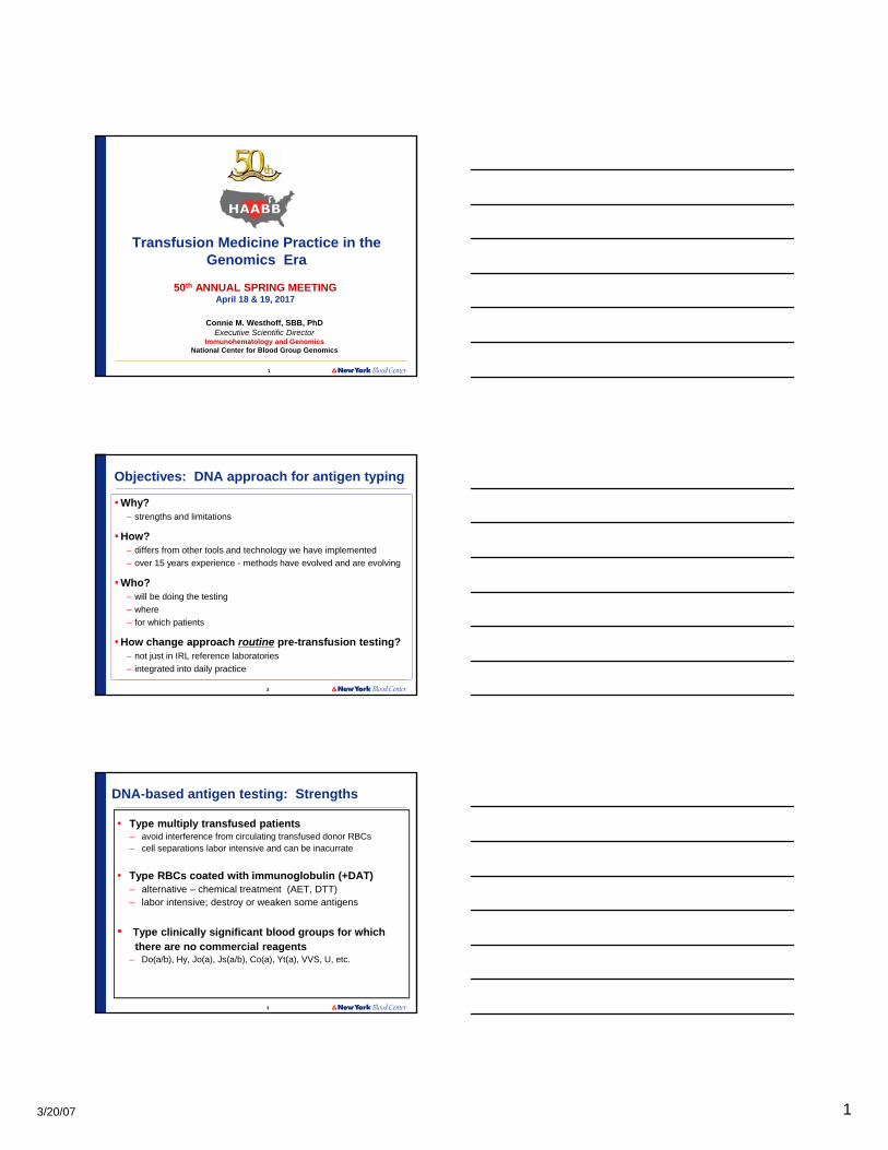
Transfusion Medicine Practice in the Genomics Era
50th ANNUAL SPRING MEETINGApril 18 & 19, 2017
Connie M. Westhoff, SBB, PhDExecutive Scientific Director
Immunohematology and GenomicsNational Center for Blood Group Genomics
2
Objectives: DNA approach for antigen typing
• Why?– strengths and limitations
• How? – differs from other tools and technology we have implemented
– over 15 years experience - methods have evolved and are evolving
• Who?– will be doing the testing
– where
– for which patients
• How change approach routine pre-transfusion testing?– not just in IRL reference laboratories
– integrated into daily practice
3
DNA-based antigen testing: Strengths
• Type multiply transfused patients – avoid interference from circulating transfused donor RBCs– cell separations labor intensive and can be inacurrate
• Type RBCs coated with immunoglobulin (+DAT)– alternative – chemical treatment (AET, DTT)– labor intensive; destroy or weaken some antigens
• Type clinically significant blood groups for whichthere are no commercial reagents
– Do(a/b), Hy, Jo(a), Js(a/b), Co(a), Yt(a), VVS, U, etc.
3/20/07 2
4
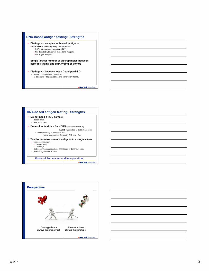
• Distinguish samples with weak antigens– FYX allele – 1-2% frequency in Caucasians
• RBCs have weak expression of Fyb
• Not detected with current monoclonal reagents
• RBCs type as Fy(b-)
Single largest number of discrepancies between serology typing and DNA typing of donors
• Distinguish between weak D and partial D– typing of females and OB women – to determine RhIg candidates and transfusion therapy
DNA-based antigen testing: Strengths
5
• Do not need a RBC sample – buccal swab – fetal amniocytes
• Determine fetal risk for HDFN (antibodies to RBCs)
NAIT (antibodies to platelet antigens)
- Paternal testing to determine risk- gene copy number (zygosity: RhD and HPA)
• Test for numerous minor antigens in a single assay– improved accuracy
• antigen typing• antibody ID
– find uncommon combinations of antigens in donor inventory– provide higher level of care
Power of Automation and Interpretation
DNA-based antigen testing: Strengths
6
Perspective
Genotype is notalways the phenotype!
Phenotype is notalways the genotype!

3/20/07 3
7
Perspective – Kell System Example Genotype is not
always the phenotype! Phenotype is not
always the genotype!
38 year old pregnant female Anti-K; titer 512
Test paternal sample to predict fetal risk
RBC phenotype: K+k–All children will be K+ and at risk
KEL genotype: Predict KEL*01/*02 K+k+
50% chance not at risk
48 year old female plasma reactivity – all cells +
DTT treated – non-reactive
RBC phenotype: K– k– Kp(b–)K0 null cell – non-reactive
Antibody to high in Kell SystemRare-Uncommon
KEL genotype: Predict K– k+, Kp(a–b+), Js(a–b+)
8
Kell null alleles (KEL*02N)
• Inability to distinguish silenced expression considered a limitation of genotyping (false positive)
• Ability to detect the presence of two alleles (K and k), even though one is silence in a paternal sample enables the accurate prediction of risk for HDFN
• K0 phenotype is very rare but chance of carrying one null (or mod) allele is higher
– European studies: 3.5 % - 7.5% of K+k– had one KEL*02Nnull silenced or mod allele
1 2 3 4 5 6 7 8 9 10 11 12 13 1415 16 17 18 19
IVS8+1g/t
578T/C 841C/C 1790T/TK+k+ Kp(a–b+) Js(a–b+)
9
DNA-based testing: Limitations
•“prediction” of presence of antigen on RBCs– silenced alleles (false positive)
• only testing region of gene that encodes the antigen• a mutation can turn “off” expression in region you are not testing
– cannot do routine ABO and RhD typing• Group O - is a silenced A or B gene • D negative - is a silenced RHD gene
• Laboratory testing environment and methods
• Test turnaround time– 5-8 hours
How to “balance” limitations
3/20/07 4
10
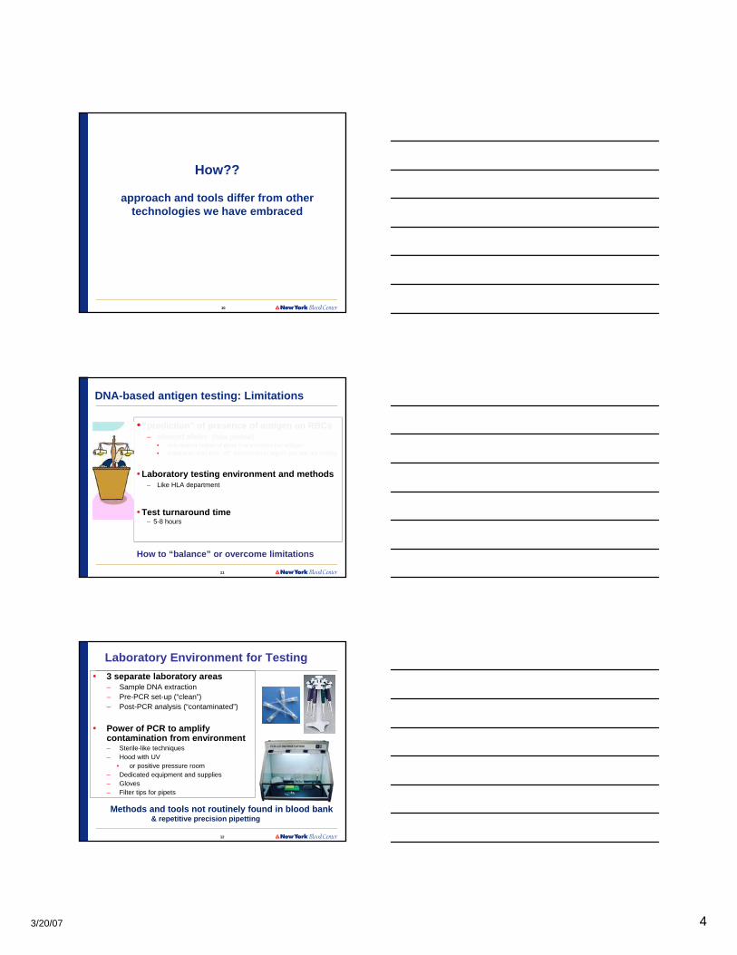
How??
approach and tools differ from othertechnologies we have embraced
11
DNA-based antigen testing: Limitations
•“prediction” of presence of antigen on RBCs– silenced alleles (false positive)
• only testing region of gene that encodes the antigen• a mutation ican turn “off” expression in region you are not testing
• Laboratory testing environment and methods– Like HLA department
• Test turnaround time– 5-8 hours
How to “balance” or overcome limitations
12
Laboratory Environment for Testing
• 3 separate laboratory areas – Sample DNA extraction– Pre-PCR set-up (“clean”)– Post-PCR analysis (“contaminated”)
• Power of PCR to amplify contamination from environment – Sterile-like techniques– Hood with UV
• or positive pressure room– Dedicated equipment and supplies– Gloves– Filter tips for pipets
Methods and tools not routinely found in blood bank& repetitive precision pipetting
3/20/07 5
13
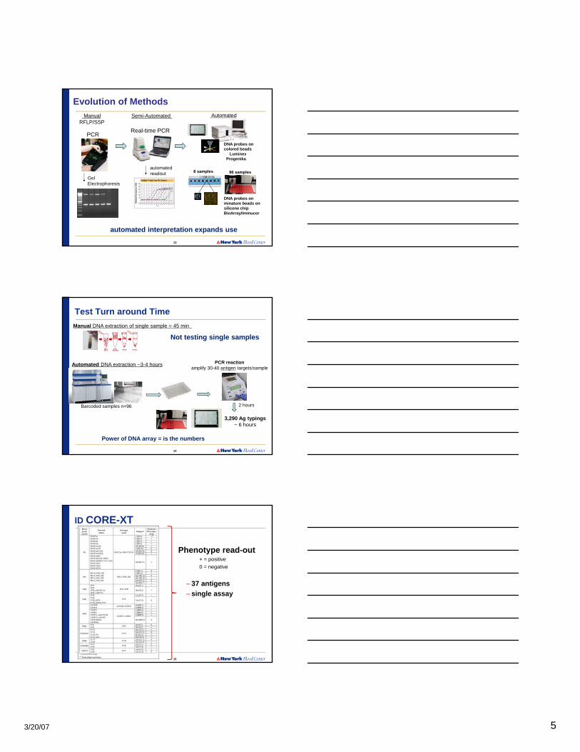
Evolution of Methods
Real-time PCR
automatedreadout
Semi-Automated ManualRFLP/SSP
PCR
GelElectrophoresis
DNA probes oncolored beads
LuminexProgenika
Automated
automated interpretation expands use
DNA probes onminature beads on silicone chipBioArray/Immucor
8 samples 96 samples
14
3,290 Ag typings~ 6 hours
Manual DNA extraction of single sample = 45 min
Test Turn around Time
Barcoded samples n=96
Automated DNA extraction ~3-4 hours
Not testing single samples
Power of DNA array = is the numbers
2 hours
PCR reactionamplify 30-40 antigen targets/sample
15
ID CORE-XT
Phenotype read-out+ = positive
0 = negative
– 37 antigens
– single assay

3/20/07 6
16
V/VS
Phenotype read-out+ = positive
0 = negative
• 34 antigens
• one tube assay
17
How to Integrate Tool into Routine Practice?
- environment needed
- economy in scale (numbers) of tests
- turn around time
- frequency of testing
18
Donor
Donor Center
Hospital Transfusion Service
Patient
Integrating DNA-based Technology into Routine Patient Care
one time testing
2X testing
3/20/07 7
19
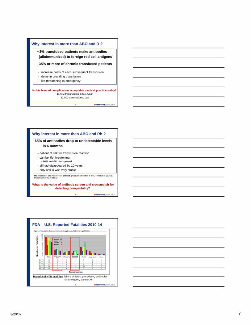
~3% transfused patients make antibodies
(alloimmunized) to foreign red cell antigens
35% or more of chronic transfused patients
- increase costs of each subsequent transfusion
- delay in providing transfusion
- life-threatening in emergency
11.6 M transfusions in U.S./year
32,000 transfusions / day
Is this level of complication acceptable medical practice today?
Why interest in more than ABO and D ?
20
65% of antibodies drop to undetectable levels
in 6 months
– patient at risk for transfusion reaction
– can be life-threatening• 90% anti-Jka disappeared
– all had disappeared by 10 years
– only anti-D was very stable
The persistence and evanescence of blood group alloantibodies in men. Tormey CA, Stack G. Transfusion 2009, 49:505-12
What is the value of antibody screen and crossmatch for detecting compatibility?
Why interest in more than ABO and Rh ?
21
FDA – U.S. Reported Fatalities 2010-14
Majority of HTR fatalities: failure to detect pre-existing antibodies or emergency transfusion
• 2005 = 16
• 2006 = 9
• 2008 = 7

3/20/07 8
22
Laboratory Pre-transfusion Testing Routine: ABO, RhD type and antibody screen
2015 -
2012 -
Approach has not changed in >60 years
2016: 36 Blood group systems (352 antigens)Cover Transfusion, Reid et al.
23
Prevention of AlloimmunizationThe most common antibody specificities: C,E, c, K
Germanymajority get CcEe & K
matched
UKFemales get K-
AustraliaFemales and children K-
NetherlandsFemales <45yr c E K
SCD/Thal CEK, Fya, Jkb, Ss
FinlandFemales CcEe & K
matched
SwitzerlandFemales CcEe & K
matchedChronic transfusion
or alloantibody match for all common
U.S.CEK matching
for SCDNot universal
24
Higher Level of Patient Care
• Blood transfusions have declined significantly over the last five years
– advances in surgical techniques
– patient blood management (PBM) programs
• Lower hgb threshold for patients (7.0 gm/dl) and limited transfusion
– Optimal RBC survival more important than ever
•Health Care Landscape– focus on outcomes – improved patient care
– personalized medicine with Genomics
3/20/07 9
25
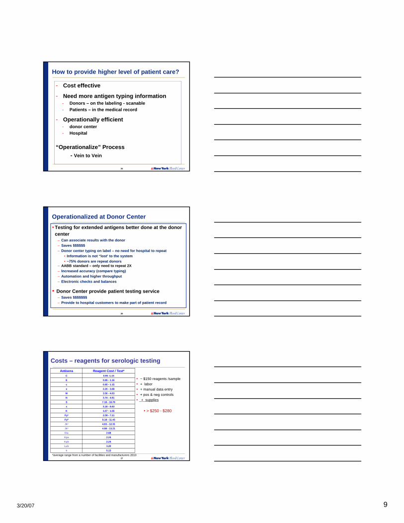
How to provide higher level of patient care?
- Cost effective
- Need more antigen typing information- Donors – on the labeling - scanable
- Patients – in the medical record
- Operationally efficient - donor center
- Hospital
“Operationalize” Process
- Vein to Vein
26
Operationalized at Donor Center
• Testing for extended antigens better done at the donor center
– Can associate results with the donor
– Saves $$$$$$
– Donor center typing on label – no need for hospital to repeat
• Information is not “lost’ to the system
• ~75% donors are repeat donors– AABB standard – only need to repeat 2X
– Increased accuracy (compare typing)
– Automation and higher throughput
– Electronic checks and balances
• Donor Center provide patient testing service– Saves $$$$$$$
– Provide to hospital customers to make part of patient record
27
Costs – reagents for serologic testing
Antisera Reagent Cost / Test*
C 0.95 -1.15
E 0.95 - 1.15
c 0.95 - 1.15
e 2.20 - 3.68
M 3.56 - 4.63
N 3.74 - 4.91
S 7.18 - 18.79
s 3.18 - 8.63
K 1.07 - 1.55
Fya 2.58 - 7.11
Fyb 9.18 - 11.43
Jka 4.63 - 12.31
Jkb 4.98 - 13.21
Dia 2.68
Kpa 2.24
Kpb 2.24
Lub 3.20
k 5.13
• ~ $150 reagents /sample
• + labor
• + manual data entry
• + pos & neg controls
• + supplies
• > $250 - $280
*average range from a number of facilities and manufacturers 2010

3/20/07 10
28
Currently
• CHALLENGE - providing extended antigen typed units• Labor intensive
• Manual activity
• Performed in IRL reference laboratory – highly trained staff
• Testing repeated each time
• Inability to scale up • No serologic reagents for some clinically significant antigens
• Consequently at many donor centers
- extended antigen typing primarily only minority donors
• Ultimate Goal - for patient care and inventory management– antigen information on all units
29
How would more information (antigen profile on patient) change approach
routine pre-transfusion testing?
30
Pre-transfusion Testing
- Every sample is “Black Box”
- Potentially has any of 300+ antibody specificities
- How would we design testing if – know what patient is at risk to make (antigen-negative)
• reduces number of possible specificities by 50%
– know what patient was exposed to
• all part of electronic medical record
– change importance of antibody screen?
3/20/07 11
31
Challenges for Current technology• Limited number of markers
– Cannot cover all antigens of potential interest
• common polymorphisms only
• cannot detect all silenced (null) alleles
• accurate ABO & D typing will require sampling entire gene
– Cannot determine “phasing” or “linkage”• which alleles are changes carried on?
• Population Diversity/Admixture– new variants
• Tests “hard-wired” if taken to FDA licensure– no pathway to add addition markers to existing design
32
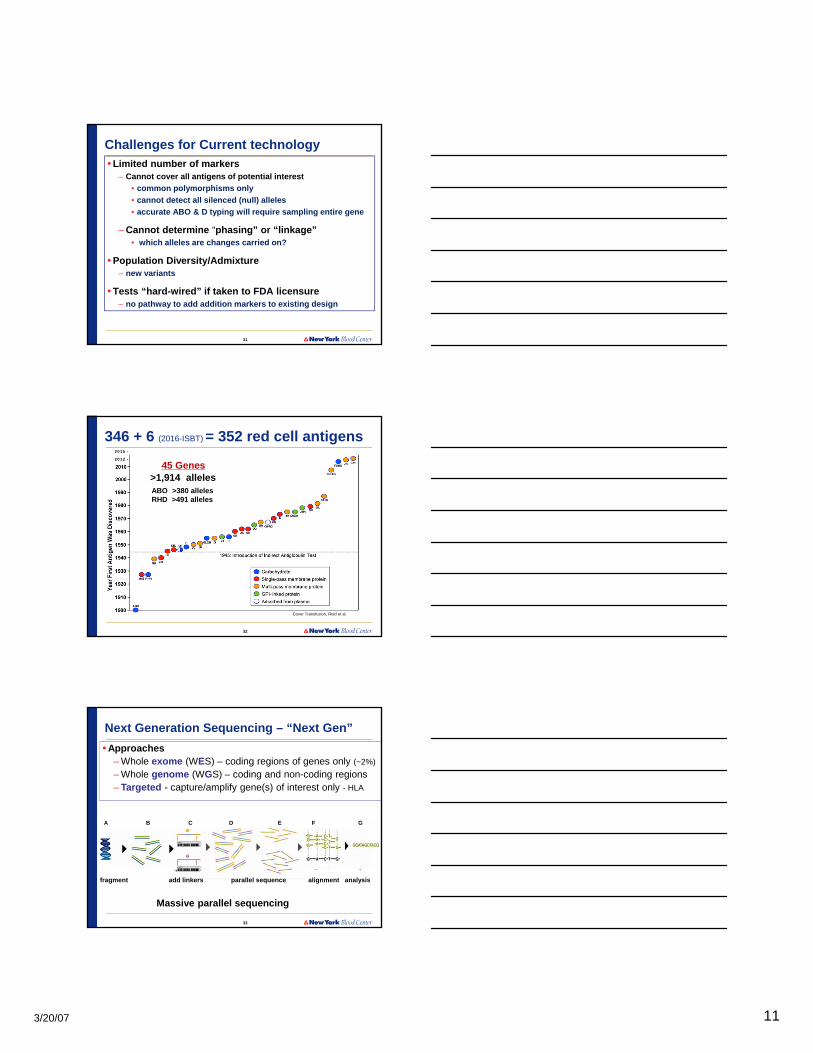
346 + 6 (2016-ISBT) = 352 red cell antigens2015 -
2012 -
45 Genes>1,914 alleles
Cover Transfusion, Reid et al.
ABO >380 allelesRHD >491 alleles
33
A B C D E F G
fragment add linkers parallel sequence alignment analysis
Next Generation Sequencing – “Next Gen”
• Approaches– Whole exome (WES) – coding regions of genes only (~2%)
– Whole genome (WGS) – coding and non-coding regions– Targeted - capture/amplify gene(s) of interest only - HLA
Massive parallel sequencing

3/20/07 12
34
Next Generation Sequencing Platforms
Roche 454 Life Technologies illumina HiSeq 2000 Pacific Biosciences RS
Nature 470:198, 2011
Ion Trrent PGM
35
Genomics Revolution
•“Sequence Once; Read Often”
• Whole genome sequence data will be available on our patients
• Especially for patients with chronic disease
• We will only need to “read” the information
36
Ilumina HiSeq - 30X coverage
45 RBC genes-346 antigens
6 platelet genes-33 antigens
Comprehensive red cell and platelet antigen prediction from whole genome sequencing: proof of principal Transfusion. 56(3):743-54, 2016 Lane WJ, Westhoff CM, Uy JM, Aguad M, Smeland-Wagman R, Kaufman RM, Rehm HL, Green RC, Silberstein LE
3/20/07 13
37
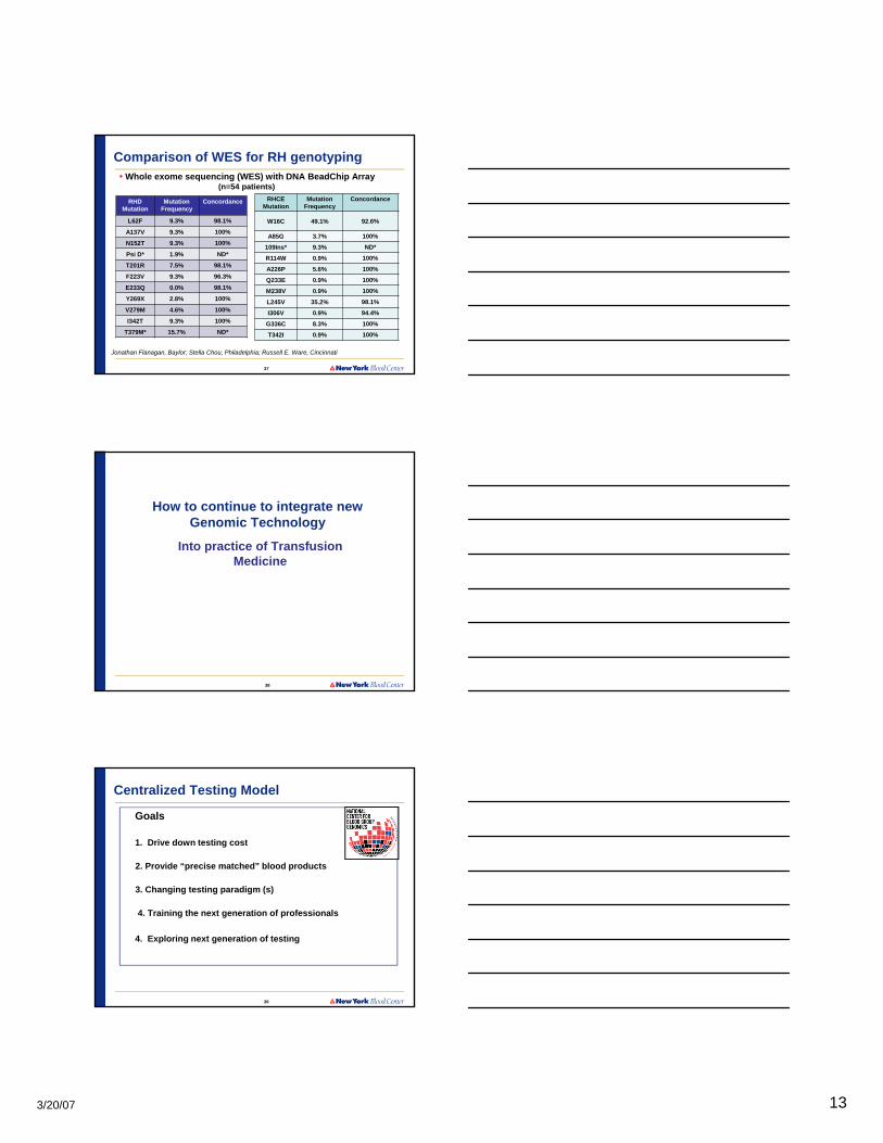
Comparison of WES for RH genotyping• Whole exome sequencing (WES) with DNA BeadChip Array
Jonathan Flanagan, Baylor; Stella Chou, Philadelphia; Russell E. Ware, Cincinnati
(n=54 patients)
RHD Mutation
Mutation Frequency
Concordance
L62F 9.3% 98.1%
A137V 9.3% 100%
N152T 9.3% 100%
Psi D* 1.9% ND*
T201R 7.5% 98.1%
F223V 9.3% 96.3%
E233Q 0.0% 98.1%
Y269X 2.8% 100%
V279M 4.6% 100%
I342T 9.3% 100%
T379M* 15.7% ND*
RHCEMutation
Mutation Frequency
Concordance
W16C 49.1% 92.6%
A85G 3.7% 100%
109Ins* 9.3% ND*
R114W 0.9% 100%
A226P 5.6% 100%
Q233E 0.9% 100%
M238V 0.9% 100%
L245V 35.2% 98.1%
I306V 0.9% 94.4%
G336C 8.3% 100%
T342I 0.9% 100%
38
How to continue to integrate new Genomic Technology
Into practice of Transfusion Medicine
39
Centralized Testing Model
Goals
1. Drive down testing cost
2. Provide “precise matched” blood products
3. Changing testing paradigm (s)
4. Training the next generation of professionals
4. Exploring next generation of testing
3/20/07 14
40
Thank You !!
New York Blood Center Community Blood Center of Kansas City NYBC CBC
Top Related