


Languages
Pages
Legal

The ‘spring’ ligament and posterior tibial tendon
function and dysfunctionMichael Ratcliffe
Sales Training Manager/Podiatrist
Cuxson Gerrard & Co Ltd.
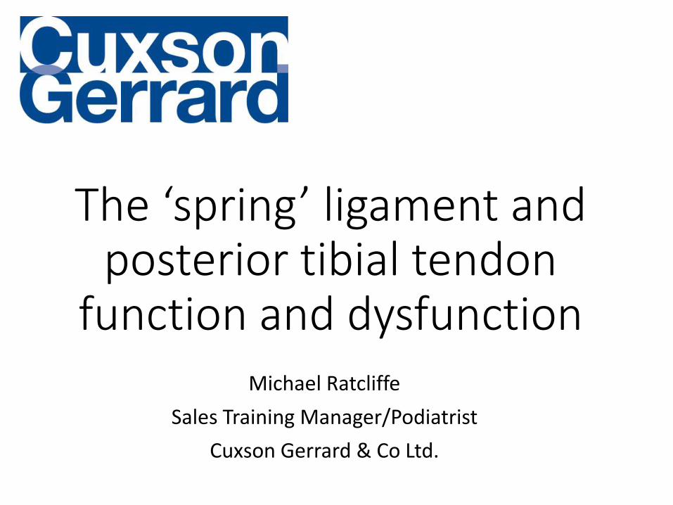
Calcaneonavicular (Spring) – Ligament Location, bony landmarks
Plantar, medial aspect of the Talar Head
Tuberosity of the Navicular
Sustumtaculum Tali of the Calcaneus
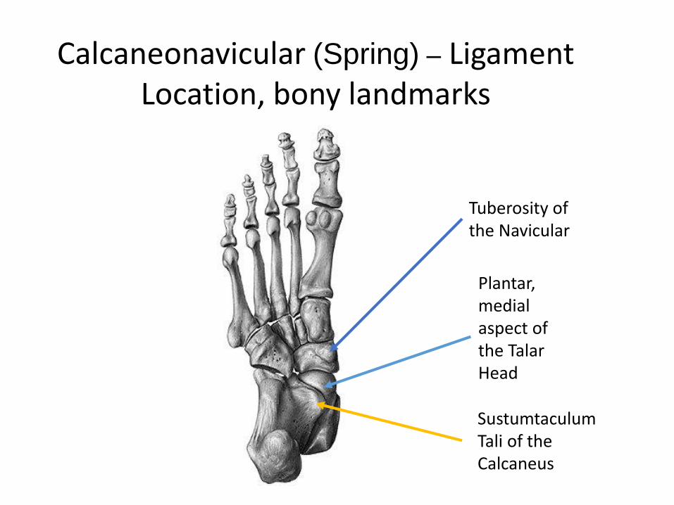
The Spring Ligament ‘covers’ the medial and plantar articular surface of the Talar Head by ‘bridging’ the gap between the posterior/plantar navicular and the anterior calcaneus
Location
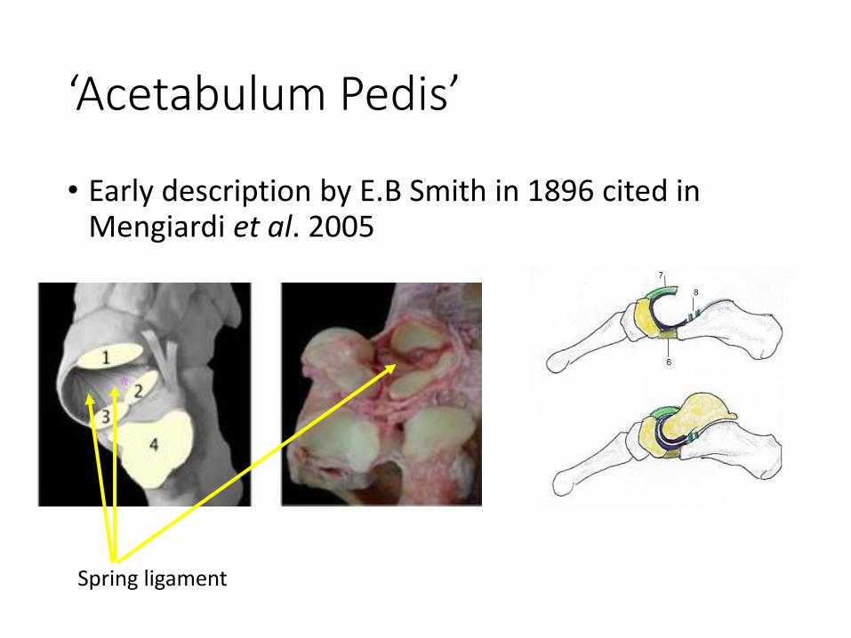
‘Acetabulum Pedis’
• Early description by E.B Smith in 1896 cited in Mengiardi et al. 2005
Spring ligament
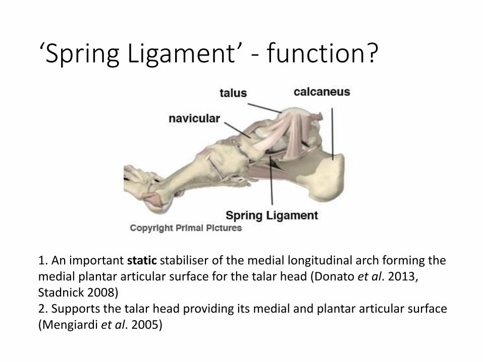
‘Spring Ligament’ - function?
1. An important static stabiliser of the medial longitudinal arch forming the medial plantar articular surface for the talar head (Donato et al. 2013, Stadnick 2008)2. Supports the talar head providing its medial and plantar articular surface (Mengiardi et al. 2005)
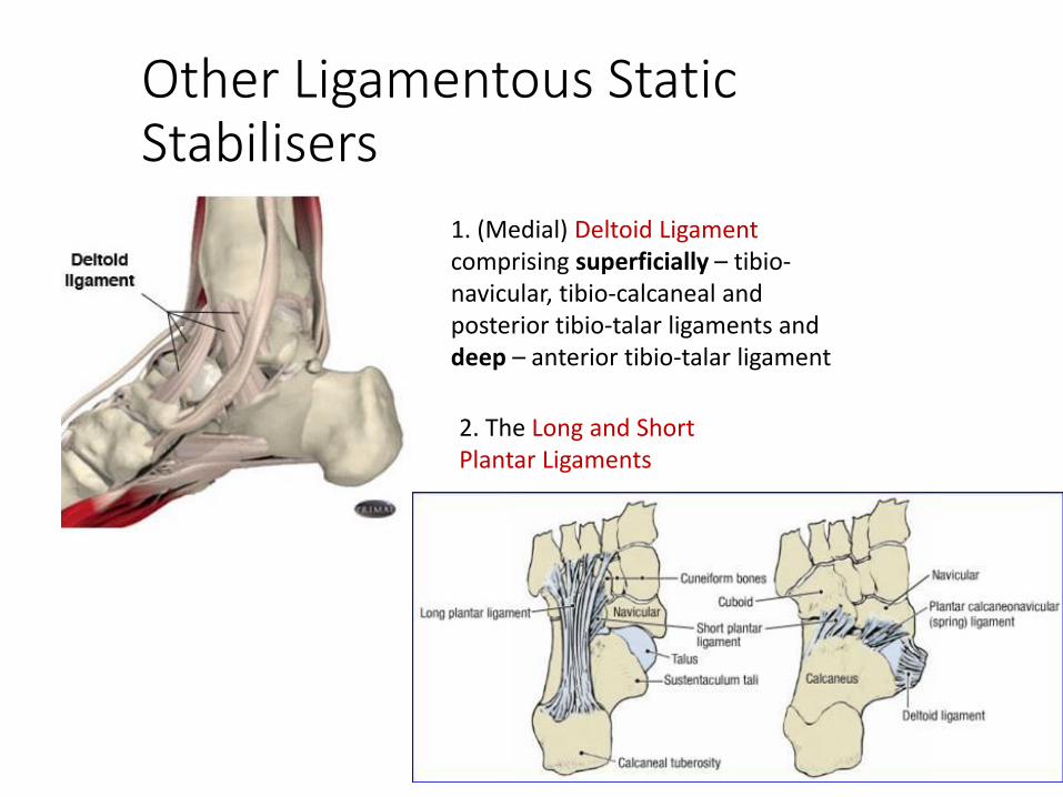
Other Ligamentous Static Stabilisers
1. (Medial) Deltoid Ligament comprising superficially – tibio-navicular, tibio-calcaneal and posterior tibio-talar ligaments and deep – anterior tibio-talar ligament
2. The Long and Short Plantar Ligaments
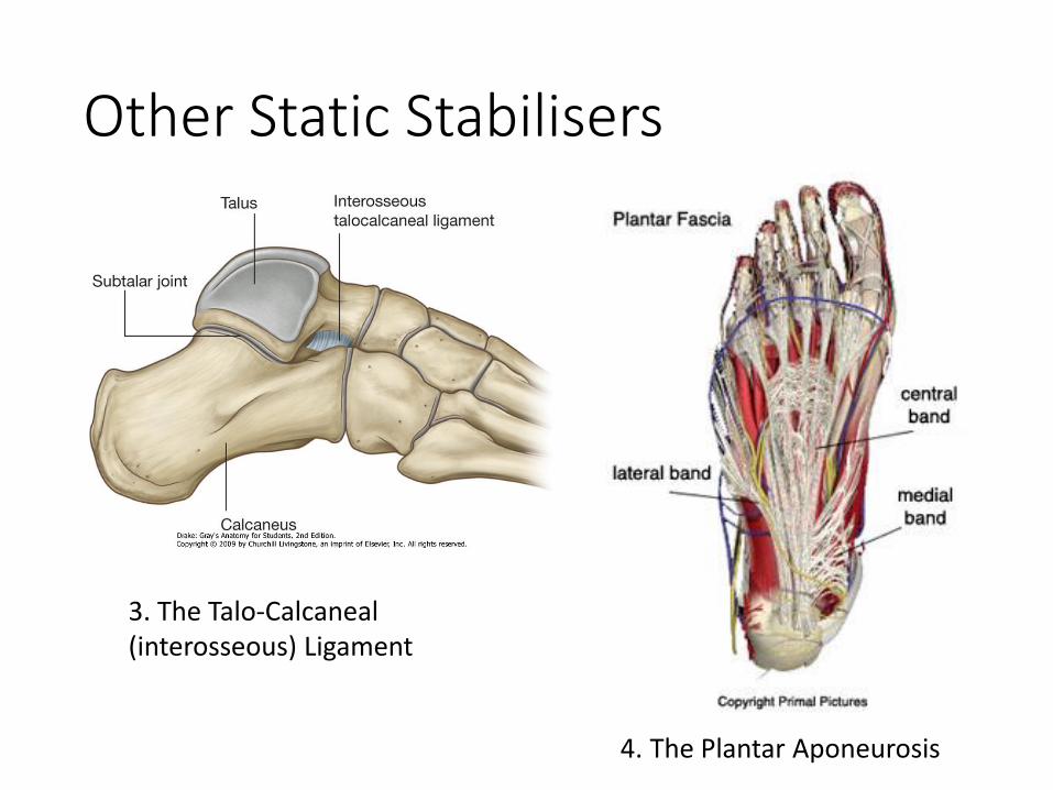
Other Static Stabilisers
3. The Talo-Calcaneal (interosseous) Ligament
4. The Plantar Aponeurosis
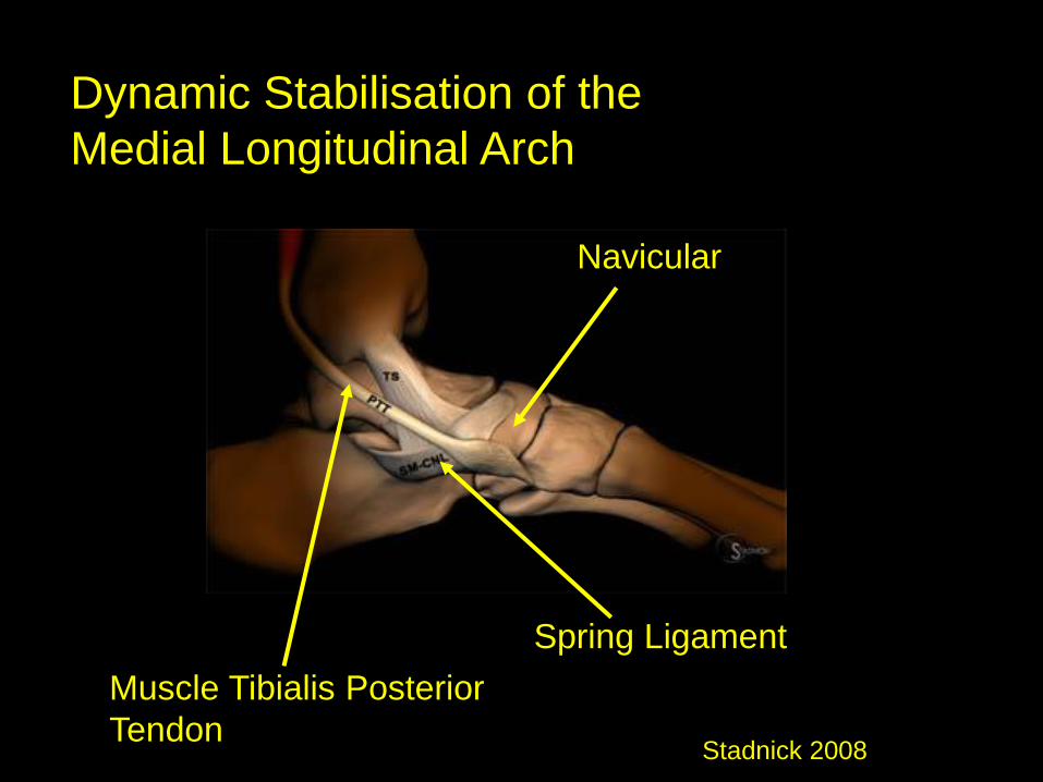
Dynamic Stabilisation of the
Medial Longitudinal Arch
Muscle Tibialis Posterior
Tendon
Spring Ligament
Navicular
Stadnick 2008
Anatomy - Spring Ligament
A multi-ligament complex comprising 3 ligaments
• Superomedial ligament
• Medioplantar oblique ligament
• Inferoplantar longitudinal ligament
Comprises Type 1 collagen which confers the toughness through anisotropy (Sherman et al. 2015)
BUT has been found not to display elasticity or contain any elastic fibres (Davis et al. 1996) unlike ligaments which are generally viscoelastic, so the label ‘spring’ may be a misnomer (Mengiardi et al. 2005)
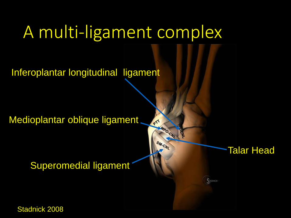
A multi-ligament complex
Superomedial ligament
Medioplantar oblique ligament
Inferoplantar longitudinal ligament
Talar Head
Stadnick 2008
23/05/2018
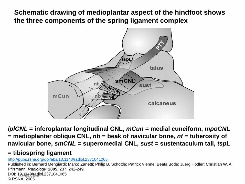
iplCNL = inferoplantar longitudinal CNL, mCun = medial cuneiform, mpoCNL
= medioplantar oblique CNL, nb = beak of navicular bone, nt = tuberosity of
navicular bone, smCNL = superomedial CNL, sust = sustentaculum tali, tspL
= tibiospring ligamenthttp://pubs.rsna.org/doi/abs/10.1148/radiol.2371041065
Published in: Bernard Mengiardi; Marco Zanetti; Philip B. Schöttle; Patrick Vienne; Beata Bode; Juerg Hodler; Christian W. A.
Pfirrmann; Radiology 2005, 237, 242-249.
DOI: 10.1148/radiol.2371041065
© RSNA, 2005
Schematic drawing of medioplantar aspect of the hindfoot shows
the three components of the spring ligament complex
23/05/2018
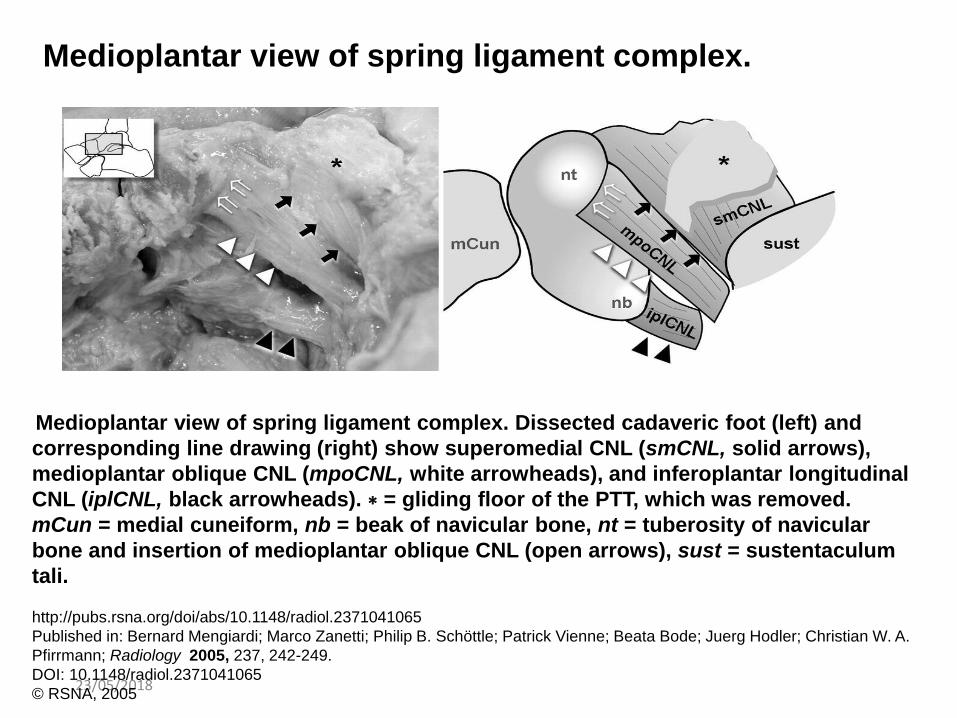
Medioplantar view of spring ligament complex. Dissected cadaveric foot (left) and
corresponding line drawing (right) show superomedial CNL (smCNL, solid arrows),
medioplantar oblique CNL (mpoCNL, white arrowheads), and inferoplantar longitudinal
CNL (iplCNL, black arrowheads). ∗ = gliding floor of the PTT, which was removed.
mCun = medial cuneiform, nb = beak of navicular bone, nt = tuberosity of navicular
bone and insertion of medioplantar oblique CNL (open arrows), sust = sustentaculum
tali.
http://pubs.rsna.org/doi/abs/10.1148/radiol.2371041065
Published in: Bernard Mengiardi; Marco Zanetti; Philip B. Schöttle; Patrick Vienne; Beata Bode; Juerg Hodler; Christian W. A.
Pfirrmann; Radiology 2005, 237, 242-249.
DOI: 10.1148/radiol.2371041065
© RSNA, 2005
Medioplantar view of spring ligament complex.
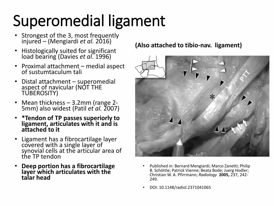
Superomedial ligament• Strongest of the 3, most frequently
injured – (Mengiardi et al. 2016)
• Histologically suited for significant load bearing (Davies et al. 1996)
• Proximal attachment – medial aspect of sustumtaculum tali
• Distal attachment – superomedialaspect of navicular (NOT THE TUBEROSITY)
• Mean thickness – 3.2mm (range 2-5mm) also widest (Patil et al. 2007)
• *Tendon of TP passes superiorly to ligament, articulates with it and is attached to it
• Ligament has a fibrocartilage layer covered with a single layer of synovial cells at the articular area of the TP tendon
• Deep portion has a fibrocartilage layer which articulates with the talar head
• Published in: Bernard Mengiardi; Marco Zanetti; Philip B. Schöttle; Patrick Vienne; Beata Bode; Juerg Hodler; Christian W. A. Pfirrmann; Radiology 2005, 237, 242-249.
• DOI: 10.1148/radiol.2371041065
(Also attached to tibio-nav. ligament)
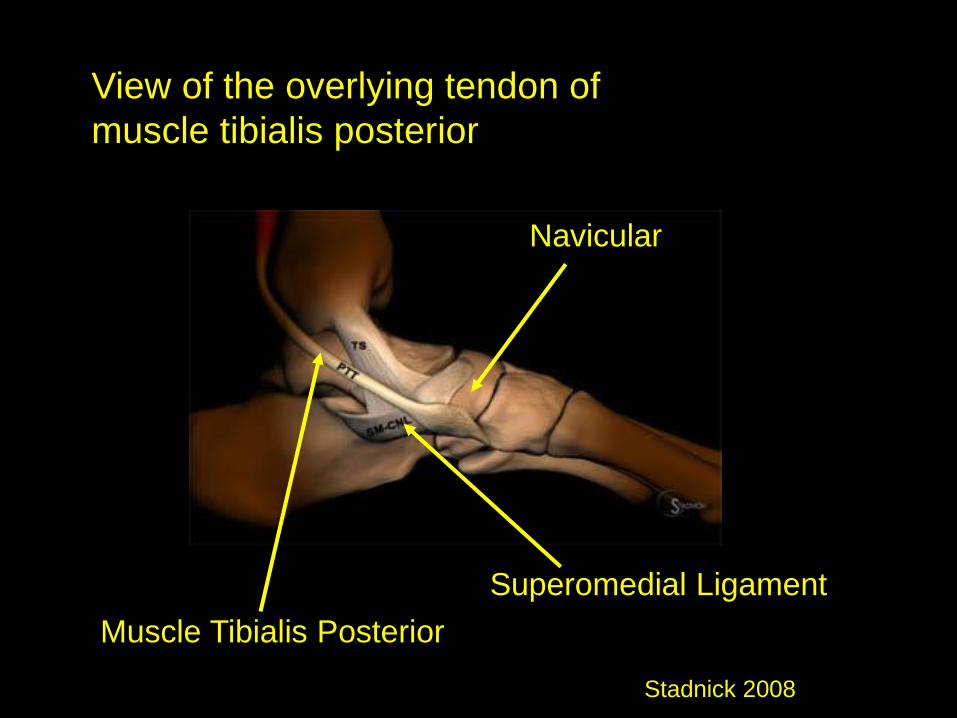
Muscle Tibialis Posterior
Superomedial Ligament
Navicular
Stadnick 2008
View of the overlying tendon of
muscle tibialis posterior
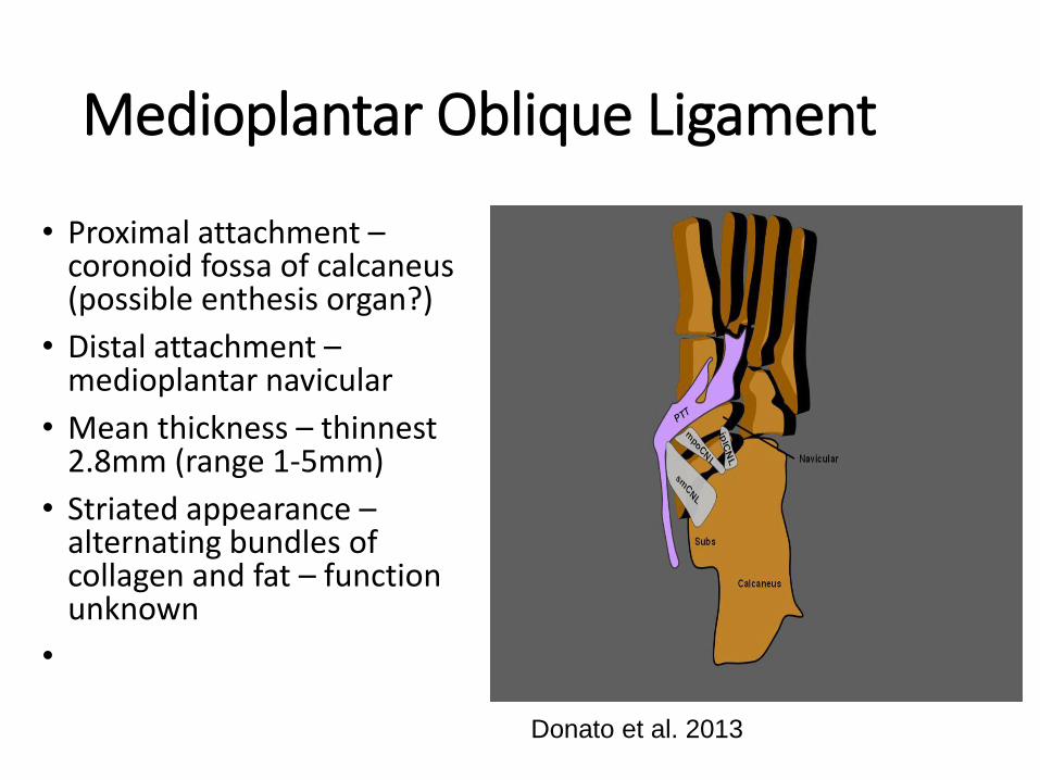
Medioplantar Oblique Ligament
• Proximal attachment –coronoid fossa of calcaneus (possible enthesis organ?)
• Distal attachment –medioplantar navicular
• Mean thickness – thinnest 2.8mm (range 1-5mm)
• Striated appearance –alternating bundles of collagen and fat – function unknown
•
Donato et al. 2013
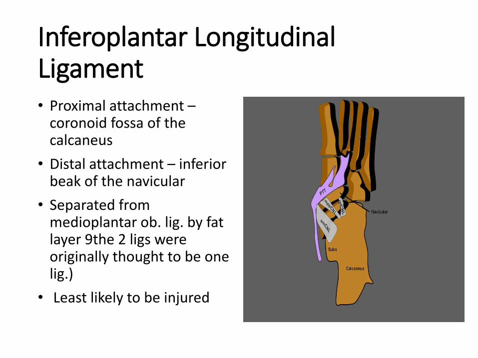
Inferoplantar Longitudinal Ligament• Proximal attachment –
coronoid fossa of the calcaneus
• Distal attachment – inferior beak of the navicular
• Separated from medioplantar ob. lig. by fat layer 9the 2 ligs were originally thought to be one lig.)
• Least likely to be injured
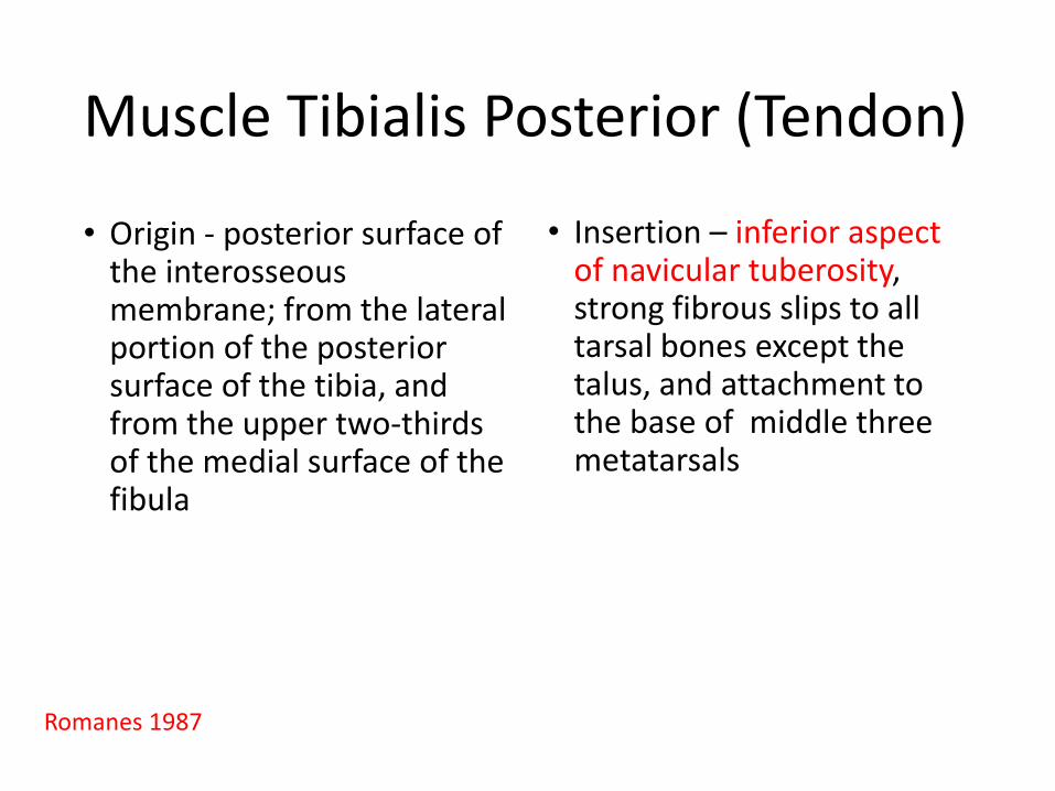
Muscle Tibialis Posterior (Tendon)
• Origin - posterior surface of the interosseous membrane; from the lateral portion of the posterior surface of the tibia, and from the upper two-thirds of the medial surface of the fibula
• Insertion – inferior aspect of navicular tuberosity, strong fibrous slips to all tarsal bones except the talus, and attachment to the base of middle three metatarsals
Romanes 1987
15
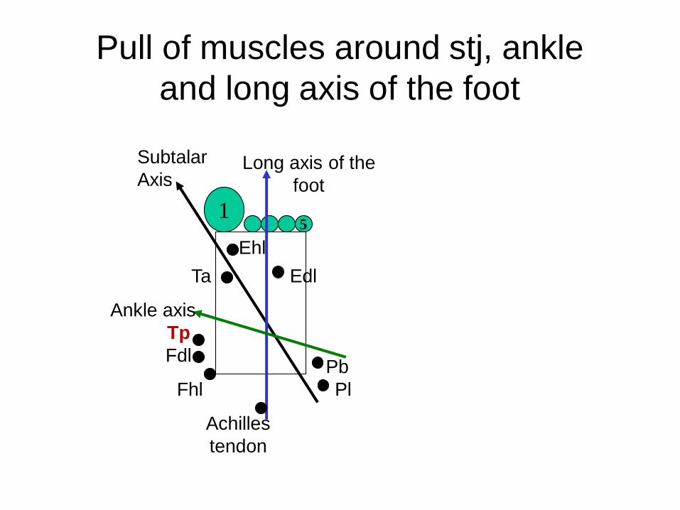
Subtalar
AxisLong axis of the
foot
Ankle axis
Pb
Pl
Achilles
tendon
Fhl
Fdl
Tp
EdlTa
Ehl
Pull of muscles around stj, ankle
and long axis of the foot
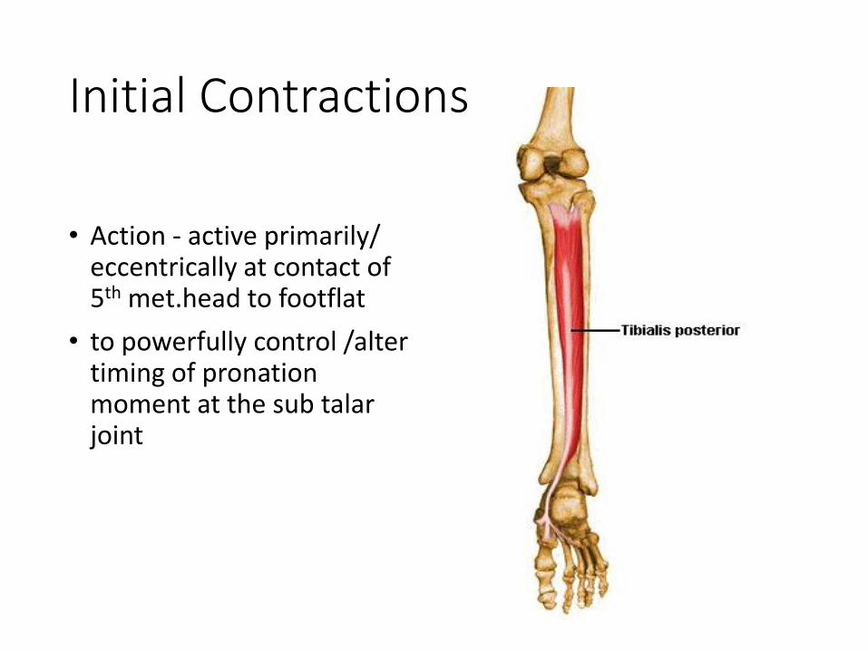
Initial Contractions
• Action - active primarily/ eccentrically at contact of 5th met.head to footflat
• to powerfully control /alter timing of pronation moment at the sub talar joint
Tarsal Cross Bracing
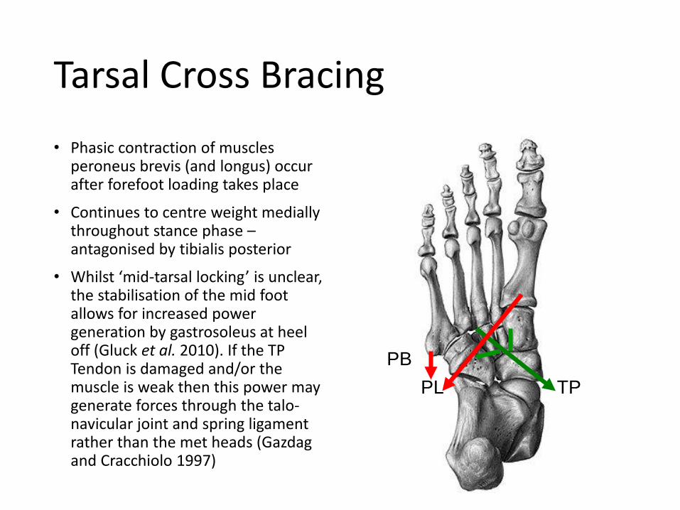
• Phasic contraction of muscles peroneus brevis (and longus) occur after forefoot loading takes place
• Continues to centre weight medially throughout stance phase –antagonised by tibialis posterior
• Whilst ‘mid-tarsal locking’ is unclear, the stabilisation of the mid foot allows for increased power generation by gastrosoleus at heel off (Gluck et al. 2010). If the TP Tendon is damaged and/or the muscle is weak then this power may generate forces through the talo-navicular joint and spring ligament rather than the met heads (Gazdag and Cracchiolo 1997)
PL TP
PB
Tibialis Posterior
• Resisting expansion at the distal tib./fib. joint (fibrous, syndesmotic joint, occasionally synovial) assists in providing ankle joint stability during plantarflexion.
• Allows movement (via quiescence) of the fibula superiorly during ankle joint dorsiflexion
Relationship between the spring ligament and the tendon of muscle tibialis posterior dysfunction• Balen and Helms (2001) report a superomedial ligament thickness of
above 5mm to be present in cases of tibialis posterior (TP) tendon dysfunction
• Mengiardi et al. (2005) found that this is nearer 4mm and also identified spring ligament injuries having a high association with TP tendon tears
• Jennings and Christensen (2008) spring ligament is a major stabiliser of the Medial Longitudinal Arch at mid-stance and the tendon of tibialis posterior cannot fully accommodate any insufficiency in it
• Orr and Nunley (2013) cautioned against rare cases where the TP tendon was normal but adult acquired flat foot existed – here spring ligament injury should be suspected
• Williams et al. (2014) identifies a strong association between spring ligament deformity (unspecified) and radiographic plano-valgus foot position
• Mengiardi et al. (2016) report that in most cases, spring ligament lesions are secondary to the tendon of tibialis posterior dysfunction
Clinical Symptoms of Spring Ligament damage• Clinical symptoms - similar to stages of posterior tibial
dysfunction. Usually poor heel lift test and everted heel position but powerful TP resisted concentric contraction
• Acute stage - vague, activity-related pain at medial ankle and foot with too many toes sign on standing
• Difficulty with balance and walking on uneven ground.
• Chronic - (as pes planovalgus deformity progresses) -activity related pain in the sinus tarsi at the lateral malleolus,
• Resistant to treatment interventions for TP tendon dysfunction
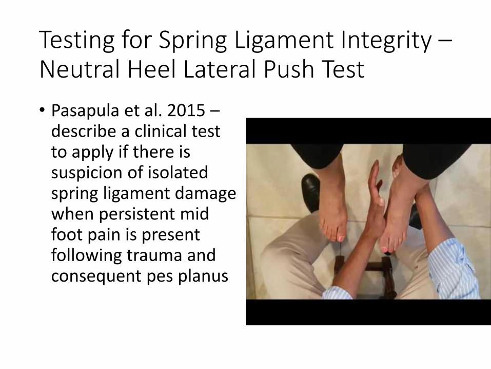
Testing for Spring Ligament Integrity –Neutral Heel Lateral Push Test
• Pasapula et al. 2015 –describe a clinical test to apply if there is suspicion of isolated spring ligament damage when persistent mid foot pain is present following trauma and consequent pes planus
Stages
• Stage 0 – Spring ligament failure/laxity/give but no tendonopathy or planovalgus
• Stage 1 – Spring ligament give with tendonopathy but normal tendon length and no deformity
• Stage 2 – Spring ligament failure with tendon lengthening and flexible planovalgus deformity
• Stage 3 – Spring ligament failure with tendon lengthening and fixed planovalgus deformity
Top Related