


Languages
Pages
Legal


Korean J Pain 2011 March; Vol. 24, No. 1: 36-43pISSN 2005-9159 eISSN 2093-0569DOI:10.3344/kjp.2011.24.1.36
| Original Article |
The Results of Cervical Nucleoplasty in Patients with Cervical Disc Disorder: A Retrospective Clinical Study of 22 Patients
Department of Anesthesiology and Pain Medicine, Konkuk University Hospital, *Seoul National University Boramae Hospital, †Seoul National University Hospital, ‡Chung-Ang University Hospital, Seoul, Korea
Sung Eun Sim, MD*, Eun Sung Ko, MD, Duk Kyung Kim, MD, Hae Kyoung Kim, MD, Yong Chul Kim, MD†, and Hwa Yong Shin, MD‡
Background:
Nucleoplasty is a minimally invasive spinal surgery using a CoblationⓇ technique that creates small voids within the disc. The purpose of this study was to evaluate the efficacy of cervical nucleoplasty in patients with cervical disc disorder.
Methods:
Between March 2008 and December 2009, 22 patients with cervical disc disorders were treated with cervical nucleoplasty after failed conservative treatment. All procedures were performed under local anesthesia, and fluoroscopic guidance and voids were created in the disc with the PercTM DC Spine WandTM. Clinical outcomes were evaluated by the Modified Macnab criteria and VAS score at preprocedure, postprocedure 1 month, and 6 months.
Results:
Six patients had one, eight patients had two and eight patients had three discs treated; a total of 46 procedures was performed. Mean VAS reduced from 9.3 at preprocedure to 3.7 at postprocedure 1 month and to 3.4 at postprocedure 6 months. There was no significant complication related to the procedure within the first month. Outcomes were good or excellent in 17/22 (77.3%) cases. Postprocedure magnetic resonance imaging was acquired in two patients after two months showing morphologic evidence of volume reduction of protruded disc material in one patient but not in the other.
Conclusions:
Percutaneous decompression with a nucleoplasty using a CoblationⓇ technique in the treatment of cervical disc disorder is a safe, minimally-invasive and less uncomfortable procedure, with an excellent short-term clinical outcome. (Korean J Pain 2011; 24: 36-43)
Key Words:
cervical, disc, diskectomy, nucleoplasty, percutaneous.
Received October 28, 2010. Revised December 15, 2010. Accepted January 6, 2011.Correspondence to: Hwa Yong Shin, MDDepartment of Anesthesiology and Pain Medicine, Chung-Ang University Hospital, 224-1, Heukseok-dong, Dongjak-gu, Seoul 100-272, KoreaTel: +82-2-6299-2577, Fax: +82-2-6298-8351, E-mail: [email protected]
This is an open-access article distributed under the terms of the Creative Commons Attribution Non-Commercial License (http:// creativecommons.org/licenses/by-nc/3.0/), which permits unrestricted non-commercial use, distribution, and reproduction in any medium, provided the original work is properly cited.Copyright ⓒ The Korean Pain Society, 2011

SE Sim, et al / Nucleoplasty in Cervical Disc Disorder 37
INTRODUCTION
Clinically, cervical disc disorders frequently cause cer-
vical axial pain, radiculopathy, and myelopathy. This kind
of pain from the intervertebral disc is known to be caused
by mechanical compression from extruded disc material,
accompanying inflammatory response, and released
chemical mediators [1].
Generally, for this kind of cervical disc disorder, con-
servative treatment such as orthosis, cervical traction, and
medication are carried out first. However, if symptoms
persist or increase in severity after 6-8 weeks of sufficient
conservative treatment, then surgical treatment is con-
sidered. Although surgical treatment is relatively well es-
tablished and known to be highly successful, it has many
drawbacks such as damage to the adjacent tissue (bone,
muscle, nerves, and blood vessels), chronic loading to ad-
jacent discs which results in damage and transformation,
and a long postoperative recovery period [1]. To supplement
these drawbacks, MIST (Minimally Invasive Spinal Techniques)
have been developed and performed for several years [1,2].
Among these, nucleopasty was developed by Arthro-
Care Corporation in the United States, approved by the
U.S. Food and Drug Administration in 1999, and first per-
formed in July of 2000. At first, Nucleopasty was devel-
oped and used to treat contained lumbar disc herniation
and protrusion with associated symptoms [1,2]. Sharps and
Isaac [3] and Singh et al. [4] reported significant pain re-
duction for up to a year after percutaneous disc decom-
pression using lumbar nucleoplasty. However, compared to
other percutaneous decompression or even lumbar nucleo-
plasty, there is a relative lack of research on and clinical
experience with cervical nucleoplasty. Cervical nucleo-
plasty has only limited evidence (level IV) in the literature
concerning the technique like lumbar nucleoplasty, and in-
dications are limited to contained herniation and protrusion
similar to the lumbar area [1,2,5]. Therefore, the goal of
this study is to evaluate on the efficacy, side effects and
patient satisfaction with cervical nucleoplasty performed
on patients with cervical disc disorders that were un-
responsive to conservative treatments.
MATERIALS AND METHODS
1. Object of study
This study was conducted on 22 patients with cervical
disc disorders who were treated with cervical nucleoplasty
between March 2008 and December 2009 at our hospital.
The study was approved by the hospital’s medical research
ethics committee, and written informed consent was ob-
tained from all patients before the nucleoplasty.
Patients were treated with nucleoplasty after con-
firmation for the existence of cervical disc disorders re-
lated to the patients’ subjective symptoms on the basis of
systemic review, physical examination, X-ray studies and
magnetic resonance imaging (MRI) examination. Physical
examination including neurological examination before the
surgery was carried out by the same pain physician. There
were no neurological deficits, such as loss of sensory, mo-
tor or reflex in any of the patients, but they all complained
of just pain. All patients had a simple radiological study
and MRI examination done at least once to confirm the ex-
istence of cervical disc disorder. The mean time period
from onset of symptoms to the nucleoplasty was 25
months, and there had been no improvement despite con-
servative pain management including medication, physical
therapy, and root blocks. All nucleoplasty was conducted
by the same pain physician.
2. Cervical nucleoplasty procedure
Before entering the operating theater, 1,000 ml of
Hartman’s solution was connected after intravenous can-
nulation, and the patient received intravenous injection of
1.0 g of cefazolin as a prophylactic antibiotic 1 hour before
the procedure after confirmation of a negative skin test.
The patient was placed in supine position on the operation
table, and to facilitate access to the cervical disc, a thin
surgical roll was placed under the neck so that it was
slightly hyperextended. The patient received the vital sign
monitoring and oxygen supply at 5 L per minute via nasal
prong throughout the procedure. All procedures were per-
formed under local anesthesia, but midazolam 2-4 mg was
administered for sedation if there was a patient’s demand.
Betadine soap and betadine solution were used to disinfect
the anterior neck as well as surrounding skin, and steri-
lized drapes were applied on the operating area.
First, the C-arm was positioned in a lateral view of
the surgical field to measure the angle of the target disc
(endplate angle). Then, the C-arm was positioned in an
antero-posterior (AP) view to confirm the target disc and
rotated axially to align the endplate at measured angle in
the prior lateral projection. Before the procedure, we

38 Korean J Pain Vol. 24, No. 1, 2011
Fig. 1. Intraoperative fluoroscopic imaging. PercTM DC Spine WandTM placement adjacent to the lesion sites within the C5-6 intervertebral disc level during ablation. (A) Antero-posterior views. (B) Oblique views. (C) Lateral views.
measured the position and angle of the herniated part
from the center of the disc with MRI examination.
According to this measured angle, the C-arm was tilted
obliquely on the opposite side to the patient’s symptoms,
and the needle entry point was marked with a surgical
marker on the anterior neck relevant to the center of the
disc to be treated. The paratracheal approach was used
in the opposite site to the patient’s symptom. After the
internal carotid artery was laterally displaced with the sur-
geon’s index and middle fingers, the needle entry site was
secured. Then, the needle was introduced in the needle en-
try point and advanced till the anterolateral annulus
fibrosus. A syringe filled with 5 ml of 2% lidocaine was
connected to the needle, and the local anesthetic was ad-
ministered from the disc to the skin while checking that
blood was not aspirated with repeated suction and
injection. Next, we used the paratracheal approach again.
After the patient’s internal carotid artery was displaced
laterally and the needle puncture site was secured, a 19
gauge 3 inch Introducer needle (ArthroCare Co., Sunnyvale,
CA, USA) was introduced in the needle entry point and ad-
vanced until it reached the annulus fibrosus. At this point,
the AP and lateral views were gained to see if the in-
troducer needle tip was in contact with the center of the
disc height. Then, the oblique view was obtained to see
that the introducer needle was directed toward the center
of the intervertebral disc and to guide the estimated pro-
gression route. The introducer needle entry angle at the
anterolateral border of annulus fibrosus was then adjusted
if needed. After the introducer needle was advanced
slightly deeper, C-arm fluoroscope images were obtained
again to see whether the introducer needle was placed in
the center of the disc according to the AP and lateral
views. The stylet of the introducer needle was withdrawn,
and the PercTM DC Spine WandTM (ArthroCare co.,
Sunnyvale, CA, USA) was replaced and fastened clockwise
to the needle hub. In the fastened state, the PercTM DC
Spine WandTM and introducer needle were advanced slightly

SE Sim, et al / Nucleoplasty in Cervical Disc Disorder 39
Table 1. Demographic Data of Patients
Data
Age (yrs)Sex (M/F)Main symptom Axial pain only Axial pain < Radicular pain Axial pain = Radicular painPreoperative MRI findings Protrusion Extrusion StenosisLevels treated (1 level/2 levels/3 levels) C3-4 C4-5 C5-6 C6-7
47.8 ± 11.915/7
4 9 9
17 2 3
6/8/8 6121414
Data for age are expressed as mean ± SD.
Fig. 2. Pain intensity at the baseline and 1, 6 months of a follow-up period after the cervical nucleoplasty. Mean ±C.I (Confidence interval). P < 0.05.
more to the estimated lesion site. Here, it should not go
past the imaginary line beyond that connects the posterior
vertebral bodies in lateral view (Fig. 1). Next, the PercTM
DC Spine WandTM was connected to the ArthroCare system
2000Ⓡ (ArthroCare Co., Sunnyvale, CA, USA), and coagu-
lation was tested with the radio-frequency controller set
at 2′ for 1-2 seconds to check that there was no movement
or paresthesia in the patient’s upper limbs. CoblationⓇ was
then carried out to remove the nucleus pulposus. By rotat-
ing the flange 180o, we ablated the disc material - de-
pending on the size and hardness of the lesion - for 20-60
seconds with the radio-frequency controller set at 2′-3′ of intensity. After this, the wand was slightly retreated
under the C-arm fluoroscope guidance, and coagulation
proceeded with controller set at 3′. When it was verified
that there was no movement or paresthesia in the pa-
tient’s upper extremities, the flange was rotated 180o and
ablation was done for 20-60 seconds with controller set
at 3′ or 4′. If the patient complained of abnormal pain dur-
ing the ablation, the needle tip was slightly retreated and
started from coagulation stage again to check nerve
stimulation. The number of voids, duration and intensity
of ablation were adjusted to the size and hardness of the
protruded disc material. After the procedure, the patient
took an absolute bed rest in the supine position for 4
hours. And an intravenous injection of 1.0 g of cefazolin
was administered again as a prophylactic antibiotic. Six
hours after the procedure, systemic symptoms were
checked and neurological examination was performed to
make sure there were no abnormalities. When the patients’
status improved, they were discharged home with the in-
structions outlining post-surgery precautions and contact
numbers for any enquiries that they might have. Patients
visited the outpatient clinic 1 month and 6 months post-op
for observation.
3. Patient assessment
Clinical improvement after nucleoplasty was assessed
with a VAS (visual analogue scale) recorded at pre-
procedure, postprocedure 1 month, and 6 months. Overall
patient satisfaction on clinical outcome was defined
‘excellent’, ‘good’, ‘fair’, ‘poor’, or ‘worse’ according to
the Modified MacNab criteria. Follow up MRI examination
were not performed in all patients due to financial
problems. Postprocedure MRI examinations were per-
formed at the 2 months follow up in only 2 patients who
did show clinical improvement.
The statistical analyses were performed using PASW
(PASW Statistics 17.0, SPSS Inc., Chicago, IL, USA). The
individual data are expressed as mean ± SD. The Wilcoxon
signed rank test was used to compare results of nucleo-
plasty at preprocedure, postprocedure 1 month, and 6
months. The level of statistical significance was set at P
< 0.05.
RESULTS
Of the 22 patients, the gender distribution was 15 male
and 7 female. The age of patients ranged from 19 to 71

40 Korean J Pain Vol. 24, No. 1, 2011
Fig. 4. Pre- and post-nucleo-plasty Magnetic Resonance Imaging for a 29-year-old man. Preoperative sagittal (A) and axial (B) images show the disc protrusion with mild cord indentation atC5-6 intervertebral disc level.Postoperative sagittal (C) and axial (D) images show the volume reduction of protruded intervertebral discsegment.
Fig. 3. Outcome of the nucleoplasty according to the modified Macnab criteria at 6 months.
years (mean 47.8 ± 11.9 years) (Table 1).
At the first presentation, 9 patients complained of a
dominant radicular pain, 4 of a dominant axial pain, and
9 of similar degree of axial and radicular pain. According
to the MRI reading by the radiologist, disc protrusion was
demonstrated in 17 patients, extrusion in 2, and stenosis
in 3. Disc disorders proven on the MRI were encountered
in our study at 4 disc levels, including C3-4 (n = 6), C4-5
(n = 12), C5-6 (n = 14), and C6-7 (n = 14). For the proven
disc disorders, 6 patients received the nucleoplasty treat-
ment at 1 level, 8 at 2 levels, 8 at 3 levels; therefore, the
mean number of disc that received nucleoplasty treatment
was 2.1 ± 0.8 level (Table 1). The nucleoplasty was suc-
cessfully carried out on all discs, and there were no abnor-
mal side effects.
The preprocedure mean VAS score was 9.3 ± 0.9 and
the mean VAS score improved to 3.7 ± 2.1 at post-
procedure 1 month, and 3.4 ± 2.3 at postprocedure 6
months. There were statistically significant differences in
VAS scores at preprocedure and postprocedure 1 month (P
< 0.05) (Fig. 2). The clinical outcomes of the nucleoplasty
according to the Modified MacNab criteria about the pa-
tient satisfaction with improvement in quality of life, pain
SE Sim, et al / Nucleoplasty in Cervical Disc Disorder 41
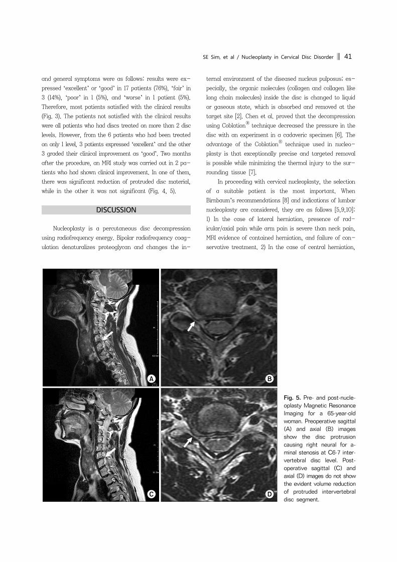
Fig. 5. Pre- and post-nucle-oplasty Magnetic ResonanceImaging for a 65-year-old woman. Preoperative sagittal(A) and axial (B) images show the disc protrusion causing right neural for a-minal stenosis at C6-7 inter-vertebral disc level. Post-operative sagittal (C) and axial (D) images do not showthe evident volume reductionof protruded intervertebral disc segment.
and general symptoms were as follows: results were ex-
pressed ‘excellent’ or ‘good’ in 17 patients (76%), ‘fair’ in
3 (14%), ‘poor’ in 1 (5%), and ‘worse’ in 1 patient (5%).
Therefore, most patients satisfied with the clinical results
(Fig. 3). The patients not satisfied with the clinical results
were all patients who had discs treated on more than 2 disc
levels. However, from the 6 patients who had been treated
on only 1 level, 3 patients expressed ‘excellent’ and the other
3 graded their clinical improvement as ‘good’. Two months
after the procedure, an MRI study was carried out in 2 pa-
tients who had shown clinical improvement. In one of them,
there was significant reduction of protruded disc material,
while in the other it was not significant (Fig. 4, 5).
DISCUSSION
Nucleoplasty is a percutaneous disc decompression
using radiofrequency energy. Bipolar radiofrequency coag-
ulation denaturalizes proteoglycan and changes the in-
ternal environment of the diseased nucleus pulposus; es-
pecially, the organic molecules (collagen and collagen like
long chain molecules) inside the disc is changed to liquid
or gaseous state, which is absorbed and removed at the
target site [2]. Chen et al. proved that the decompression
using CoblationⓇ technique decreased the pressure in the
disc with an experiment in a cadaveric specimen [6]. The
advantage of the CoblationⓇ technique used in nucleo-
plasty is that exceptionally precise and targeted removal
is possible while minimizing the thermal injury to the sur-
rounding tissue [7].
In proceeding with cervical nucleoplasty, the selection
of a suitable patient is the most important. When
Birnbaum’s recommendations [8] and indications of lumbar
nucleoplasty are considered, they are as follows [5,9,10]:
1) In the case of lateral herniation, presence of rad-
icular/axial pain while arm pain is severe than neck pain,
MRI evidence of contained herniation, and failure of con-
servative treatment. 2) In the case of central herniation,

42 Korean J Pain Vol. 24, No. 1, 2011
presence of axial neck pain, unresponsive to 3 months of
conservative treatment, MRI evidence of contained hernia-
tion, and the disc height more than 75%. Exclusion criteria
include that the disc height less than 50%, extruded or se-
questrated disc, spinal fracture or tumor, spinal stenosis,
complete disruption of the annulus fibrosis, central myel-
opathy, instability from degeneration, and extrusion more
than 1/3 of the spinal canal.
Nardi et al. [1] argued that patient who can be a can-
didate for cervical nucleoplasty must have contained her-
niation or focal bulging proven on MRI, but Bonaldi et al.
[2] reported that the cervical nucleoplasty was performed
in the patients who had a bulging, protruding, or soft ex-
truded disc which was not sequestrated or migrated de-
termined by MRI or CT studies. In our study, we performed
cervical nucleoplasty on patients showing extrusion and
stenosis in addition to protrusion on their MRI; who had
no improvement from more than 3 months of conservative
treatment including appropriate pain management; who
keenly want non-surgical treatment over surgical treat-
ment; and after adequate explanation on the possibility of
failure, possible side-effects and costs of nucleoplasty. In
MRI studies taken before the procedure, only 17 patients
from the total 22 patients had disc protrusion suitable for
indications, and from the 5 patients who had non-in-
dication, 2 patients showed extrusion while the other 3 pa-
tients showed degenerative spinal stenosis. From these
patients who had disc extrusion, one expressed ‘excellent’
according to the Modified McNab criteria after the cervical
nucleoplasty and the other one was also satisfied and ex-
pressed ‘good’. From the 3 patients who had degenerative
spinal stenosis, one expressed ‘excellent’ while the other
2 expressed ‘fair’. Considering these results, although
more clinical experience should be accumulated, nucleo-
plasty could be considered in disc disorders like soft disc
extrusions and stenosis prior to surgical treatment if the
patient wants non-surgical treatment; however, cautious
application of cervical nucleoplasty should be taken after
consideration of cost and possible side-effects [5].
In this study, postprocedure MRI studies were acquired
in 2 patients who had shown clinical improvement after 2
months follow up. MRI study in one patient showed a mor-
phologic evidence of volume reduction of protruded disc
material but the other did not. Nardi et al. [1] had reported
that regression of the herniated disc shown on the MRI
has a correlation with clinical resolution, but Bonaldi et al.
[2] reported that clinical improvement did not always ac-
company the regression of the herniated disc on MRI. Even
in cases where there seems to be no regression of the
herniated disc material, a minute reduction in the disc ad-
jacent to the nerve root can cause nerve root pressure to
fall below the critical point. If the postprocedure MRI had
been acquired in all patients, we could have gained more
information from the results. However, it was not possible
due to the economic circumstances of the patients.
Few side effects concerning the cervical nucleoplasty
have been reported and no serious side effects have been
known. Side effects reported in the study of Bonaldi et al.
[2] included 1 case of infectious discitis out of 55 patients,
temporary side effects related to local anesthetic
(bradycardia, Horner’s syndrome, hoarseness, etc.), and
retrosternal and retropharyngeal pain in patients treated
on 3 levels but they responded well to conservative treat-
ment [11,12]. There was no significant complication related
to the procedure within the first month in our study.
Nardi et al. [1] and Birnbaum [8] had proven the effi-
cacy of nucleoplasty by comparing the cervical nucleo-
plasty group and the conservative treatment group. Bonaldi
et al. [2] performed cervical nucleoplasty on 55 patients
and reported that it was successful in 85% of them. Li et
al. [13] reported that 83.73% from the 126 patients treated
reported ‘excellent’ or ‘good’ according to the MacNab
criteria. In our study, 76% of the patients expressed sat-
isfaction by answering ‘good’ or ‘excellent’. These results
of cervical nucleoplasty appear to be much better than the
results of lumbar nucleoplasty [3,4,7]. However, the rea-
sons that the nucleoplasty treatment may be more effec-
tive at the cervical level than at the lumbar level are not
clear. One possible explanation could be anatomic: The
cervical nerve root is confined to a relatively smaller space
than its lumbar counterpart so the cervical nerve root re-
spond more sensitively to even a minute reduction. For this
reason, even if the pressure of the disc is reduced slightly,
the decompression on the nerve root and reduction in clin-
ical symptoms can be easily obtained. Another reason
could be the topography of the lesion and direction from
which it is approached for treatment. In lumbar nucleo-
plasty, we use a posterolateral approach from the lesion.
But in cervical nucleoplasty, we use anterolateral approach
to the disc, and so, the SpineWandTM could be accurately
positioned in the lesion site posteriorly located. In other
words, since symptomatic herniation is directed posterior,

SE Sim, et al / Nucleoplasty in Cervical Disc Disorder 43
cervical nucleoplasty can effectively approach the lesion
site because it uses the anterior approach [2,5].
Another benefits of cervical nucleoplasty are that it
does not have any influence on the stability of the cervical
vertebrae compared to surgical treatment [13]; it is mini-
mally invasive since it uses a 19-gauge introducer needle,
which is smaller compared to other percutaneous decom-
pression; the PercTM DCTM SpineWand that is handled by
the surgeon is small and hard so it could be operated more
precisely; it only takes a 10-12 minutes to treat one level
of disc; and there is a markedly low possibility of damage
to the surrounding tissue [5,8].
Upon retrospective examination of the medical records
of the 22 patients that were treated in our hospital, clinical
improvement in symptoms within a month after cervical
nucleoplasty were seen in most patients. Most of the pa-
tients reported the reduced subjective symptoms, such as
radicular and axial pain, and an improved quality of life.
Consequently, they expressed satisfaction with the cervical
nucleoplasty.
In conclusion, in the treatment of cervical disc dis-
orders, cervical nucleoplasty is minimally invasive, easy
and has fewer complications than the surgical treatment.
Therefore, when there is no response to conservative
treatment, cervical nucleoplasty can be considered as a
suitable alternative prior to open surgical treatment.
However, to obtain good clinical results, appropriate se-
lection of patients according to the indications of nucleo-
plasty must precede the procedure, and more experience
and research should be accumulated for other indications
such as extrusion and stenosis.
REFERENCES
1. Nardi PV, Cabezas D, Cesaroni A. Percutaneous cervical nucleoplasty using coblation technology. Clinical results in fifty
consecutive cases. Acta Neurochir Suppl 2005; 92: 73-8.2. Bonaldi G, Baruzzi F, Facchinetti A, Fachinetti P, Lunghi S.
Plasma radio-frequency-based diskectomy for treatment of cervical herniated nucleus pulposus: feasibility, safety, and preliminary clinical results. AJNR Am J Neuroradiol 2006; 27: 2104-11.
3. Sharps LS, Isaac Z. Percutaneous disc decompression using nucleoplasty. Pain Physician 2002; 5: 121-6.
4. Singh V, Piryani C, Liao K, Nieschulz S. Percutaneous disc decompression using coblation (nucleoplasty) in the treatment of chronic discogenic pain. Pain Physician 2002; 5: 250-9.
5. Ro JY, Oh CS, Kim OS, Kim DK, Woo NS, Shin HY. Nucle-oplasty in a patient with cervical disc extrusion with radiating pain of an upper extremity. Anesth Pain Med 2009; 4: 203-7.
6. Chen YC, Lee SH, Chen D. Intradiscal pressure study of percutaneous disc decompression with nucleoplasty in human cadavers. Spine 2003; 28: 661-5.
7. Boswell MV, Trescot AM, Datta S, Schultz DM, Hansen HC, Abdi S, et al. Interventional techniques: evidence-based practice guidelines in the management of chronic spinal pain. Pain Physician 2007; 10: 7-111.
8. Birnbaum K. Percutaneous cervical disc decompression. Surg Radiol Anat 2009; 31: 379-87.
9. Calisaneller T, Ozdemir O, Karadeli E, Altinors N. Six months post-operative clinical and 24 hour post-operative MRI examinations after nucleoplasty with radiofrequency energy. Acta Neurochir (Wien) 2007; 149: 495-500.
10. Singh V. Scientific basis for nucleoplasty. Tech Reg Anesth Pain Manag 2005; 9: 13-24.
11. Bhagia SM, Slipman CW, Nirschl M, Isaac Z, El-Abd O, Sharps LS, et al. Side effects and complications after percutaneous disc decompression using coblation technology. Am J Phys Med Rehabil 2006; 85: 6-13.
12. Oh WS, Park YO, Shim WS, Wie HW, Woo CH. Psoas abscess and discitis after intradiscal nucleoplasty. Korean J Anesthesiol 2002; 42: 844-9.
13. Li J, Yan DL, Zhang ZH. Percutaneous cervical nucleoplasty in the treatment of cervical disc herniation. Eur Spine J 2008; 17: 1664-9.
Top Related