


Languages
Pages
Legal

www.sciencemag.org/content/348/6235/707/suppl/DC1
Supplementary Materials for
Engineering of a light-gated potassium channel
Cristian Cosentino, Laura Alberio, Sabrina Gazzarrini, Marco Aquila, Edoardo Romano, Solei Cermenati, Paolo Zuccolini, Jan Petersen, Monica Beltrame, James L. Van Etten,
John M. Christie, Gerhard Thiel, Anna Moroni*
*Corresponding author. E-mail: [email protected]
Published 8 May 2015, Science 348, 707 (2015) DOI: 10.1126/science.aaa2787
This PDF file includes:
Materials and Methods Figs. S1 to S9 Table S1 References
Other Supplementary Material for this manuscript includes the following: (available at www.sciencemag.org/cgi/content/full/348/6235/707/DC1)
Movie S1
1
Supplementary Materials for
Engineering of a light-gated potassium channel
Cristian Cosentino, Laura Alberio, Sabrina Gazzarrini, Marco Aquila, Edoardo Romano, Solei Cermenati, Paolo Zuccolini, Jan Petersen, Monica Beltrame, James L. Van Etten, John M.
Christie, Gerhard Thiel and Anna Moroni
This section includes:
Materials and Methods
Figures S1-S9
Tables S1
Movie S1
Supplementary References
Materials and Methods:
Cloning procedures and plasmids
All constructs were prepared by adding the LOV2 domain, aa 404-546 of Avena sativa Phototropin
1 (NPH1-1 GenBank: AAC05083.1) (24) to Kcv, aa 2-94 of chlorovirus PBCV-1-Kcv
(NP_048599.1) (11) by overlapping PCR. The complete list of mutations, deletions and insertions,
which were introduced in each construct, is reported in Table S1. Mutations introduced either in the
LOV domain or in Kcv are numbered on the original sequences of phototropin and Kcv. Overlapping
PCR was used to add the N-terminal myristoylation and palmitoylation sequence (MGCTVSAE) (14)
to the N terminus of the construct and QuikChange XL II (Agilent Technologies) was used to
introduce point mutations. Coding sequences were cloned in pYES2-Met25 vector (25) for S.
2
cerevisiae expression; in pSGEM (a modified version of pGEM-HE vector) for expression in Xenopus
laevis oocytes; in pCDNA3.1+ for HEK293T cells expression; in a modified version of pET-21d
containing a 7His-Strep II-SUMO tag (26) for expression in E. coli and in pCS2+ for zebrafish
expression. The plasmid for expression in SF9 Spodoptera frugiperda cells was built by Gibson
assembly (27) using pAcHLT-A digested with SphI and SacI as backbone, a fragment amplified from
pAcHLT-A (base pairs 215-2203) and the BLINK1 coding sequence amplified with a 3’ primer
creating a C-terminal 6His-tag.
Yeast functional complementation assays
Functional complementation assays were performed using the S. cerevisiae strain SGY1528 (MATa
ade2–1 can1–100 his3–11,15 leu2–3,112 trp1–1 ura3–1 trk1::HIS3 trk2::TRP1 (25, 28) This strain
lacks the endogenous K+ uptake system and it is not able to grow on media supplemented with K+
lower than 100 mM.
To test the functionality of the clones, yeast colonies were inoculated in non-selective medium (SD -
Ura, 100 mM KCl) and grown overnight to stationary phase. Cultures were then diluted to OD600 =
0.8 and 10-fold serial dilutions were spotted (7μl) onto selective plates (SEL -Ura -Met with 0.5, 1, 4
mM KCl). Twin plates were prepared for each K+ concentration and were either exposed to blue light
or grown in the dark. After 3 and 4 days at 30°C images were taken and plates were screened for
colonies with differential growth between Blue light and dark in the presence of low KCl
concentration. To confirm functional complementation, cultures from overnight stationary phase
were also inoculated in liquid selective media (SEL -Ura -Met with 4, 7, 10, 100 mM KCl) and grown
for up to 117 h in presence of blue light or in the dark. OD600 of the cultures were measured to
determine the growth ratio between the two conditions at 21, 45, 52 and 117 h after inducing protein
expression. Positive clones were selected for plasmid isolation and DNA amplification in E. coli for
sequencing. Non-selective and selective media were prepared as previously described (25). Solid
media were obtained adding 15g/l agar.
3
Yeast random library screening
Randomization was performed on the sequence coding the myristoylation and palmitoylation domain,
the whole LOV domain and the Kcv channel up to the first transmembrane region, where the channel
DNA sequence has an endogenous SmaI restriction site. Random mutagenized libraries were
generated using the Gene Morph II random mutagenesis kit (Agilent Technologies) in three steps:
two samples with 50 ng of myLK N538A were amplified by PCR (30 cycles) with PAGE-purified
60-mer primers (IDT-Tema Ricerca). The forward primer is complementary to the upstream plasmid
sequence while the reverse primer is complementary to the Kcv sequence downstream of the SmaI
restriction site (corresponding to the first transmembrane region). Two μl of each PCR reaction were
used as template for a subsequent amplification reaction (30 cycles). This was repeated twice. The
end products of the final six reactions were gel-purified, grouped and gap-repaired in yeast SGY1528
strain with SmaI linearized pYES2-Met25 plasmid containing wild- type myLK N538A. Co-
transformation was performed with a Frozen-EZ yeast transformation II kit (Zymo Research). The
library was first plated on non-selective medium (SD –Ura with 100 mM KCl) and grown for 3 days
at 30 °C, in dark, yielding roughly 20000 transformed colonies. Non-selective plates were then
replica-plated twice onto selective plates (SEL –Ura -Met with 0.5 mM KCl) and grown for 3 days at
30 °C under blue light or dark conditions. Colonies showing differential growth between the two
conditions were selected for subsequent functional complementation assays.
Electrophysiology
Xenopus oocytes - cRNAs were transcribed in vitro using T7 RNA polymerase (Promega) as
described (11). Oocytes were prepared according to standard methods and injected with 50 nl of water
or 50 nl of cRNA (0.8 µg/µl) and incubated in the dark at 19°C in ND96 solution : 96 mM NaCl, 2
mM KCl, 1.8 mM CaCl2, 1mM MgCl2, 5 mM HEPES–NaOH buffer (pH 7.5), 5 mM sodium
pyruvate, 50 µg /ml gentamicin). Measurements were performed 2-5 days after injection. Currents
4
were recorded by two-electrode voltage clamp, using the Gene-Clamp 500 amplifier (Axon
Instruments) under control of pCLAMP7. Oocytes were perfused at room temperature with a solution
containing: 100 mM KCl, 1.8 mM CaCl2, 1 mM MgCl2, and 5 mM HEPES-KOH buffer (pH 7.4).
When different KCl concentrations were used, the osmolarity was adjusted to 215 mOsm with
mannitol. The voltage protocols applied consisted in a step protocol of 20 mV steps from + 60 to -
180 mV or in a gap free protocol at -60 or -80 mV. The channel blocker BaCl2 was applied to the
external solution at a concentration of 1 or 5 mM, as indicated.
HEK293T cells - HEK293T cells were cultured in Dulbecco’s modified Eagle’s medium (Euroclone)
supplemented with 10% fetal bovine serum (Euroclone), 100 IU/mL of penicillin and 100 µg/ml of
streptomycin, and stored in a 37 °C humidified incubator with 5% CO2. BLINK1 and myLK33
cDNAs were co-transfected in HEK293T with a plasmid containing green fluorescent protein (GFP).
Transfections were performed with TurboFect Transfection Reagent (Thermo Scientific). One to two
days after transfection, HEK293T cells were dispersed by trypsin treatment and placed on 35-mm
plastic petri dishes. Cells showing a clear fluorescence signal were selected for patch clamp analysis.
Membrane currents were recorded in whole cell configuration using a Dagan 3900A patch-clamp
amplifier and digitized using a Digidata 1322A controlled by pCLAMP 9.2. The pipette solution
contained: 10 mM NaCl, 130 mM KCl, 2 mM MgCl2, and 5 mM HEPES–KOH buffer (pH 7.2). The
extracellular bath’s solution contained: 100 mM KCl, 80 mM D-Mannitol, 1.8 mM CaCl2, 1 mM
MgCl2 and 5 mM HEPES–KOH buffer (pH 7.4). When different KCl concentrations were used, the
osmolarity was adjusted to 290 mOsm with mannitol. The channel blocker BaCl2 was applied to the
external solution at a concentration of 1 or 5 mM.
Light stimulation
Yeast functional complementation assays were performed by means of a custom-made array of light
emitting diodes (LEDs) (Royal-Blue LED, λ 447 ± 10 nm, LUXEON Rebel LED) for homogeneous
5
illumination of the samples (2 ± 0.2 μW/mm2) in a controlled temperature chamber. The same system
was used for illumination of zebrafish embryos at 80 μW/mm2. Red light was also provided at 80
μW/mm2 by means of Red-Orange LED, λ 617 nm (MR-H2060-20T, LUXEON Rebel LEDs).
Transfected HEK293T cells and injected oocytes were protected from ambient light to prevent
receptor activation prior to the assays and all manipulations were performed under red light (MR-
H2060-20T, LUXEON Rebel LEDs Red-Orange (617 nm)) in the Faraday cage shielded with a 580
nm cutoff filter (Rosco Supergel, Deep Amber #22). Blue light was provided by a light-emitting diode
(Royal Blue, 455 nm, High-Power LED; Thorlabs) at intensity indicated in figure legends. Light
power was measured using a handheld light power meter (Thorlabs).
Protein expression and spectroscopic analysis
For protein expression and purification, E. coli RosettaBL21(DE3)pLysS (Novagen) cells were
grown in LB medium to an OD600 of 0.6, induced with 0.5 mM IPTG and incubated overnight at
24 °C. Cells were harvested and tandem affinity purified with subsequent SUMO protease cleavage
of the tag as described (26).
Sf9 insect cells were grown in a monolayer with TC100 medium containing 10% FBS at 27 °C. For
the construction of recombinant virus the transfer vector was co-transfected into cells with linearized
baculovirus DNA using the BacMagic DNA Kit (Novagen). Transfected cells were incubated for 5
days to raise the primary viral stock. Two additional rounds of virus propagation (each for 3 days)
were performed. High titer virus was used for protein expression. Cells were incubated in medium
supplemented with 20 μM riboflavin for 3 days in darkness prior to harvest. For fluorescence emission
and excitation measurements, the cells were washed to remove residual riboflavin and resuspended
in Dulbecco's PBS at a density of 1.5 x 10⁷ cells/ml.

6
Absorption spectra of purified protein from E. coli were collected with a Shimadzu MultiSpec-1501
diode array spectrophotometer at room temperature. In vivo fluorescence emission and excitation
spectra of insect cells were recorded using a Perkin-Elmer LS-55 luminescence spectrometer.
Fluorescence excitation spectra were recorded by monitoring the emission at 520 nm. Fluorescence
emission spectra were obtained using an excitation wavelength of 450 nm. For absorbance and
fluorescence measurements, a blue-light emitting diode (λ 455 nm) provided the excitation pulse.
Immunocytochemistry
HEK293T cells transfected with BLINK1 and Kcv-GFP (29) channels were treated with blocking
solution containing 2% BSA in PBS. Cells were incubated 1 hour with anti-Kcv monoclonal antibody
(30) washed briefly with blocking solution and incubated 1 hour with Alexa Fluor® 594 AffiniPure
secondary antibody and Hoechst dye. Confocal microscopy analyses were performed using Leica SP2
(http://www.leica-microsystems.com) laser scanning confocal imaging system.
Microinjections and touch response assays in zebrafish
pCS2+BLINK1 and pCS2+GFP constructs were linearized with NotI and transcribed with Sp6 RNA
polymerase, using the mMESSAGE mMACHINE® SP6 Transcription Kit (Ambion) and following
manufacturer’s instructions. Embryos were injected at the 1- to 2-cell stage with 200pg/embryo of
RNA in water (W3500, Sigma-Aldrich), supplemented with rhodamine-dextran (D1817, Molecular
Probes) as a tracer. Injected embryos were raised in E3 medium (5 mM NaCl, 0.17 mM KCl, 0.33
mM MgSO4, 0.33 mM CaCl2) at 28.5 °C and manually dechorionated at 1 day post-fertilization (1
dpf). The screening for touch-evoked escape response was performed at 2 days post-fertilization
(unless differently specified), following standardized procedures. Using a hand-broken microloader
tip, a gentle stimulus was applied to the tail of the embryos and their reaction observed. Wild-type
embryos at this developmental stage swim away from the source of the stimulus (31).
7
Western blot
Yeast microsome preparation-Yeast cells grown in liquid culture in selective medium for 3 days at
30 °C were pelleted (1,500 x g, 20 min). Pellet was resuspended in Breaking Buffer (50 mM
NaH2PO4*H2O, 1 mM EDTA, 5 % (w/v) glycerol, 5μM Leupeptin, 1μM Pepstatin, 0.1mM PMSF,
5mM DTT). Cells were broken by vortexing with glass beads (425-600 μm, Sigma-Aldrich) and
centrifugated (4000 x g, 5 min) to remove cell debris. Then, the Eppendorf tube was centrifuged
(15,000 x g, 1 hour) at 4°C and the supernatant was discarded. The microsomes were resuspended in
Breaking Buffer and stored at -80 °C.
Preparation of zebrafish lysates- Deyolked embryos were resuspended in modified RIPA buffer
(50mM Tris, 150mM KCl, 0.1%SDS, 5%DOC, 0.1% NP-40, filter sterilized) and homogenized using
IKA T10 basic homogenizer. Lysates were quantified and stored at -80°C.
Insect cell protein extract - Cells were harvested (1,000 x g, 1min) and resuspended in Dulbecco's
phosphate buffered saline supplemented with cOmplete EDTA-free protease inhibitor (Roche). Lysis
of cells was performed by sonication with subsequent centrifugation (14,000 x g, 5 min) to remove
cell debris.
Western blot analysis- 60 μg (zebrafish) or 13μg (yeast) /lane of total protein were separated on 4-
20% tris-glycine denaturating gel (Novex). Proteins were blotted on a PVDF membrane and blots
were blocked in 5% dry milk in TBST solution for 2 hours and incubated with primary anti-Kcv
monoclonal antibody (30) at 4 °C overnight. Then the blots were rinsed in TBST for 3 X 10 min and
incubated with alkaline phosphatase-conjugated secondary antibody for 1 hour. Antibody binding
was detected using SIGMAFAST BCIP/NBT tablet (Sigma).
For immunodetection of BLINK1 protein expressed in insect cells, 20 μg of total protein were
separated by SDS-PAGE on a 4-20% gradient gel (BioRad) and transferred to a nitrocellulose
membrane. The membrane was blocked for 1 h with TBST containing 4% milk powder, followed by
1 h incubation with the first antibody (anti-Kcv). Afterwards the membrane was washed 3 x TBST

8
for 10 min each, incubated with the secondary HRP conjugated antibody for 45 min and then washed
again as described before. Detection of immune complexes was made with ECL (Pierce) according
to manufacturer’s instructions and a Fusion Fx imager (peqlab) used to record the signal.
9
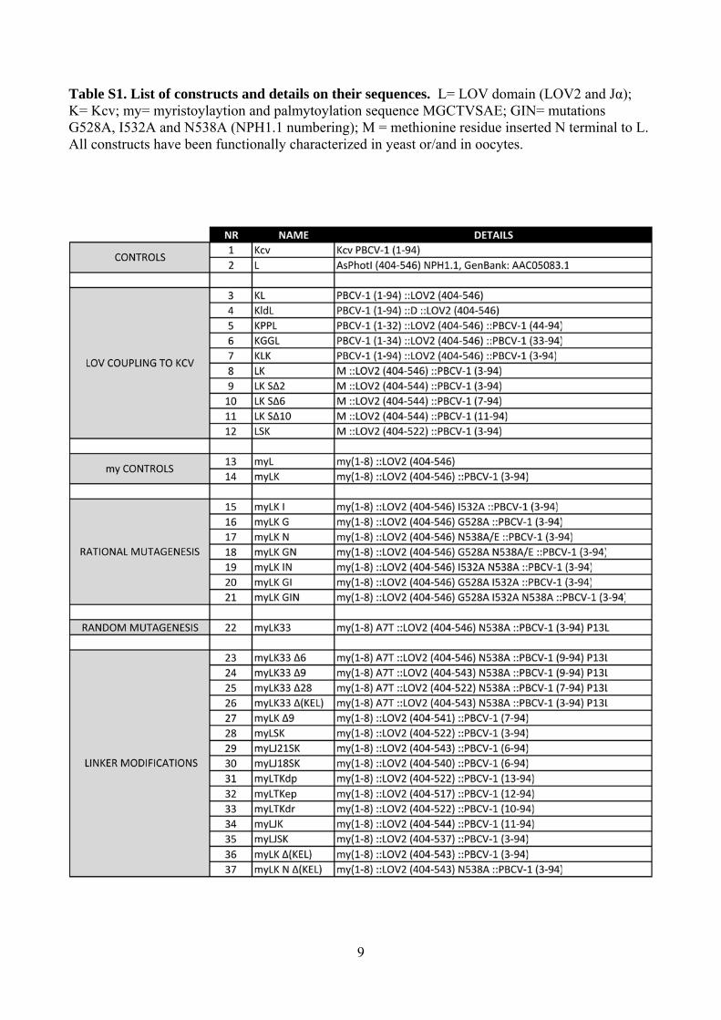
Table S1. List of constructs and details on their sequences. L= LOV domain (LOV2 and Jα); K= Kcv; my= myristoylaytion and palmytoylation sequence MGCTVSAE; GIN= mutations G528A, I532A and N538A (NPH1.1 numbering); M = methionine residue inserted N terminal to L. All constructs have been functionally characterized in yeast or/and in oocytes.
10
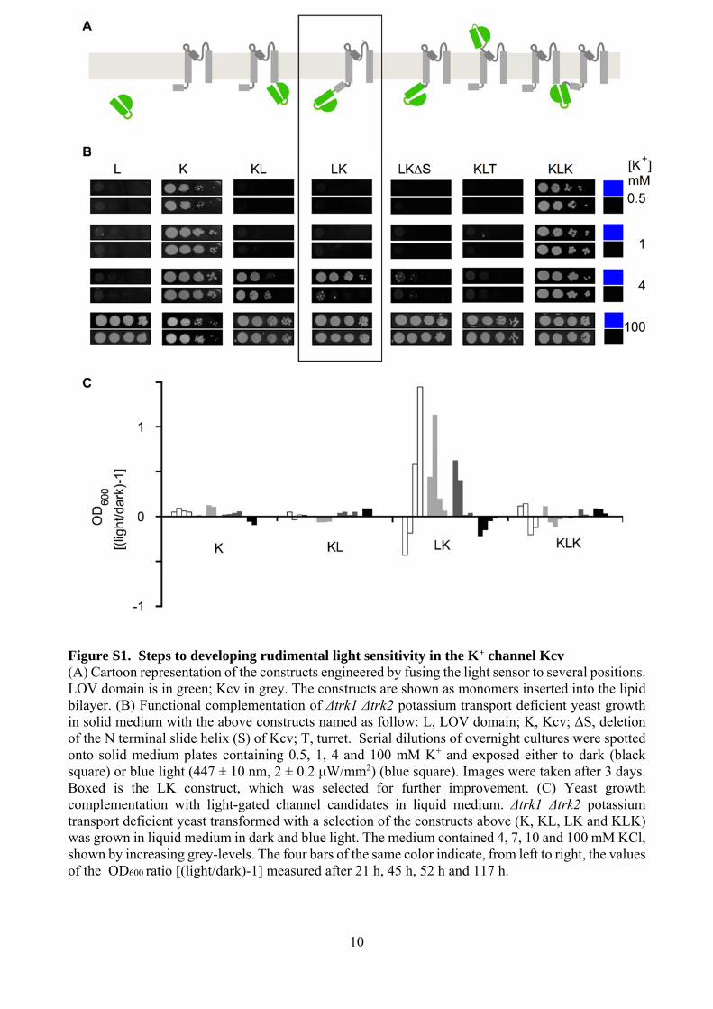
Figure S1. Steps to developing rudimental light sensitivity in the K+ channel Kcv (A) Cartoon representation of the constructs engineered by fusing the light sensor to several positions. LOV domain is in green; Kcv in grey. The constructs are shown as monomers inserted into the lipid bilayer. (B) Functional complementation of Δtrk1 Δtrk2 potassium transport deficient yeast growth in solid medium with the above constructs named as follow: L, LOV domain; K, Kcv; ΔS, deletion of the N terminal slide helix (S) of Kcv; T, turret. Serial dilutions of overnight cultures were spotted onto solid medium plates containing 0.5, 1, 4 and 100 mM K+ and exposed either to dark (black square) or blue light (447 ± 10 nm, 2 ± 0.2 μW/mm2) (blue square). Images were taken after 3 days. Boxed is the LK construct, which was selected for further improvement. (C) Yeast growth complementation with light-gated channel candidates in liquid medium. Δtrk1 Δtrk2 potassium transport deficient yeast transformed with a selection of the constructs above (K, KL, LK and KLK) was grown in liquid medium in dark and blue light. The medium contained 4, 7, 10 and 100 mM KCl, shown by increasing grey-levels. The four bars of the same color indicate, from left to right, the values of the OD600 ratio [(light/dark)-1] measured after 21 h, 45 h, 52 h and 117 h.
11
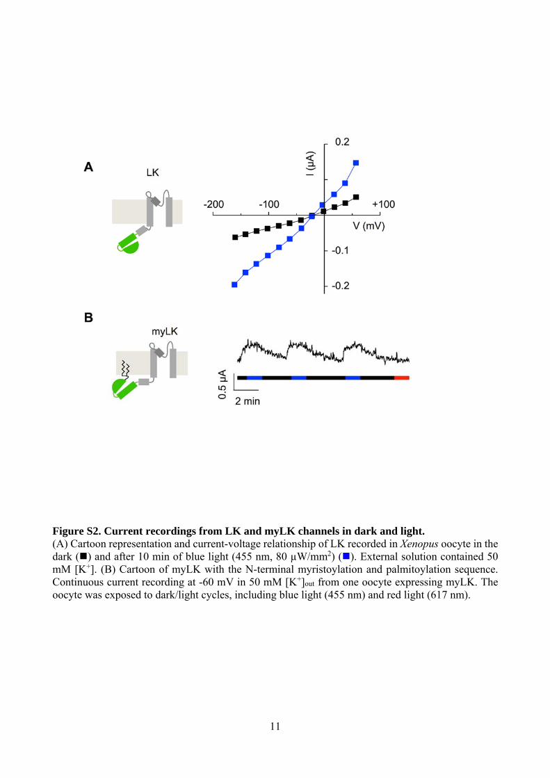
Figure S2. Current recordings from LK and myLK channels in dark and light. (A) Cartoon representation and current-voltage relationship of LK recorded in Xenopus oocyte in the dark () and after 10 min of blue light (455 nm, 80 µW/mm2) (). External solution contained 50 mM [K+]. (B) Cartoon of myLK with the N-terminal myristoylation and palmitoylation sequence. Continuous current recording at -60 mV in 50 mM [K+]out from one oocyte expressing myLK. The oocyte was exposed to dark/light cycles, including blue light (455 nm) and red light (617 nm).
12
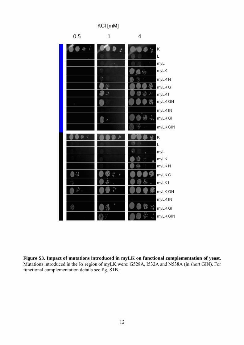
Figure S3. Impact of mutations introduced in myLK on functional complementation of yeast. Mutations introduced in the J region of myLK were: G528A, I532A and N538A (in short GIN). For functional complementation details see fig. S1B.
13
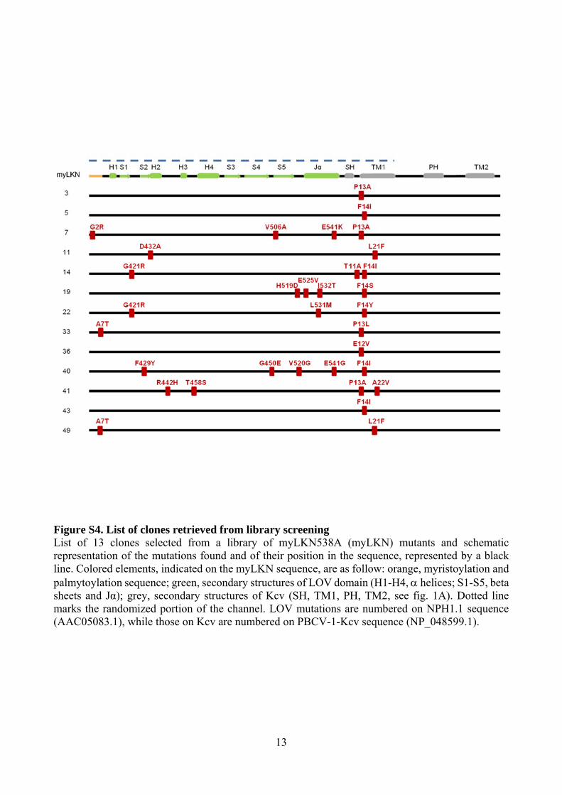
Figure S4. List of clones retrieved from library screening List of 13 clones selected from a library of myLKN538A (myLKN) mutants and schematic representation of the mutations found and of their position in the sequence, represented by a black line. Colored elements, indicated on the myLKN sequence, are as follow: orange, myristoylation and palmytoylation sequence; green, secondary structures of LOV domain (H1-H4, helices; S1-S5, beta sheets and Jα); grey, secondary structures of Kcv (SH, TM1, PH, TM2, see fig. 1A). Dotted line marks the randomized portion of the channel. LOV mutations are numbered on NPH1.1 sequence (AAC05083.1), while those on Kcv are numbered on PBCV-1-Kcv sequence (NP_048599.1).
14
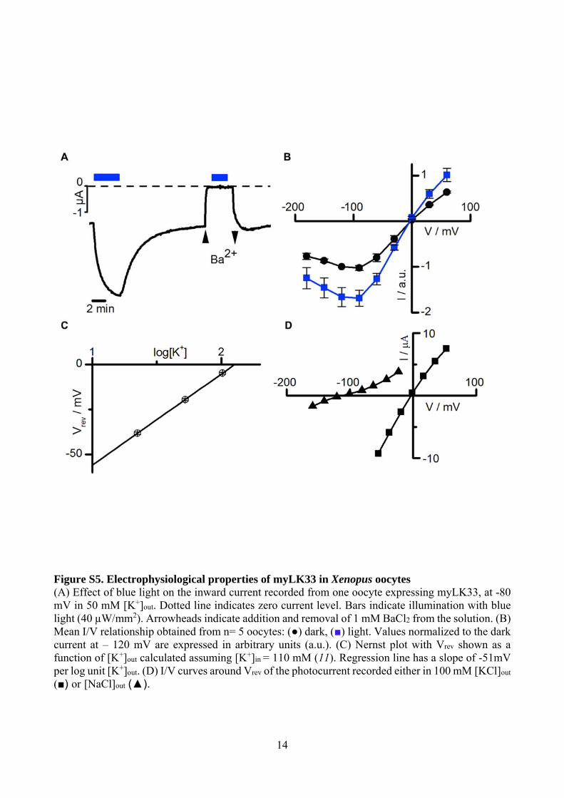
Figure S5. Electrophysiological properties of myLK33 in Xenopus oocytes (A) Effect of blue light on the inward current recorded from one oocyte expressing myLK33, at -80 mV in 50 mM [K+]out. Dotted line indicates zero current level. Bars indicate illumination with blue light (40 µW/mm2). Arrowheads indicate addition and removal of 1 mM BaCl2 from the solution. (B) Mean I/V relationship obtained from n= 5 oocytes: (●) dark, (■) light. Values normalized to the dark current at – 120 mV are expressed in arbitrary units (a.u.). (C) Nernst plot with Vrev shown as a function of [K+]out calculated assuming [K+]in = 110 mM (11). Regression line has a slope of -51mV per log unit [K+]out. (D) I/V curves around Vrev of the photocurrent recorded either in 100 mM [KCl]out (■) or [NaCl]out (▲).
15
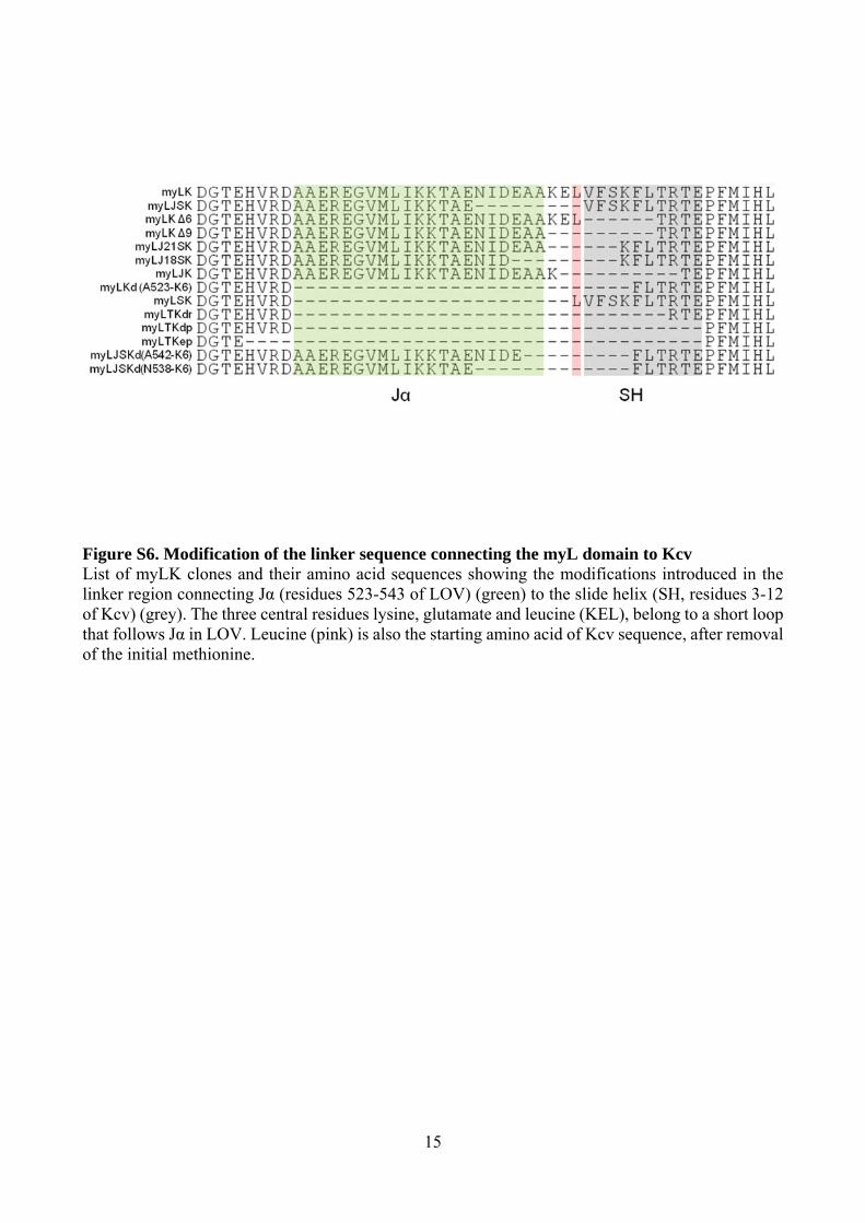
Figure S6. Modification of the linker sequence connecting the myL domain to Kcv List of myLK clones and their amino acid sequences showing the modifications introduced in the linker region connecting Jα (residues 523-543 of LOV) (green) to the slide helix (SH, residues 3-12 of Kcv) (grey). The three central residues lysine, glutamate and leucine (KEL), belong to a short loop that follows Jα in LOV. Leucine (pink) is also the starting amino acid of Kcv sequence, after removal of the initial methionine.
16
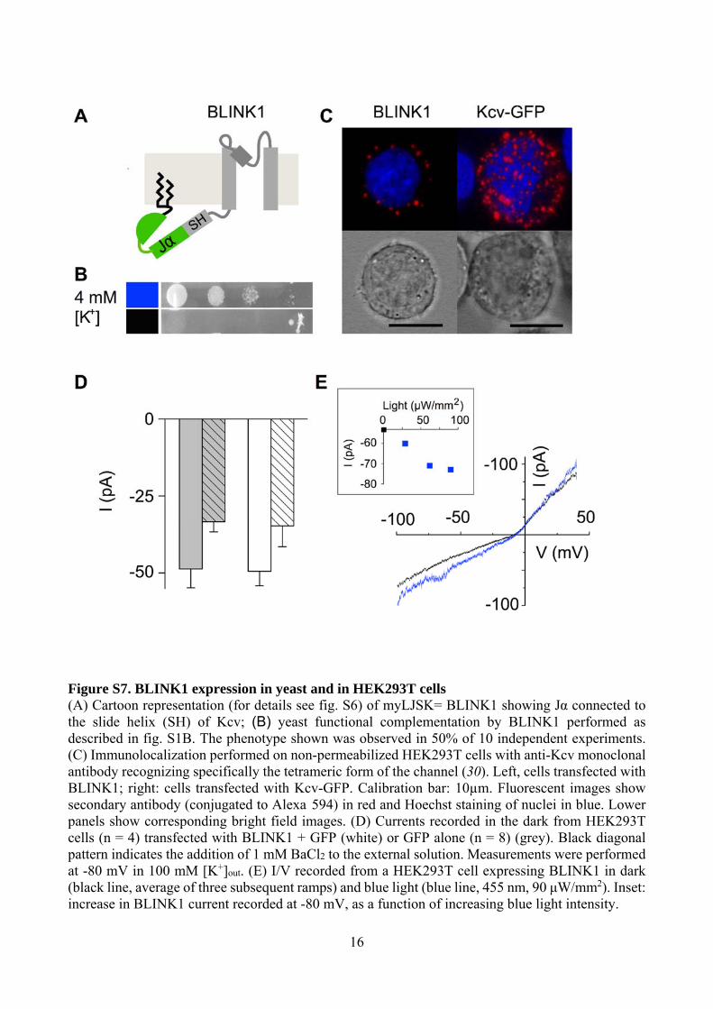
Figure S7. BLINK1 expression in yeast and in HEK293T cells (A) Cartoon representation (for details see fig. S6) of myLJSK= BLINK1 showing Jα connected to the slide helix (SH) of Kcv; (B) yeast functional complementation by BLINK1 performed as described in fig. S1B. The phenotype shown was observed in 50% of 10 independent experiments. (C) Immunolocalization performed on non-permeabilized HEK293T cells with anti-Kcv monoclonal antibody recognizing specifically the tetrameric form of the channel (30). Left, cells transfected with BLINK1; right: cells transfected with Kcv-GFP. Calibration bar: 10µm. Fluorescent images show secondary antibody (conjugated to Alexa 594) in red and Hoechst staining of nuclei in blue. Lower panels show corresponding bright field images. (D) Currents recorded in the dark from HEK293T cells (n = 4) transfected with BLINK1 + GFP (white) or GFP alone (n = 8) (grey). Black diagonal pattern indicates the addition of 1 mM BaCl2 to the external solution. Measurements were performed at -80 mV in 100 mM [K+]out. (E) I/V recorded from a HEK293T cell expressing BLINK1 in dark (black line, average of three subsequent ramps) and blue light (blue line, 455 nm, 90 μW/mm2). Inset: increase in BLINK1 current recorded at -80 mV, as a function of increasing blue light intensity.
17
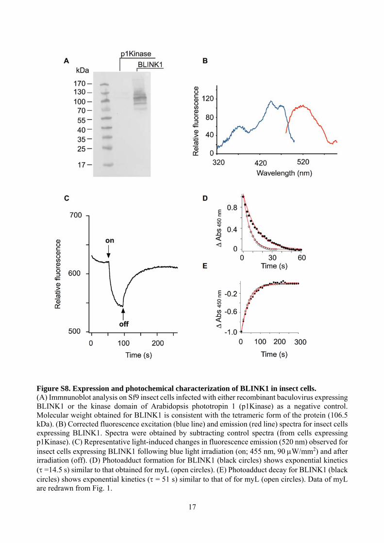
Figure S8. Expression and photochemical characterization of BLINK1 in insect cells. (A) Immnunoblot analysis on Sf9 insect cells infected with either recombinant baculovirus expressing BLINK1 or the kinase domain of Arabidopsis phototropin 1 (p1Kinase) as a negative control. Molecular weight obtained for BLINK1 is consistent with the tetrameric form of the protein (106.5 kDa). (B) Corrected fluorescence excitation (blue line) and emission (red line) spectra for insect cells expressing BLINK1. Spectra were obtained by subtracting control spectra (from cells expressing p1Kinase). (C) Representative light-induced changes in fluorescence emission (520 nm) observed for insect cells expressing BLINK1 following blue light irradiation (on; 455 nm, 90 W/mm2) and after irradiation (off). (D) Photoadduct formation for BLINK1 (black circles) shows exponential kinetics ( =14.5 s) similar to that obtained for myL (open circles). (E) Photoadduct decay for BLINK1 (black circles) shows exponential kinetics ( = 51 s) similar to that of for myL (open circles). Data of myL are redrawn from Fig. 1.
18
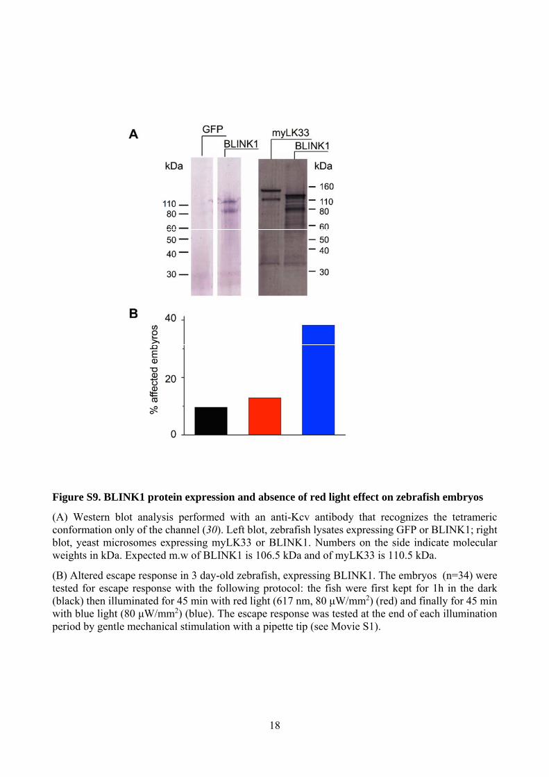
Figure S9. BLINK1 protein expression and absence of red light effect on zebrafish embryos
(A) Western blot analysis performed with an anti-Kcv antibody that recognizes the tetrameric conformation only of the channel (30). Left blot, zebrafish lysates expressing GFP or BLINK1; right blot, yeast microsomes expressing myLK33 or BLINK1. Numbers on the side indicate molecular weights in kDa. Expected m.w of BLINK1 is 106.5 kDa and of myLK33 is 110.5 kDa.
(B) Altered escape response in 3 day-old zebrafish, expressing BLINK1. The embryos (n=34) were tested for escape response with the following protocol: the fish were first kept for 1h in the dark (black) then illuminated for 45 min with red light (617 nm, 80 μW/mm2) (red) and finally for 45 min with blue light (80 μW/mm2) (blue). The escape response was tested at the end of each illumination period by gentle mechanical stimulation with a pipette tip (see Movie S1).

19
Movie S1. Touch response of zebrafish embryos expressing BLINK1. (GFP) 2 day-old control embryo (GFP-injected), exposed to blue light for 1 h and then tested by mechanical stimulation with the tip of a pipette. (BLINK1) BLINK1-injected embryo, same treatment as the GFP-injected one.

References and Notes
1. S. B. Long, E. B. Campbell, R. Mackinnon, Crystal structure of a mammalian voltage-dependent Shaker family K+ channel. Science 309, 897–903 (2005). Medline doi:10.1126/science.1116269
2. R. Latorre, F. J. Morera, C. Zaelzer, Allosteric interactions and the modular nature of the voltage- and Ca2+-activated (BK) channel. J. Physiol. 588, 3141–3148 (2010). Medline doi:10.1113/jphysiol.2010.191999
3. U. M. Ohndorf, R. MacKinnon, Construction of a cyclic nucleotide-gated KcsA K+ channel. J. Mol. Biol. 350, 857–865 (2005). Medline doi:10.1016/j.jmb.2005.05.050
4. C. Arrigoni, I. Schroeder, G. Romani, J. L. Van Etten, G. Thiel, A. Moroni, The voltage-sensing domain of a phosphatase gates the pore of a potassium channel. J. Gen. Physiol. 141, 389–395 (2013). Medline doi:10.1085/jgp.201210940
5. M. R. Banghart, M. Volgraf, D. Trauner, Engineering light-gated ion channels. Biochemistry 45, 15129–15141 (2006). Medline doi:10.1021/bi0618058
6. M. Banghart, K. Borges, E. Isacoff, D. Trauner, R. H. Kramer, Light-activated ion channels for remote control of neuronal firing. Nat. Neurosci. 7, 1381–1386 (2004). Medline doi:10.1038/nn1356
7. H. Janovjak, S. Szobota, C. Wyart, D. Trauner, E. Y. Isacoff, A light-gated, potassium-selective glutamate receptor for the optical inhibition of neuronal firing. Nat. Neurosci. 13, 1027–1032 (2010). Medline doi:10.1038/nn.2589
8. J. Y. Kang, D. Kawaguchi, I. Coin, Z. Xiang, D. D. O’Leary, P. A. Slesinger, L. Wang, In vivo expression of a light-activatable potassium channel using unnatural amino acids. Neuron 80, 358–370 (2013). Medline doi:10.1016/j.neuron.2013.08.016
9. D. Schmidt, P. W. Tillberg, F. Chen, E. S. Boyden, A fully genetically encoded protein architecture for optical control of peptide ligand concentration. Nat. Commun. 5, 3019 (2014). Medline doi:10.1038/ncomms4019
10. J. M. Christie, Phototropin blue-light receptors. Annu. Rev. Plant Biol. 58, 21–45 (2007). Medline doi:10.1146/annurev.arplant.58.032806.103951
11. B. Plugge, S. Gazzarrini, M. Nelson, R. Cerana, J. L. Van Etten, C. Derst, D. DiFrancesco, A. Moroni, G. Thiel, A potassium channel protein encoded by chlorella virus PBCV-1. Science 287, 1641–1644 (2000). Medline doi:10.1126/science.287.5458.1641
12. J. M. Christie, J. Gawthorne, G. Young, N. J. Fraser, A. J. Roe, LOV to BLUF: Flavoprotein contributions to the optogenetic toolkit. Mol. Plant 5, 533–544 (2012). Medline doi:10.1093/mp/sss020
13. F. C. Chatelain, S. Gazzarrini, Y. Fujiwara, C. Arrigoni, C. Domigan, G. Ferrara, C. Pantoja, G. Thiel, A. Moroni, D. L. Minor Jr., Selection of inhibitor-resistant viral potassium channels identifies a selectivity filter site that affects barium and amantadine block. PLOS ONE 4, e7496 (2009). Medline doi:10.1371/journal.pone.0007496
20
14. C. Aicart-Ramos, R. A. Valero, I. Rodriguez-Crespo, Protein palmitoylation and subcellular trafficking. Biochim. Biophys. Acta 1808, 2981–2994 (2011). Medline doi:10.1016/j.bbamem.2011.07.009
15. D. Strickland, X. Yao, G. Gawlak, M. K. Rosen, K. H. Gardner, T. R. Sosnick, Rationally improving LOV domain-based photoswitches. Nat. Methods 7, 623–626 (2010). Medline doi:10.1038/nmeth.1473
16. B. Hertel, S. Tayefeh, T. Kloss, J. Hewing, M. Gebhardt, D. Baumeister, A. Moroni, G. Thiel, S. M. Kast, Salt bridges in the miniature viral channel Kcv are important for function. Eur. Biophys. J. 39, 1057–1068 (2010). Medline doi:10.1007/s00249-009-0451-z
17. J. Mattis, K. M. Tye, E. A. Ferenczi, C. Ramakrishnan, D. J. O’Shea, R. Prakash, L. A. Gunaydin, M. Hyun, L. E. Fenno, V. Gradinaru, O. Yizhar, K. Deisseroth, Principles for applying optogenetic tools derived from direct comparative analysis of microbial opsins. Nat. Methods 9, 159–172 (2011). Medline doi:10.1038/nmeth.1808
18. R. M. Colwill, R. Creton, Imaging escape and avoidance behavior in zebrafish larvae. Rev. Neurosci. 22, 63–73 (2011). Medline doi:10.1515/rns.2011.008
19. F. Zhang, L. P. Wang, M. Brauner, J. F. Liewald, K. Kay, N. Watzke, P. G. Wood, E. Bamberg, G. Nagel, A. Gottschalk, K. Deisseroth, Multimodal fast optical interrogation of neural circuitry. Nature 446, 633–639 (2007). Medline doi:10.1038/nature05744
20. X. Han, E. S. Boyden, Multiple-color optical activation, silencing, and desynchronization of neural activity, with single-spike temporal resolution. PLOS ONE 2, e299 (2007). Medline doi:10.1371/journal.pone.0000299
21. A. Berndt, S. Y. Lee, C. Ramakrishnan, K. Deisseroth, Structure-guided transformation of channelrhodopsin into a light-activated chloride channel. Science 344, 420–424 (2014). Medline doi:10.1126/science.1252367
22. J. Wietek, J. S. Wiegert, N. Adeishvili, F. Schneider, H. Watanabe, S. P. Tsunoda, A. Vogt, M. Elstner, T. G. Oertner, P. Hegemann, Conversion of channelrhodopsin into a light-gated chloride channel. Science 344, 409–412 (2014). Medline doi:10.1126/science.1249375
23. S. Tayefeh, T. Kloss, M. Kreim, M. Gebhardt, D. Baumeister, B. Hertel, C. Richter, H. Schwalbe, A. Moroni, G. Thiel, S. M. Kast, Model development for the viral Kcv potassium channel. Biophys. J. 96, 485–498 (2009). Medline doi:10.1016/j.bpj.2008.09.050
24. E. Huala, P. W. Oeller, E. Liscum, I. S. Han, E. Larsen, W. R. Briggs, Arabidopsis NPH1: A protein kinase with a putative redox-sensing domain. Science 278, 2120–2123 (1997). Medline doi:10.1126/science.278.5346.2120
25. D. L. Minor Jr., S. J. Masseling, Y. N. Jan, L. Y. Jan, Transmembrane structure of an inwardly rectifying potassium channel. Cell 96, 879–891 (1999). Medline doi:10.1016/S0092-8674(00)80597-8
26. J. M. Christie, A. S. Arvai, K. J. Baxter, M. Heilmann, A. J. Pratt, A. O’Hara, S. M. Kelly, M. Hothorn, B. O. Smith, K. Hitomi, G. I. Jenkins, E. D. Getzoff, Plant UVR8
21
photoreceptor senses UV-B by tryptophan-mediated disruption of cross-dimer salt bridges. Science 335, 1492–1496 (2012). Medline doi:10.1126/science.1218091
27. D. G. Gibson, L. Young, R. Y. Chuang, J. C. Venter, C. A. Hutchison 3rd, H. O. Smith, Enzymatic assembly of DNA molecules up to several hundred kilobases. Nat. Methods 6, 343–345 (2009). Medline doi:10.1038/nmeth.1318
28. W. Tang, A. Ruknudin, W. P. Yang, S. Y. Shaw, A. Knickerbocker, S. Kurtz, Functional expression of a vertebrate inwardly rectifying K+ channel in yeast. Mol. Biol. Cell 6, 1231–1240 (1995). Medline doi:10.1091/mbc.6.9.1231
29. A. Moroni, C. Viscomi, V. Sangiorgio, C. Pagliuca, T. Meckel, F. Horvath, S. Gazzarrini, P. Valbuzzi, J. L. Van Etten, D. DiFrancesco, G. Thiel, The short N-terminus is required for functional expression of the virus-encoded miniature K+ channel Kcv. FEBS Lett. 530, 65–69 (2002). Medline doi:10.1016/S0014-5793(02)03397-5
30. G. Romani, A. Piotrowski, S. Hillmer, J. Gurnon, J. L. Van Etten, A. Moroni, G. Thiel, B. Hertel, A virus-encoded potassium ion channel is a structural protein in the chlorovirus Paramecium bursaria chlorella virus 1 virion. J. Gen. Virol. 94, 2549–2556 (2013). Medline doi:10.1099/vir.0.055251-0
31. M. Granato, F. J. van Eeden, U. Schach, T. Trowe, M. Brand, M. Furutani-Seiki, P. Haffter, M. Hammerschmidt, C. P. Heisenberg, Y. J. Jiang, D. A. Kane, R. N. Kelsh, M. C. Mullins, J. Odenthal, C. Nüsslein-Volhard, Genes controlling and mediating locomotion behavior of the zebrafish embryo and larva. Development 123, 399–413 (1996). Medline
22
Top Related