


Languages
Pages
Legal

1
SUPPLEMENTAL APPENDIX Autosomal Dominant Polycystic Kidney Disease (ADPKD): Report from a Kidney Disease: Improving Global Outcomes (KDIGO) Controversies Conference Arlene B. Chapman1, Olivier Devuyst*2; Kai-Uwe Eckardt3, Ron T. Gansevoort4, Tess Harris5, Shigeo Horie6, Bertram L. Kasiske**7, Dwight Odland8, York Pei9, Ronald D. Perrone10, Yves Pirson11, Robert W. Schrier12, Roser Torra13, Vicente E. Torres*14, Terry Watnick15, David C. Wheeler**16; for Conference Participants*** 1Emory University School of Medicine, Atlanta, Georgia, USA; 2University of Zurich, Switzerland; 3University of Erlangen- Nürnberg, Erlangen, Germany; 4University Medical Center Groningen, Groningen, The Netherlands; 5PKD International, Geneva, Switzerland; 6Juntendo University Graduate School of Medicine, Bunkyou, Tokyo, Japan; 7Hennepin County Medical Center, Minneapolis, Minnesota, USA; 8PKD Foundation, Kansas City, Missouri, USA; 9University Health Network and University of Toronto, Toronto, Ontario, Canada; 10 Tufts Medical Center and Tufts University School of Medicine, Boston, Massachusetts, USA; 11Université Catholique de Louvain, Brussels, Belgium; 12University of Colorado, Denver, Colorado, USA; 13Fundació Puigvert, REDinREN, Universitat Autónoma de Barcelona, Barcelona, Spain; 14Mayo Clinic, Rochester, Minnesota, USA; 15University of Maryland School of Medicine, Baltimore, Maryland, USA; 16University College London, London, UK. * Conference Co-Chairs ** KDIGO Co-Chairs *** Other conference participants: Curie Ahn, Korea; Ahsan Alam, Canada; Béatrice Aussilhou,
France; Kyongtae T Bae, USA; William M Bennett, USA; Carsten Bergmann, Germany; Daniel G Bichet, Canada; Klemens Budde, Germany; Dominique Chauveau, France; Benjamin Cowley, USA; Brenda de Coninck, The Netherlands; Katherine M Dell, USA; Joost PH Drenth, The Netherlands; Tevfik Ecder, Turkey; Francesco Emma, Italy; Claude Férec, France; Bruno Flamion, Belgium; Flavia Galletti, Switzerland; Bernice Gitomer, USA; Jared J Grantham, USA; Nicole Harr, USA; Peter C Harris, USA; Eiji Higashihara, Japan; Eiko Hodouchi, Japan; Marie C Hogan, USA; Vivek Jha, India; Uwe Korst, Germany; Corinne Lagrafeuil, France; Rodolfo S Martin, Argentina; Changlin Mei, China; Michal Mrug, USA; Gregorio T Obrador, Mexico; Albert CM Ong, UK; Luiz F Onuchic, Brazil; Luisa Sternfeld Pavia, Italy; Gopala K Rangan, Australia; Richard Sandford, UK; Andreas L Serra, Switzerland; Theodore I Steinman, USA;; Svend Strandgaard, Denmark; Gerd Walz, Germany; Christopher G Winearls, UK; Kaori Yamane Winston, Japan
2
Running title: ADPKD: A KDIGO report Keywords: ADPKD; diagnosis; end-stage renal disease; management; patient support; polycystic kidney disease Corresponding authors: Vicente E. Torres Division of Nephrology and Hypertension Mayo Clinic 200 First Street SW Rochester, MN 55905 email: [email protected] Olivier Devuyst Institute of Physiology Zurich Center for Integrative Human Physiology University of Zurich Winterthurerstrasse 190 CH-8057 Zürich, Switzerland. Email: [email protected]
3
ABSTRACT
Autosomal Dominant Polycystic Kidney Disease (ADPKD) affects up to 12 million
individuals and is the 4th most common cause for renal replacement therapy worldwide.
There have been many recent advances in the understanding of its molecular genetics
and biology, and in the diagnosis and management of its manifestations. Yet, diagnosis,
evaluation, prevention and treatment vary widely and there are no broadly accepted
practice guidelines. Barriers to translation of basic science breakthroughs to clinical
care exist, with considerable heterogeneity across countries. The Kidney Disease:
Improving Global Outcomes Controversies Conference on ADPKD brought together a
panel of multidisciplinary clinical expertise and engaged patients to identify areas of
consensus, gaps in knowledge, and research and health care priorities related to
diagnosis; monitoring of kidney disease progression; management of hypertension,
renal function decline and complications; end-stage renal disease; extrarenal
complications; and practical integrated patient support. These are summarized in this
report.
4
INTRODUCTION
ADPKD, an inherited kidney disease that affects 12.5 million people worldwide in all
ethnic groups, is responsible for up to 10% of patients in end-stage renal disease
(ESRD), and is a major burden for public health.1 It is characterized by relentless
development and growth of cysts causing progressive kidney enlargement associated
with hypertension, abdominal fullness and pain, episodes of cyst hemorrhage, gross
hematuria, nephrolithiasis, cyst infections, and reduced quality of life (QOL).2-4 Despite
continuous destruction of renal parenchyma, compensatory hyperfiltration of the
surviving glomeruli maintains renal function within the normal range for decades.5,6 Only
when the majority of nephrons have been destroyed, renal function declines, typically
after the fourth decade of life, and ESRD eventually ensues. ADPKD is a systemic
disorder affecting other organs with potentially serious complications such as massive
hepatomegaly and intracranial aneurysm (ICA) rupture.2
Mutations in two genes (i.e., PKD1 and PKD2) account for the overwhelming majority of
ADPKD cases.7,8 There is no convincing evidence for the existence of a third PKD
gene.9,10 Disease severity is highly variable, in part due to a strong genic effect.11,12
Compared to PKD1, subjects affected with PKD2 mutations have milder renal disease
with fewer renal cysts, delayed onset of hypertension and ESRD by almost two decades
and longer patient survival.11,13 More recent studies have delineated a significant allelic
effect in PKD1 with milder disease associated with non-truncating compared to
truncating mutations.14-17 A previous gene linkage analysis of European families
suggested that ~85% and ~15% of the cases were due to PKD1 and PKD2 mutations,
respectively.18 However, population-based studies from Canada and United States have
documented a higher PKD2 prevalence of 26% and 36%, respectively.19,20
Since polycystic kidney disease (PKD) has been known for over 300 years, it has been
considered a rare and incurable disease.21 With the medical advances of the last
century, ADPKD is now diagnosed more frequently and there are several strategies
through which QOL and life-span have improved. These include early detection and
5
treatment of hypertension, lifestyle modifications, treatment of renal and extrarenal
complications, management of chronic kidney disease (CKD)-related complications and
renal replacement therapy (RRT). However, approaches to the diagnosis, evaluation,
prevention and treatment of ADPKD vary substantially and at present there are no
widely accepted practice guidelines. Basic and translational research on PKD has
increased exponentially in the last three decades, particularly after the discovery of the
PKD1 and PKD2 genes in 1994 and 1996,22 respectively. Molecular genetic diagnosis in
government approved labs is now available. Many therapeutic targets have been
identified and tested in animal models and several clinical trials demonstrate
encouraging results. The relatively low frequency of de novo mutations, dominant
pattern of inheritance, accurate measurement of cyst burden through renal imaging, and
slow disease progression make ADPKD an ideal candidate for nephroprevention.
The objective of this KDIGO conference was to assess the current state of knowledge
related to the evaluation, management and treatment of ADPKD, to pave the way to
harmonize and standardize the care of ADPKD patients, to identify knowledge gaps,
and to propose a research agenda to resolve controversial issues. The following
sections summarize the areas of consensus and controversy discussed by a global
interdisciplinary expert panel on diagnosis; monitoring of kidney disease progression;
management of hypertension, renal function decline and renal complications;
management of ESRD including transplantation and dialysis; management of extrarenal
complications; and practical integrated patient support. Additional information about the
conference can also be found online at: http://kdigo.org/home/conferences/adpkd/.
1. DIAGNOSIS OF ADPKD
Pre-symptomatic screening of patients at risk for ADPKD.
ADPKD is a Mendelian autosomal dominant disorder where at-risk individuals have a
50% chance of inheriting the disease. Throughout this report, we define at-risk
individuals as first-degree relatives of individuals diagnosed or suspected to have
6
ADPKD. Pre-symptomatic diagnosis of adults at risk for ADPKD is most commonly
performed by ultrasonography (US) which is inexpensive and widely available.8 Pre-
symptomatic screening of at-risk children is not currently recommended based on the
potential for adverse psychological consequences, denial of future insurance coverage,
and the lack of evidence that such screening would improve outcomes. The possible
implications of a positive diagnosis should be discussed beforehand and results clearly
explained to the patient and to their parents in the case of minors.
Simple cysts occur more frequently with increasing age in the general population. Age-
dependent US criteria for diagnosis and disease exclusion were initially established for
PKD1,23 and have been subsequently refined for PKD2 and for at-risk adults of
unknown gene type. “Unified Criteria” (Table 1) have been established for both
diagnosis and exclusion of ADPKD.24 Specifically, the presence of “a total of three or
more renal cysts” for at-risk subjects aged 15-39 years and “two cysts or more in each
kidney” for at-risk subjects aged 40-59 years are sufficient for a diagnosis of ADPKD.
Conversely, the “absence of any renal cyst” is sufficient for disease exclusion only in at-
risk subjects aged 40 years or older. These criteria were derived from a large cohort of
at-risk subjects from PKD1 and PKD2 families by comparing their molecular genetic
results and US findings using scanners with the capability of detecting cysts 1 cm or
more in diameter.24 High-resolution US using modern scanners which have imaging
resolution enabling routine detection of renal cysts down to 2-3 mm will most likely
result in a revision of the cyst number required for a diagnosis of ADPKD.
Subjects at risk for ADPKD are often evaluated as potential living kidney donors.
Ultrasonography is a reasonable first test for excluding affected subjects. However, the
“absence of any renal cyst” by conventional US is not sufficient for disease exclusion in
at-risk subjects younger than 40 years of age without genetic information. As part of
living donor evaluations, transplant centers include magnetic resonance imaging (MRI)
or contrast-enhanced computerized tomography (CT). In this setting, the finding of a
total of less than of 5 renal cysts by magnetic resonance imaging (MRI) is sufficient for
disease exclusion.25

7
Table 1. Performance of ultrasound-based unified criteria for diagnosis or exclusion of ADPKD Diagnostic confirmation
Age (years)
PKD1 PKD2 Unknown gene type
15-29 A total of 3 cysts*: PPV=100%; SEN=94.3%
PPV=100%; SEN=69.5%
PPV=100%; SEN=81.7%
30-39 A total of 3 cysts*: PPV=100%; SEN=96.6%
PPV=100%; SEN=94.9%
PPV=100%; SEN=95.5%
40-59 2 cysts in each kidney: PPV=100%; SEN=92.6%
PPV=100%; SEN=88.8%
PPV=100%; SEN=90%
Disease exclusion
Age (years)
PKD1 PKD2 Unknown gene type
15-29 No renal cyst: NPV=99.1%; SPEC=97.6%
NPV=83.5%; SPEC=96.6%
NPV=90.8%; SPEC=97.1%
30-39 No renal cyst: NPV=100%; SPEC=96%
NPV=96.8%; SPEC=93.8%
NPV=98.3%; SPEC=94.8%
40-59 No renal cyst: NPV=100%; SPEC=93.9%
NPV=100%; SPEC=93.7%
NPV=100%; SPEC=93.9%
Abbreviations: ADPKD, autosomal-dominant polycystic kidney disease; NPV, negative predictive value; PPV, positive predictive value; SEN, sensitivity; SPEC, specificity. *Unilateral or bilateral. Testing of symptomatic subjects at risk for ADPKD
Imaging with US, CT or MRI, depending on the clinical setting, is indicated in at-risk
subjects who present with medical complications (e.g., abdominal/flank pain,
hypertension, hematuria, proteinuria, or increased serum creatinine). The implications of
a positive diagnosis should be discussed beforehand and results clearly explained to
the patients and their parents in the case of minors. When US-based testing is
performed, the Unified Criteria can be used for diagnosis and exclusion of ADPKD.24
Whether these criteria can be extrapolated to CT (contrast-enhanced) or MRI for
evaluation of at-risk subjects using the number of cysts measuring 1 cm or more in size
is unknown.
8
A positive family history is absent in 10-15% of patients with ADPKD. A family history
may be absent due to de novo mutations, mosaicism, mild disease from PKD2 and non-
truncating PKD1 mutations, or unavailability of parental medical records.26 Reviewing
the medical records and US screening of parents and older relatives may be useful. In
the absence of other findings to suggest a different cystic disease, a patient with
bilaterally enlarged kidneys and innumerable cysts most likely has ADPKD. Otherwise,
the differential diagnosis needs to be broadened to include other cystic kidney diseases
(see Table 2). However, kidney size can be close to normal with low cyst number in
ADPKD and therefore mutation-based diagnostic workup may be required. There is no
consensus on a diagnostic algorithm that integrates clinical findings with renal imaging
and molecular genetic testing.
Newborns or children with renal cysts comprise a heterogeneous diagnostic group of
common and rare cystic disorders.8 US is commonly used in this setting due to its non-
invasiveness and may provide specific diagnostic clues (e.g., dysplastic kidneys,
glomerulocystic disease, and tuberous sclerosis complex). Thorough clinical
assessment for extrarenal manifestations (for syndromic forms of PKD or ARPKD) and
careful review for family history of renal cystic disease are the most important first steps.
US screening of the parents and/or grandparents should be considered in the setting of
a negative family history. Consultation with a specialist with expertise in hereditary renal
disease is strongly encouraged as genetic testing is often required.
9
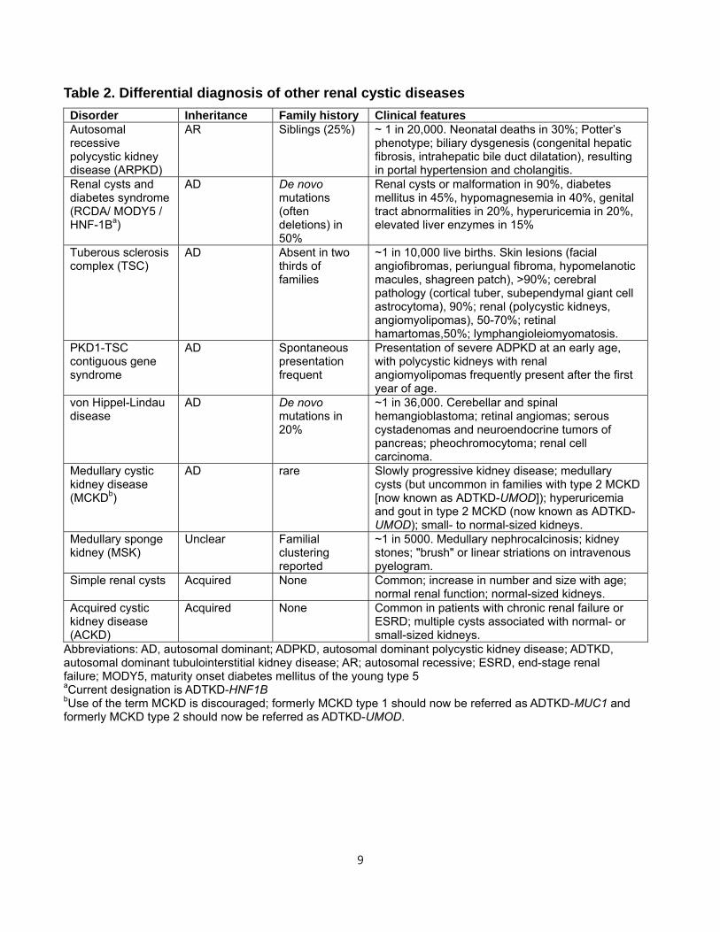
Table 2. Differential diagnosis of other renal cystic diseases
Disorder Inheritance Family history Clinical features Autosomal recessive polycystic kidney disease (ARPKD)
AR Siblings (25%) ~ 1 in 20,000. Neonatal deaths in 30%; Potter’s phenotype; biliary dysgenesis (congenital hepatic fibrosis, intrahepatic bile duct dilatation), resulting in portal hypertension and cholangitis.
Renal cysts and diabetes syndrome (RCDA/ MODY5 / HNF-1Ba)
AD De novo mutations (often deletions) in 50%
Renal cysts or malformation in 90%, diabetes mellitus in 45%, hypomagnesemia in 40%, genital tract abnormalities in 20%, hyperuricemia in 20%, elevated liver enzymes in 15%
Tuberous sclerosis complex (TSC)
AD Absent in two thirds of families
~1 in 10,000 live births. Skin lesions (facial angiofibromas, periungual fibroma, hypomelanotic macules, shagreen patch), >90%; cerebral pathology (cortical tuber, subependymal giant cell astrocytoma), 90%; renal (polycystic kidneys, angiomyolipomas), 50-70%; retinal hamartomas,50%; lymphangioleiomyomatosis.
PKD1-TSC contiguous gene syndrome
AD Spontaneous presentation frequent
Presentation of severe ADPKD at an early age, with polycystic kidneys with renal angiomyolipomas frequently present after the first year of age.
von Hippel-Lindau disease
AD De novo mutations in 20%
~1 in 36,000. Cerebellar and spinal hemangioblastoma; retinal angiomas; serous cystadenomas and neuroendocrine tumors of pancreas; pheochromocytoma; renal cell carcinoma.
Medullary cystic kidney disease (MCKDb)
AD rare Slowly progressive kidney disease; medullary cysts (but uncommon in families with type 2 MCKD [now known as ADTKD-UMOD]); hyperuricemia and gout in type 2 MCKD (now known as ADTKD-UMOD); small- to normal-sized kidneys.
Medullary sponge kidney (MSK)
Unclear Familial clustering reported
~1 in 5000. Medullary nephrocalcinosis; kidney stones; "brush" or linear striations on intravenous pyelogram.
Simple renal cysts Acquired None Common; increase in number and size with age; normal renal function; normal-sized kidneys.
Acquired cystic kidney disease (ACKD)
Acquired None Common in patients with chronic renal failure or ESRD; multiple cysts associated with normal- or small-sized kidneys.
Abbreviations: AD, autosomal dominant; ADPKD, autosomal dominant polycystic kidney disease; ADTKD, autosomal dominant tubulointerstitial kidney disease; AR; autosomal recessive; ESRD, end-stage renal failure; MODY5, maturity onset diabetes mellitus of the young type 5 aCurrent designation is ADTKD-HNF1B bUse of the term MCKD is discouraged; formerly MCKD type 1 should now be referred as ADTKD-MUC1 and formerly MCKD type 2 should now be referred as ADTKD-UMOD.
10
Molecular diagnosis of ADPKD
Historically, linkage analysis of polymorphic markers flanking the two disease genes has
been used for a molecular diagnosis of ADPKD, but requires multiple (preferably 4 or
more) affected family members to be informative.8 Moreover, the test results are indirect
and can be confounded by de novo mutations, mosaicism, and bilineal disease9,10
Presently, mutation-based screening by Sanger sequencing of all exons and splice
junctions of the PKD1 and PKD2 genes is the method of choice for molecular diagnosis
of ADPKD.20 Linkage analysis is rarely performed except for screening embryos in pre-
implantation genetic diagnostics (PGD) where genotyping several markers associated
with the familial mutation can provide assurance against problems associated with
screening a very small amount of DNA, such as allele dropout. PKD1 is a large complex
gene with its first 33 exons duplicated in six pseudogenes (PKD1P1-PKD1P6) with high
sequence identity, making mutation screening highly challenging.7 By contrast, PKD2 is
a single copy gene which is highly amenable to conventional mutation screening.
Comprehensive screening for PKD1 mutations is now possible using protocols that
exploit rare mismatches between the duplicated region and the PKD1P1-P6 loci for
PKD1-specific PCR (polymerase chain reaction).20 This approach, however, is labor-
intensive and costly.7 In sequencing-negative cases, multiplex ligation-dependent probe
amplification (MLPA) can be used as a follow-up test to detect large gene re-
arrangements in less than 5% of cases.27
To date, more than 1270 and 200 pathogenic mutations have been reported for PKD1
and PKD2, respectively (http://pkdb.mayo.edu). These results indicate extensive allelic
heterogeneity, especially for PKD1, with no apparent mutation “hot-spots” or common
recurrent mutations. Up to 15% of patients with suspected ADPKD are mutation-
negative despite a comprehensive screen. Some of these patients with very mild or
asymmetric PKD of de novo onset may have somatic mosaicism resulting from a
disease-causing mutation affecting an oligopotent progenitor cell during early
embryogenesis.28 The hallmark of mosaicism is the presence of more than one
genetically distinct cell line in an individual.28 The difference between somatic and
germline mosaicism is based on the findings of genetically distinct populations of cells in
11
the somatic and germline tissues, respectively.29 Mosaicism is a well-recognized cause
of variable disease expressivity in more than 30 Mendelian disorders but one that is
very difficult to diagnose by Sanger sequencing. However, Sanger sequencing of an
affected offspring of the mosaic individual may uncover the pathogenic mutation. Recent
advances in resequencing (i.e., Next-Generation Sequencing [NGS]) technologies have
enabled high-throughput mutation screening of both PKD1 and PKD2 with a recent
“proof-of-principle” study showing promising results.30 The adaptation of this new
technology to molecular diagnostics in ADPKD is expected to facilitate mutation
screening while reducing the costs at the same time.31
Marked discordant renal disease severity among affected family members has been
well documented suggesting a role for both genetic and environmental modifiers.32-35 In
several of these families, two (homozygous or compound heterozygous) non-truncating
mutations on different copies of PKD1 have been found in affected subjects with
atypical or severe renal disease while other family members with one non-truncating
mutation have mild disease.14 In other families, a truncating and a non-truncating
mutation on different copies of PKD1 or a non-truncating PKD1 mutation in combination
with a mutation in another cystogene (e.g., HNF-1β or PKHD1) has been found in
patients diagnosed in utero or with severe renal disease.16,36 Comprehensive mutation
screening of PKD1 and PKD2 as well as other cystogenes has the potential to account
for some of the within-family variability of disease severity, refine genotype-phenotype
correlations and provide useful clinical prognostic information.11-17,36
Current approach and indications of genetic testing
Most patients with ADPKD do not need molecular genetic testing. When indicated,
gene-based mutation screening of PKD1 and PKD2 by Sanger sequencing followed by
MLPA to detect gene rearrangement in sequencing-negative cases is the method of
choice but is laborious and expensive.8 Molecular genetic testing is not required for
most patients but may be considered in cases of equivocal or atypical renal imaging
findings (e.g., markedly asymmetric PKD, renal failure without significant kidney
enlargement); marked discordant disease within family; very mild PKD; sporadic PKD
12
with no family history; early and severe PKD or PKD with syndromic features; and
reproductive counseling.
Molecular genetic testing plays a greater role in childhood where PKD can be due to
autosomal recessive polycystic kidney disease (ARPKD), ADPKD or a number of rare
genetic diseases. Genetic testing of childhood PKD may be considered in cases of early
and severe PKD and in PKD with syndromic features. Genetic testing in this setting
requires consideration of diseases beyond ADPKD and should be performed by
physicians/geneticists in centers with appropriate experience and expertise.
Future role of molecular diagnostics in ADPKD
The role of molecular diagnostics in clinical medicine is rapidly evolving. Recent
advances in NGS which provides high-throughput and comprehensive diagnostic
screening at low cost compared to Sanger sequencing can be readily applied to
ADPKD.30,31 Recent studies that employed comprehensive mutation screening of PKD1,
PKD2 and other cystogenes (e.g., PKHD1, HNF1β) have identified allelic and genic
interactions that can modulate renal disease severity in ADPKD.13-17 Targeted or whole
exome sequencing will likely play an important role in the molecular diagnostics of
childhood PKD in the future. Standardized and informative reporting as well as
physician education is needed.
Pre-Implantation genetic diagnosis
PGD has been successfully applied in more than 300 genetic disorders for selecting
healthy embryos created by in-vitro fertilization for implantation. Currently, PGD is most
commonly used in severe genetic diseases with early manifestations such as cystic
fibrosis, ARPKD, among many others.37-39 PGD should be included in the discussion of
reproductive choices with patients with ADPKD, but it is only available in certain
countries and the acceptance of this technique is influenced by personal values as well
as the severity of the disease.40-42
Identification of embryos harboring a pathogenic mutation requires a biopsy. The most
13
common approach is the biopsy of cleavage-stage embryos in which one blastomere is
removed from the embryo on day 3 of development. PCR amplification of DNA from a
single cell is subject to two major pitfalls: (i) amplification failure and (ii) amplification of
only one of the two alleles present in the cell, so called ‘allele drop-out’ which can lead
to misdiagnosis.38,39 A haplotype-based screening using flanking and intragenic
microsatellite markers and multiplex PCR can be used to provide assurance against this
complication and has been successfully applied to ADPKD.43 An alternative biopsy
method (blastocyst biopsy) targets the trophectoderm on day 5 of development.44,45 This
approach removes multiple cells for analysis without sacrificing any part of the embryo
proper. The larger DNA yield compared to the single blastomere method facilitates the
molecular diagnosis. It is usually combined with cryopreservation and thawed embryo
transfer to allow more time for the genetic testing.
2. MONITORING KIDNEY DISEASE PROGRESSION IN ADPKD
Clinical trials
Treatments proven to extend kidney survival in ADPKD do not currently exist. Ideally,
treatment should start early, when the kidney parenchyma is relatively preserved.46 At
later stages, other pathologic mechanisms independent of ADPKD likely become
dominant. Nevertheless, treatments in later stage disease are also important to
preserve kidney function and their efficacy and safety should also be determined.
Randomized clinical trials (RCTs) should ideally include patients with a high likelihood of
disease progression. At early stages of ADPKD and for several decades, glomerular
filtration rate (GFR) is normal and therefore not informative. However, kidney volume in
relation to age3,4,47,48 can identify patients with progressive disease.
Adopting GFR as an outcome in trials that include patients at early stages would require
long periods of follow-up and are unrealistic. Conversely, change in total kidney volume
(TKV) or change in volume of specific kidney compartments may be a valid primary or
secondary outcome. TKV is an accurate estimate of kidney cyst burden and associates
14
with many renal manifestations of ADPKD including pain, hypertension, gross
hematuria, and proteinuria or albuminuria. While there is broad consensus for the value
of TKV as a prognostic biomarker, most regulatory agencies do not currently accept
TKV as a primary endpoint in clinical trials for ADPKD.
Total kidney volume
TKV increases exponentially in virtually every ADPKD patient. The rate of increase is
highly variable and unique for each individual. Average rates of increase of TKV in
adults are 5-6%/year.3,49,50 Elevated TKV, particularly when used together with age and
kidney function, identifies individuals who are at highest risk for progression to
advanced stage CKD and ESRD and conversely, those who will most likely never lose
kidney function or progress to ESRD.47,48
TKV can be measured using a variety of imaging modalities (US, MRI and CT). Precise
measurements of TKV necessary in clinical trials to assess the impact of therapeutic
interventions over short periods of time51 can be obtained by planimetry or stereology
analysis of MR or CT images.52,53 MRI and CT are equivalent with regard to precision
and reproducibility,48 but CT imaging is associated with radiation exposure. MRI
measurements can be done using either T1 or T2 weighted images; however, T2
weighted images provide information regarding total cyst volume and do not require
gadolinium, eliminating the risk for nephrogenic systemic fibrosis.
Although less expensive, US measurements of TKV are operator-dependent, less
reproducible and less precise, and can overestimate TKV compared to MRI and CT.54,55
US measurement of TKV typically is calculated utilizing the ellipsoid equation
(π/6xLxWxD), by measuring maximum orthogonal length, width and depth of the
kidney.56 Although less precise, US has been used successfully to measure disease
progression in studies with long periods of follow-up.57 The ellipsoid equation can also
be applied to kidney dimensions obtained from MRI or CT images for rapid calculations
of TKV that can be used to select study populations in clinical trials or to help clinically
in the determination of prognosis.48
15
In clinical practice, imaging of the kidneys should be obtained as an initial evaluation of
a patient with ADPKD. Radiology reports should be standardized for all imaging
modalities and include a maximum kidney length, width and depth, and an estimate of
TKV. Given that there is no currently approved medical therapy to slow disease
progression, repeated measurements of TKV in asymptomatic patients are not currently
indicated. When approved disease-modifying therapies become available or if lifestyle
modifications are shown to alter disease progression, repeated imaging may become an
informative tool.
Other imaging parameters
Standardized reporting of imaging findings should also include the exact number of
cysts when there are less than 10 in each kidney and the liver; minimal and maximal
size of cysts in both organs; presence of complex cyst(s) and exophytic cyst(s); and the
dominant pattern (i.e., cortical, medullary, or diffuse) for each kidney. However, the
prognostic value of these data has not been adequately studied.
Other studies have underlined the importance of the non-cystic tissue as an indicator of
disease severity. One group has taken advantage of advanced image processing
techniques to subdivide non-cystic tissue on contrast-enhanced CT into two separate
components, fully enhanced parenchyma and hypoenhanced (“intermediate”)
compartment. The latter is thought to represent fibrotic tissue.58,59 The ratio of
intermediate volume relative to parenchymal volume significantly correlates with
baseline and longitudinal changes in GFR. Several MRI technologies such as diffusion
weighted and diffusion tensor MRI and MR elastography have been used to assess the
state of the parenchyma in various renal conditions60,61 but have not yet been evaluated
in ADPKD.
Although the potential of MRI as a non-invasive method for measuring blood flow in vivo
is well-established, measurement of renal blood flow (RBF) by MRI is challenging.
Several technological innovations have made it possible to measure RBF accurately
and reproducibly.62 At present, the methodology to measure RBF is not widely available.
16
In the Consortium for Radiologic Imaging Studies of Polycystic Kidney Disease (CRISP)
study, reduction in RBF measured by MRI paralleled the increase in TKV, preceded the
decline in GFR and predicted disease progression.63,64
Glomerular filtration rate
Estimation of GFR using equations including CKD-EPI and the MDRD equation (eGFR)
is acceptable for clinical care of ADPKD patients. In specific circumstances,
measurement of GFR (mGFR) using the clearance of inulin, iothalamate,
diethylenetriaminepentaacetic acid (DTPA) or iohexol is warranted. A case in point is
the timing of a potential living kidney donation procedure in an ADPKD patient with an
abnormal muscle mass for age and gender in whom eGFR may be unreliable. In this
instance, it may be necessary to assess mGFR using one of the aforementioned “gold-
standard” techniques.
Whether estimation of GFR by equations is adequate for use in clinical trials of ADPKD
has been debated. One report questioned the reliability of eGFR using the MDRD and
CKD-EPI equations to reflect actual GFR values and suggested that use of eGFR may
fail to detect changes in kidney function over time.65 This concern is based on the
theoretical rationale that ADPKD is a tubular disease and that tubular secretion of
creatinine may be different in this disease when compared to non-ADPKD individuals.
Another study reported that tubular creatinine secretion was indeed increased in
ADPKD patients when compared to healthy controls at similar mGFR level (measured
by a “gold standard” method, in this case iothalamate).66 However, this effect was
limited to those with a high-normal mGFR. Consequently, in this study the CKD-EPI and
MDRD Study equations performed relatively well in estimating GFR and change in
eGFR. These conclusions are corroborated by a third study, which added that using
cystatin C in combination with creatinine to determine eGFR might even be better.67 In
addition, the relationship between mGFR and eGFR in the MDRD Study where patients
had established renal insufficiency was not different in ADPKD as compared to other
kidney disease populations. Therefore, eGFR is in general acceptable for clinical trials.
When feasible, however, mGFR is preferable. Methods for mGFR are more
17
cumbersome, associated with considerable costs, and impractical in clinical trials with a
large number of participating centers. Whether a limited number of mGFRs outperforms
a larger number of eGFRs to assess change in kidney function over time in clinical trials
is an unanswered question. Importantly, when developing novel medical treatments, it
should be investigated whether such treatments interfere with tubular creatinine
secretion. When this is the case, baseline pretreatment eGFR should be compared with
off-treatment eGFR after study completion, or mGFR should be used.
Proteinuria
Proteinuria (greater than 300 mg/day), occurs in approximately 25% of adults diagnosed
with ADPKD,68 but typically does not exceed 1 gm/day. Its origin and glomerular versus
tubular pattern have not been thoroughly ascertained.69 Presence and level of
proteinuria are associated with larger TKV, faster decline of renal function and earlier
onset of ESRD, and therefore have prognostic value. Maximum reduction in proteinuria
in ADPKD is the treatment goal. Strategies to reach these goals include appropriate
blood pressure control and use of inhibitors of the renin-angiotensin system including
ACE inhibitors and angiotensin receptor blockers as in other chronic kidney diseases.70
In patients with nephrotic range proteinuria, the presence of a second kidney disorder
and a renal biopsy should be considered if access to renal parenchyma is feasible.
Patient reported outcomes and QOL
Instruments such as patient-reported outcome measures (PROM) are useful as end-
points for clinical trials.51 They can also be used to improve patient care but there are
gaps in knowledge about their usefulness.71 There is no current validated PROM for
ADPKD. The physical and psychological burdens to ADPKD patients are significant, yet
they are incompletely characterized and difficult to quantify. Patients with ADPKD have
not been found to score differently from the general population in standardized
questionnaires (SF36) evaluating QOL.71 Since the SF36 questionnaire was developed
to evaluate individuals with more immediate life threatening disorders, it may not be
sufficiently sensitive to characterize the domains of suffering in a chronic slowly
progressive disease such as ADPKD. A large cohort (n = 1,043) of hypertensive ADPKD
18
individuals enrolled in the Polycystic Kidney Disease Treatment Network HALT clinical
trials who completed SF36 questionnaire and the Wisconsin pain survey prior to
randomization revealed no reduction in mental or physical SF 36 scores compared to
the general population.72 In patients with early disease (eGFR > 60mL/min/1.73 m2),
there was no association between pain and height adjusted TKV (htTKV), except in
patients with large kidneys (htTKV> 1,000mL/m). Comparing across eGFR levels
patients with eGFRs of 20-44mL/min/1.73m2 were significantly more likely to report that
pain impacted on their daily lives and had lower SF-36 scores than patients with eGFRs
of 45-60 and ≥60mL/min/1.73 m2.
3. MANAGEMENT of HYPERTENSION, RENAL FUNCTION DECLINE and RENAL
COMPLICATIONS
Treatment of hypertension in the adult ADPKD population
Patients with ADPKD are at increased risk for hypertension, cardiovascular events and
cardiovascular mortality when compared to the general population.73,74 The increase in
blood pressure (BP) in this patient group has been attributed to several causes,
including increased activity of the renin-angiotensin-aldosterone system (RAAS), and
increase in sympathetic tone and primary vascular dysfunction.75-79
At present, there is no consensus on whether disease-specific BP targets apply to
ADPKD. At least, the general advice of the 2012 KDIGO Clinical Practice Guideline for
the Management of BP in CKD should be followed, suggesting a BP target ≤140/90
mmHg.80,81 In accordance with this guideline, blood pressure targets should be
individualized taking comorbidities into account. In conditions such as left ventricular
dysfunction, ICA, diabetes or proteinuria, lower BP targets are advised (≤130/80
mmHg).80,81 A RCT in 79 adult hypertensive patients with left ventricular hypertrophy
indicated that strict BP control (≤120/80 mmHg) versus regular BP control (≤140/90
mmHg) was more effective in reducing left ventricular mass.82 The recently published
19
results of the HALT PKD clinical trials suggest that blood pressure targets below those
recommended by current guidelines may be advantageous in young hypertensive
ADPKD patients with CKD stages 1 or 2 and without diabetes mellitus or significant
cardiovascular comorbidities (see below).
Home BP monitoring is relatively easy to accomplish, cost-effective and expected to
result in better treatment adherence and BP control than BP measurement during clinic
visits only.83 24h ambulatory BP measurement (ABPM) can identify subjects who do not
show a normal BP decrease during night time (non-dippers) and thus may benefit from
more intensive antihypertensive drug treatment, or drug dosing during evening hours;
this issue warrants further study.84
BP control can be achieved by lifestyle modification and medical treatment. Although
not formally studied in ADPKD patients, it is expected that achieving or maintaining a
“healthy” weight (i.e., body mass index (BMI) 20-25 kg/m2), undertaking an exercise
program (aiming for at least 30 minutes 5 times per week), and lowering salt intake (≤90
mmol/day of sodium, corresponding to ≤5 g/day of sodium chloride and ≤2 g/day of
sodium) will lower BP and consequently improve long-term cardiovascular outcome.
The case for a salt-restricted diet is strengthened by observations that ADPKD patients
have been shown to be sodium overloaded, have sodium-sensitive hypertension,76,85
and the association between higher sodium intake and increased TKV in the CRISP
study.86
It is generally accepted that agents that interfere with the RAAS should be first-line BP-
lowering agents based on evidence of a hyperactive RAAS in ADPKD patients, the
observation that these agents lower albuminuria and left ventricular mass more than
other BP-lowering agents, and limited clinical evidence suggesting more
renoprotection.82,87-89 Angiotensin-converting enzyme inhibitor (ACEi) and angiotensin-
receptor blocker (ARB) are regarded equivalent, although formal evidence is limited,
and either can be used at the discretion of the treating physician. A small study (n=20)
suggests that the ARB telmisartan is equivalent to enalapril in lowering BP, but has
20
more potent antiproteinuric, anti-inflammatory and antioxidative effects in ADPKD
hypertensive patients with microalbuminuria.90 RAAS blockade should be combined with
a sodium-restricted diet to enhance the BP lowering, cardioprotective and potential
renoprotective effects.88 In the HALT PKD clinical trial, the administration of an ACE
inhibitor alone was sufficient to achieve blood pressure control in the majority of
patients, supporting the utilization of this class of antihypertensive agents as first-line
blood pressure lowering agent. The utilization of an ACEi and ARB combination did not
confer any additional benefit compared to an ACEi alone.50 The place of
mineralocorticoid receptor antagonists in ADPKD has not been ascertained and is
worthy of study because they may exert anti-fibrotic effects89 and interstitial fibrosis is
an essential part of later stage ADPKD.47
There is controversy as to which second-line BP-lowering agents should be used. Large
RCTs in non-ADPKD populations suggested that calcium channel blockers and diuretics
may be preferred over beta-blockers for cardiovascular protection.88 On the other hand,
there are theoretical concerns that argue against using these agents in ADPKD.
Calcium channel blockers may lower intracellular calcium concentration in collecting
duct cells. This may result in an increase in tubular cell proliferation and fluid secretion,
in turn leading to accelerated cyst growth and kidney function decline.90 Diuretics
increase plasma arginine vasopressin concentration (AVP), and there is experimental
and clinical evidence suggesting that higher levels of AVP are also associated with more
rapid kidney and cyst enlargement.91,92 Furthermore, these agents may also increase
uric acid and increase the activity of the RAAS in ADPKD, which in turn, could lead to
accelerated disease progression. Comorbid conditions may influence the choice for a
specific class. For instance, in patients with angina, beta-blockers may be preferred,
and in subjects with prostate hypertrophy, alpha-blockers would be appropriate.
21
Diagnosis and management of hypertension in pediatric patients
Vascular abnormalities in ADPKD are evident from a young age. Epidemiological
studies indicate an increased risk for hypertension as well as increased left ventricular
mass index (LVMI), even in children with BPs in the prehypertensive or “borderline”
range.93,94 It is therefore recommended to screen children with a family history of
ADPKD for hypertension, despite ethical implications of positive findings.
The approach to hypertension screening is dependent on country-specific
circumstances. For instance, in some countries, all children undergo regular medical
check-ups (including BP measurement) at school. In other countries, children routinely
visit a pediatrician and BP may be checked as part of routine well childcare. In countries
where children are not regularly seen by a physician and/or BP measurements are not
standard practice, it is advised that children with a family history of ADPKD have their
BP checked by a practitioner with experience in BP measurement in children. There is
no consensus at what age such screening should be started, nor what the frequency
should be. Screening from the age of 5 years onward, with an interval of 3 years in
cases in which no hypertension is found, seems prudent. The diagnosis of hypertension
is made when systolic or diastolic BP is >95th percentile for age, height and sex, in
accordance with prevailing pediatric guidelines.95
When hypertension is diagnosed in children with a family history of ADPKD, ADPKD is
the most likely underlying cause. Screening for other causes of secondary hypertension,
therefore, will probably have limited utility and US will likely demonstrate polycystic
kidneys. While establishing the diagnosis of ADPKD in a hypertensive child at risk for
ADPKD may impact management (e.g., referral to specialist, choice of anti-hypertensive
medication), it is important to recognize that the diagnosis may have significant
psychological and economic consequences for the child and parents. Additional
diagnostic testing, specifically US, should therefore be undertaken only after careful
discussion of the possible consequences with the parents.
22
Treatment of hypertension in the pediatric population should follow prevailing pediatric
guidelines. Based on data from the adult population (outlined above) and limited clinical
evidence in the pediatric population,96 RAAS blockade by either an ACEi or ARB is
preferred as first-line treatment but should be used with caution in female adolescents
at risk for teen-age pregnancies because of their teratogenic effects even in the first
trimester of pregnancy.97
“Conventional” renoprotective treatments
Most ADPKD patients develop progressive renal insufficiency that eventually leads to
ESRD between 40 to 70 years of age.98 While several renoprotective strategies have
been identified in non-ADPKD CKD (e.g., strict BP control, RAAS inhibition and low-
protein diets), testing of such interventions in ADPKD has led to disappointing
results.82,99,100 However, many of these studies were underpowered, had short periods
of follow-up or included patients in early disease stages at low risk for progression and
with relatively stable renal function, in whom it is difficult to detect potential beneficial
effects.
Recently, the results of the HALT PKD clinical trials have been published.50,101 In study
A, 558 hypertensive patients with ADPKD (15 to 49 years of age, with an eGFR >60 ml
per minute per 1.73 m2) were randomly assigned to either a standard blood-pressure
target (120/70 to 130/80 mm Hg) or a low blood-pressure target (95/60 to 110/75 mm
Hg) and to either lisinopril plus telmisartan or lisinopril plus placebo.50 In study B, 486
patients hypertensive patients with ADPKD (18 to 64 years of age, with eGFR 25 to 60
ml per minute per 1.73 m2) were randomly assigned to receive lisinopril plus telmisartan
or lisinopril plus placebo, with the doses adjusted to achieve a blood pressure of 110/70
to 130/80 mm Hg.101 Both studies showed that an ACE inhibitor alone can adequately
control hypertension in most patients justifying its use as first-line treatment for
hypertension in this disease. Study A showed that lowering blood pressure to levels
below those recommended by current guidelines in young patients with good kidney
function reduced the rate of increase in kidney volume (by 14%), the increase in renal
vascular resistance, urine albumin excretion (all identified in CRISP as predictors of
23
renal function decline), left ventricular mass index, and marginally (after the first four
months of treatment) the rate of decline in estimated glomerular filtration rate (eGFR).
The overall effect of low blood pressure on eGFR, however, was not statistically
significant, possibly because the reduction of blood pressure to low levels was
associated with an acute reduction in eGFR within the first four months of treatment.
Although these results may not be unanimously viewed as positive, they do underline
the importance of early detection and treatment of hypertension in ADPKD. The addition
of an ARB to an ACE inhibitor did not confer additional benefit.
Two observational studies have suggested that in ADPKD patients, the average age at
start of RRT has increased considerably during the last two decades.102,103 A recent
study of ERA-EDTA Registry data on patients starting RRT between 1991 and 2010,
spanning 12 European countries with 208 million inhabitants, also showed that mean
age at onset of RRT among ADPKD patients (n=20,596) has increased, albeit
considerably less than in the two aforementioned studies from 56.6 to 58.0 years.1
While the RRT incidence did not change among ADPKD patients less than 50 years of
age, it increased among older ADPKD patients (above the age of 70). These data
suggest that the increased age of ADPKD patients at the onset of RRT may be
explained by increased access of elderly to RRT, or by lower competing risk of mortality
risk prior to the start of RRT, rather than the consequence of effective renoprotective
therapies.104 No changes in age or alterations in male to female ratio were observed
among ADPKD patients who have started RRT in Catalonia between 1984 and 2009.105
Although a low-protein diet did not show an effect on the rate of renal function decline in
ADPKD patients,100 lowering protein intake to 0.8 g/kg/day is still recommended when
eGFR is less than 30 ml/min/1.73 m2 to avoid uremic complications in accordance with
the 2012 KDIGO Guideline on CKD Evaluation and Management.80 Prescribing a
protein restricted diet should be done with appropriate patient education, preferably by a
renal dietician, and patients on such a diet should be monitored for malnutrition,
especially those patients with high total kidney and liver volumes, for whom dietary
intake of nutrients may become insufficient.
24
“Novel” ADPKD specific renoprotective treatments
Based on better knowledge of pathophysiological processes, a large number of novel
targets for lifestyle and medical interventions have been proposed.106 In the past
decades, experimental and epidemiological studies have suggested a detrimental role
of the antidiuretic hormone AVP in ADPKD. V2 receptor activation by AVP in vitro
increases intracellular cAMP levels, and consequently is believed to lead to cyst
formation and cyst growth.91,107-110 Serum levels of AVP and its surrogate copeptin are
elevated in ADPKD patients and their levels have been associated with disease severity
in cross-sectional studies111 and disease progression in longitudinal studies.92 These
observations provided the rationale to study interventions that inhibit this cAMP-
mediated pathway via increased water intake or use of vasopressin V2 receptor
antagonists.
While beneficial effects of increased water intake in ADPKD have been suggested by
animal studies,112 confirming clinical data in humans are lacking. Given the theoretical
background and the evidence from experimental data, we advise patients to increase
their water intake. There is a controversy on how to identify ADPKD patients that may
benefit from increased water intake, and the level to which water intake should be
increased. Some have advised to increase the intake of water to achieve an average
urine osmolality of 250 mOsm/kg.113 Whether an increase in water intake can be
sustained over long periods of time remains to be determined.114 The risk of
hyponatremia has to be considered, particularly in patients who have impaired kidney
function and are also on a sodium restricted diet and receiving diuretics or drugs that
can inappropriately stimulate the release of vasopressin or potentiate its action, such as
serotonin reuptake inhibitors and tricyclic antidepressants.113 It should also be noted
that a recent study in 34 patients failed to demonstrate a beneficial effect of increased
hydration in ADPKD.115 Because the study was not randomized, lasted only one year,
and the patients in the high water group had a higher salt intake (reflected by higher
urine sodium excretion), it needs to be interpreted with caution. Long-term randomized
studies of enhanced hydration in ADPKD are needed.
25
Given the importance of dietary interventions for the treatment of hypertension, as well
as prevention of uremic symptoms and possibly to prevent renal function decline, we
advise that dietary compliance be monitored with 24h urine collections to measure urine
volume and excretions of sodium and urea nitrogen. Caffeine is a methylxanthine that
increases intracellular cAMP levels in cultured ADPKD renal epithelial cells.116 However,
the clinical effects of caffeine restriction have been insufficiently investigated in ADPKD
to support a firm recommendation on the limits of intake. A cross-sectional study of 102
ADPKD patients and healthy controls showed a low level of caffeine consumption by
ADPKD patients likely due to awareness of the recommendation for caffeine restriction
and no association between caffeine intake and kidney volume within the range of
caffeine intake by ADPKD patients in this study (0-471 mg/day).117 For now, avoiding
high caffeine intake seems justified as a general principle.
There are exciting developments with respect to medical treatments to manage renal
disease progression in ADPKD.118 There is overwhelming evidence for enhanced
mTORC1 signaling in PKD cystic tissues, and preclinical trials of mTOR-inhibiting
rapalogs (sirolimus and everolimus) in rodent models have been mostly encouraging. At
doses and blood levels achievable in humans, sirolimus and everolimus were effective
in a rat model of PKD affecting proximal tubules119,120 but not in a model of ARPKD
affecting the distal nephron and collecting duct.121 Mice tolerate much higher doses and
blood levels than rats and humans, and these high doses of rapalogs were consistently
effective in orthologous and nonorthologous mouse models.122,123 However, the results
of clinical trials in ADPKD stages with early, as well as later stage CKD have been
discouraging,124-126 likely because blood levels capable of inhibiting mTOR in peripheral
blood mononuclear cells do not inhibit mTOR in the kidney.127 Several strategies to
overcome the systemic toxicity and limited renal bioavailability of rapalogs deserve
further study.128-130
The TEMPO 3:4 trial studied the effects of the vasopressin V2 receptor antagonist
tolvaptan in 1445 ADPKD patients with an estimated creatinine clearance ≥60 mL/min
and a TKV of ≥750 mL.49 This RCT demonstrated a significant beneficial effect on the
26
rate of growth of TKV (-48%) and rate of eGFR decline (-26%) in patients with ADPKD.
Tolvaptan was approved in March 2014 by the regulatory authorities in Japan for the
suppression of progression of ADPKD in patients with increased and rapid rate of
increase in TKV.131 In the United States the FDA requested the manufacturer of
tolvaptan to provide additional data to further evaluate the efficacy and safety of this
drug in patients with ADPKD.132 Concerns raised during the initial review process
included: 1) not accepting TKV as an established surrogate; 2) uncertainty introduced
by missing data and a post-treatment baseline for the key secondary endpoint; 3)
potential risk for hepatotoxicity; and 4) the “small” 1 mL/min/1.73 m2/year (26%)
improvement in renal function decline. Applications for approval of tolvaptan for the
treatment of ADPKD are currently under review by the European Medicines Agency
(EMA) and Health Canada.
Somatostatin analogues, such as lanreotide and octreotide, have been studied for their
effects on liver volume in ADPKD patients with symptomatic polycystic livers. Three
placebo controlled RCTs all indicated a favorable effect on this outcome, and also
suggested beneficial kidney volume growth reducing effects and preservation of kidney
function.133-136 These trials were of short duration and included a relatively small number
of patients. Therefore, they do not allow firm conclusions. The recently published
ALADIN study137 included 79 ADPKD patients with an eGFR ≥40 mL/min/1.73 m2
randomized to intramuscular injections of octreotide-LAR or placebo. The primary
outcome variable, a mean increase in TKV at three years of follow-up, showed
numerically smaller growth in the octreotide-LAR group than in the placebo group (220
versus 454 mL). The difference, however, was not statistically significant. A favorable
effect was noted on the secondary outcome of kidney function, but this endpoint also
did not reach statistical significance. These findings provide support for larger RCTs to
test the protective effect of somatostatin analogues against renal function loss. At least
one of such trials that includes 300 ADPKD patients with CKD stages 3a and 3b is
ongoing.138 Until the results of larger trials become available, somatostatin analogues
should not be prescribed for renoprotection outside of a research study.
27
Lastly, an RCT of HMG-CoA reductase inhibition with pravastatin in ADPKD children
with an estimated creatinine clearance ≥ 80 ml/min/1.73m2 showed slower kidney
volume growth and reduced loss of kidney function.139 These promising data need
confirmation also in the adult ADPKD population. A two-year, randomized open-label
clinical trial of pravastatin in 49 adult ADPKD patients with all levels of renal function
showed no significant differences in the rate of GFR decline or level of proteinuria
between the active treatment and placebo arms despite a significant fall in total serum
cholesterol in the pravastatin-treated patients.140 Larger, longer duration studies are
needed.
Hematuria and cyst hemorrhage
Cyst hemorrhage or gross hematuria occur in approximately 60% of patients. Cyst
hemorrhage can be associated with fever and differentiation from cyst infection may be
difficult. Gross hematuria can result from cyst hemorrhage, nephrolithiasis, infection and
rarely renal cell or urothelial carcinoma, but often no specific cause can be identified. In
young individuals with ADPKD, gross hematuria is commonly seen following impact
trauma associated with sports and physical activity. Hematuria is positively associated
with increased kidney volume and cyst wall calcifications. Microscopic hematuria also
occurs in ADPKD but its frequency has not been well defined.
Hematuria can be asymptomatic or painless, or it can associate with acute pain
syndromes necessitating medical attention and narcotic analgesics. Episodes of cyst
hemorrhage or gross hematuria are usually self-limited and resolve within 2 to 7 days. If
symptoms last longer than 1 week or if the initial episode of hematuria occurs after age
50 years, investigation to exclude neoplasm should be undertaken. Rarely, bleeding can
be persistent or severe, sometimes with extensive subcapsular or retroperitoneal
hematomas, requiring hospitalization. Temporary discontinuation of RAAS inhibitors and
diuretics to avoid acute kidney injury during an episode of acute cyst hemorrhage has
been suggested.141 The antifibrinolytic agent tranexamic acid has been used
successfully to treat the hemorrhagic complications of ADPKD, but no controlled studies
have been performed.142
28
Nephrolithiasis
Nephrolithiasis and cyst wall calcifications are common in ADPKD. Increased urinary
stasis and metabolic factors (reduced urine pH, ammonium excretion and urinary
citrate) account for the increased frequency of stones.143-146 Whether nephrolithiasis
associates with an increased risk for renal insufficiency, as it has been reported in the
general population, is uncertain.147 CT is the best imaging technique for the detection
and evaluation of kidney stones. Dual energy CT can differentiate uric acid from calcium
containing stones.148,149 Medical treatment of nephrolithiasis in patients with ADPKD is
the same as in patients without ADPKD. Potassium citrate is the treatment of choice in
the three stone-forming conditions associated with ADPKD: uric acid nephrolithiasis,
hypocitraturic calcium oxalate nephrolithiasis, and distal acidification defects.
Information on indications and results of surgical interventions for nephrolithiasis are
limited to reports of center experiences and therefore subjected to substantial bias.
Nevertheless these reports suggest that extracorporeal shock wave lithotripsy and
percutaneous nephrostolithotomy can be used in most patients with ADPKD without
increased complications compared to patients without ADPKD.150 Flexible
ureterorenoscopy with laser fragmentation has also been used safely and effectively
with less risk for traumatic nephron loss.151,152
Management of renal cyst infection
Recent meta-analyses highlight the course and successful management of both renal
and liver cyst infections.153 The presence of fever, abdominal pain, and high
sedimentation rate or level of C-reactive protein (CRP) should raise the suspicion of a
cyst infection, but the differential diagnosis is broad and a definitive diagnosis is
hindered by the lack of specificity of conventional imaging studies.154,155 Blood and urine
cultures may be negative and cyst aspiration for culture should be considered if a
complex cyst in the right setting is identified. By some reports 18 F-fluorodeoxyglucose-
positron emission tomography (FDG-PET) is particularly helpful in identifying infected
cysts,153 but it is not widely available or reimbursed for this indication in some countries
and there is no consensus on whether it provides additional information that changes
medical decision making.156,157 Lipid-permeable anti-microbial agents such as
29
fluoroquinolones and trimethoprim-sulfamethoxazole, depending on sensitivity (if
available), remain the standard treatment for cyst infections.158 Once antibiotic therapy
has been initiated, there is wide variability regarding duration of treatment and
indications and timing of percutaneous or surgical draining; however extended antibiotic
therapy is often warranted. Efficacy of antibiotic treatment and infection eradication are
defined by the disappearance of fever, normalization of CRP levels, and at least two
negative blood and/or urine cultures. Cyst infection may recur even after adequate
periods of antibiotic therapy.
Management of chronic pain
Kidney pain is common in patients with ADPKD and it can be severe and disabling.159-
161 It may develop after an episode of acute pain and is likely maintained by aberrant
activity of sensory and autonomic neurons innervating the kidney, renal pelvis and
ureter. It has a negative impact on sleep, activity, mental status, and social
relationships. Health care providers often fail to discuss pain during encounters with
patients, leading to suboptimal management. Ongoing support to patients is essential
for the management of chronic pain. Careful history taking and physical exam (location
and characterization of the pain) are the initial steps.160,161 Differential diagnosis should
be sought by a multidisciplinary workup with radiologists, physical therapists, and pain
specialists. Pre-medication therapy needs to be initiated with consultation of the patient
and physical therapist. If needed, a sequential medication approach should be based on
the WHO’s pain relief ladder.160,161 Percutaneous cyst aspiration is helpful as a
diagnostic procedure to determine whether a more permanent intervention such as cyst
sclerosis or laparoscopic cyst fenestration is worth pursuing.162,163 Celiac plexus
blockade, radiofrequency ablation, and spinal cord stimulation have also been used.164
Thoracoscopic sympathosplanchnicectomy may be helpful in some patients with
disabling pain but it is invasive and has potential complications such as pneumothorax
and orthostatic hypotension.165 Laparoscopic renal denervation has been helpful in a
small series of patients.166 Recently, percutaneous transluminal catheter-based
denervation has also been shown to be effective for the treatment of kidney pain in
single case reports and deserves further evaluation in ADPKD.167,168
30
Reproductive issues
All women of reproductive potential should receive counseling on potential aggravation
of polycystic liver disease (PLD) with exogenous estrogen or progesterone exposure.
Counseling for both parents should also discuss the risk of passing on the disease to
their offspring, and the risks to both the baby and mother should pregnancy take place.
Preemptive discontinuation of RAAS inhibitors is necessary due to the potential
teratogenicity and increased risk of acute renal failure in the developing fetus. Utilization
of appropriate antihypertensive medications documented to be safe in pregnancy is
important.
Most of the available information on maternal and fetal outcomes during pregnancy in
ADPKD was collected retrospectively in the 1980’s and 1990’s.169,170 In general, ADPKD
women with normal BP and kidney function have a favorable course during pregnancy.
Nevertheless, pregnancy induced hypertension and preeclampsia occur more
frequently. These rates increase when hypertension is present prior to the pregnancy.
Recent data indicate that preeclampsia is a risk factor for future development of ESRD
in the general population, but its contribution to disease progression in ADPKD has not
been studied.171 Multiple pregnancies (> 3) have been reported to be associated with a
greater risk for decline in kidney function in ADPKD.172
Similar to general CKD, ADPKD women with established renal insufficiency are at
increased risk for early fetal loss, difficulty in controlling hypertension and accelerated
loss of kidney function.173 Because of ADPKD pregnancies are associated with a higher
frequency of new onset hypertension, pre-eclampsia, intrauterine growth retardation
and premature delivery, referral to a high-risk obstetrician is recommended especially in
patients with hypertension or elevated creatinine level.
New fetal US technology and improved imaging, specifically with regard to fetal kidneys
and liver, presents an opportunity for prenatal screening for ADPKD. Currently this is
not recommended due to ethical concerns of assigning a diagnosis when no proven
therapy is available; lack of data regarding the application of prognosis and diagnosis to
31
abnormal kidney or liver fetal US findings; and limitations of semi-quantitative measures
of amniotic fluid levels with regard to renal prognosis. Given the importance of the intra-
uterine environment on terminal nephron differentiation and birth weight, a known risk
factor for the development of CKD, further research into the role of intra-uterine
environment in contributing to disease severity in ADPKD should be conducted.
4. MANAGEMENT of ESRD
ADPKD leads to renal failure in most affected individuals. While several aspects of
ESRD management can be inferred from data in non-ADPKD patient populations, there
are some issues which are specifically relevant for ADPKD patients.
Optimal choice of RRT
Kidney transplantation is the optimal choice of RRT in appropriate patients with ADPKD.
This recommendation is based on the presumed applicability of data in the general
ESRD population to ADPKD patients and on observational data in single centers and
national or regional registries in France,174 Denmark,103 the US,175 Italy,176 and
Catalonia.105 Furthermore, the degree of comorbidity is generally lower in ADPKD than
in other types of ESRD patients, and thus a higher percentage of the former is likely to
benefit from renal transplantation. As for patients with other kidney disease etiologies, a
direct comparison of the prognosis of transplanted and non-transplanted patients is
difficult, due to strong selection bias. A comparison of the prognosis of transplanted
patients with patients who are equally qualified for transplantation but still on the waiting
list, has shown a benefit of transplantation in the general ESRD population.177
As in the general ESRD population, living kidney donation, ideally performed as
preemptive transplantation is likely to be associated with best outcomes in ADPKD
patients.178 However, a direct comparison between the results of preemptive and later
transplantation has not been performed in ADPKD patients and the time on dialysis
associated with a worsening of prognosis is unknown. The long course of ADPKD, the
32
high level of family awareness and the predictable rate of loss of renal function facilitate
arrangements for preemptive or at least early transplantation from a living donor. The
limited number of potential donors in some affected families raises the question about
donation priorities, in particular when children already have reduced kidney function at
the time when one of their parents develops ESRD. Appropriate individual and family
counseling is required to support decision making in such situations.
When transplantation is not an option, or for those waiting for transplantation, either
hemodialysis (HD) or peritoneal dialysis (PD) are suitable treatment modalities.
Although intra-abdominal space restrictions, increased risk for abdominal wall hernias
and increased prevalence of colonic diverticula may pose challenges, ADPKD is not a
contraindication for PD. The most convincing evidence supporting this conclusion
comes from Hong Kong, where a general policy for starting ESRD therapy with PD is
being implemented for all ESRD patients: ADPKD patients were not found to experience
an increased risk of treatment failure.179 Others have also reported the feasibility of PD
in ADPKD.180,181 Nevertheless determining risk factors for PD failure and complications
based on patient history and measurements of total kidney and liver size and abdominal
cavity volume are desirable to support rational decision making.
Preparation for transplantation, nephrectomy prior to kidney transplantation
In preparing ADPKD patients for kidney transplantation, the removal of one kidney is
frequently considered. However, nephrectomy in ADPKD patients, even when
performed as elective surgery, is associated with significant morbidity, potential need for
blood transfusions, and procedure-related mortality.158,182-185 Therefore the indication
should be based on a risk-benefit analysis and kidneys should not be routinely removed
prior to transplantation. Hand assisted laparoscopic nephrectomy may be better
tolerated, although conversion to open nephrectomy may be necessary for very large
cystic kidneys.186-188 Possible indications include recurrent and/or severe infection,
symptomatic nephrolithiasis, recurrent and/or severe bleeding, intractable pain, and
suspicion of renal cancer. Insufficient space for insertion of a kidney graft may represent
an indication for native nephrectomy, but establishing this need is difficult and practices
33
vary widely, with pre-transplant nephrectomy rates between 3% and 100%.174,182,184,189
While no direct comparisons of different strategies are available, on average less than
one third of patients in published series undergo pre-transplant nephrectomy,174,189,190 a
figure that may serve as a benchmark for transplant programs. The decision for or
against nephrectomy should also take into account that native kidney size typically
declines after transplantation.191 Space considerations are usually an indication for
unilateral rather than bilateral nephrectomy. Experience with both, prior and
simultaneous nephrectomy has been reported189,192 but both practices have not been
directly compared in a prospective and randomized fashion. Transcatheter artery
embolization has been suggested as an alternative to nephrectomy to obtain sufficient
volume reduction for graft implantation.193
Apart from the consideration of nephrectomy and, in very rare cases, combined
liver/kidney transplantation, the evaluation of ADPKD patients prior to transplantation is
the same as for non-ADPKD candidates. Some centers screen patients for ICA prior to
transplantation, but the risk-benefit relationship of this approach remains unknown.
Practice varies also with respect to screening for diverticular disease. Evaluation of BMI
needs to take into account the weight of severely enlarged organs.
Post-transplant complications in ADPKD patients
There is no evidence to suggest that ADPKD patients should be treated with different
immunosuppressive protocols as compared to other transplant recipients.
Overall, post-transplant morbidity appears not to be increased in ADPKD patients as
compared to other, non-diabetic transplant recipients. However, specific complications
have been reported to be more frequent, including new onset diabetes,174
gastrointestinal (GI) complications,194,195 erythrocytosis,174 urinary tract infections,174,196
thromboembolic complications,174 and hemorrhagic stroke.197
34
Use of kidneys from ADPKD patients for transplantation
Occasionally the question arises whether kidneys from a deceased ADPKD patient can
be offered for transplantation. Under specific circumstances the use of ADPKD kidneys
with acceptable kidney function and size is an option, provided there is fully informed
consent of the recipient. The success of such an approach has been reported.198
However, the optimal donor, organ, and recipient characteristics needed to make this an
acceptable strategy have not been defined.
Risk for renal cancer in ADPKD with renal failure
The incidence of clinically significant renal cell carcinoma (RCC) in ADPKD patients on
dialysis or after transplantation is not known to be increased as compared to patients
with other kidney disease etiology.199,200 A recent study from the Scientific Registry of
Transplant Recipients of 10,166 and 107,339 kidney recipients with and without
ADPKD, respectively, found no increased risk of RCC associated with this diagnosis.201
However, examination of ADPKD kidneys after nephrectomy of dialysis patients
revealed a 5% to 8% incidence of RCC, most measuring ≤2 cm in diameter.202,203
Although this observation raises concerns about the potential for malignant
transformation in ADPKD kidneys, there is currently insufficient evidence for systematic
screening in asymptomatic patients. Furthermore, the diagnostic value of non-invasive
US is limited in ADPKD kidneys and the appropriate screening methodology (i.e.,
contrast-enhanced CT) is associated with costs and potential harm. Given the increased
risk of nephrectomy in ADPKD patients, the optimal management of suspicious lesions
(i.e., observation vs. intervention) remains unknown and as such decisions should be
taken individually. In any case, visible hematuria requires evaluation of the entire urinary
tract for cause.
Hemoglobin, BP, and lipid targets in ADPKD patients on dialysis
There is no evidence that therapeutic targets for BP, lipids or hemoglobin should be
different in ADPKD compared to other patients on dialysis. Due to better preserved
erythropoietin production, anemia is on average less severe in ADPKD patients than in
35
other CKD patients 180 and some patients spontaneously maintain hemoglobin levels
above current treatment targets without receiving ESAs.204 In general such patients do
not appear to be at increased risk for thromboembolic complications. The threshold for
intervention by phlebotomy can therefore be higher than the hemoglobin target range of
patients treated with ESAs.
Anticoagulation
There is insufficient evidence to recommend a specific management of anticoagulation
in ADPKD patients with ESRD. The history of bleeding and/or macrohematuria episodes
should influence treatment decisions and trigger work-up in individual patients. Whether
and to what extent the risk and/or severity of bleeding from ICA are increased by
systemic anticoagulation is unknown.
5. MANAGEMENT of EXTRARENAL COMPLICATIONS
ADPKD is a systemic disorder, associated with numerous extrarenal manifestations that
can be a significant cause of morbidity and mortality.205 ICA and PLD are among the
most common and debilitating of these manifestations.
Intracranial aneurysms
ICA rupture is one of the most serious complications of ADPKD, resulting in combined
morbidity and mortality rates of 35-50%.206-208 Given the safety and accuracy of current
imaging methods for screening along with the availability of less invasive treatment
modalities, early pre-symptomatic detection is desirable. However, major questions
include: Is widespread screening for ICA of all patients with ADPKD justified? If not,
which patients should be screened? If screening is negative, should patients be
screened again and if so, at what time interval? When an unruptured ICA (UIA) is
detected, what are the indications to intervene? If an UIA is recommended for
conservative management, what are the recommendations for follow-up to reduce the
risk of rupture?
36
There is limited information with respect to the natural history of ICA in ADPKD and
most of our knowledge comes from small, single center observational studies. These
studies suggest that the prevalence of asymptomatic ICA detected by magnetic
resonance angiography (MRA) among patients with ADPKD is ~9-12%, compared with
~2-3% in the general population.209-214208-213 The prevalence rates of ICA vary between
~20-27% in ADPKD patients with a positive family history vs. ~6% in those lacking a
family history.209-213,215
There are no clear risk factors for ICA rupture in patients with ADPKD, other than family
history of an ICA. The average age at ICA rupture is ~40 years, approximately 10 years
earlier than in the general population.206,207,216-218 Overall there appears to be no
difference in the rate or size of ICA rupture between ADPKD and the general
population.216 One large international study of UIA (the ISUIA study) found that the rate
of ICA rupture correlated with increasing size, location in the posterior vs. anterior
circulation and prior history of subarachnoid hemorrhage.208 Pre-symptomatic screening
for ICA in the ADPKD population shows that 80-90% of ICA are in the anterior
circulation of the circle of Willis, nearly all <7mm. If the findings from the ISUIA study are
extrapolated to ADPKD, then most ICAs that are detected by pre-symptomatic
screening would fall into a low risk category for rupture. However, there are reports of
ICA rupturing at small sizes in ADPKD.206,215,216,218 It has been suggested that small
ICAs that form rapidly may be at the greatest risk for rupture soon after they develop.208
Whether this process evolves differently in ADPKD is unknown.
It is often assumed that patients with ADPKD are not at increased risk of intracranial
hemorrhage once they are on RRT. A retrospective analysis of the US Renal Data
Service database, however, revealed that ADPKD is a significant predictor of this
complication in ESRD, with an adjusted hazard ratio of 1.63 vs. non-ADPKD.219 The
authors observed that the increased risk did not manifest until approximately 3 years
after starting dialysis and they surmised that the risk was mitigated after kidney
transplantation.
ICA can be associated with mutations in both PKD1 and PKD2.220 Some series, but not
others, have also reported that the median PKD1 mutation position is more 5’ (N-
37
terminal) in individuals with a vascular phenotype compared with controls.220,221
Additional analyses in larger cohorts would be needed to determine whether mutation
class could be used reliably for risk stratification.
A decision making analysis based on the ICA prevalence and risk of rupture209 revealed
that screening the ADPKD population at age 30 would result in a gain of quality-
adjusted life years only if the 5-year rupture rate exceeds 8.5%, a figure that is far
higher than the actual rate reported in ADPKD.197
Based on the data summarized above, ADPKD patients with a family history of ICA or a
personal history of ICA rupture should be screened for asymptomatic ICA. Patients with
no or unknown family history should be counseled about the risks of ICA that are
associated with ADPKD as well as the pros and cons of pre-symptomatic screening.
Those individuals who remain anxious about their risk should be offered screening.
Screening should also be considered in individuals with high-risk professions (e.g.,
pilots) where ICA rupture might place lives of others at risk. Routine, long-standing
headaches are not an indication for screening. However, the sudden occurrence of
atypical, suddenly intense headache (often described as a thunder clap headache)
possibly coupled with other neurologic symptoms, should be considered a neurologic
emergency and requires urgent evaluation.207
Traditionally, angiographic methods have been the gold standard for diagnosis of ICA.
However recent advances in technology and the desire to avoid iodinated contrast
media in patients with renal disease have made MRI the screening method of choice. A
recent study showed that 3D time-of-flight (TOF) MRA with a 3T magnet had a
screening sensitivity of 67% for ICAs <3mm, 87% for those 3-5mm and 95% for those
>5mm.222 This was equivalent to the sensitivity of CT angiography where patients
experience both radiation and contrast exposure. Therefore, TOF MRA without
gadolinium enhancement, preferably with a 3T-imaging platform, should be used for
pre-symptomatic screening.223
There is only one study that followed individuals with negative initial screening MRAs.
Schrier et al. found that 2 out of 76 patients (one patient having a family history of ICA
38
rupture) with a negative MRA developed an ICA on re-screening after 10 years.224
Based on this limited evidence, it appears prudent that individuals with a positive family
history and a negative screening MRA should be re-screened at 5-10 year intervals
while there is no need to re-screen those with a negative family history. Larger studies
are needed to support this recommendation.
Management of UIA should be discussed with a multidisciplinary team including the
treating nephrologist, neurosurgeon and interventional neuroradiologist. The size and
location of the ICA, the general health and age of the affected individual, and the risk for
rupture should determine the therapeutic approach.210,223 Overall endovascular
procedures appear to have lower associated morbidity and mortality in comparison with
surgical approaches.223,225,226 Nevertheless there remains concern with respect to the
durability of coil embolization. Treatment is best performed in expert centers with large
procedure volumes.
The natural history of UIA in ADPKD has been evaluated in a limited number of small
studies,210,224,227 suggesting that the risk of growth or rupture of small ICAs detected by
pre-symptomatic screening in ADPKD patients is quite low. It is therefore reasonable to
re-evaluate individuals with small untreated UIAs at intervals that are determined with
neurosurgical consultation but usually ranging from 6 months (initially) to every two to
three years (after stability has been established). Additional modifiable risk factors
including smoking cessation, BP control, limited heavy alcohol use and control of
cardiovascular risk factors such as hyperlipidemia should also be addressed. It is
recognized that these risk factors for ICA have not been specifically evaluated in the
ADPKD population.228
Despite the consensus of this panel that widespread screening for ICA is not indicated,
other opinions are published,223 and different attitudes exist across centers. Some
centers screen ADPKD patients with either a de novo ADPKD (i.e., no family history) or
an incomplete family history as well as those undergoing major elective surgery (e.g.,
cardiac surgery, hepatic resection and kidney/liver transplantation) or before
transplantation.
39
Diagnosis of PLD and implications for contraception
Liver cysts are the most common extrarenal manifestation of ADPKD, with a prevalence
>80% by the age of 30.229 Liver cyst burden increases with age and is greater in women
compared with men. Approximately 20% of patients with ADPKD go on to develop
symptomatic PLD.230
The use of liver imaging to determine the extent of PLD should be part of the initial
assessment of all patients with ADPKD. Most patients with PLD are asymptomatic and
can be managed conservatively.230,231 Typically, liver involvement does not cause
hepatic dysfunction. However, massive liver enlargement can result in compression of
surrounding organs. Compressive symptoms include abdominal pain and distension,
back pain, early satiety potentially causing malnutrition, gastroesophageal reflux,
compromised lung function with dyspnea or recurrent pneumonia, and hepatic venous-
outflow obstruction.230,232,233 Additional complications include infections, cyst rupture,
and hemorrhage.
A number of studies have shown that PLD tends to be more severe in women.229,234-237
Risk factors for severe PLD include multiple pregnancies and use of exogenous
estrogens. In one small prospective non-randomized study, polycystic liver volumes
increased over one year in post-menopausal women taking estrogens.237 There was
consensus that exogenous hormones or hormone-containing contraceptives should be
avoided in women who have symptomatic or severe PLD. Progesterone, like estrogens,
stimulates the proliferation of cholangiocytes, therefore, contraceptives containing only
progestogens may not be safer than those containing estrogens.238
What are the indications and modalities for intervention in PLD?
Treatment options for PLD include conservative management, surgical intervention, or
medical therapy. The indications for intervention include symptomatic PLD with
significantly diminished QOL. Several types of surgical approaches can be used to
decrease cyst burden. The choice of a specific intervention should be tailored to the
40
individual patient depending on liver anatomy, stage of PLD, concomitant renal disease
and expertise of the medical center.
The surgical options that can be considered for PLD have been reviewed in detail.239
Aspiration and sclerotherapy involve percutaneous drainage of a cyst with subsequent
instillation of an agent such as ethanol that destroys cyst lining cells so that fluid is no
longer produced. The main indication for aspiration and sclerotherapy is a large
dominant cyst that is causing symptoms. Fenestration involves drainage and
surgical/laparoscopic deroofing of cysts. Multiple cysts can be drained at the same time
using this procedure, although certain areas of the liver are not amenable to
laparoscopic visualization. Partial or segmental liver resection can also be considered in
severe, symptomatic PLD when one lobe is relatively spared and the vascular anatomy
of the preserved liver is deemed suitable. This procedure can result in substantial
morbidity. In one large series (N=124 cases) the perioperative morbidity and mortality
were 63% and 3%, respectively.240 Nonetheless liver resection can provide
considerable and sustained symptomatic relief. In the same study, performance status
had normalized or improved in ~75% of patients after a mean follow-up of 9 years.
Given the complexity and risk of this surgery, partial hepatic resection should only be
performed in centers that have extensive experience with this procedure.
Liver transplantation is a last option for selected patients with severe PLD who are not
candidates for partial liver resection. This may be a particularly relevant option for
ESRD patients who are candidates for renal transplantation. In some countries
allocation of liver grafts is based strictly on the MELD (Model for End Stage Liver
Disease) score, which doesn’t accurately reflect liver disease severity in PLD. Since
patients with severe PLD have intact liver synthetic function, they lack markers of liver
dysfunction (e.g., international normalized ratio and serum albumin) that contribute to
the MELD score, and thus priority on the liver transplant list is usually low. Data from the
European Transplant Registry suggests that liver transplant outcomes in PLD patients
are comparable to those of non-PLD liver recipients.241 It must be noted that liver
explantation can be challenging in patients with PLD if they develop adhesions as the
result of prior liver procedures. Many transplant surgeons are reluctant to transplant
41
patients who have previously had liver resection due to the potential for serious
complications.
Transcatheter arterial embolization of hepatic artery branches that supply major liver
cysts has been reported to decrease total liver volume in small series.242,243 There is
limited experience with this procedure at most centers and therefore larger, controlled
studies are needed before this therapy can be recommended.
Somatostatin analogues are a promising new avenue to reduce liver cyst volume in
PLD.234 These agents bind to somatostatin receptors and are thought to act by
decreasing cAMP levels in biliary epithelial cells. Two long-acting somatostatin
analogues, octreotide and lanreotide have been tested in clinical trials and yield a small
but reproducible and clinically significant decrement in liver volume over 1-2 years of
treatment (~ ‒4% - ‒6%) compared with ~ 0% - +1.6% in placebo groups.135,136,244-247
These agents have been relatively well tolerated, but with side effects including
diarrhea, nausea, hyperglycemia and cholelithiasis. Several studies have suggested
that most of the benefit is seen over the first year of treatment and that liver cyst volume
begins to increase again once the drug is stopped.245 The response to somatostatin
analogues is quite variable but a pooled analysis suggests that women under the age of
48 exhibit the most benefit.248 Although a small retrospective study suggested that
sirolimus used in the post-kidney transplant setting might slow PLD,249 adding
everolimus in a prospective fashion to octreotide did not confer any additional benefit.250
Thus far, somatostatin analogues have not been approved by regulatory agencies for
treatment of PLD and therefore can only be recommended for use in a clinical trial
setting. If compassionate, off-label use is contemplated in highly symptomatic cases,
then liver volume should be followed using MRI or CT to document efficacy.
How to diagnose and treat liver cyst infections?
Liver cyst infections are a common complication of PLD that can pose diagnostic and
therapeutic challenges.155 Clinical features are non-specific and include fever, right
upper quadrant tenderness and laboratory data consistent with inflammation.251,252
42
Serum and cyst fluid CA19-9 levels have been reported to be elevated in liver cyst
infection but the levels are variable and diagnostic cut-off values have not been
established.155,253 Based on retrospective data, optimal treatment includes drainage of
the infected cyst(s) and appropriate antimicrobial therapy.254 Sampling and culture of
infected cyst fluid is important for guiding antimicrobial therapy. MRI and CT are not
sufficiently specific for identifying infected cysts since chronic parenchymal injury is
usually present at baseline.154,155 There are reports showing that FDG-PET may be a
sensitive alternative for determining which cyst among many is infected.254-256 The
rationale for this approach is that metabolically active inflammatory cells take up high
amounts of glucose. Patients with liver cyst infections may require a prolonged course
of antibiotics to treat recurrent or persistent infections.154 In general the choice of an
antimicrobial agent will be guided by the culture results but antibiotics that have good
cyst penetration such as fluoroquinolones are strongly advised.251
Additional extrarenal manifestations
Extrarenal complications from ADPKD such as cardiac and vascular abnormalities and
development of cysts in other organs have also been reported. These are discussed in
greater detail below and summarized in Table 3.
Cardiac valvular abnormalities. Mitral valve prolapse is found in up to 25% of ADPKD
patients.257,258 Aortic insufficiency may be found in association with dilatation of the
aortic root. Although these lesions can progress with time, they rarely require valve
replacement.2 Pericardial effusion can be detected in up to 35% of ADPKD patients, but
it is well tolerated and usually clinically insignificant.259 We do not recommend screening
echocardiography in ADPKD unless a murmur is detected or there are other
cardiovascular signs or symptoms.
Extracranial aneurysms. Dissections and aneurysms of many large arteries including
ascending aorta,34,260 popliteal,261 coronary,262,263 and splenic arteries264 have been
reported in ADPKD. Abdominal aortic aneurysms, however, do not appear to be
43
increased in patients with ADPKD.265 Clinicians should be aware that there may be a
predisposition to a vascular phenotype in some ADPKD patients.
Arachnoid membrane cysts are found in 8 to 12% of ADPKD patients.266,267 Arachnoid
cysts are typically asymptomatic. In rare circumstances, arachnoid cysts have been
associated with an increased risk for subdural hematoma.268-270 Chronic subdural
hematoma may present with headaches and focal neurologic deficits requiring surgical
drainage.268
Spinal meningeal cysts are observed in 1.7% of ADPKD patients.271 They rarely present
with features of intracranial hypotension (orthostatic headache, diplopia, hearing loss,
ataxia) that is caused by cerebrospinal fluid loss.
Pancreatic cysts are found in about 10% of patients with ADPKD.272 They are usually
asymptomatic but cystic compression of the pancreatic duct may rarely cause chronic
pancreatitis.273
Diverticular disease. The prevalence of colonic diverticulosis in ADPKD may depend on
renal status. In one series of ADPKD patients that had not reached ESRD, there was no
increase in diverticulosis compared with controls.274 However, in a smaller series of
patients who had reached ESRD, diverticulosis was found in 50% (7 of 14) of ADPKD
patients vs. 15% (13 of 86) of non-ADPKD controls.275 In another retrospective study of
ADPKD patients with ESRD, diverticulitis was found in 20% (12 of 59) of ADPKD
patients versus 3% (4 of 125) in non-ADPKD controls.276 There are case reports of
diverticular disease in other regions of the GI tract as well.277 We do not recommend
routine screening for diverticulosis but one should be aware of a possible increased
occurrence of diverticulosis in ADPKD patients who have reached ESRD.
Abdominal hernias. A retrospective study identified abdominal wall hernias (inguinal,
incisional or para-umbilical) in 38 of 85 (45%) ADPKD patients on RRT vs. 8% in a
matched non-ADPKD ESRD cohort.278 This should be kept in mind when PD is
contemplated in an ADPKD patient.
44
Seminal vesicle cysts are found in about 40% of male ADPKD patients but are not
correlated with semen abnormalities.279 Seminal vesicle cysts are rarely symptomatic
and we do not recommend routine screening.
Infertility. A few studies have associated male infertility and sperm abnormalities
(asthenozoospermia, defect in flagella) with ADPKD.279,280 Whether infertility is more
common in males with ADPKD remains unknown. Female infertility has not been
associated with ADPKD.
Bronchiectasis. In one retrospective analysis of 95 ADPKD patients, CT scan revealed
radiographic bronchiectasis in 37% of the ADPKD group versus 13% of the CKD control
group.281 Bronchiectasis was generally mild with no clinical consequences. Therefore,
we do not recommend routine screening and imaging should be guided by symptoms.
Congenital hepatic fibrosis. Congenital hepatic fibrosis complicated by portal
hypertension, a constant association of ARPKD, is a rare but potentially life-threatening
complication of ADPKD.282 Increased liver echogenicity on US, decreased platelet
count, enlarged left lobe of liver, or enlarged spleen should alert physicians to this
possibility. While in these families liver cysts occur in multiple generations, the
association with congenital hepatic fibrosis is restricted to single individuals or siblings,
suggesting the importance of modifier genes. Upon diagnosis of an index case, siblings
should be evaluated for this association.
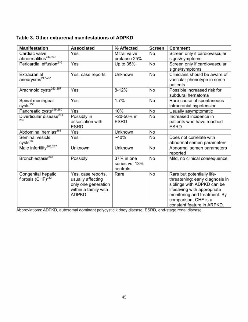
45
Table 3. Other extrarenal manifestations of ADPKD
Abbreviations: ADPKD, autosomal dominant polycystic kidney disease; ESRD, end-stage renal disease
Manifestation Associated % Affected Screen Comment Cardiac valve abnormalities244,245
Yes Mitral valve prolapse 25%
No Screen only if cardiovascular signs/symptoms
Pericardial effusion246 Yes Up to 35% No Screen only if cardiovascular signs/symptoms
Extracranial aneurysms247-251
Yes, case reports Unknown No Clinicians should be aware of vascular phenotype in some patients
Arachnoid cysts253-257 Yes 8-12% No Possible increased risk for subdural hematoma
Spinal meningeal cysts258
Yes 1.7% No Rare cause of spontaneous intracranial hypotension
Pancreatic cysts259,260 Yes 10% No Usually asymptomatic Diverticular disease261-
265 Possibly in association with ESRD
~20-50% in ESRD
No Increased incidence in patients who have reached ESRD
Abdominal hernias265 Yes Unknown No Seminal vesicle cysts266
Yes ~40% No Does not correlate with abnormal semen parameters
Male infertility266,267 Unknown Unknown No Abnormal semen parameters reported
Bronchiectasis268 Possibly 37% in one series vs. 13% controls
No Mild, no clinical consequence
Congenital hepatic fibrosis (CHF)282
Yes, case reports, usually affecting only one generation within a family with ADPKD
Rare No Rare but potentially life-threatening; early diagnosis in siblings with ADPKD can be lifesaving with appropriate monitoring and treatment. By comparison, CHF is a constant feature in ARPKD.
46
6. PRACTICAL INTEGRATED PATIENT SUPPORT
Despite ADPKD being the most common inherited kidney disease encountered by every
nephrologist worldwide, there is a lack of knowledge about the needs of and care for
patients outside the clinic environment and routine care.
What should a doctor tell or give a patient at first diagnosis?
The first diagnosis contact between the patient and physician has major importance.
Studies in cancer patients show that anxiety and distress can be lessened if the
physician can recognize the patient's psychosocial needs and convey reassurance and
support in that first consultation.283 However, there are no studies about the ways to
communicate about ADPKD to the newly diagnosed and how the latter react to the
diagnosis. There is ample evidence to suggest that primary care providers (PCPs) do
not have adequate training and knowledge about ADPKD and not all newly-diagnosed
patients are referred automatically to a nephrologist. Furthermore, many nephrologists
lack time and ability to explain the diagnosis and all its complex implications (e.g.,
medical, personal, insurance, genetics, etc.).284 Patient education programs and tools
for patients with CKD are available,285 but there is little research about their
implementation and effectiveness, and their relevance for ADPKD patients.
Evidence from one-to-one conversations or online community forums suggests that
'shock' is a common reaction of the newly-diagnosed. A patient may feel 'sad' and
sometimes 'angry' at not being told previously by a parent.286 Individual patient
responses will also vary widely according to personality and age, ranging from
determination to learn everything about ADPKD to willingness to 'put it away' and forget.
There is consensus that all patients need simple, disease-specific information initially,
including a printed material that could be read later or at home. Practical implications
such as potential impact on work, insurance, lifestyle and family planning should be
included. Where possible, patients should be automatically referred to local or national
support groups, online references and be encouraged to find someone to talk to. Each
consultation should be individualized, reassuring and tailored to the patient's literacy
47
level and culture/language. Throughout the consultation, the physician should focus on
the possibilities, not the problems, and retain a positive attitude.
Nephrologists should be familiar with the extrarenal associations of ADPKD and
appropriately educate their patients. These have been covered above and will not be
discussed in detail here. Women should be advised about the increased prevalence of
PLD in women and that multiple pregnancies or exposure to estrogens may increase
the risk for complications in those with many liver cysts.235 Physicians should disclose
the potential risk for cysts in other organs to provide reassurance and avoid
unnecessary investigations. Despite evidence about the low risk of ICA, patients remain
fearful particularly when there is family history of ICA rupture. Patients should be
advised that common headaches are not due to an ICA/UIA. Conversely, if the
headache is a 'thunderclap' type, seeking emergency medical advice is essential.
Screening should be offered if the patient requires reassurance or is highly stressed
about ICA risk.
Physicians should show empathy towards patients who present with pain. Pain should
be addressed quickly to prevent the body becoming sensitized to it. Strategies for pain
management in ADPKD have also been covered above.
Family planning
Family planning issues fall broadly into three areas: contraception/family planning,
genetic counseling, and PGD/in vitro fertilization (IVF). Considerations include ethical,
moral, legal, financial, and religious perspectives. Nephrologists and genetic counselors
should be objective in their communication of information and options. A patient (and
partner when relevant) should feel sufficiently informed and empowered to make their
own decisions. Information about contraception should be provided and individualized,
particularly on the risk of PLD in women. Female patients should be referred for a scan
to ascertain the severity of the PLD.
For genetic counseling, consistent with current clinical practice, kidney imaging is
considered sufficient. Patients should receive precise information about the extent of the
disease. With increasing availability of genetic testing and clinical relevance of such
48
testing for prognosis,17 physicians and counselors will need to be trained to
communicate the potential benefits and limitations of these analyses. Globally, there is
a wide variation in awareness, provision and exclusion of reimbursement of genetic
counseling services exists throughout the world.
The PGD/IVF subject is controversial but there was consensus that these services
should be available to all patients. Cost should not be a consideration for refusal as the
long-term cost to society of ADPKD is potentially significant. There is wide variation
across countries (from tightly restricted to available and unregulated) in the licensing of
PGD for ADPKD.
Talking to children about ADPKD and deciding when to screen
Parents with at-risk but undiagnosed children have three options: 1) screen the children
as young as possible and disclose the results to the entire family; 2) screen and
disclose results only to the parents; 3) do not screen. Parents have the right to choose.
It is preferable to be open with children if parents choose to disclose that one parent has
ADPKD,287 rather than face potential adverse consequences of diagnosis later in life.
Parents are advised to disclose the diagnosis to a child, strengthen their knowledge and
set a good example by including the entire family in a healthy lifestyle program.
Adolescents in particular may experience a range of difficulties (with friendships,
relationships, talking about ADPKD, bullying, self-worth, their future, and exclusion from
sports) and in need of special support.
What dietary and lifestyle modifications should be recommended?
Patients with ADPKD are advised to avoid high salt/sodium intake, not smoke,288 eat a
healthy diet, keep hydrated, moderate their alcohol intake and take exercise.
Additionally, patients should be encouraged and supported to self-monitor their BP and
weight. Physicians should explain the implications of blood test results and offer referral
to a renal dietitian as necessary.
49
Despite general consensus on these recommendations, little is known about the
effectiveness of these ADPKD-specific ‘healthy’ lifestyle modifications to delaying
disease progression and about how to motivate patients to adhere to such a 'healthy'
diet/lifestyle. The majority of ADPKD patients and families are confused about what they
should, can and cannot eat or drink. It is recommended to have some measures of
lifestyle change effectiveness studies, in order to encourage patient adherence.
Although there are a few PKD-friendly cookbooks available for all stages of the disease,
more are needed and they should be offered in all languages. Renal dietitians tend to
care for patients on dialysis who often follow very strict dietary regimens and the
tendency is for negative messages, e.g., 'Don't eat this and that'. More comprehensive
patient education, with focus on positive messages about diet and lifestyle are required.
An example would be “You can qualify for a transplant if you lose weight.” Patient socio-
demographics play a role in adherence289 and this should be factored into
dietary/lifestyle guidance. Where possible, patients could be self-empowered through
web-apps and research into these types of patient support programs is encouraged.
Similarly, there is controversy about soy in the diet. It is known that soy is a suitable
substitute for animal protein in CKD290 (and ADPKD by extrapolation) but nothing is
known about its action on PLD. There is a general lack of patient lifestyle guidance for
PLD.
Impact of hobbies and sports
There is a large void of evidence on the impact of exercise in ADPKD patients.291
Exercise is generally recommended, but an individual risk assessment should be
carried out for concerns related to enlarged kidneys and/or liver and a disposition to cyst
rupture. Historically, patients have always been told to 'avoid contact sports' but no
definitions are given of these. A large retrospective study of 4.4 million 'athlete-
exposures' in high school athletes with a single (non-ADPKD) kidney showed very few
kidney related injuries.292 Patients should be cautious of 'hard' contact sports, e.g.,
American football or rugby, or other potentially problematic activities such as horseback
riding, particularly when enlarged kidneys can be felt on physical examination. However,
the final decision should be made by the patient or parents.
50
Do we need to provide doctors with psychological guidelines for the care of
ADPKD patients?
Physicians should actively listen and have empathy for psychological and emotional
concerns of ADPKD patients, including anxiety about the condition and its impact on
normal life, body image/dysmorphic issues, and sexual dysfunction. It is known that
anxiety and depression are highly prevalent in CKD patients and are related to lowered
life expectancy.293 Dialysis disfigurement can lead to insecurity and relationship stress.
Many couples split during these difficult times, further increasing stress and poor
outcomes. Physicians should be trained in asking psychosocial questions to ADPKD
patients. A set of indirect questions for healthcare providers may be developed to
circumvent the difficulties inherent in asking such questions. Similarly, a set of prompts
for these difficult conversations could also be developed for patients to ask their
physicians.
There is a lack of knowledge and evidence about the impact of these psychological
factors on the QOL of ADPKD patients. Some studies have shown that anxiety is
present even in the newly diagnosed, symptomless patients.294 Another study found
depression in over 60% of ADPKD patients.295 There is no validated tool to measure
these factors in ADPKD patients. One study using the SF-36 tool concluded that it
lacked the sensitivity and specificity to detect QOL changes, in particular the growing
burden of cystic kidneys.71 Strategies for managing anxiety exist for other chronic
conditions296,297 and should be tested in ADPKD.
Financial impacts of ADPKD: careers, income, life and health insurance
Insurers' actuarial algorithms are out of date and do not take account of improvements
in symptom management, especially hypertension.298 There is inequitable access to
insurance with international/regional variation. Diagnosed adults cannot purchase life
insurance in many countries and positive diagnosis can impact ability to buy a house in
some. Patients and physicians may not disclose it on forms but many insurers now
explicitly ask about PKD or 'inherited conditions'. It is recommended that an up-to-date,
standardized and endorsed statement about ADPKD be produced that patients could
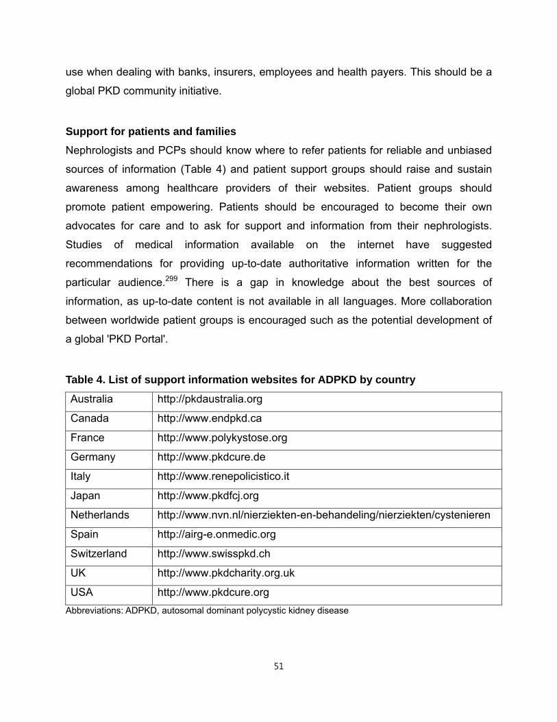
51
use when dealing with banks, insurers, employees and health payers. This should be a
global PKD community initiative.
Support for patients and families
Nephrologists and PCPs should know where to refer patients for reliable and unbiased
sources of information (Table 4) and patient support groups should raise and sustain
awareness among healthcare providers of their websites. Patient groups should
promote patient empowering. Patients should be encouraged to become their own
advocates for care and to ask for support and information from their nephrologists.
Studies of medical information available on the internet have suggested
recommendations for providing up-to-date authoritative information written for the
particular audience.299 There is a gap in knowledge about the best sources of
information, as up-to-date content is not available in all languages. More collaboration
between worldwide patient groups is encouraged such as the potential development of
a global 'PKD Portal'.
Table 4. List of support information websites for ADPKD by country
Australia http://pkdaustralia.org
Canada http://www.endpkd.ca
France http://www.polykystose.org
Germany http://www.pkdcure.de
Italy http://www.renepolicistico.it
Japan http://www.pkdfcj.org
Netherlands http://www.nvn.nl/nierziekten-en-behandeling/nierziekten/cystenieren
Spain http://airg-e.onmedic.org
Switzerland http://www.swisspkd.ch
UK http://www.pkdcharity.org.uk
USA http://www.pkdcure.org
Abbreviations: ADPKD, autosomal dominant polycystic kidney disease
52
Benefit of recognized 'PKD Center or Clinic' vs. single nephrologist
There is consensus about the value of a multidisciplinary team approach to care, with all
relevant specialties provided in one center or clinic to ADPKD patients. Benefits include:
a) opportunities for patients to meet other patients; b) opportunities to publicize research
studies and speed up recruitment to trials; c) potential for physicians to build up an
expert network; d) potential for reduction in inefficiencies from multiple clinic
appointments and fragmented care; e) potential for improved patient outcomes. There is
evidence of integration benefits and impressive cost savings in the care of patients with
rare diseases in general.300
There are challenges in the uneven geographical distribution of expertise and patients.
Establishment of specialized services in centers of excellence would inevitably result in
some patients having to travel long distances to attend. This difficulty could be
addressed by the growth in telemedicine, which is implemented across a number of
complex care specialties in the US and elsewhere.301 A 'telenephrology' approach to
care in CKD has been proposed and could be adapted for ADPKD.
CONCLUSION AND PERSPECTIVES
As a major genetic disorder affecting up to 12 million individuals worldwide and the 4th
most common global cause for RRT, ADPKD is of critical importance for nephrology
and society at large. Improvements in the diagnosis and management of the disease
manifestations paralleled general advances in medicine during the last century. More
recently, improved understanding of the molecular genetics and biology of ADPKD have
raised the hope that slowing renal disease progression or even making it
inconsequential is within reach. Yet, approaches to the diagnosis, evaluation,
prevention and treatment of the renal and extrarenal manifestations of ADPKD vary
widely and there are no broadly accepted practice guidelines. Like other types of
inherited kidney disorders, challenges and barriers to the translation of breakthroughs in
basic science to clinical care of patients with ADPKD exist, with considerable
53
heterogeneity across various countries.302 Similarly, the perceptions of the disease are
also widely divergent between physicians and patients worldwide.
The KDIGO controversy conference on ADPKD represents the first global initiative that
brought together a panel of multi-disciplinary clinical expertise and engaged patients
from 20 countries to perform a detailed analysis of the literature and to identify areas of
consensus, gaps in knowledge, and research and healthcare priorities. To this end, this
conference report has proposed an extensive research agenda with the goal to close up
these said gaps and resolve outstanding controversies (Table 5). Current knowledge
and the large volume of ongoing clinical trials and large collaborative studies warrant
the development of practice guidelines/best practice policies for ADPKD. Facing the
identification of priorities for clinical research, there is a need for a global, academic
network to prioritize, facilitate, coordinate and avoid duplication of such trials. Patient
support organizations play a key role in closing the gap between disease understanding
and the development of effective education tools, new treatments, and improved health
policies.
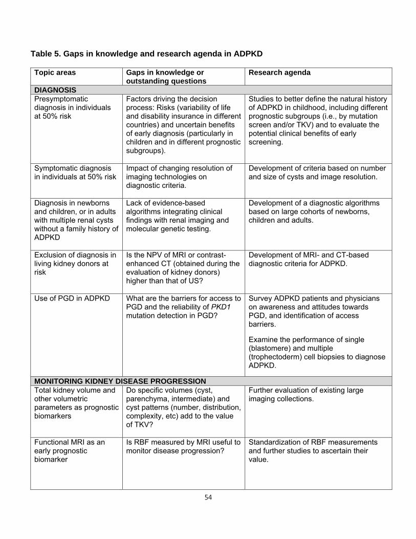
54
Table 5. Gaps in knowledge and research agenda in ADPKD
Topic areas Gaps in knowledge or outstanding questions
Research agenda
DIAGNOSIS Presymptomatic diagnosis in individuals at 50% risk
Factors driving the decision process: Risks (variability of life and disability insurance in different countries) and uncertain benefits of early diagnosis (particularly in children and in different prognostic subgroups).
Studies to better define the natural history of ADPKD in childhood, including different prognostic subgroups (i.e., by mutation screen and/or TKV) and to evaluate the potential clinical benefits of early screening.
Symptomatic diagnosis in individuals at 50% risk
Impact of changing resolution of imaging technologies on diagnostic criteria.
Development of criteria based on number and size of cysts and image resolution.
Diagnosis in newborns and children, or in adults with multiple renal cysts without a family history of ADPKD
Lack of evidence-based algorithms integrating clinical findings with renal imaging and molecular genetic testing.
Development of a diagnostic algorithms based on large cohorts of newborns, children and adults.
Exclusion of diagnosis in living kidney donors at risk
Is the NPV of MRI or contrast- enhanced CT (obtained during the evaluation of kidney donors) higher than that of US?
Development of MRI- and CT-based diagnostic criteria for ADPKD.
Use of PGD in ADPKD What are the barriers for access to PGD and the reliability of PKD1 mutation detection in PGD?
Survey ADPKD patients and physicians on awareness and attitudes towards PGD, and identification of access barriers.
Examine the performance of single (blastomere) and multiple (trophectoderm) cell biopsies to diagnose ADPKD.
MONITORING KIDNEY DISEASE PROGRESSION Total kidney volume and other volumetric parameters as prognostic biomarkers
Do specific volumes (cyst, parenchyma, intermediate) and cyst patterns (number, distribution, complexity, etc) add to the value of TKV?
Further evaluation of existing large imaging collections.
Functional MRI as an early prognostic biomarker
Is RBF measured by MRI useful to monitor disease progression?
Standardization of RBF measurements and further studies to ascertain their value.
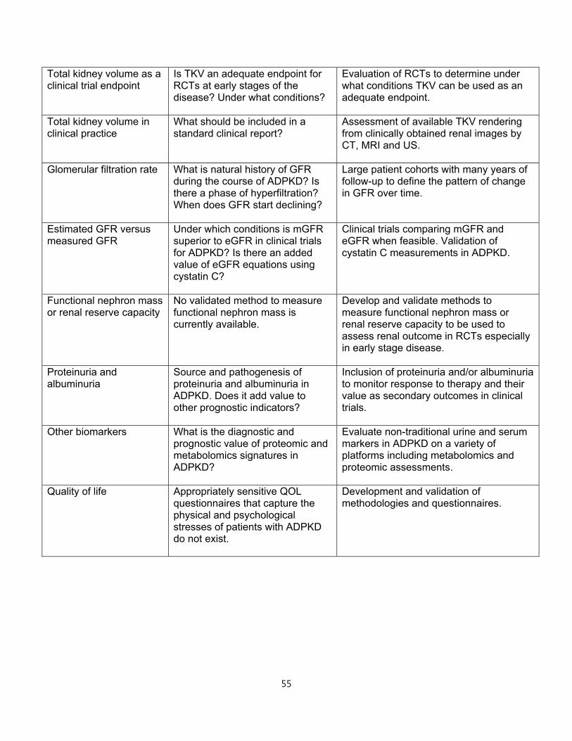
55
Total kidney volume as a clinical trial endpoint
Is TKV an adequate endpoint for RCTs at early stages of the disease? Under what conditions?
Evaluation of RCTs to determine under what conditions TKV can be used as an adequate endpoint.
Total kidney volume in clinical practice
What should be included in a standard clinical report?
Assessment of available TKV rendering from clinically obtained renal images by CT, MRI and US.
Glomerular filtration rate What is natural history of GFR during the course of ADPKD? Is there a phase of hyperfiltration? When does GFR start declining?
Large patient cohorts with many years of follow-up to define the pattern of change in GFR over time.
Estimated GFR versus measured GFR
Under which conditions is mGFR superior to eGFR in clinical trials for ADPKD? Is there an added value of eGFR equations using cystatin C?
Clinical trials comparing mGFR and eGFR when feasible. Validation of cystatin C measurements in ADPKD.
Functional nephron mass or renal reserve capacity
No validated method to measure functional nephron mass is currently available.
Develop and validate methods to measure functional nephron mass or renal reserve capacity to be used to assess renal outcome in RCTs especially in early stage disease.
Proteinuria and albuminuria
Source and pathogenesis of proteinuria and albuminuria in ADPKD. Does it add value to other prognostic indicators?
Inclusion of proteinuria and/or albuminuria to monitor response to therapy and their value as secondary outcomes in clinical trials.
Other biomarkers What is the diagnostic and prognostic value of proteomic and metabolomics signatures in ADPKD?
Evaluate non-traditional urine and serum markers in ADPKD on a variety of platforms including metabolomics and proteomic assessments.
Quality of life
Appropriately sensitive QOL questionnaires that capture the physical and psychological stresses of patients with ADPKD do not exist.
Development and validation of methodologies and questionnaires.
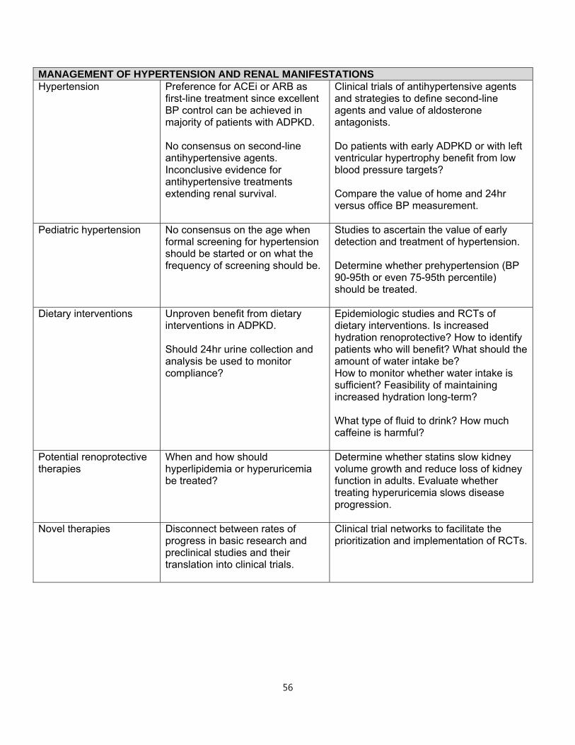
56
MANAGEMENT OF HYPERTENSION AND RENAL MANIFESTATIONS Hypertension Preference for ACEi or ARB as
first-line treatment since excellent BP control can be achieved in majority of patients with ADPKD. No consensus on second-line antihypertensive agents. Inconclusive evidence for antihypertensive treatments extending renal survival.
Clinical trials of antihypertensive agents and strategies to define second-line agents and value of aldosterone antagonists. Do patients with early ADPKD or with left ventricular hypertrophy benefit from low blood pressure targets? Compare the value of home and 24hr versus office BP measurement.
Pediatric hypertension No consensus on the age when formal screening for hypertension should be started or on what the frequency of screening should be.
Studies to ascertain the value of early detection and treatment of hypertension. Determine whether prehypertension (BP 90-95th or even 75-95th percentile) should be treated.
Dietary interventions Unproven benefit from dietary interventions in ADPKD. Should 24hr urine collection and analysis be used to monitor compliance?
Epidemiologic studies and RCTs of dietary interventions. Is increased hydration renoprotective? How to identify patients who will benefit? What should the amount of water intake be? How to monitor whether water intake is sufficient? Feasibility of maintaining increased hydration long-term? What type of fluid to drink? How much caffeine is harmful?
Potential renoprotective therapies
When and how should hyperlipidemia or hyperuricemia be treated?
Determine whether statins slow kidney volume growth and reduce loss of kidney function in adults. Evaluate whether treating hyperuricemia slows disease progression.
Novel therapies Disconnect between rates of progress in basic research and preclinical studies and their translation into clinical trials.
Clinical trial networks to facilitate the prioritization and implementation of RCTs.
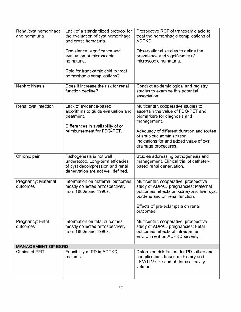
57
Renal/cyst hemorrhage and hematuria
Lack of a standardized protocol for the evaluation of cyst hemorrhage and gross hematuria. Prevalence, significance and evaluation of microscopic hematuria. Role for tranexamic acid to treat hemorrhagic complications?
Prospective RCT of tranexamic acid to treat the hemorrhagic complications of ADPKD. Observational studies to define the prevalence and significance of microscopic hematuria.
Nephrolithiasis Does it increase the risk for renal function decline?
Conduct epidemiological and registry studies to examine this potential association.
Renal cyst infection Lack of evidence-based algorithms to guide evaluation and treatment. Differences in availability of or reimbursement for FDG-PET.
Multicenter, cooperative studies to ascertain the value of FDG-PET and biomarkers for diagnosis and management. Adequacy of different duration and routes of antibiotic administration. Indications for and added value of cyst drainage procedures.
Chronic pain Pathogenesis is not well understood. Long-term efficacies of cyst decompression and renal denervation are not well defined.
Studies addressing pathogenesis and management. Clinical trial of catheter-based renal denervation.
Pregnancy: Maternal outcomes
Information on maternal outcomes mostly collected retrospectively from 1980s and 1990s.
Multicenter, cooperative, prospective study of ADPKD pregnancies: Maternal outcomes, effects on kidney and liver cyst burdens and on renal function. Effects of pre-eclampsia on renal outcomes.
Pregnancy: Fetal outcomes
Information on fetal outcomes mostly collected retrospectively from 1980s and 1990s.
Multicenter, cooperative, prospective study of ADPKD pregnancies: Fetal outcomes; effects of intrauterine environment on ADPKD severity.
MANAGEMENT OF ESRD Choice of RRT Feasibility of PD in ADPKD
patients. Determine risk factors for PD failure and complications based on history and TKV/TLV size and abdominal cavity volume.
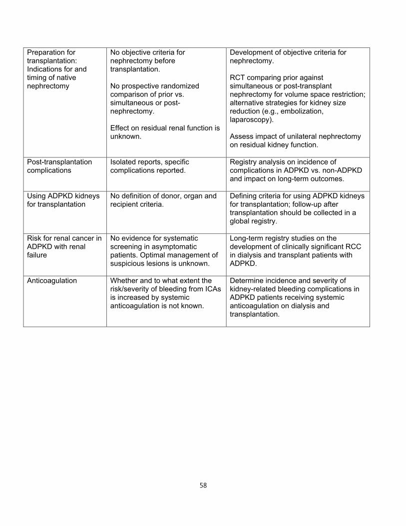
58
Preparation for transplantation: Indications for and timing of native nephrectomy
No objective criteria for nephrectomy before transplantation. No prospective randomized comparison of prior vs. simultaneous or post- nephrectomy. Effect on residual renal function is unknown.
Development of objective criteria for nephrectomy. RCT comparing prior against simultaneous or post-transplant nephrectomy for volume space restriction; alternative strategies for kidney size reduction (e.g., embolization, laparoscopy). Assess impact of unilateral nephrectomy on residual kidney function.
Post-transplantation complications
Isolated reports, specific complications reported.
Registry analysis on incidence of complications in ADPKD vs. non-ADPKD and impact on long-term outcomes.
Using ADPKD kidneys for transplantation
No definition of donor, organ and recipient criteria.
Defining criteria for using ADPKD kidneys for transplantation; follow-up after transplantation should be collected in a global registry.
Risk for renal cancer in ADPKD with renal failure
No evidence for systematic screening in asymptomatic patients. Optimal management of suspicious lesions is unknown.
Long-term registry studies on the development of clinically significant RCC in dialysis and transplant patients with ADPKD.
Anticoagulation Whether and to what extent the risk/severity of bleeding from ICAs is increased by systemic anticoagulation is not known.
Determine incidence and severity of kidney-related bleeding complications in ADPKD patients receiving systemic anticoagulation on dialysis and transplantation.
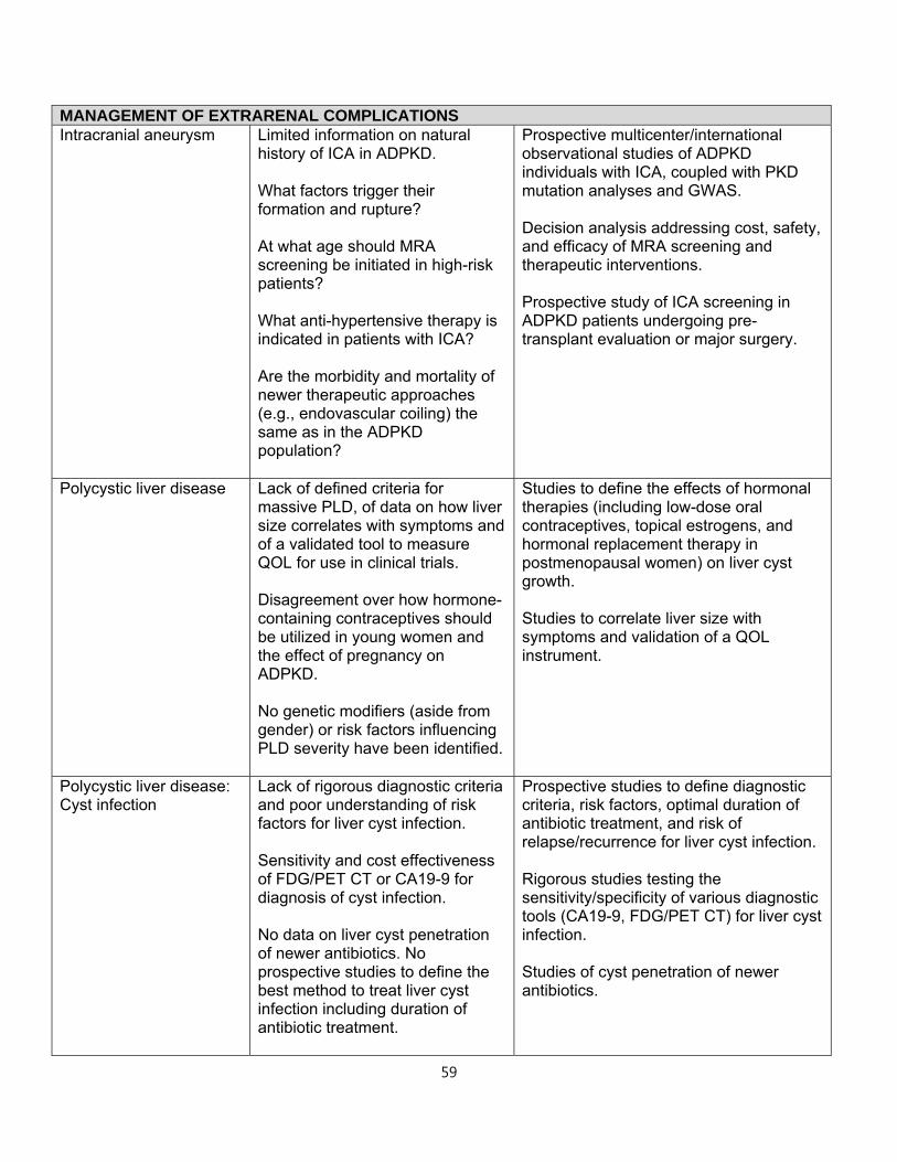
59
MANAGEMENT OF EXTRARENAL COMPLICATIONS Intracranial aneurysm Limited information on natural
history of ICA in ADPKD. What factors trigger their formation and rupture? At what age should MRA screening be initiated in high-risk patients? What anti-hypertensive therapy is indicated in patients with ICA? Are the morbidity and mortality of newer therapeutic approaches (e.g., endovascular coiling) the same as in the ADPKD population?
Prospective multicenter/international observational studies of ADPKD individuals with ICA, coupled with PKD mutation analyses and GWAS. Decision analysis addressing cost, safety, and efficacy of MRA screening and therapeutic interventions. Prospective study of ICA screening in ADPKD patients undergoing pre-transplant evaluation or major surgery.
Polycystic liver disease Lack of defined criteria for massive PLD, of data on how liver size correlates with symptoms and of a validated tool to measure QOL for use in clinical trials. Disagreement over how hormone-containing contraceptives should be utilized in young women and the effect of pregnancy on ADPKD. No genetic modifiers (aside from gender) or risk factors influencing PLD severity have been identified.
Studies to define the effects of hormonal therapies (including low-dose oral contraceptives, topical estrogens, and hormonal replacement therapy in postmenopausal women) on liver cyst growth. Studies to correlate liver size with symptoms and validation of a QOL instrument.
Polycystic liver disease: Cyst infection
Lack of rigorous diagnostic criteria and poor understanding of risk factors for liver cyst infection. Sensitivity and cost effectiveness of FDG/PET CT or CA19-9 for diagnosis of cyst infection. No data on liver cyst penetration of newer antibiotics. No prospective studies to define the best method to treat liver cyst infection including duration of antibiotic treatment.
Prospective studies to define diagnostic criteria, risk factors, optimal duration of antibiotic treatment, and risk of relapse/recurrence for liver cyst infection. Rigorous studies testing the sensitivity/specificity of various diagnostic tools (CA19-9, FDG/PET CT) for liver cyst infection. Studies of cyst penetration of newer antibiotics.
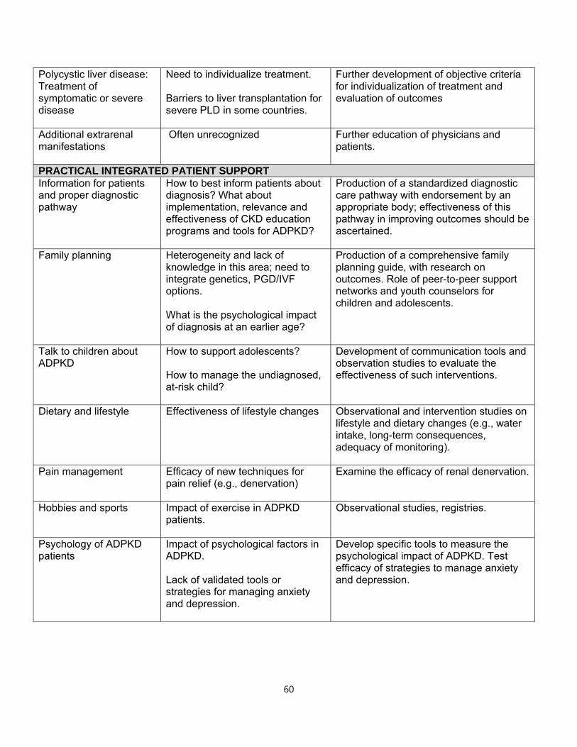
60
Polycystic liver disease: Treatment of symptomatic or severe disease
Need to individualize treatment. Barriers to liver transplantation for severe PLD in some countries.
Further development of objective criteria for individualization of treatment and evaluation of outcomes
Additional extrarenal manifestations
Often unrecognized Further education of physicians and patients.
PRACTICAL INTEGRATED PATIENT SUPPORTInformation for patients and proper diagnostic pathway
How to best inform patients about diagnosis? What about implementation, relevance and effectiveness of CKD education programs and tools for ADPKD?
Production of a standardized diagnostic care pathway with endorsement by an appropriate body; effectiveness of this pathway in improving outcomes should be ascertained.
Family planning Heterogeneity and lack of knowledge in this area; need to integrate genetics, PGD/IVF options. What is the psychological impact of diagnosis at an earlier age?
Production of a comprehensive family planning guide, with research on outcomes. Role of peer-to-peer support networks and youth counselors for children and adolescents.
Talk to children about ADPKD
How to support adolescents? How to manage the undiagnosed, at-risk child?
Development of communication tools and observation studies to evaluate the effectiveness of such interventions.
Dietary and lifestyle Effectiveness of lifestyle changes Observational and intervention studies on lifestyle and dietary changes (e.g., water intake, long-term consequences, adequacy of monitoring).
Pain management Efficacy of new techniques for pain relief (e.g., denervation)
Examine the efficacy of renal denervation.
Hobbies and sports Impact of exercise in ADPKD patients.
Observational studies, registries.
Psychology of ADPKD patients
Impact of psychological factors in ADPKD. Lack of validated tools or strategies for managing anxiety and depression.
Develop specific tools to measure the psychological impact of ADPKD. Test efficacy of strategies to manage anxiety and depression.
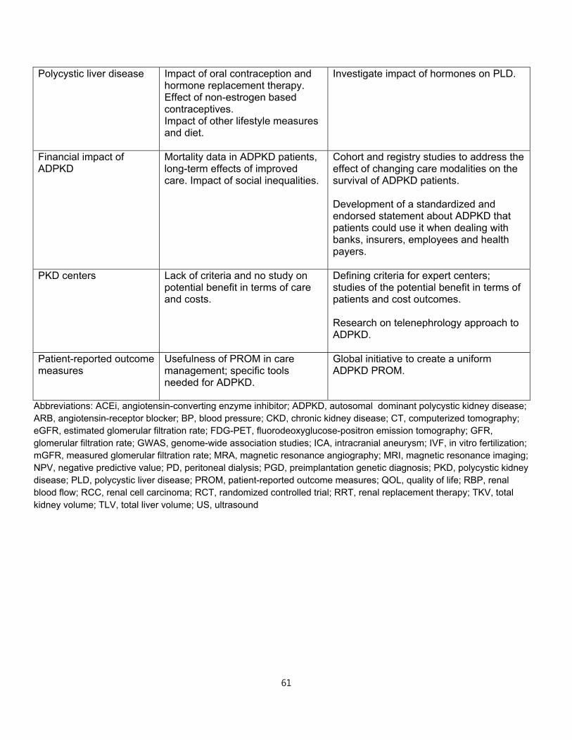
61
Polycystic liver disease Impact of oral contraception and hormone replacement therapy. Effect of non-estrogen based contraceptives. Impact of other lifestyle measures and diet.
Investigate impact of hormones on PLD.
Financial impact of ADPKD
Mortality data in ADPKD patients, long-term effects of improved care. Impact of social inequalities.
Cohort and registry studies to address the effect of changing care modalities on the survival of ADPKD patients. Development of a standardized and endorsed statement about ADPKD that patients could use it when dealing with banks, insurers, employees and health payers.
PKD centers Lack of criteria and no study on potential benefit in terms of care and costs.
Defining criteria for expert centers; studies of the potential benefit in terms of patients and cost outcomes. Research on telenephrology approach to ADPKD.
Patient-reported outcome measures
Usefulness of PROM in care management; specific tools needed for ADPKD.
Global initiative to create a uniform ADPKD PROM.
Abbreviations: ACEi, angiotensin-converting enzyme inhibitor; ADPKD, autosomal dominant polycystic kidney disease; ARB, angiotensin-receptor blocker; BP, blood pressure; CKD, chronic kidney disease; CT, computerized tomography; eGFR, estimated glomerular filtration rate; FDG-PET, fluorodeoxyglucose-positron emission tomography; GFR, glomerular filtration rate; GWAS, genome-wide association studies; ICA, intracranial aneurysm; IVF, in vitro fertilization; mGFR, measured glomerular filtration rate; MRA, magnetic resonance angiography; MRI, magnetic resonance imaging; NPV, negative predictive value; PD, peritoneal dialysis; PGD, preimplantation genetic diagnosis; PKD, polycystic kidney disease; PLD, polycystic liver disease; PROM, patient-reported outcome measures; QOL, quality of life; RBP, renal blood flow; RCC, renal cell carcinoma; RCT, randomized controlled trial; RRT, renal replacement therapy; TKV, total kidney volume; TLV, total liver volume; US, ultrasound
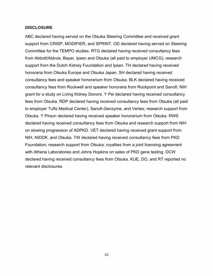
62
DISCLOSURE
ABC declared having served on the Otsuka Steering Committee and received grant
support from CRISP, MODIFIER, and SPRINT. OD declared having served on Steering
Committee for the TEMPO studies. RTG declared having received consultancy fees
from Abbott/Abbvie, Bayer, Ipsen and Otsuka (all paid to employer UMCG); research
support from the Dutch Kidney Foundation and Ipsen. TH declared having received
honoraria from Otsuka Europe and Otsuka Japan. SH declared having received
consultancy fees and speaker honorarium from Otsuka. BLK declared having received
consultancy fees from Rockwell and speaker honoraria from Rockpoint and Sanofi; NIH
grant for a study on Living Kidney Donors. Y Pei declared having received consultancy
fees from Otsuka. RDP declared having received consultancy fees from Otsuka (all paid
to employer Tufts Medical Center), Sanofi-Genzyme, and Vertex; research support from
Otsuka. Y Pirson declared having received speaker honorarium from Otsuka. RWS
declared having received consultancy fees from Otsuka and research support from NIH
on slowing progression of ADPKD. VET declared having received grant support from
NIH, NIDDK, and Otsuka. TW declared having received consultancy fees from PKD
Foundation; research support from Otsuka; royalties from a joint licensing agreement
with Athena Laboratories and Johns Hopkins on sales of PKD gene testing. DCW
declared having received consultancy fees from Otsuka. KUE, DO, and RT reported no
relevant disclosures.
63
REFERENCES
1. Spithoven E, Kramer A, Meijer E, et al. Renal replacement therapy for autosomal
dominant polycystic kidney disease ︵ADPKD ︶ in Europe: prevalence and
survival-an analysis of data from the ERA-EDTA Registry. Nephrol Dial Transplant
2014; 29: 15-25.
2. Torres VE, Harris PC, Pirson Y. Autosomal dominant polycystic kidney disease.
Lancet 2007; 369: 1287-1301.
3. Grantham JJ, Torres VE, Chapman AB, et al. Volume progression in polycystic
kidney disease. N Engl J Med 2006; 354: 2122-2130.
4. Chapman AB, Bost JE, Torres VE, et al. Kidney Volume and Functional Outcomes
in Autosomal Dominant Polycystic Kidney Disease. Clin J Am Soc Nephrol 2012;
7: 479-486.
5. Franz KA, Reubi FC. Rate of functional deterioration in polycystic kidney disease.
Kidney Int 1983; 23: 526-529.
6. Grantham JJ, Chapman AB, Torres VE. Volume progression in autosomal
dominant polycystic kidney disease: The major factor determining clinical
outcomes. Clin J Am Soc Nephrol 2006; 1: 148-157.
7. Harris PC, Rossetti S. Determinants of renal disease variability in ADPKD. Adv
Chronic Kidney Dis 2010; 17: 131-139.
64
8. Pei Y, Watnick T. Diagnosis and screening of autosomal dominant polycystic
kidney disease. Adv Chronic Kidney Dis 2010; 17: 140-152.
9. Pei Y, Paterson AD, Wang KR, et al. Bilineal disease and trans-heterozygotes in
autosomal dominant polycystic kidney disease. Am J Hum Genet 2001; 68: 355-
363.
10. Paul BM, Consugar MB, Ryan Lee M, et al. Evidence of a third ADPKD locus is not
supported by re-analysis of designated PKD3 families. Kidney Int 2014; 85: 383-
392.
11. Hateboer N, van Dijk MA, Bogdanova N, et al. Comparison of phenotypes of
polycystic kidney disease types 1 and 2. Lancet 1999; 353: 103-107.
12. Dicks E, Ravani P, Langman D, et al. Incident renal events and risk factors in
autosomal dominant polycystic kidney disease: a population and family-based
cohort followed for 22 years. Clin J Am Soc Nephrol 2006; 1: 710-717.
13. Harris PC, Bae KT, Rossetti S, et al. Cyst number but not the rate of cystic growth
is associated with the mutated gene in autosomal dominant polycystic kidney
disease. J Am Soc Nephrol 2006; 17: 3013-3019.
14. Rossetti S, Kubly V, Consugar M, et al. Incompletely penetrant PKD1 alleles
associated with mild, homozygous and in utero onset polycystic kidney disease
Kidney Int 2009; 75: 848-855.
65
15. Vujic M, Heyer CM, Ars E, et al. Incompletely penetrant PKD1 alleles mimic the
renal manifestations of ARPKD. J Am Soc Nephrol 2010; 21: 1097-1102.
16. Pei Y, Lan Z, Wang K, et al. A missense mutation in PKD1 attenuates the severity
of renal disease. Kidney Int 2012; 81: 412-417.
17. Cornec-Le Gall E, Audrezet MP, Chen JM, et al. Type of PKD1 Mutation Influences
Renal Outcome in ADPKD. J Am Soc Nephrol 2013; 24: 1006-1013.
18. Peters DJM, Sandkuijl LA. Genetic heterogeneity of polycystic kidney disease in
Europe. In: Breuning MH, Devoto M, Romeo G ︵eds ︶. Contributions to
Nephrology: Polycystic Kidney Disease, vol. 97. Karger: Basel, 1992, pp 128-139.
19. Barua M, Cil O, Paterson AD, et al. Family history of renal disease severity
predicts the mutated gene in ADPKD. J Am Soc Nephrol 2009; 20: 1833-1838.
20. Rossetti S, Consugar MB, Chapman AB, et al. Comprehensive molecular
diagnostics in autosomal dominant polycystic kidney disease. J Am Soc Nephrol
2007; 18: 2143-2160.
21. Torres V, Watson M. Polycystic kidney disease antiquity to the 20th century.
Nephrol Dial Transplant 1998; 13: 2690-2696.
22. Mochizuki T, Wu G, Hayashi T, et al. PKD2, a gene for polycystic kidney disease
that encodes an integral membrane protein. Science 1996; 272: 1339-1342.
66
23. Ravine D, Gibson RN, Walker RG, et al. Evaluation of ultrasonographic diagnostic
criteria for autosomal dominant polycystic kidney disease 1. Lancet 1994; 343:
824-827.
24. Pei Y, Obaji J, Dupuis A, et al. Unified criteria for ultrasonographic diagnosis of
ADPKD. J Am Soc Nephrol 2009; 20: 205-212.
25. Pei Y, Hwang YH, Conklin J, et al. Imaging-based diagnosis of autosomal
dominant polycystic kidney disease. J Am Soc Nephrol 2015; 26: 746-753.
26. Reed B, McFann K, Kimberling WJ, et al. Presence of de novo mutations in
autosomal dominant polycystic kidney disease patients without family history. Am J
Kidney Dis 2008; 52: 1042-1050.
27. Consugar MB, Wong WC, Lundquist PA, et al. Characterization of large
rearrangements in autosomal dominant polycystic kidney disease and the
PKD1/TSC2 contiguous gene syndrome. Kidney Int 2008; 74: 1468-1479.
28. Youssoufian H, Pyeritz RE. Mechanisms and consequences of somatic mosaicism
in humans. Nat Rev Genet 2002; 3: 748-758.
29. Gottlieb B, Beitel LK, Trifiro MA. Somatic mosaicism and variable expressivity.
Trends Genet 2001; 17: 79-82.
67
30. Rossetti S, Hopp K, Sikkink RA, et al. Identification of gene mutations in autosomal
dominant polycystic kidney disease through targeted resequencing. J Am Soc
Nephrol 2012; 23: 915-933.
31. Tan AY, Michaeel A, Liu G, et al. Molecular diagnosis of autosomal dominant
polycystic kidney disease using next-generation sequencing. J Mol Diagn 2014;
16: 216-228.
32. Persu A, Duyme M, Pirson Y, et al. Comparison between siblings and twins
supports a role for modifier genes in ADPKD. Kidney Int 2004; 66: 2132-2136.
33. Paterson AD, Magistroni R, He N, et al. Progressive loss of renal function is an
age-dependent heritable trait in type 1 autosomal dominant polycystic kidney
disease. J Am Soc Nephrol 2005; 16: 755-762.
34. Liu M, Shi S, Senthilnathan S, et al. Genetic variation of DKK3 may modify renal
disease severity in ADPKD. J Am Soc Nephrol 2010; 21: 1510-1520.
35. Bergmann C. ARPKD and early manifestations of ADPKD: the original polycystic
kidney disease and phenocopies. Pediatr Nephrol 2015; 30: 15-30.
36. Bergmann C, von Bothmer J, Ortiz Bruchle N, et al. Mutations in multiple PKD
genes may explain early and severe polycystic kidney disease. J Am Soc Nephrol
2011; 22: 2047-2056.
68
37. Harper JC, Wilton L, Traeger-Synodinos J, et al. The ESHRE PGD Consortium: 10
years of data collection. Hum Reprod Update 2012; 18: 234-247.
38. Rechitsky S, Verlinsky O, Kuliev A. PGD for cystic fibrosis patients and couples at
risk of an additional genetic disorder combined with 24-chromosome aneuploidy
testing. Reprod Biomed Online 2013; 26: 420-430.
39. Gigarel N, Frydman N, Burlet P, et al. Preimplantation genetic diagnosis for
autosomal recessive polycystic kidney disease. Reprod Biomed Online 2008; 16:
152-158.
40. Malek J, Daar J. The case for a parental duty to use preimplantation genetic
diagnosis for medical benefit. Am J Bioeth 2012; 12: 3-11.
41. de Melo-Martin I. A parental duty to use PGD: more than we bargained for? Am J
Bioeth 2012; 12: 14-15.
42. Goldsammler M, Jotkowitz A. The ethics of PGD: what about the physician? Am J
Bioeth 2012; 12: 28-29.
43. De Rycke M, Georgiou I, Sermon K, et al. PGD for autosomal dominant polycystic
kidney disease type 1. Mol Hum Reprod 2005; 11: 65-71.
69
44. Chang LJ, Huang CC, Tsai YY, et al. Blastocyst biopsy and vitrification are
effective for preimplantation genetic diagnosis of monogenic diseases. Hum
Reprod 2013; 28: 1435-1444.
45. Collins SC. Preimplantation genetic diagnosis: technical advances and expanding
applications. Curr Opin Obstet Gynecol 2013; 25: 201-206.
46. Grantham JJ, Mulamalla S, Swenson-Fields KI. Why kidneys fail in autosomal
dominant polycystic kidney disease. Nat Rev Nephrol 2011; 7: 556-566.
47. Perrone RD, Coons SJ, Cavanaugh K, et al. Patient-reported outcomes in clinical
trials of CKD-related therapies: report of a symposium sponsored by the national
kidney foundation and the U.S. Food and Drug Administration. Am J Kidney Dis
2013; 62: 1046-1057.
48. Irazabal MV, Rangel LJ, Bergstralh EJ, et al. Imaging classification of ADPKD: A
simple model for selecting patients for clinical trials. J Am Soc Nephrol 2014; 1:
160-172.
49. Torres VE, Chapman AB, Devuyst O, et al. Tolvaptan in Patients with Autosomal
Dominant Polycystic Kidney Disease. N Engl J Med 2012; 367: 2407-2418.
50. Schrier RS, Abebe KZ, Perrone RD, et al. Angiotensin Blockade, Blood Pressure
and Autosomal Dominant Polycystic Kidney Disease. N Engl J Med 2014; 371:
2255-2266.
70
51. Kistler AD, Poster D, Krauer F, et al. Increases in kidney volume in autosomal
dominant polycystic kidney disease can be detected within 6 months. Kidney Int
2009; 75: 235-241.
52. King BF, Reed JE, Bergstralh EJ, et al. Quantification and longitudinal trends of
kidney, renal cyst, and renal parenchyma volumes in autosomal dominant
polycystic kidney disease. J Am Soc Nephrol 2000; 11: 1505-1511.
53. Sise C, Kusaka M, Wetzel LH, et al. Volumetric determination of progression in
autosomal dominant polycystic kidney disease by computed tomography. Kidney
Int 2000; 58: 2492-2501.
54. O'Neill WC, Robbin ML, Bae KT, et al. Sonographic assessment of the severity
and progression of autosomal dominant polycystic kidney disease: the Consortium
of Renal Imaging Studies in Polycystic Kidney Disease ︵CRISP ︶. Am J Kidney
Dis 2005; 46: 1058-1064.
55. Bakker J, Olree M, Kaatee R, et al. Renal volume measurements: accuracy and
repeatability of US compared with that of MR imaging. Radiology 1999; 211: 623-
628.
56. Hricak H, Lieto RP. Sonographic determination of renal volume. Radiology 1983;
148: 311-312.
71
57. Fick-Brosnahan GM, Belz MM, McFann KK, et al. Relationship between renal
volume growth and renal function in autosomal dominant polycystic kidney
disease: a longitudinal study. Am J Kidney Dis 2002; 39: 1127-1134.
58. Caroli A, Antiga L, Conti S, et al. Intermediate volume on computed tomography
imaging defines a fibrotic compartment that predicts glomerular filtration rate
decline in autosomal dominant polycystic kidney disease patients. Am J Pathol
2011; 179: 619-627.
59. Antiga L, Piccinelli M, Fasolini G, et al. Computed tomography evaluation of
autosomal dominant polycystic kidney disease progression: a progress report. Clin
J Am Soc Nephrol 2006; 1: 754-760.
60. Inoue T, Kozawa E, Okada H, et al. Noninvasive evaluation of kidney hypoxia and
fibrosis using magnetic resonance imaging. J Am Soc Nephrol 2011; 22: 1429-
1434.
61. Wang WJ, Pui MH, Guo Y, et al. 3T magnetic resonance diffusion tensor imaging
in chronic kidney disease. Abdom Imaging 2014; 39: 770-775.
62. Dambreville S, Chapman AB, Torres VE, et al. Renal arterial blood flow
measurement by breath-held MRI: Accuracy in phantom scans and reproducibility
in healthy subjects. Magn Reson Med 2010; 63: 940-950.
72
63. King BF, Torres VE, Brummer ME, et al. Magnetic resonance measurements of
renal blood flow as a marker of disease severity in autosomal-dominant polycystic
kidney disease. Kidney Int 2003; 64: 2214-2221.
64. Torres VE, King BF, Chapman AB, et al. Magnetic resonance measurements of
renal blood flow and disease progression in autosomal dominant polycystic kidney
disease. Clin J Am Soc Nephrol 2007; 2: 112-120.
65. Ruggenenti P, Gaspari F, Cannata A, et al. Measuring and estimating GFR and
treatment effect in ADPKD patients: results and implications of a longitudinal
cohort study. PloS one 2012; 7: e32533.
66. Spithoven EM, Meijer E, Boertien WE, et al. Tubular secretion of creatinine in
autosomal dominant polycystic kidney disease: consequences for cross-sectional
and longitudinal performance of kidney function estimating equations. AM J Kidney
Dis 2013; 62: 531-540.
67. Orskov B, Borresen ML, Feldt-Rasmussen B, et al. Estimating glomerular filtration
rate using the new CKD-EPI equation and other equations in patients with
autosomal dominant polycystic kidney disease. Am J Nephrol 2010; 31: 53-57.
68. Chapman A, Johnson A, Gabow P, et al. Overt proteinuria and microalbuminuria in
autosomal dominant polycystic kidney disease. J Am Soc Nephrol 1994; 5: 1349-
1354.
73
69. Obermuller N, Kranzlin B, Blum WF, et al. An endocytosis defect as a possible
cause of proteinuria in polycystic kidney disease. Am J Physiol Renal Physiol
2001; 280: F244-253.
70. Jafar TH, Stark PC, Schmid CH, et al. The effect of angiotensin-converting-enzyme
inhibitors on progression of advanced polycystic kidney disease. Kidney Int 2005;
67: 265-271.
71. Rizk D, Jurkovitz C, Veledar E, et al. Quality of life in autosomal dominant
polycystic kidney disease patients not yet on dialysis. Clin J Am Soc Nephrol 2009;
4: 560-566.
72. Miskulin DC, Abebe KZ, Chapman AB, et al. Health-related quality of life in
patients with autosomal dominant polycystic kidney disease and CKD stages 1-4: a
cross-sectional study. Am J Kidney Dis 2014; 63: 214-226.
73. Schrier RW. Hypertension and autosomal dominant polycystic kidney disease. Am
J Kidney Dis 2011; 57: 811-813.
74. Ecder T. Cardiovascular complications in autosomal dominant polycystic kidney
disease. Curr Hypertens Rev 2013; 9: 2-11.
75. Chapman AB, Johnson A, Gabow PA, et al. The renin-angiotensin-aldosterone
system and autosomal dominant polycystic kidney disease. N Eng J Med 1990;
323: 1091-1096.
74
76. Torres VE, Wilson DM, Burnett JCJ, et al. Effect of inhibition of converting enzyme
on renal hemodynamics and sodium management in polycystic kidney disease.
Mayo Clin Proc 1991; 66: 1010-1017.
77. Wang D, Iversen J, Wilcox CS, et al. Endothelial dysfunction and reduced nitric
oxide in resistance arteries in autosomal-dominant polycystic kidney disease.
Kidney Int 2003; 64: 1381-1388.
78. Loghman-Adham M, Soto CE, Inagami T, et al. The intrarenal renin-angiotensin
system in autosomal dominant polycystic kidney disease. Am J Physiol Renal
Physiol 2004; 287: F775-788.
79. Rahbari-Oskoui F, Williams O, Chapman A. Mechanisms and management of
hypertension in autosomal dominant polycystic kidney disease. Nephrol Dial
Transplant 2014; 29: 2194-2201.
80. Kidney Disease: Improving Global Outcomes ︵KDIGO ︶ Blood Pressure Work
Group. KDIGO Clinical Practice Guideline for the Management of Blood Pressure
in Chronic Kidney Disease. Kidney Int Suppl 2012; 2: 337-414.
81. Kidney Disease: Improving Global Outcomes ︵KDIGO ︶ CKD Work Group.
KDIGO Clinical Practice Guideline for the Evaluation and Management of Chronic
Kidney Disease. Kidney Int Suppl 2013; 3: 1-150.
75
82. Schrier R, McFann K, Johnson A, et al. Cardiac and renal effects of standard
versus rigorous blood pressure control in autosomal-dominant polycystic kidney
disease: results of a seven-year prospective randomized study. J Am Soc Nephrol
2002; 13: 1733-1739.
83. Cohen DL, Huan Y, Townsend RR. Home Blood Pressure Monitoring in CKD. Am
J Kidney Dis 2014; 63: 835-842.
84. Rahbari-Oskoui FF, Miskulin DC, Hogan MC, et al. Short-term reproducibility of
ambulatory blood pressure monitoring in autosomal dominant polycystic kidney
disease. Blood Press Monit 2011; 16: 47-54.
85. Harrap S, Davies D, MacNicol A, et al. Renal, cardiovascular and hormonal
characteristics of young adults with autosomal dominant polycystic kidney disease.
Kidney Int 1991; 40: 501-508.
86. Torres VE, Grantham JJ, Chapman AB, et al. Potentially modifiable factors
affecting the progression of autosomal dominant polycystic kidney disease. Clin J
Am Soc Nephrol 2011; 6: 640-647.
87. Nutahara K, Higashihara E, Horie S, et al. Calcium channel blocker versus
angiotensin II receptor blocker in autosomal dominant polycystic kidney disease.
Nephron Clin Pract 2005; 99: c18-23.

76
88. The Task Force for the management of arterial hypertension of the European
Society of Hypertension ︵ESH ︶ and of the European Society of Cardiology ︵ESC ︶. ESH/ESC Guidelines for the management of arterial hypertension. J
Hypertens 2013 31: 1281-1357.
89. Brown NJ. Contribution of aldosterone to cardiovascular and renal inflammation
and fibrosis. Nat Rev Nephrol 2013; 9: 459-469.
90. Nagao S, Nishii K, Yoshihara D, et al. Calcium channel inhibition accelerates
polycystic kidney disease progression in the Cy/+ rat. Kidney Int 2008; 73: 269-
277.
91. Belibi FA, Reif G, Wallace DP, et al. Cyclic AMP promotes growth and secretion in
human polycystic kidney epithelial cells. Kidney Int 2004; 66: 964-973.
92. Boertien WE, Meijer E, Li J, et al. Relationship of Copeptin, a Surrogate Marker for
Arginine Vasopressin, With Change in Total Kidney Volume and GFR Decline in
Autosomal Dominant Polycystic Kidney Disease: Results From the CRISP Cohort.
Am J Kidney Dis 2013; 61: 420-429.
93. Cadnapaphornchai MA, McFann K, Strain JD, et al. Increased left ventricular mass
in children with autosomal dominant polycystic kidney disease and borderline
hypertension. Kidney Int 2008; 74: 1192-1196.
77
94. Zeier M, Geberth S, Schmidt KG, et al. Elevated blood pressure profile and left
ventricular mass in children and young adults with autosomal dominant polycystic
kidney disease. J Am Soc Nephrol 1993; 3: 1451-1457.
95. National High Blood Pressure Education Program Working Group on High Blood
Pressure in Children and Adolescents. The fourth report on the diagnosis,
evaluation, and treatment of high blood pressure in children and adolescents. .
Pediatrics 2004; 114: 555-576.
96. Cadnapaphornchai MA, McFann K, Strain JD, et al. Prospective change in renal
volume and function in children with ADPKD. Clin J Am Soc Nephrol 2009; 4: 820-
829.
97. Cooper WO, Hernandez-Diaz S, Arbogast PG, et al. Major congenital
malformations after first-trimester exposure to ACE inhibitors. N Eng J Med 2006;
354: 2443-2451.
98. Gabow P. Autosomal dominant polycystic kidney disease. N Engl J Med 1993;
329: 323-342.
99. van Dijk MA, Breuning MH, Duiser R, et al. No effect of enalapril on progression in
autosomal dominant polycystic kidney disease. Nephrol Dial Transplant 2003; 18:
2314-2320.
78
100. Klahr S, Breyer J, Beck G, et al. Dietary protein restriction, blood pressure control,
and the progression of polycystic kidney disease modification of diet in renal
disease study group. J Am Soc Nephrol 1995; 5: 2037-2047.
101. Torres VE, Abebe KZ, Chapman AB, et al. Angiotensin Blockade in Late
Autosomal Dominant Polycystic Kidney Disease. New Engl J Med 2014; 371:
2267-2276.
102. Schrier RW, McFann KK, Johnson AM. Epidemiological study of kidney survival in
autosomal dominant polycystic kidney disease. Kidney Int 2003; 63: 678-685.
103. Orskov B, Romming Sorensen V, Feldt-Rasmussen B, et al. Improved prognosis in
patients with autosomal dominant polycystic kidney disease in Denmark. Clin J Am
Soc Nephrol 2010; 5: 2034-2039.
104. Spithoven E, Kramer A, Meijer E, et al. Analysis of data from the ERA-EDTA
Registry indicates that conventional treatments for chronic kidney disease do not
reduce the need for renal replacement therapy in autosomal dominant polycystic
kidney disease. Kidney Int 2014; 86: 1244-1252.
105. Martinez V, Comas J, Arcos E, et al. Renal replacement therapy in ADPKD
patients: a 25-year survey based on the Catalan registry. BMC Nephrol 2013; 14:
186.
106. Chang MY, Ong AC. New treatments for autosomal dominant polycystic kidney
disease. Br J Clin Pharmacol 2013; 76: 524-535.
79
107. Gattone VH, Maser RL, Tian C, et al. Developmental expression of urine
concentration-associated genes and their altered expression in murine infantile-
type polycystic kidney disease. Develop Gen 1999; 24: 309-318.
108. Gattone VH, Wang X, Harris PC, et al. Inhibition of renal cystic disease
development and progression by a vasopressin V2 receptor antagonist. Nature
Med 2003; 9: 1323-1326.
109. Torres VE, Wang X, Qian Q, et al. Effective treatment of an orthologous model of
autosomal dominant polycystic kidney disease. Nature Med 2004; 10: 363-364.
110. Wang X, Wu Y, Ward CJ, et al. Vasopressin directly regulates cyst growth in
polycystic kidney disease. J Am Soc Nephrol 2008; 19: 102-108.
111. Meijer E, Bakker SJ, van der Jagt EJ, et al. Copeptin, a surrogate marker of
vasopressin, is associated with disease severity in autosomal dominant polycystic
kidney disease. Clin J Am Soc Nephrol 2011; 6: 361-368.
112. Nagao S, Nishii K, Katsuyama M, et al. Increased water intake decreases
progression of polycystic kidney disease in the PCK rat. J Am Soc Nephrol 2006;
17: 2220-2227.
113. Torres VE, Bankir L, Grantham JJ. A Case for Water in the Treatment of Polycystic
Kidney Disease. Clin J Am Soc Nephrol 2009; 4: 1140-1150.
80
114. Wang CJ, Creed C, Winklhofer FT, et al. Water prescription in autosomal dominant
polycystic kidney disease: a pilot study. Clin J Am Soc Nephrol 2011; 6: 192-197.
115. Higashihara E, Nutahara K, Tanbo M, et al. Does increased water intake prevent
disease progression in autosomal dominant polycystic kidney disease? Nephrol
Dial Transplant 2014; 29: 1710-1719.
116. Belibi FA, Wallace DP, Yamaguchi T, et al. The effect of caffeine on renal epithelial
cells from patients with autosomal dominant polycystic kidney disease. J Am Soc
Nephrol 2002; 13: 2723-2729.
117. Vendramini LC, Nishiura JL, Baxmann AC, et al. Caffeine intake by patients with
autosomal dominant polycystic kidney disease. Braz J Med Biol Res 2012; 45:
834-840.
118. Harris PC, Torres VE. Genetic mechanisms and signaling pathways in autosomal
dominant polycystic kidney disease. J Clin Invest 2014; 124: 2315-2324.
119. Tao Y, Kim J, Schrier RW, et al. Rapamycin markedly slows disease progression
in a rat model of polycystic kidney disease. J Am Soc Nephrol 2005; 16: 46-51.
120. Shillingford JM, Murcia NS, Larson CH, et al. The mTOR pathway is regulated by
polycystin-1, and its inhibition reverses renal cystogenesis in polycystic kidney
disease. Proc Natl Acad Sci U S A 2006; 103: 5466-5471.
81
121. Renken C, Fischer DC, Kundt G, et al. Inhibition of mTOR with sirolimus does not
attenuate progression of liver and kidney disease in PCK rats. Nephrol Dial
Transplant 2011; 26: 92-100.
122. Gattone VH, 2nd, Sinders RM, Hornberger TA, et al. Late progression of renal
pathology and cyst enlargement is reduced by rapamycin in a mouse model of
nephronophthisis. Kidney Int 2009; 76: 178-182.
123. Shillingford JM, Piontek KB, Germino GG, et al. Rapamycin Ameliorates PKD
Resulting from Conditional Inactivation of Pkd1. J Am Soc Nephrol 2010; 21: 489-
497.
124. Serra AL, Poster D, Kistler AD, et al. Sirolimus and kidney growth in autosomal
dominant polycystic kidney disease. N Engl J Med 2010; 363: 820-829.
125. Walz G, Budde K, Mannaa M, et al. Everolimus in patients with autosomal
dominant polycystic kidney disease. N Engl J Med 2010; 363: 830-840.
126. Perico N, Antiga L, Caroli A, et al. Sirolimus therapy to half the progression of
ADPKD. J Am Soc Nephrol 2010; 21: 1031-1040.
127. Canaud G, Knebelmann B, Harris PC, et al. Therapeutic mTOR inhibition in
autosomal dominant polycystic kidney disease: What is the appropriate serum
level? Am J Transplant 2010; 10: 1701-1706.
82
128. Shillingford JM, Leamon CP, Vlahov IR, et al. Folate-conjugated rapamycin slows
progression of polycystic kidney disease. J Am Soc Nephrol 2012; 23: 1674-1681.
129. Liu Y, Kach A, Ziegler U, et al. The role of phospholipase D in modulating the
MTOR signaling pathway in polycystic kidney disease. PloS one 2013; 8: e73173.
130. Wander SA, Hennessy BT, Slingerland JM. Next-generation mTOR inhibitors in
clinical oncology: how pathway complexity informs therapeutic strategy. J Clin
Invest 2011; 121: 1231-1241.
131. Torres VE. Vasopressin receptor antagonists, heart failure and autosomal
dominant polycystic kidney disease. Annu Rev Med 2015; 66:195-210.
132. FDA. Cardiovascular and Renal Drug Advisory Committee Meeting. August 5,
2013.
133. Ruggenenti P, Remuzzi A, Ondei P, et al. Safety and efficacy of long-acting
somatostatin treatment in autosomal dominant polcysytic kidney disease. Kidney
Int 2005; 68: 206-216.
134. van Keimpema L, Nevens F, Vanslembrouck R, et al. Lanreotide reduces the
volume of polycystic liver: A randomized, double-blind, placebo-controlled trial.
Gastroenterology 2009; 137: 1661-1668.
83
135. Caroli A, Antiga L, Cafaro M, et al. Reducing polycystic liver volume in ADPKD:
effects of somatostatin analogue octreotide. Clin J Am Soc Nephrol 2010; 5: 783-
789.
136. Hogan MC, Masyuk TV, Page LJ, et al. Randomized clinical trial of long-acting
somatostatin for autosomal dominant polycystic kidney and liver disease. J Am
Soc Nephrol 2010; 21: 1052-1061.
137. Caroli A, Perico N, Perna A, et al. Effect of long acting somatostatin analogue on
kidney and cyst growth in autosomal dominant polycystic kidney disease
︵ALADIN ︶: a randomised, placebo-controlled, multicentre trial. Lancet 2013; 382:
1485-1495.
138. Meijer E, Drenth JP, d'Agnolo H, et al. Rationale and design of the DIPAK 1 study:
a randomized controlled clinical trial assessing the efficacy of lanreotide to halt
disease progression in autosomal dominant polycystic kidney disease. Am J
Kidney Dis 2014; 63: 446-455.
139. Cadnapaphornchai M, George D, Wang W, et al. Effect of pravastatin on total
kidney volume, left ventricular mass index, and microalbuminuria in Pediatric
Autosomal Dominant Polycystic Kidney Disease. Clin J Am Soc Nephrol 2014 9:
889-896.
84
140. Fassett RG, Coombes JS, Packham D, et al. Effect of pravastatin on kidney
function and urinary protein excretion in autosomal dominant polycystic kidney
disease. Scand J Urol Nephrol 2010; 44: 56-61.
141. Chapman A, Gabow P, Schrier R. Reversible renal failure associated with
angiotensin-converting enzyme inhibitors in polycystic kidney disease. Ann Intern
Med 1991; 115: 769-773.
142. Peces R, Aguilar A, Vega C, et al. Medical therapy with tranexamic acid in
autosomal dominant polycystic kidney disease patients with severe haematuria.
Nefrologia 2012; 32: 160-165.
143. Grampsas SA, Chandhoke PS, Fan J, et al. Anatomic and metabolic risk factors
for nephrolithiasis in patients with autosomal dominant polycystic kidney disease.
Am J Kidney Dis 2000; 36: 53-57.
144. Torres V, Keith D, Offord K, et al. Renal ammonia in autosomal dominant
polycystic kidney disease. Kidney Int 1994; 45: 1745-1753.
145. Torres VE, Wilson DM, Hattery RR, et al. Renal stone disease in autosomal
dominant polycystic kidney disease. Am J Kidney Dis 1993; 22: 513-519.
146. Nishiura JL, Neves RF, Eloi SR, et al. Evaluation of nephrolithiasis in autosomal
dominant polycystic kidney disease patients. Clin J Am Soc Nephrol 2009; 4: 838-
844.
85
147. Keddis MT, Rule AD. Nephrolithiasis and loss of kidney function. Curr Opin
Nephrol Hypertens 2013; 22: 390-396.
148. Levine E, Grantham JJ. Calcified renal stones and cyst calcifications in autosomal
dominant polycystic kidney disease: clinical and CT study in 84 patients. Am J
Roentgenol 1992; 159: 77-81.
149. Qu M, Ramirez-Giraldo JC, Leng S, et al. Dual-energy dual-source CT with
additional spectral filtration can improve the differentiation of non-uric acid renal
stones: an ex vivo phantom study. Am J Roentgenol 2011; 196: 1279-1287.
150. Umbreit EC, Childs MA, Patterson DE, et al. Percutaneous nephrolithotomy for
large or multiple upper tract calculi and autosomal dominant polycystic kidney
disease. J Urol 2010; 183: 183-187.
151. Mufti UB, Nalagatla SK. Nephrolithiasis in autosomal dominant polycystic kidney
disease. J Endourol 2010; 24: 1557-1561.
152. Yili L, Yongzhi L, Ning L, et al. Flexible ureteroscopy and holmium laser lithotripsy
for treatment of upper urinary tract calculi in patients with autosomal dominant
polycystic kidney disease. Urol Res 2012; 40: 87-91.
153. Lantinga MA, Drenth JP, Gevers TJ. Diagnostic criteria in renal and hepatic cyst
infection. Nephrol Dial Transplant 2014 June 20;
http://www.ncbi.nlm.nih.gov/pubmed/24950937.
86
154. Sallee M, Rafat C, Zahar JR, et al. Cyst infections in patients with autosomal
dominant polycystic kidney disease. Clin J Am Soc Nephrol 2009; 4: 1183-1189.
155. Jouret F, Lhommel R, Devuyst O, et al. Diagnosis of cyst infection in patients with
autosomal dominant polycystic kidney disease: attributes and limitations of the
current modalities. Nephrol Dial Transplant 2012; 27: 3746-3751.
156. Bleeker-Rovers CP, Vos FJ, Corstens FH, et al. Imaging of infectious diseases
using [18F] fluorodeoxyglucose PET. Q J Nucl Med Mol Imaging 2008; 52: 17-29.
157. Soussan M, Sberro R, Wartski M, et al. Diagnosis and localization of renal cyst
infection by 18F-fluorodeoxyglucose PET/CT in polycystic kidney disease. Ann
Nucl Med 2008; 22: 529-531.
158. Bennett WM, Elzinga L, Pulliam JP, et al. Cyst fluid antibiotic concentrations in
autosomal-dominant polycystic kidney disease. Am J Kid Dis 1985; 6: 400-404.
159. Grantham JJ. Renal pain in polycystic kidney disease: when the hurt won't stop. J
Am Soc Nephrol 1992; 2: 1161-1162.
160. Bajwa ZH, Gupta S, Warfield CA, et al. Pain management in polycystic kidney
disease. Kidney Int 2001; 60: 1631-1644.
161. Hogan MC, Norby SM. Evaluation and management of pain in autosomal dominant
polycystic kidney disease. Adv Chronic Kidney Dis 2010; 17: e1-e16.
87
162. Agarwal MM, Hemal AK. Surgical management of renal cystic disease. Current
urology reports 2011; 12: 3-10.
163. Haseebuddin M, Tanagho YS, Millar M, et al. Long-term impact of laparoscopic
cyst decortication on renal function, hypertension and pain control in patients with
autosomal dominant polycystic kidney disease. J Urol 2012; 188: 1239-1244.
164. Walsh N, Sarria JE. Management of chronic pain in a patient with autosomal
dominant polycystic kidney disease by sequential celiac plexus blockade,
radiofrequency ablation, and spinal cord stimulation. Am J Kidney Dis 2012; 59:
858-861.
165. Chapuis O, Sockeel P, Pallas G, et al. Thoracoscopic renal denervation for
intractable autosomal dominant polycystic kidney disease-related pain. Am J
Kidney Dis 2004; 43: 161-163.
166. Valente JF. Laparoscopic renal denervation for intractable ADPKD-related pain.
Neph Dial Transplant 2001; 16: 160.
167. Shetty SV, Roberts TJ, Schlaich MP. Percutaneous transluminal renal denervation:
a potential treatment option for polycystic kidney disease-related pain? Int J
Cardiol 2013; 162: e58-59.
168. Casteleijn NF, de Jager RL, Neeleman MP, et al. Chronic Kidney Pain in
Autosomal Dominant Polycystic Kidney Disease: A Case Report of Successful
88
Treatment by Catheter-Based Renal Denervation. Am J Kidney Dis 2014 63: 1019-
1021.
169. Milutinovic J, Fialkow PJ, Agodoa LY, et al. Fertility and pregnancy complications
in women with autosomal dominant polycystic kidney disease. Obstet Gynecol
1983; 61: 566-569.
170. Chapman AB, Johnson AM, Gabow PA. Pregnancy outcome and its relationship to
progression of renal failure in autosomal dominant polycystic kidney disease. J Am
Soc Nephrol 1994; 5: 1178-1185.
171. Vikse BE. Pre-eclampsia and the risk of kidney disease. Lancet 2013; 382: 104-
106.
172. Gabow PA. Polycystic kidney disease: clues to pathogenesis. Kidney Int 1991; 40:
989-996.
173. Nevis IF, Reitsma A, Dominic A, et al. Pregnancy outcomes in women with chronic
kidney disease: a systematic review. Clin J Am Soc Nephrol 2011; 6: 2587-2598.
174. Jacquet A, Pallet N, Kessler M, et al. Outcomes of renal transplantation in patients
with autosomal dominant polycystic kidney disease: a nationwide longitudinal
study. Transpl Int 2011; 24: 582-587.
89
175. Perrone RD, Ruthazer R, Terrin NC. Survival after end-stage renal disease in
autosomal dominant polycystic kidney disease: contribution of extrarenal
complications to mortality. Am J Kidney Dis 2001; 38: 777-784.
176. Mosconi G, Persici E, Cuna V, et al. Renal transplant in patients with polycystic
disease: the Italian experience. Transplant Proc 2013; 45: 2635-2640.
177. Wolfe RA, Ashby VB, Milford EL, et al. Comparison of mortality in all patients on
dialysis, patients on dialysis awaiting transplantation, and recipients of a first
cadaveric transplant. N Engl J Med 1999; 341: 1725-1730.
178. Meier-Kriesche HU, Port FK, Ojo AO, et al. Effect of waiting time on renal
transplant outcome. Kidney Int 2000; 58: 1311-1317.
179. Li L, Szeto CC, Kwan BC, et al. Peritoneal dialysis as the first-line renal
replacement therapy in patients with autosomal dominant polycystic kidney
disease. Am J Kidney Dis 2011; 57: 903-907.
180. Abbott KC, Agodoa LY. Polycystic kidney disease in patients on the renal
transplant waiting list: trends in hematocrit and survival. BMC Nephrol 2002; 3: 7.
181. Kumar S, Fan SL, Raftery MJ, et al. Long term outcome of patients with autosomal
dominant polycystic kidney diseases receiving peritoneal dialysis. Kidney Int 2008;
74: 946-951.
90
182. Patel P, Horsfield C, Compton F, et al. Native nephrectomy in transplant patients
with autosomal dominant polycystic kidney disease. Ann R Coll Surg Engl 2011;
93: 391-395.
183. Kirkman MA, van Dellen D, Mehra S, et al. Native nephrectomy for autosomal
dominant polycystic kidney disease: before or after kidney transplantation? BJU Int
2011; 108: 590-594.
184. Rozanski J, Kozlowska I, Myslak M, et al. Pretransplant nephrectomy in patients
with autosomal dominant polycystic kidney disease. Transplant Proc 2005; 37:
666-668.
185. Neeff HP, Pisarski P, Tittelbach-Helmrich D, et al. One hundred consecutive
kidney transplantations with simultaneous ipsilateral nephrectomy in patients with
autosomal dominant polycystic kidney disease. Nephrol Dial Transplant 2013; 28:
466-471.
186. Verhoest G, Delreux A, Mathieu R, et al. Transperitoneal laparoscopic
nephrectomy for autosomal dominant polycystic kidney disease. J Soc
Laparoendo Surg 2012; 16: 437-442.
187. Lipke MC, Bargman V, Milgrom M, et al. Limitations of laparoscopy for bilateral
nephrectomy for autosomal dominant polycystic kidney disease. J Urol 2007; 177:
627-631.
91
188. Lee DI, Clayman RV. Hand-assisted laparoscopic nephrectomy in autosomal
dominant polycystic kidney disease. J Endourol 2004; 18: 379-382.
189. Fuller TF, Brennan TV, Feng S, et al. End stage polycystic kidney disease:
indications and timing of native nephrectomy relative to kidney transplantation. J
Urol 2005; 174: 2284-2288.
190. ERBP Guideline on the Management and Evaluation of the Kidney Donor and
Recipient. Nephrol Dial Transplant 2013; 28 ii1-71.
191. Yamamoto T, Watarai Y, Kobayashi T, et al. Kidney volume changes in patients
with autosomal dominant polycystic kidney disease after renal transplantation.
Transplantation 2012; 93: 794-798.
192. Kramer A, Sausville J, Haririan A, et al. Simultaneous bilateral native nephrectomy
and living donor renal transplantation are successful for polycystic kidney disease:
the University of Maryland experience. J Urol 2009; 181: 724-728.
193. Cornelis F, Couzi L, Le Bras Y, et al. Embolization of polycystic kidneys as an
alternative to nephrectomy before renal transplantation: a pilot study. Am J
Transplant 2010; 10: 2363-2369.
92
194. Andreoni KA, Pelletier RP, Elkhammas EA, et al. Increased incidence of
gastrointestinal surgical complications in renal transplant recipients with polycystic
kidney disease. Transplantation 1999; 67: 262-266.
195. Pourfarziani V, Mousavi-Nayeeni SM, Ghaheri H, et al. The outcome of
diverticulosis in kidney recipients with polycystic kidney disease. Transplant Proc
2007; 39: 1054-1056.
196. Stiasny B, Ziebell D, Graf S, et al. Clinical aspects of renal transplantation in
polycystic kidney disease. Clin Nephrol 2002; 58: 16-24.
197. Abedini S, Holme I, Fellstrom B, et al. Cerebrovascular events in renal transplant
recipients. Transplantation 2009; 87: 112-117.
198. Eng MK, Zorn KC, Harland RC, et al. Fifteen-year follow-up of transplantation of a
cadaveric polycystic kidney: a case report. Transplant Proc 2008; 40: 1747-1750.
199. Bonsib SM. Renal cystic diseases and renal neoplasms: a mini-review. Clin J Am
Soc Nephrol 2009; 4: 1998-2007.
200. Orskov B, Sorensen VR, Feldt-Rasmussen B, et al. Changes in causes of death
and risk of cancer in Danish patients with autosomal dominant polycystic kidney
disease and end-stage renal disease. Nephrol Dial Transplant 2012; 27: 1607-
1613.
93
201. Wetmore JB, Calvet JP, Yu AS, et al. Polycystic kidney disease and cancer after
renal transplantation. J Am Soc Nephrol 2014; 25: 2335-2341.
202. Hajj P, Ferlicot S, Massoud W, et al. Prevalence of renal cell carcinoma in patients
with autosomal dominant polycystic kidney disease and chronic renal failure.
Urology 2009; 74: 631-634.
203. Jilg CA, Drendel V, Bacher J, et al. Autosomal dominant polycystic kidney disease:
prevalence of renal neoplasias in surgical kidney specimens. Nephron Clinical
practice 2013; 123: 13-21.
204. Kaynar K, Dilli UD, Akdogan R, et al. Erythrocytosis in a patient on hemodialysis
for thirteen years. Mt Sinai J Med 2006; 73: 1095-1097.
205. Luciano RL, Dahl NK. Extra-renal manifestations of autosomal dominant polycystic
kidney disease ︵ADPKD ︶: considerations for routine screening and
management. Nephrol Dial Transplant 2014; 29: 247-254.
206. Chauveau D, Pirson Y, Verellen-Dumoulin C, et al. Intracranial aneurysms in
autosomal dominant polycystic kidney disease. Kidney Int 1994; 45: 1140-1146.
207. Pirson Y, Chauveau D, Torres VE. Management of cerebral aneurysms in
autosomal dominant polycystic kidney disease: unruptured asymptomatic
intracranial aneurysms. J Am Soc Nephrol 2002; 13: 269-276.
94
208. Wiebers DO, Whisnant JP, Huston J, 3rd, et al. Unruptured intracranial aneurysms:
natural history, clinical outcome, and risks of surgical and endovascular treatment.
Lancet 2003; 362: 103-110.
209. Ruggieri P, Poulos N, Masaryk T, et al. Occult intracranial aneurysms in polycystic
kidney disease: screening with MR angiography. Radiology 1994; 191: 33-39.
210. Irazabal MV, Huston J, 3rd, Kubly V, et al. Extended follow-up of unruptured
intracranial aneurysms detected by presymptomatic screening in patients with
autosomal dominant polycystic kidney disease. Clin J Am Soc Nephrol 2011; 6:
1274-1285.
211. Huston J, III., Torres V, Sulivan P, et al. Value of magnetic resonance angiography
for the detection of intracranial aneurysms in autosomal dominant polycystic kidney
disease. J Am Soc Nephrol 1993; 3: 1871-1877.
212. Xu HW, Yu SQ, Mei CL, et al. Screening for intracranial aneurysm in 355 patients
with autosomal-dominant polycystic kidney disease. Stroke 2011; 42: 204-206.
213. Graf S, Schischma A, Eberhardt KE, et al. Intracranial aneurysms and
dolichoectasia in autosomal dominant polycystic kidney disease. Nephrol Dial
Transplant 2002; 17: 819-823.
95
214. Vlak MH, Algra A, Brandenburg R, et al. Prevalence of unruptured intracranial
aneurysms, with emphasis on sex, age, comorbidity, country, and time period: a
systematic review and meta-analysis. Lancet Neurol 2011; 10: 626-636.
215. Chapman AB, Rubinstein D, Hughes R, et al. Intracranial aneurysms in autosomal
dominant polycystic kidney disease. N Eng J Med 1992; 327: 916-920.
216. Schievink W, Torres V, Piepgras D, et al. Saccular intracranial aneurysms in
autosomal dominant polycystic kidney disease. J Am Soc Nephrol 1992; 3: 88-95.
217. Chauveau D, Pirson Y, Le Moine A, et al. Extrarenal manifestations in autosomal
dominant polycystic kidney disease. Adv Nephrol Necker Hosp 1997; 26: 265-289.
218. Lozano AM, Leblanc R. Familial intracranial aneurysms. J Neurosurg 1987; 66:
522-528.
219. Yoo DJ, Agodoa L, Yuan CM, et al. Risk of intracranial hemorrhage associated
with autosomal dominant polycystic kidney disease in patients with end stage renal
disease. BMC Nephrol 2014; 15: 39.
220. Rossetti S, Chauveau D, Kubly V, et al. Association of mutation position in
polycystic kidney disease 1 ︵PKD1 ︶ gene and development of a vascular
phenotype. Lancet 2003; 361: 2196-2201.
96
221. Neumann HP, Malinoc A, Bacher J, et al. Characteristics of intracranial aneurysms
in the else kroner-fresenius registry of autosomal dominant polycystic kidney
disease. Cerebrovasc Dis Extra 2012; 2: 71-79.
222. Hiratsuka Y, Miki H, Kiriyama I, et al. Diagnosis of unruptured intracranial
aneurysms: 3T MR angiography versus 64-channel multi-detector row CT
angiography. Magn Reson Med Sci 2008; 7: 169-178.
223. Rozenfeld MN, Ansari SA, Shaibani A, et al. Should patients with autosomal
dominant polycystic kidney disease be screened for cerebral aneurysms? Am J
Neuroradiol 2014; 35: 3-9.
224. Schrier RW, Belz MM, Johnson AM, et al. Repeat imaging for intracranial
aneurysms in patients with autosomal dominant polycystic kidney disease with
initially negative studies: a prospective ten-year follow-up. J Am Soc Nephrol 2004;
15: 1023-1028.
225. Alshekhlee A, Mehta S, Edgell RC, et al. Hospital mortality and complications of
electively clipped or coiled unruptured intracranial aneurysm. Stroke 2010; 41:
1471-1476.
226. Brinjikji W, Rabinstein AA, Nasr DM, et al. Better outcomes with treatment by
coiling relative to clipping of unruptured intracranial aneurysms in the United
States, 2001-2008. Am J Neuroradiol 2011; 32: 1071-1075.
97
227. Jiang T, Wang P, Qian Y, et al. A follow-up study of autosomal dominant polycystic
kidney disease with intracranial aneurysms using 3.0 T three-dimensional time-of-
flight magnetic resonance angiography. Eur J Radiol 2013; 82: 1840-1845.
228. Clarke M. Systematic review of reviews of risk factors for intracranial aneurysms.
Neuroradiology 2008; 50: 653-664.
229. Bae KT, Zhu F, Chapman AB, et al. Magnetic resonance imaging evaluation of
hepatic cysts in early autosomal-dominant polycystic kidney disease: the
Consortium for Radiologic Imaging Studies of Polycystic Kidney Disease
︵CRISP ︶ cohort. Clin J Am Soc Nephrol 2006; 1: 64-69.
230. Abu-Wasel B, Walsh C, Keough V, et al. Pathophysiology, epidemiology,
classification and treatment options for polycystic liver diseases. World J
Gastroenterol 2013; 19: 5775-5786.
231. Everson GT, Helmke SM, Doctor B. Advances in management of polycystic liver
disease. Expert Rev Gastroenterol Hepatol 2008; 2: 563-576.
232. Wijnands TF, Neijenhuis MK, Kievit W, et al. Evaluating health-related quality of life
in patients with polycystic liver disease and determining the impact of symptoms
and liver volume. Liver Int 2013; 10: 1578-1583.
233. Torres V, Rastogi S, King B, et al. Hepatic venous outflow obstruction in autosomal
dominant polycystic kidney disease. J Am Soc Nephrol 1994; 5: 1186-1192.
98
234. Gevers TJ, Drenth JP. Diagnosis and management of polycystic liver disease. Nat
Rev Gastroenterol Hepatol 2013; 10: 101-108.
235. Chapman AB. Cystic disease in women: clinical characteristics and medical
management. Adv Ren Replace Ther 2003; 10: 24-30.
236. Hoevenaren IA, Wester R, Schrier RW, et al. Polycystic liver: clinical
characteristics of patients with isolated polycystic liver disease compared with
patients with polycystic liver and autosomal dominant polycystic kidney disease.
Liver Int 2008; 28: 264-270.
237. Sherstha R, McKinley C, Russ P, et al. Postmenopausal estrogen therapy
selectively stimulates hepatic enlargement in women with autosomal dominant
polycystic kidney disease. Hepatology 1997; 26: 1282-1286.
238. Glaser S, DeMorrow S, Francis H, et al. Progesterone stimulates the proliferation
of female and male cholangiocytes via autocrine/paracrine mechanisms. Am J
Physiol Gastrointest Liver Physiol 2008; 295: G124-G136.
239. Drenth JP, Chrispijn M, Nagorney DM, et al. Medical and surgical treatment
options for polycystic liver disease. Hepatology 2010; 52: 2223-2230.
240. Schnelldorfer T, Torres VE, Zakaria S, et al. Polycystic liver disease: a critical
appraisal of hepatic resection, cyst fenestration, and liver transplantation. Ann Surg
2009; 250: 112-118.
99
241. van Keimpema L, Nevens F, Adam R, et al. Excellent survival after liver
transplantation for isolated polycystic liver disease: an European Liver Transplant
Registry study. Transpl Int 2011; 24: 1239-1245.
242. Hoshino J, Ubara Y, Suwabe T, et al. Intravascular embolization therapy in
patients with enlarged polycystic liver. Am J Kidney Dis 2014; 63: 937-944.
243. Takei R, Ubara Y, Hoshino J, et al. Percutaneous Transcatheter Hepatic Artery
Embolism for Patients with Polycystic Liver Disease. Am J Kidney Dis 2007; 49:
744-752.
244. Hogan MC, Masyuk TV, Page L, et al. Somatostatin analog therapy for severe
polycystic liver disease: results after 2 years. Nephrol Dial Transplant 2012; 27:
3532-3539.
245. Chrispijn M, Nevens F, Gevers TJ, et al. The long-term outcome of patients with
polycystic liver disease treated with lanreotide. Aliment Pharmacol Ther 2012; 35:
266-274.
246. Temmerman F, Gevers T, Ho TA, et al. Safety and efficacy of different lanreotide
doses in the treatment of polycystic liver disease: pooled analysis of individual
patient data. Aliment Pharmacol Ther 2013; 38: 397-406.
100
247. van Keimpema L, Nevens F, Vanslembrouck R, et al. Lanreotide reduces the
volume of polycystic liver: a randomized, double-blind, placebo-controlled trial.
Gastroenterology 2009; 137: 1661-1668.
248. Gevers TJ, Inthout J, Caroli A, et al. Young women with polycystic liver disease
respond best to somatostatin analogues: a pooled analysis of individual patient
data. Gastroenterology 2013; 145: 357-365.
249. Qian Q, Du H, King BF, et al. Sirolimus reduces polycystic liver volume in ADPKD
patients. J Am Soc Nephrol 2008; 19: 631-638.
250. Chrispijn M, Gevers TJ, Hol JC, et al. Everolimus does not further reduce
polycystic liver volume when added to long acting octreotide: Results from a
randomized controlled trial. J Hepatol 2013; 59: 153-159.
251. Telenti A, Torres V, Gross J, Jr., et al. Hepatic cyst infection in autosomal
dominant polycystic kidney disease. Mayo Clin Proc 1990; 65: 933-942.
252. Suwabe T, Ubara Y, Sumida K, et al. Clinical features of cyst infection and
hemorrhage in ADPKD: new diagnostic criteria. Clin Exp Nephrol 2012; 16: 892-
902.
253. Kanaan N, Goffin E, Pirson Y, et al. Carbohydrate antigen 19-9 as a diagnostic
marker for hepatic cyst infection in autosomal dominant polycystic kidney disease.
Am J Kidney Dis 2010; 55: 916-922.
101
254. Bleeker-Rovers CP, de Sevaux RG, van Hamersvelt HW, et al. Diagnosis of renal
and hepatic cyst infections by 18-F-fluorodeoxyglucose positron emission
tomography in autosomal dominant polycystic kidney disease. Am J Kidney Dis
2003; 41: E18-21.
255. Jouret F, Lhommel R, Beguin C, et al. Positron-emission computed tomography in
cyst infection diagnosis in patients with autosomal dominant polycystic kidney
disease. Clin J Am Soc Nephrol 2011; 6: 1644-1650.
256. Piccoli GB, Arena V, Consiglio V, et al. Positron emission tomography in the
diagnostic pathway for intracystic infection in adpkd and "cystic" kidneys. a case
series. BMC Nephrol 2011; 12: 48.
257. Hossack KF, Leddy CL, Johnson AM, et al. Echocardiographic findings in
autosomal dominant polycystic kidney disease. N Eng J Med 1988; 319: 907-912.
258. Lumiaho A, Ikaheimo R, Miettinen R, et al. Mitral valve prolapse and mitral
regurgitation are common in patients with polycystic kidney disease type 1. Am J
Kidney Dis 2001; 38: 1208-1216.
259. Qian Q, Hartman RP, King BF, et al. Increased occurrence of pericardial effusion
in patients with autosomal dominant polycystic kidney disease. Clin J Am Soc
Nephrol 2007; 2: 1223-1227.
102
260. Somlo SR, G. Giuffra, LA. Reeders, ST. Cugino, A. Whittier, FC. A kindred
exhibiting cosegregation of an overlap connective tissue disorder and the
chromosome 16 linked form of autosomal dominant polycystic kidney disease. J
Am Soc Nephrol 1993; 4: 1371-1378.
261. Al-Hakim W, Goldsmith DJ. Bilateral popliteal aneurysms complicating adult
polycystic kidney disease in a patient with a marfanoid habitus. Postgrad Med J
2003; 79: 474-475.
262. Hadimeri H, Lamm C, Nyberg G. Coronary aneurysms in patients with autosomal
dominant polycystic kidney disease. J Am Soc Nephrol 1998; 9: 837-841.
263. Ohara K, Kimura T, Karasawa T, et al. A large coronary aneurysm and its probable
precursor lesions in a patient with autosomal dominant polycystic kidney disease:
an implication for the process of aneurysmogenesis. Pathol Int 2012; 62: 758-762.
264. Kanagasundaram NS, Perry EP, Turney JH. Aneurysm of the splenic artery in a
patient with autosomal dominant polycystic kidney disease. Nephrol Dial
Transplant 1999; 14: 183-184.
265. Torra R, Nicolau C, Badenas C, et al. Abdominal aortic aneurysms and autosomal
dominant polycystic kidney disease. J Am Soc Nephrol 1996; 7: 2483-2486.
266. Alehan FK, Gurakan B, Agildere M. Familial arachnoid cysts in association with
autosomal dominant polycystic kidney disease. Pediatrics 2002; 110: e13.
103
267. Schievink W, Huston J, Torres V, et al. Intracranial cysts in autosomal dominant
polycystic kidney disease. J Neurosurg 1995; 83: 1004-1007.
268. Wijdicks EF, Torres VE, Schievink WI. Chronic subdural hematoma in autosomal
dominant polycystic kidney disease. Am J Kidney Dis 2000; 35: 40-43.
269. Abderrahim E, Hedri H, Laabidi J, et al. Chronic subdural haematoma and
autosomal polycystic kidney disease: report of two new cases. Nephrology 2004;
9: 331-333.
270. Leung GK, Fan YW. Chronic subdural haematoma and arachnoid cyst in
autosomal dominant polycystic kidney disease ︵ADPKD ︶. J Clin Neurosci 2005;
12: 817-819.
271. Schievink W, Torres V. Spinal meningeal diverticula in autosomal dominant
polycystic kidney disease. Lancet 1997; 349: 1223-1224.
272. Torra RN, C. Badenas, C. Navarro, S. Perez, L. Estivill, X. Darnell, A.
Ultrasonographic study of pancreatic cysts in autosomal dominant polycystic
kidney disease. Clin Neph 1997; 47: 19-22.
273. Malka D, Hammel P, Vilgrain V, et al. Chronic obstructive pancreatitis due to a
pancreatic cyst in a patient with autosomal dominant polycystic kidney disease.
Gut 1998; 42: 131-134.
104
274. Sharp CK, Zeligman BE, Johnson AM, et al. Evaluation of colonic diverticular
disease in autosomal dominant polycystic kidney disease without end-stage renal
disease. Am J Kidney Dis 1999; 34: 863-868.
275. McCune TR, Nylander WA, Van Buren DH, et al. Colonic screening prior to renal
transplantation and its impact on post-transplant colonic complications. Clin
Transplant 1992; 6: 91-96.
276. Lederman ED, McCoy G, Conti DJ, et al. Diverticulitis and polycystic kidney
disease. Am Surg 2000; 66: 200-203.
277. Kumar S, Adeva M, King BF, et al. Duodenal diverticulosis in autosomal dominant
polycystic kidney disease. Nephrol Dial Transplant 2006; 21: 3576-3578.
278. Morris-Stiff G, Coles G, Moore R, et al. Abdominal wall hernia in autosomal
dominant polycystic kidney disease. Br J Surg 1997; 84: 615-617.
279. Torra R, Sarquella J, Calabia J, et al. Prevalence of cysts in seminal tract and
abnormal semen parameters in patients with autosomal dominant polycystic kidney
disease. Clin J Am Soc Nephrol 2008; 3: 790-793.
280. Okada H, Fujioka H, Tatsumi N, et al. Assisted reproduction for infertile patients
with 9 + 0 immotile spermatozoa associated with autosomal dominant polycystic
kidney disease. Hum Reprod 1999; 14: 110-113.
105
281. Driscoll JA, Bhalla S, Liapis H, et al. Autosomal dominant polycystic kidney
disease is associated with an increased prevalence of radiographic bronchiectasis.
Chest 2008; 133: 1181-1188.
282. O'Brien K, Font-Montgomery E, Lukose L, et al. Congenital hepatic fibrosis and
portal hypertension in autosomal dominant polycystic kidney disease. J Pediatr
Gastroenterol Nutr 2012; 54: 83-89.
283. Fogarty LA, Curbow BA, Wingard JR, et al. Can 40 seconds of compassion reduce
patient anxiety? J Clin Oncol 1999; 17: 371-379.
284. European Kidney Patients Federation ︵CEAPIR ︶. Pilot survey on the treatment
of end stage renal disease from the patients perspective. 2013.
285. Wright Nunes JA. Education of patients with chronic kidney disease at the interface
of primary care providers and nephrologists. Adv Chronic Kidney Dis 2013; 20:
370-378.
286. Schipper K, Abma TA, Hene RJ, et al. Polycystic kidney disease. Brit Med J 2009;
338: b1595.
287. Metcalfe A, Plumridge G, Coad J, et al. Parents' and children's communication
about genetic risk: a qualitative study, learning from families' experiences. Eur J
Hum Genet 2011; 19: 640-646.
106
288. Shankar A, Klein R, Klein BE. The association among smoking, heavy drinking,
and chronic kidney disease. Am J Epidemiol 2006; 164: 263-271.
289. Gadkari AS, McHorney CA. Unintentional non-adherence to chronic prescription
medications: how unintentional is it really? BMC Health Serv Res 2012; 12: 98.
290. Zhang J, Liu J, Su J, et al. The effects of soy protein on chronic kidney disease: a
meta-analysis of randomized controlled trials. Eur J Clin Nutr 2014; 68: 987-993.
291. Patel DR, Raj VM, Torres A. Chronic kidney disease, exercise, and sports in
children, adolescents, and adults. Phys Sportsmed 2009; 37: 11-19.
292. Grinsell MM, Butz K, Gurka MJ, et al. Sport-related kidney injury among high
school athletes. Pediatrics 2012; 130: e40-45.
293. Palmer S, Vecchio M, Craig JC, et al. Prevalence of depression in chronic kidney
disease: systematic review and meta-analysis of observational studies. Kidney Int
2013; 84: 179-191.
294. Perez Dominguez TS, Rodriguez Perez A, Buset Rios N, et al. Psychonephrology:
psychological aspects in autosomal dominant polycystic kidney disease. Nefrologia
2011; 31: 716-722.
107
295. de Barros BP, Nishiura JL, Heilberg IP, et al. Anxiety, depression, and quality of
life in patients with familial glomerulonephritis or autosomal dominant polycystic
kidney disease. J Bras Nefrol 2011; 33: 120-128.
296. Duncan LG, Moskowitz JT, Neilands TB, et al. Mindfulness-based stress reduction
for HIV treatment side effects: a randomized, wait-list controlled trial. J Pain
Symptom Manage 2012; 43: 161-171.
297. Fang A, Sawyer AT, Aderka IM, et al. Psychological treatment of social anxiety
disorder improves body dysmorphic concerns. J Anxiety Disord 2013; 27: 684-691.
298. Patch C, Charlton J, Roderick PJ, et al. Use of antihypertensive medications and
mortality of patients with autosomal dominant polycystic kidney disease: a
population-based study. Am J Kidney Dis 2011; 57: 856-862.
299. Ogah I, Wassersug RJ. How reliable are "reputable sources" for medical
information on the Internet? The case of hormonal therapy to treat prostate cancer.
Urol Oncol 2013; 31: 1546-1552.
300. Olauson A. The Agrenska centre: a socioeconomic case study of rare diseases.
Pharmacoeconomics 2002; 20 Suppl 3: 73-75.
301. Gordon EJ, Fink JC, Fischer MJ. Telenephrology: a novel approach to improve
coordinated and collaborative care for chronic kidney disease. Nephrol Dial
Transplant 2013; 28: 972-981.

108
302. Devuyst O, Knoers NV, Remuzzi G, et al. Rare inherited kidney diseases:
challenges, opportunities and perspectives. Lancet 2014; 383: 1844-1859.
Top Related