


Languages
Pages
Legal

Effect of custom-made probiotic chocolates on
Streptococcus mutans, plaque pH, salivary pH and buffering
capacity in children- A randomized controlled trial
Dissertation submitted to
THE TAMIL NADU Dr M.G.R. MEDICAL UNIVERSITY
In partial fulfilment for the degree of
MASTER OF DENTAL SURGERY
BRANCH – VIII
PEDODONTICS AND PREVENTIVE DENTISTRY
MAY 2018
KSR INSTITUTE OF DENTAL SCIENCE AND RESEARCH
DEPARTMENT OF PEDODONTICS AND PREVENTIVE DENTISTRY
CERTIFICATE
This is to certify that the dissertation titled “Effect of custom-made probiotic chocolates on
Streptococcus mutans, plaque pH, salivary pH and buffering capacity in children- A
randomized controlled trial”is a bonafide work done by Dr.JANANI RG, Postgraduate
student, during the course of the study for the degree of “Master of Dental Surgery” in
Department of Pedodontics and Preventive Dentistry, KSR Institute of Dental Science and
Research,Tiruchengode during the period of 2015-2018.
Date: Dr. G.S. Kumar, M.D.S.,
Place: Tiruchengode Principal
KSR INSTITUTE OF DENTAL SCIENCE AND RESEARCH
DEPARTMENT OF PEDODONTICS AND PREVENTIVE DENTISTRY
CERTIFICATE
This is to certify that the dissertation titled “Effect of custom-made probiotic chocolates on
Streptococcus mutans, plaque pH, salivary pH and buffering capacity in children- A
randomized controlled trial”is a bonafide work done by Dr.JANANI RG, Postgraduate
student, during the course of the study for the degree of “Master of Dental Surgery” in
Department of Pedodonticsand Preventive Dentistry, KSR Institute of Dental Science and
Research,Tiruchengode during the period of 2015-2018.
Date: Dr. Sharath Asokan, M.D.S., Ph.D
Place: Tiruchengode Professor and Head
DECLARATION BY THE CANDIDATE
TITLE OF DISSERTATION
Effect of custom-made probiotic
chocolates on Streptococcus mutans,
plaque pH, salivary pH and buffering
capacity in children- A randomized
controlled trial
PLACE OF STUDY
K.S.R Institute of Dental Science and Research
DURATION OF COURSE
3 Years (2015-2018)
NAME OF THE GUIDE
Dr. GeethaPriya PR, M.D.S
HEAD OF THE DEPARTMENT
Dr. Sharath Asokan, M.D.S, PhD
I hereby declare that no part of the dissertation will be utilized for gaining financial
assistance for research or other promotions without obtaining prior permission from the
principal, K.S.R Institute of Dental Science and Research, Tiruchengode. In addition, I declare
that no part of this work will be published either in print or electronic without the guide who has
been actively involved in this dissertation. The author has the rights reserved for publishing the
work solely with prior permission of the principal, K.S.R Institute of Dental Science and
Research, Tiruchengode.
Head of the Department Signature of candidate
Acknowlegement
I am extremely grateful to my mother and best friend Late Mrs. R. Rajeswari,
for all the care, love and affection she had given me all her life. Nothing and nobody
can replace her role for building me up personally and professionally. I soulfully
thank her for encouraging me constantly and accepting all my mistakes. My mere
expression of thanks would not suffice for all the debt I am yet to pay her.
It’s my privilege to thank my father Mr. K. Gopalakrishnan, for providing me
with a congenial atmosphere to pursue my studies. My heartfelt thanks for his belief
in my aptitude and his encouragement to pursue my dreams. I thank him for all the
sacrifices he has done all his life to make me happy.
I express my sincere thanks to Dr. G. S. Kumar, M.D.S.,Principal for
providing me an opportunity for doing post-graduation in this college.
I consider it my privilege and honour to express the most sincere gratitude to
my incomparable teacher Prof. Dr. Sharath Asokan, M.D.S, PhD., Head of the
Department, who with his dynamic character and literary efficiency has always been
a source of inspiration. His recommendations and instructions have enabled me to
assemble and complete the dissertation effectively. Also I take this opportunity to
thank him for the guidance and support he has offered me at all stages of the PG
course. I thank him for trusting in my abilities. Beyond a teacher he was a great
mentor for all his care and valuable advices for life.
I express my sincere gratitude to my guide Prof.Dr. GeethaPriya PR, M.D.S.,
for her valuable guidance, scholarly inputs and the consistent encouragement I
received throughout my preparation of the thesis. Words wouldn’t be enough to
thank her for the concern and care she has shown me all through my PG course.
I would like to express my sincere thanks to Dr.T.D.Yogesh Kumar,
Dr.G.Thiruvenkadam and Dr.Logeshwari my senior lecturers, for being a pillar of
support and encouragement throughout the course. Thank you for all your valuable
advices and clinical tips. I would also like to thank Dr. Lakshmi Prabha, for her
support and motivation.
At this juncture I extend my deepest gratitude in thanking my lovely sister
Mrs. A.S.Latha Mahaeswari, my brother-in-law Mr. R. Saravana Kumar and my little
bundle of joy Master S.L. Aniruth Nithin for all the love, encouragement, advices
and happiness they have extended me throughout life.
I would like to extend my gratitude to all the school principals. I thank the
little stars of my thesis, the children who participated in my study. Thank you for
believing in my work and enjoying my chocolates.
I would like to extend my heartfelt thanks to Mr. Padmanaban PhD.,
Karunya University, Coimbatore for helping me with the invitro trials. I also thank
Prof. Mr. Sharath Chandekar, Head of the department of microbiology, KSR
institiute of dental sciences and Mr. Munusamy Msc., for helping me with the invivo
cultures.
I also wish to thank my seniors Dr.Seby Thomas, Dr.Saravana Kumar
and Dr. kameshwaran for their advice, motivation and help they have extended all
through my PG course. It’s my immense pleasure in thanking my dearest friends
Dr.Allwyn Sameul , Dr.Vijayasankari V, Dr.Jijo Mon, Dr.Chitravadhana V,
Dr.Sudhandra V, Dr.Kesavaraj B, for being my family who constantly supported,
helped and cared me throughout my PG course. Thank you all for the love which
will be remembered and cherished forever.
Words wouldn’t be enough to thanks my best friends Dr.K. Dhanyashri, and
Mr. R. Prasanth, for listening to all my lamentations patiently. Thank you for being
catalysts and activating me from within. I would also like to thanks my friends Dr.
M.Varshini, Dr. P.Parthiban, Dr. J.Balaji and Dr. Nandini for being a sympathetic
ear in times of dire need.
I would also like to thank my fellow PG Dr. M.Kanmani for being my
most valuable well wisher. Thank you for all the care you have shown me when it
was needed the most. I would also thank my juniors Dr. Sangavi, Dr. K. Govarthini,
Dr. N.R. Hema and Dr. R.Priyanka for their constant love and support.
Thank you all for making me the person I am today which enabled me to
work with peace of mind. This PG experience has given me the best life lessons
along with a family away from home.
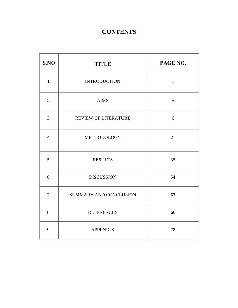
CONTENTS
S.NO
TITLE
PAGE NO.
1.
INTRODUCTION
1
2.
AIMS
5
3.
REVIEW OF LITERATURE
6
4.
METHODOLOGY
21
5.
RESULTS
35
6.
DISCUSSION
54
7.
SUMMARY AND CONCLUSION
63
8.
REFERENCES
66
9.
APPENDIX
78

INTRODUCTION
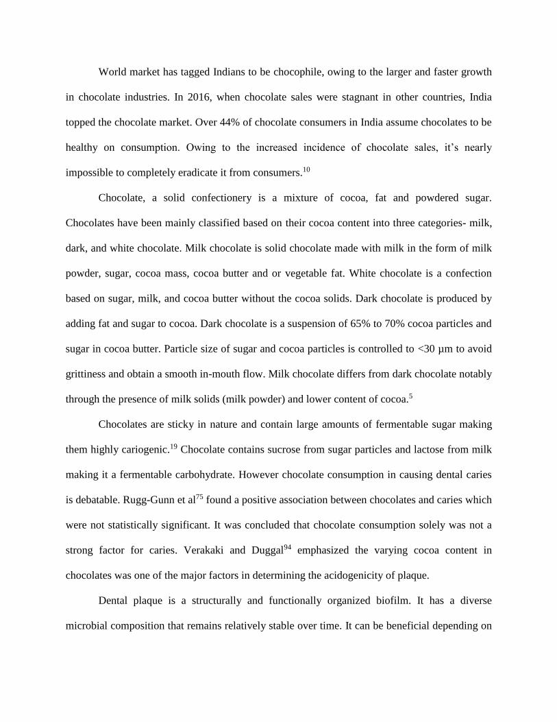
World market has tagged Indians to be chocophile, owing to the larger and faster growth
in chocolate industries. In 2016, when chocolate sales were stagnant in other countries, India
topped the chocolate market. Over 44% of chocolate consumers in India assume chocolates to be
healthy on consumption. Owing to the increased incidence of chocolate sales, it’s nearly
impossible to completely eradicate it from consumers.10
Chocolate, a solid confectionery is a mixture of cocoa, fat and powdered sugar.
Chocolates have been mainly classified based on their cocoa content into three categories- milk,
dark, and white chocolate. Milk chocolate is solid chocolate made with milk in the form of milk
powder, sugar, cocoa mass, cocoa butter and or vegetable fat. White chocolate is a confection
based on sugar, milk, and cocoa butter without the cocoa solids. Dark chocolate is produced by
adding fat and sugar to cocoa. Dark chocolate is a suspension of 65% to 70% cocoa particles and
sugar in cocoa butter. Particle size of sugar and cocoa particles is controlled to <30 µm to avoid
grittiness and obtain a smooth in-mouth flow. Milk chocolate differs from dark chocolate notably
through the presence of milk solids (milk powder) and lower content of cocoa.5
Chocolates are sticky in nature and contain large amounts of fermentable sugar making
them highly cariogenic.19 Chocolate contains sucrose from sugar particles and lactose from milk
making it a fermentable carbohydrate. However chocolate consumption in causing dental caries
is debatable. Rugg-Gunn et al75 found a positive association between chocolates and caries which
were not statistically significant. It was concluded that chocolate consumption solely was not a
strong factor for caries. Verakaki and Duggal94 emphasized the varying cocoa content in
chocolates was one of the major factors in determining the acidogenicity of plaque.
Dental plaque is a structurally and functionally organized biofilm. It has a diverse
microbial composition that remains relatively stable over time. It can be beneficial depending on
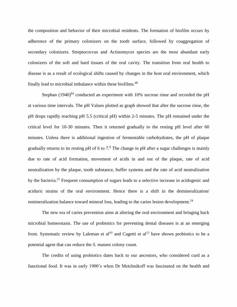
the composition and behavior of their microbial residents. The formation of biofilm occurs by
adherence of the primary colonizers on the tooth surface, followed by coaggregation of
secondary colonizers. Streptococcus and Actinomyces species are the most abundant early
colonizers of the soft and hard tissues of the oral cavity. The transition from oral health to
disease is as a result of ecological shifts caused by changes in the host oral environment, which
finally lead to microbial imbalance within these biofilms.48
Stephan (1940)84 conducted an experiment with 10% sucrose rinse and recorded the pH
at various time intervals. The pH Values plotted as graph showed that after the sucrose rinse, the
pH drops rapidly reaching pH 5.5 (critical pH) within 2-5 minutes. The pH remained under the
critical level for 10-30 minutes. Then it returned gradually to the resting pH level after 60
minutes. Unless there is additional ingestion of fermentable carbohydrates, the pH of plaque
gradually returns to its resting pH of 6 to 7.8 The change in pH after a sugar challenges is mainly
due to rate of acid formation, movement of acids in and out of the plaque, rate of acid
neutralization by the plaque, tooth substance, buffer systems and the rate of acid neutralization
by the bacteria.25 Frequent consumption of sugars leads to a selective increase in acidogenic and
aciduric strains of the oral environment. Hence there is a shift in the demineralization/
remineralization balance toward mineral loss, leading to the caries lesion development.24
The new era of caries prevention aims at altering the oral environment and bringing back
microbial homeostasis. The use of probiotics for preventing dental diseases is at an emerging
front. Systematic review by Laleman et al43 and Cagetti et al12 have shown probiotics to be a
potential agent that can reduce the S. mutans colony count.
The credits of using probiotics dates back to our ancestors, who considered curd as a
functional food. It was in early 1900’s when Dr Metchnikoff was fascinated on the health and
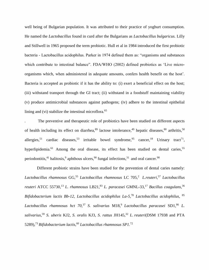
well being of Bulgarian population. It was attributed to their practice of yoghurt consumption.
He named the Lactobacillus found in curd after the Bulgarians as Lactobacillus bulgaricus. Lilly
and Stillwell in 1965 proposed the term probiotic. Hull et al in 1984 introduced the first probiotic
bacteria - Lactobacillus acidophilus. Parker in 1974 defined them as: “organisms and substances
which contribute to intestinal balance”. FDA/WHO (2002) defined probiotics as ‘Live micro-
organisms which, when administered in adequate amounts, confers health benefit on the host’.
Bacteria is accepted as probiotic if it has the ability to: (i) exert a beneficial effect on the host;
(iii) withstand transport through the GI tract; (ii) withstand in a foodstuff maintaining viability
(v) produce antimicrobial substances against pathogens; (iv) adhere to the intestinal epithelial
lining and (vi) stabilize the intestinal microflora.65
. The preventive and therapeutic role of probiotics have been studied on different aspects
of health including its effect on diarrhea,89 lactose intolerance,49 hepatic diseases,80 arthritis,50
allergies,51 cardiac diseases,53 irritable bowel syndrome,95 cancer,18 Urinary tract71,
hyperlipidemia.61 Among the oral disease, its effect has been studied on dental caries,55
periodontitis,41 halitosis,9 aphthous ulcers,90 fungal infections,31 and oral cancer.98
Different probiotic strains have been studied for the prevention of dental caries namely:
Lactobacillus rhamnosus GG,55 Lactobacillus rhamnosus LC 705,2 L.reuteri,57 Lactobacillus
reuteri ATCC 55730,13 L. rhamnosus LB21,83 L. paracasei GMNL-33,17 Bacillus coagulans,36
Bifidobacterium lactis Bb-12, Lactobacillus acidophilus La-5,78 Lactobacillus acidophilus, 85
Lactobacillus rhamnosus hct 70,37 S. salivarius M18,9 Lactobacillus paracasei SD1,86 L.
salivarius,59 S. uberis KJ2, S. oralis KJ3, S. rattus JH145,32 L. reuteri(DSM 17938 and PTA
5289),73 Bifidobacterium lactis,60 Lactobacillus rhamnosus SP1.72

Probiotics has been used with different vehicles such as cheese,2 milk,55,83 yoghurt,57,85
lozenges,15 capsule,52 ice creams,3,78 straw,13 tablet,17 gum,14 powder36 and drink.20 Ideally its
delivery should be suitable for all ages to receive its benefit. Possemiers et al69 added probiotics
to dark and milk chocolates. It was observed that coating probiotics in chocolates was an
excellent solution to protect it from stress conditions and for optimal delivery into the GI tract.
Kanafari et al39 in an invitro experiment proved the effectiveness of probiotic dark chocolate
against S.mutans. Literature search did not reveal any invivo trials of probiotic chocolates in
general as well as in dental research. Considering the growing sales of chocolate, a pragmatic
idea would be to manufacture a tooth friendly chocolate.
Hence the aim of this study was to make a customized probiotic chocolate and evaluate
its effectiveness in the oral cavity against change in pH and S. mutans level.

AIMS
AIMS
To formulate 3 types (milk, white and dark) of chocolates with probiotics.
To compare the plaque pH, salivary pH and buffering capacity of the three probiotic
chocolates with conventional chocolates in children
To evaluate the antimicrobial efficacy of the 3 custom made probiotic chocolates against
Streptococcus mutans.

REVIEW OF LITERATURE
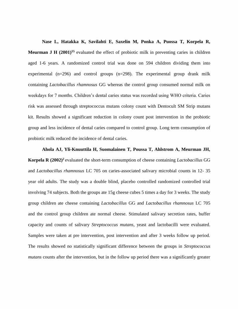
Nase L, Hatakka K, Savilahti E, Saxelin M, Ponka A, Puossa T, Korpela R,
Meurman J H (2001)55 evaluated the effect of probiotic milk in preventing caries in children
aged 1-6 years. A randomized control trial was done on 594 children dividing them into
experimental (n=296) and control groups (n=298). The experimental group drank milk
containing Lactobacillus rhamnosus GG whereas the control group consumed normal milk on
weekdays for 7 months. Children’s dental caries status was recorded using WHO criteria. Caries
risk was assessed through streptococcus mutans colony count with Dentocult SM Strip mutans
kit. Results showed a significant reduction in colony count post intervention in the probiotic
group and less incidence of dental caries compared to control group. Long term consumption of
probiotic milk reduced the incidence of dental caries.
Ahola AJ, Yli-Knuuttila H, Suomalainen T, Poussa T, Ahlstrom A, Meurman JH,
Korpela R (2002)2 evaluated the short-term consumption of cheese containing Lactobacillus GG
and Lactobacillus rhamnosus LC 705 on caries-associated salivary microbial counts in 12- 35
year old adults. The study was a double blind, placebo controlled randomized controlled trial
involving 74 subjects. Both the groups ate 15g cheese cubes 5 times a day for 3 weeks. The study
group children ate cheese containing Lactobacillus GG and Lactobacillus rhamnosus LC 705
and the control group children ate normal cheese. Stimulated salivary secretion rates, buffer
capacity and counts of salivary Streptococcus mutans, yeast and lactobacilli were evaluated.
Samples were taken at pre intervention, post intervention and after 3 weeks follow up period.
The results showed no statistically significant difference between the groups in Streptococcus
mutans counts after the intervention, but in the follow up period there was a significantly greater
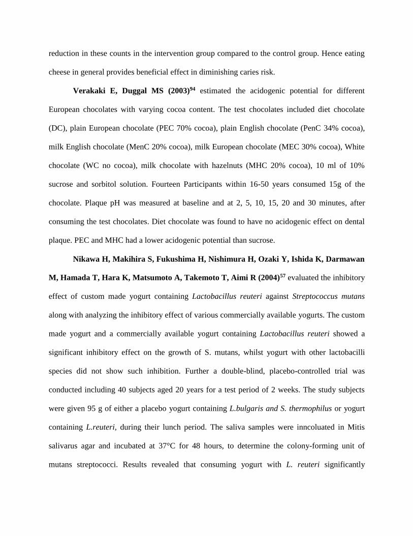
reduction in these counts in the intervention group compared to the control group. Hence eating
cheese in general provides beneficial effect in diminishing caries risk.
Verakaki E, Duggal MS (2003)94 estimated the acidogenic potential for different
European chocolates with varying cocoa content. The test chocolates included diet chocolate
(DC), plain European chocolate (PEC 70% cocoa), plain English chocolate (PenC 34% cocoa),
milk English chocolate (MenC 20% cocoa), milk European chocolate (MEC 30% cocoa), White
chocolate (WC no cocoa), milk chocolate with hazelnuts (MHC 20% cocoa), 10 ml of 10%
sucrose and sorbitol solution. Fourteen Participants within 16-50 years consumed 15g of the
chocolate. Plaque pH was measured at baseline and at 2, 5, 10, 15, 20 and 30 minutes, after
consuming the test chocolates. Diet chocolate was found to have no acidogenic effect on dental
plaque. PEC and MHC had a lower acidogenic potential than sucrose.
Nikawa H, Makihira S, Fukushima H, Nishimura H, Ozaki Y, Ishida K, Darmawan
M, Hamada T, Hara K, Matsumoto A, Takemoto T, Aimi R (2004)57 evaluated the inhibitory
effect of custom made yogurt containing Lactobacillus reuteri against Streptococcus mutans
along with analyzing the inhibitory effect of various commercially available yogurts. The custom
made yogurt and a commercially available yogurt containing Lactobacillus reuteri showed a
significant inhibitory effect on the growth of S. mutans, whilst yogurt with other lactobacilli
species did not show such inhibition. Further a double-blind, placebo-controlled trial was
conducted including 40 subjects aged 20 years for a test period of 2 weeks. The study subjects
were given 95 g of either a placebo yogurt containing L.bulgaris and S. thermophilus or yogurt
containing L.reuteri, during their lunch period. The saliva samples were inncoluated in Mitis
salivarus agar and incubated at 37°C for 48 hours, to determine the colony-forming unit of
mutans streptococci. Results revealed that consuming yogurt with L. reuteri significantly
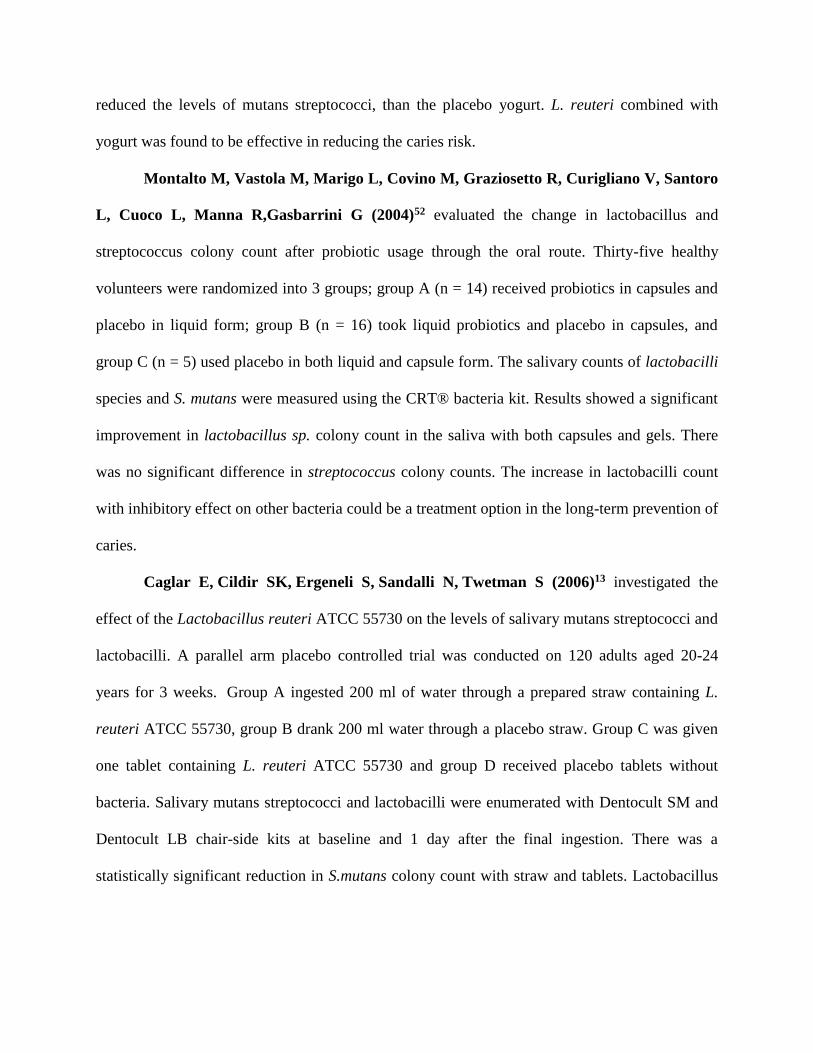
reduced the levels of mutans streptococci, than the placebo yogurt. L. reuteri combined with
yogurt was found to be effective in reducing the caries risk.
Montalto M, Vastola M, Marigo L, Covino M, Graziosetto R, Curigliano V, Santoro
L, Cuoco L, Manna R,Gasbarrini G (2004)52 evaluated the change in lactobacillus and
streptococcus colony count after probiotic usage through the oral route. Thirty-five healthy
volunteers were randomized into 3 groups; group A (n = 14) received probiotics in capsules and
placebo in liquid form; group B (n = 16) took liquid probiotics and placebo in capsules, and
group C (n = 5) used placebo in both liquid and capsule form. The salivary counts of lactobacilli
species and S. mutans were measured using the CRT® bacteria kit. Results showed a significant
improvement in lactobacillus sp. colony count in the saliva with both capsules and gels. There
was no significant difference in streptococcus colony counts. The increase in lactobacilli count
with inhibitory effect on other bacteria could be a treatment option in the long-term prevention of
caries.
Caglar E, Cildir SK, Ergeneli S, Sandalli N, Twetman S (2006)13 investigated the
effect of the Lactobacillus reuteri ATCC 55730 on the levels of salivary mutans streptococci and
lactobacilli. A parallel arm placebo controlled trial was conducted on 120 adults aged 20-24
years for 3 weeks. Group A ingested 200 ml of water through a prepared straw containing L.
reuteri ATCC 55730, group B drank 200 ml water through a placebo straw. Group C was given
one tablet containing L. reuteri ATCC 55730 and group D received placebo tablets without
bacteria. Salivary mutans streptococci and lactobacilli were enumerated with Dentocult SM and
Dentocult LB chair-side kits at baseline and 1 day after the final ingestion. There was a
statistically significant reduction in S.mutans colony count with straw and tablets. Lactobacillus

counts were not statistically significant. Both modes of deliveries probiotic were effective in
reducing the Streptococcus mutans colony count.
Caglar E, Kavaloglu SC, Kuscu OO, Sandalli N, Holgerson PL, Twetman S (2007)14
carried out a randomized controlled trial to evaluate the effectiveness of probiotic and xylitol
against mutans streptococcus (MS) and lactobaccili (LB). About 80 children aged 21-24 years
were randomly assigned to one of the 4 groups: group A- probiotic gum; group B- xylitol gum;
group C- probiotic and xylitol gum and group D- placebo gum. The probiotic gum contained
Lactobacilli reuteri ATCC 55730 and ATCC PTA 5289. Children received the gums for a period
of 3 weeks. Baseline saliva and 1 day post intervention saliva was collected and tested for MS
and LB with chair-side kits. A statistically significant reduction of salivary MS was seen in
group A and B after the intervention when compared with baseline. Group C also showed
reduction in colony count which was not statistically significant. There was no change in the
colony counts in group D. They concluded that use of probiotic and xylitol chewing gums
showed reduction in mutans streptococcus but they were ineffective when used in combination.
Caglar E, Kuscu OO, Cildir SK, Kuvvetli SS, Sandalli N (2008)15 investigated the
effect of Lactobacillus reuteri on salivary Streptococcus mutans colony count. A pacifier like
medical device was used to deliver the probiotic lozenges. Twenty adults were randomly divided
into experimental and control group with 10 in each group. The experimental group sucked
probiotic lozenge with L. reuteri (ATCC 55730) and L. reuteri (ATCC PTA 5289), once daily
for 10 days. While the control subjects received placebo medical devices. Salivary mutans
streptococci and lactobacilli were evaluated with chair-side kits at baseline and 1 day after the
final ingestion. Salivary S. mutans levels in the experimental group significantly reduced.
Lozenges were shown to be an effective means of probiotic delivery.
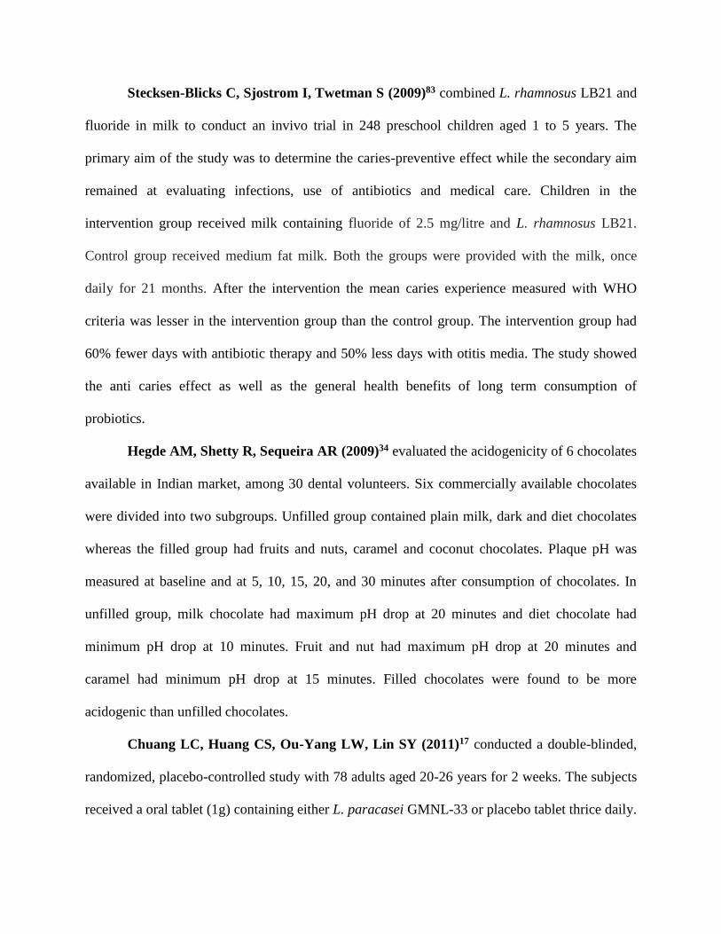
Stecksen-Blicks C, Sjostrom I, Twetman S (2009)83 combined L. rhamnosus LB21 and
fluoride in milk to conduct an invivo trial in 248 preschool children aged 1 to 5 years. The
primary aim of the study was to determine the caries-preventive effect while the secondary aim
remained at evaluating infections, use of antibiotics and medical care. Children in the
intervention group received milk containing fluoride of 2.5 mg/litre and L. rhamnosus LB21.
Control group received medium fat milk. Both the groups were provided with the milk, once
daily for 21 months. After the intervention the mean caries experience measured with WHO
criteria was lesser in the intervention group than the control group. The intervention group had
60% fewer days with antibiotic therapy and 50% less days with otitis media. The study showed
the anti caries effect as well as the general health benefits of long term consumption of
probiotics.
Hegde AM, Shetty R, Sequeira AR (2009)34 evaluated the acidogenicity of 6 chocolates
available in Indian market, among 30 dental volunteers. Six commercially available chocolates
were divided into two subgroups. Unfilled group contained plain milk, dark and diet chocolates
whereas the filled group had fruits and nuts, caramel and coconut chocolates. Plaque pH was
measured at baseline and at 5, 10, 15, 20, and 30 minutes after consumption of chocolates. In
unfilled group, milk chocolate had maximum pH drop at 20 minutes and diet chocolate had
minimum pH drop at 10 minutes. Fruit and nut had maximum pH drop at 20 minutes and
caramel had minimum pH drop at 15 minutes. Filled chocolates were found to be more
acidogenic than unfilled chocolates.
Chuang LC, Huang CS, Ou-Yang LW, Lin SY (2011)17 conducted a double-blinded,
randomized, placebo-controlled study with 78 adults aged 20-26 years for 2 weeks. The subjects
received a oral tablet (1g) containing either L. paracasei GMNL-33 or placebo tablet thrice daily.
Dentocult SM Strip kit was used to check the bacterial counts of salivary S. mutans, Dentocult
LB Dip Slide for lactobacillus and Dentobuff Strip for salivary buffering capacity of saliva. The
measurements were done at the baseline (T1), the completion (T2) of study period and 2 weeks
post intervention (T3). Though not significant, a reduction in the salivary S. mutans count was
detected between T2 and T3 of the intervention group. There was no significant difference in the
lactobacillus count and buffering capacity of saliva. Short term consumption of probiotic tablet
reduced the risk for caries.
Jindal G, Pandey RK, Agarwal J, Singh M (2011)36 conducted a study with 150
children aged 7-14 years. The children were divided into 3 groups; group A received placebo
powder, group B received freeze drided Lactobacillus rhamnosus and Bifidobacterium while
group C received Bacillus coagulans. The children were instructed to mix the preparation with
20ml of water to swish and swallow for 14 days. Mutans streptococcus colony count was
checked on mitis salivarius bacitracin agar at baseline and 14 days post intervention. There was a
significant reduction of colony count in both group B and C at 14 days post intervention. Dietary
supplements with probiotics could be a cheap alternative to reduce the incidence of dental caries.
Singh RP, Damle SG, Chawla A (2011)78 conducted a study with ice cream
containing Bifidobacterium lactis Bb-12 and Lactobacillus acidophilus La-5. It was a double
blind, placebo controlled study which included 40 children aged 12-14 years. Children were
given ice cream weighing 52 g for 10 days after which there was a wash out period of 2 weeks.
In the next phase, the ice creams were inter changed between groups. The S.mutans colony count
was measured using Dentocult SM at baseline and post intervention. There was significant
reduction in the colony count post consumption of probiotic ice cream compared to baseline in
both the groups. Ice creams can serve as a vehicle for delivering probiotics effectively.
Sudhir R, Praveen P, Anantharaj A, Venkataraghavan K (2012)85 compared the
effect of probiotic curd containing Lactobacillus acidophilus vs normal curd consumption in
children. Forty children were randomly divided into 2 groups for an interventional period of 30
days. Baseline and post intervention saliva samples were collected and cultured in Mitis
salivarius bacitracin agar to estimate the S.mutans colony count. There was a significant
reduction in the colony count after 30 days of probiotic curd consumption. Short‑term
consumption of probiotic curds can reduce oral S. Mutans counts.
Juneja A, Kakade A (2012)37 conducted a study to determine the effectiveness of short
time probiotic intake on 40 children aged 12-15 years for 9 weeks. The study was divided into 3
phases of 3 weeks each. First phase was baseline, while in the second phase children were given
either milk containing Lactobacillus rhamnosus hct 70 or placebo milk. The third phase was
follow up. After each phase salivary samples were collected. The intervention group had a
statistically significant reduction of S.mutans counts at post intervention and follow up period.
The study suggested a protective and preventive role of probiotic Lactobacillus rhamnosus hct
70 against dental caries.
Khanafari A, Hosseini Porgham S, Tajabadi Ebrahimi M (2012)39 evaluated the
inhibitory potential of S. mutans using three probiotic strains Lactobaccilus plantarum, L.
rhamnosus and L. acidophilus. The antimicrobial effect of the probiotic strain against S.mutans
was determined by deferred cross streak method and susceptibility was tested by disk diffusion
method, in which L. plantarum was identified to be having the maximal antimicrobial effect.
Probiotic strains were then added to dark chocolate at 108CFU/mL and their antimicrobial effect
against S.mutans was evaluated using disk diffusion susceptibility test. Probiotic chocolate
containing L.plantarum showed the maximal inhibitory zone. Chocolates can be used as a means
for delivering probiotics.
Dhawan R, Dhawan S (2013)21 assessed the effectiveness of commercially available
probiotic on plaque, gingivitis, and salivary Streptococcus mutans levels in subjects with chronic
gingivitis. Commercially available probiotic BIFILAC- HP capsule containing Lactobacillus
sporogenes, Streptococcus faecalis T‑110JPC, Clostridium butyrium TO‑A, and Bacillus
mesentericus TO‑A JPC was used. The experimental group (n=17) probiotic capsule and the
control group was given placebo capsule to swallow for a period of 2 weeks. Plaque index,
gingival index and streptococcus mutans colony count was measured at baseline, post
intervention and 2 weeks post intervention. Probiotic group showed a statistically significant
difference from the placebo group in all the parameters assessed, throughout the study period.
The use of probiotics could decrease the disease processes in oral cavity effectively.
Chinnappa A, Konde H, Konde S, Raj S, Beena JP (2013)16 conducted a clinical trial
to compare Streptococcus mutans count after consuming probiotic curd or ice cream. Forty
children aged 12-14 years were randomly divided into probiotic ice cream, plain ice cream,
probiotic yogurt or plain yogurt group. All the groups received 100ml of the product for 7 days.
Saliva samples were assessed at baseline, 1 hour and 7 days post intervention using Mitis
salivarius Bacitracin agar. There was a reduction in colony count at 1 hour post intervention for
all the groups. Reduction in colony count was significant in the probiotic groups at 7 days
compared to baseline. However there was no significant difference between probiotic yogurt and
ice cream at 1 hour and 7 days. The use of probiotic products can be an alternative strategy for
displacing pathogenic micro organisms.
Burton JP, Drummond BK, Chilcott CN, Tagg JR, Thomson WM, Hale JD,
Wescombe PA (2013)9 evaluated the effect of probiotic lozenges on plaque, gingival health and
oral microflora. Children aged 5- 10 year were divided into probiotic group (n=40) and placebo
group (n=43). Probiotic group received 2 lozenges per day containing S. salivarius M18 for 3
months and the placebo group received similar lozenges without probiotic. Plaque, gingival
health assessment and salivary assessments were done at 1, 3 and 7 months. Chromogenic agar
for Candida spp, Rogosa SL agar for lactobacilli, Mitis Salivarius agar for S. salivarius and
Tryptone Yeast Cystine Sucrose Bacitracin (TYCSB) agar for S.mutans was used. Gingival
health and S.salivarius colony count improved while S.mutans colony count reduced in the
probiotic group. S. salivarius M18 can provide oral health benefits on long term consumption.
Taipale T, Pienihakkinen K, Alanen P, Jokela J, Soderling E (2013)86 evaluated the
effect of Lactobacillus paracasei SD1 on the number of salivary mutans streptococci,
lactobacilli, and yeasts. The study consisted of 40 adults randomly divided into probiotic milk
and standard milk group. The probiotic group received milk containing Lactobacillus paracasei
SD1 and the control group received standard milk once daily for 4 weeks. Salivary samples were
collected at baseline and once a week for 4 weeks post intervention. The colony count of salivary
mutans streptococci, lactobacilli, and yeasts were evaluated using mitis salivarius bacitracin agar,
MRS, and Sabouraud Dextrose agar respectively. A statistically significant reduction in mutans
streptococci count occurred in probiotic group compared to the baseline. A significant increase in
lactobacilli count also occurred and was detected up to 4 weeks post intervention. No significant
reduction in yeast count was observed. Short term daily intake of Lactobacillus paracasei SD1
reduced the S.mutans colony count.
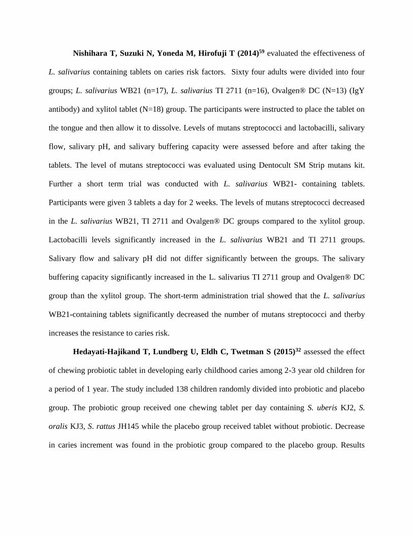
Nishihara T, Suzuki N, Yoneda M, Hirofuji T (2014)59 evaluated the effectiveness of
L. salivarius containing tablets on caries risk factors. Sixty four adults were divided into four
groups; L. salivarius WB21 (n=17), L. salivarius TI 2711 (n=16), Ovalgen® DC (N=13) (IgY
antibody) and xylitol tablet (N=18) group. The participants were instructed to place the tablet on
the tongue and then allow it to dissolve. Levels of mutans streptococci and lactobacilli, salivary
flow, salivary pH, and salivary buffering capacity were assessed before and after taking the
tablets. The level of mutans streptococci was evaluated using Dentocult SM Strip mutans kit.
Further a short term trial was conducted with L. salivarius WB21- containing tablets.
Participants were given 3 tablets a day for 2 weeks. The levels of mutans streptococci decreased
in the L. salivarius WB21, TI 2711 and Ovalgen® DC groups compared to the xylitol group.
Lactobacilli levels significantly increased in the L. salivarius WB21 and TI 2711 groups.
Salivary flow and salivary pH did not differ significantly between the groups. The salivary
buffering capacity significantly increased in the L. salivarius TI 2711 group and Ovalgen® DC
group than the xylitol group. The short-term administration trial showed that the L. salivarius
WB21-containing tablets significantly decreased the number of mutans streptococci and therby
increases the resistance to caries risk.
Hedayati-Hajikand T, Lundberg U, Eldh C, Twetman S (2015)32 assessed the effect
of chewing probiotic tablet in developing early childhood caries among 2-3 year old children for
a period of 1 year. The study included 138 children randomly divided into probiotic and placebo
group. The probiotic group received one chewing tablet per day containing S. uberis KJ2, S.
oralis KJ3, S. rattus JH145 while the placebo group received tablet without probiotic. Decrease
in caries increment was found in the probiotic group compared to the placebo group. Results
suggested that early childhood caries development could be reduced through administration of
probiotic tablets.
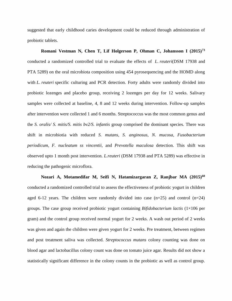
Romani Vestman N, Chen T, Lif Holgerson P, Ohman C, Johansson I (2015)73
conducted a randomized controlled trial to evaluate the effects of L. reuteri(DSM 17938 and
PTA 5289) on the oral microbiota composition using 454 pyrosequencing and the HOMD along
with L. reuteri specific culturing and PCR detection. Forty adults were randomly divided into
probiotic lozenges and placebo group, receiving 2 lozenges per day for 12 weeks. Salivary
samples were collected at baseline, 4, 8 and 12 weeks during intervention. Follow-up samples
after intervention were collected 1 and 6 months. Streptococcus was the most common genus and
the S. oralis/ S. mitis/S. mitis bv2/S. infantis group comprised the dominant species. There was
shift in microbiotia with reduced S. mutans, S. anginosus, N. mucosa, Fusobacterium
periodicum, F. nucleatum ss vincentii, and Prevotella maculosa detection. This shift was
observed upto 1 month post intervention. L.reuteri (DSM 17938 and PTA 5289) was effective in
reducing the pathogenic microflora.
Nozari A, Motamedifar M, Seifi N, Hatamizargaran Z, Ranjbar MA (2015)60
conducted a randomized controlled trial to assess the effectiveness of probiotic yogurt in children
aged 6-12 years. The children were randomly divided into case (n=25) and control (n=24)
groups. The case group received probiotic yogurt containing Bifidobacterium lactis (1×10⁶ per
gram) and the control group received normal yogurt for 2 weeks. A wash out period of 2 weeks
was given and again the children were given yogurt for 2 weeks. Pre treatment, between regimen
and post treatment saliva was collected. Streptococcus mutans colony counting was done on
blood agar and lactobacillus colony count was done on tomato juice agar. Results did not show a
statistically significant difference in the colony counts in the probiotic as well as control group.
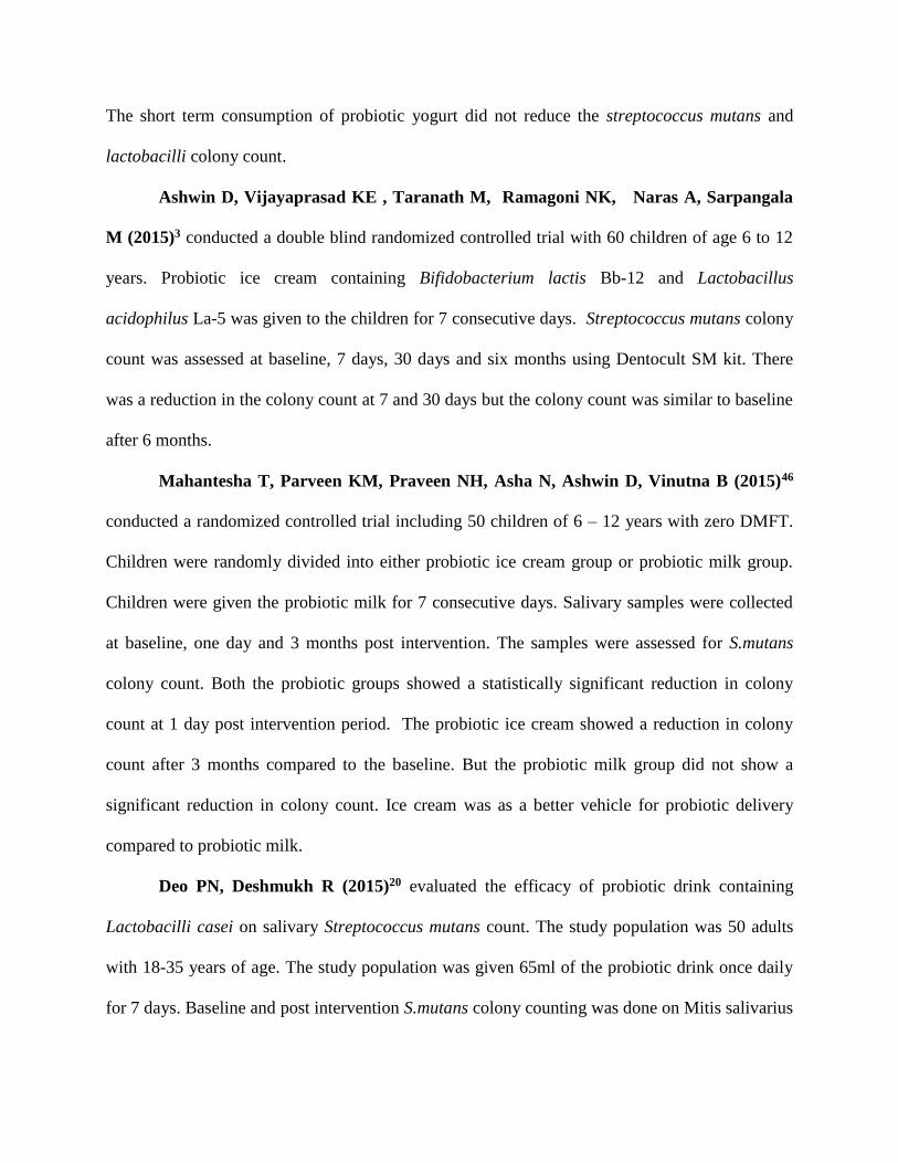
The short term consumption of probiotic yogurt did not reduce the streptococcus mutans and
lactobacilli colony count.
Ashwin D, Vijayaprasad KE , Taranath M, Ramagoni NK, Naras A, Sarpangala
M (2015)3 conducted a double blind randomized controlled trial with 60 children of age 6 to 12
years. Probiotic ice cream containing Bifidobacterium lactis Bb-12 and Lactobacillus
acidophilus La-5 was given to the children for 7 consecutive days. Streptococcus mutans colony
count was assessed at baseline, 7 days, 30 days and six months using Dentocult SM kit. There
was a reduction in the colony count at 7 and 30 days but the colony count was similar to baseline
after 6 months.
Mahantesha T, Parveen KM, Praveen NH, Asha N, Ashwin D, Vinutna B (2015)46
conducted a randomized controlled trial including 50 children of 6 – 12 years with zero DMFT.
Children were randomly divided into either probiotic ice cream group or probiotic milk group.
Children were given the probiotic milk for 7 consecutive days. Salivary samples were collected
at baseline, one day and 3 months post intervention. The samples were assessed for S.mutans
colony count. Both the probiotic groups showed a statistically significant reduction in colony
count at 1 day post intervention period. The probiotic ice cream showed a reduction in colony
count after 3 months compared to the baseline. But the probiotic milk group did not show a
significant reduction in colony count. Ice cream was as a better vehicle for probiotic delivery
compared to probiotic milk.
Deo PN, Deshmukh R (2015)20 evaluated the efficacy of probiotic drink containing
Lactobacilli casei on salivary Streptococcus mutans count. The study population was 50 adults
with 18-35 years of age. The study population was given 65ml of the probiotic drink once daily
for 7 days. Baseline and post intervention S.mutans colony counting was done on Mitis salivarius
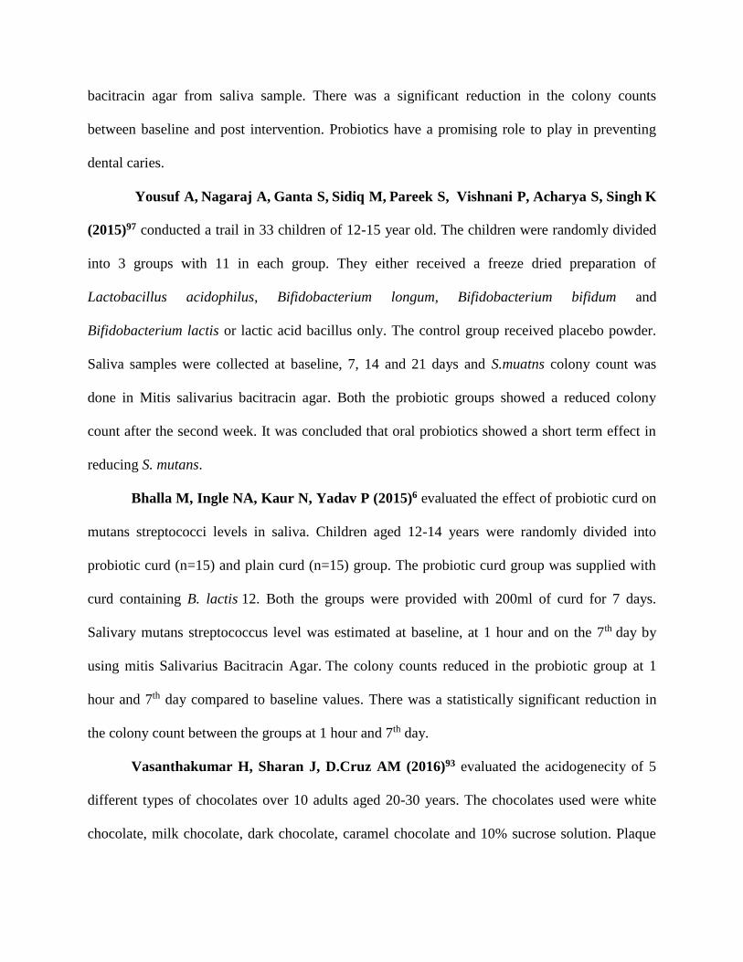
bacitracin agar from saliva sample. There was a significant reduction in the colony counts
between baseline and post intervention. Probiotics have a promising role to play in preventing
dental caries.
Yousuf A, Nagaraj A, Ganta S, Sidiq M, Pareek S, Vishnani P, Acharya S, Singh K
(2015)97 conducted a trail in 33 children of 12-15 year old. The children were randomly divided
into 3 groups with 11 in each group. They either received a freeze dried preparation of
Lactobacillus acidophilus, Bifidobacterium longum, Bifidobacterium bifidum and
Bifidobacterium lactis or lactic acid bacillus only. The control group received placebo powder.
Saliva samples were collected at baseline, 7, 14 and 21 days and S.muatns colony count was
done in Mitis salivarius bacitracin agar. Both the probiotic groups showed a reduced colony
count after the second week. It was concluded that oral probiotics showed a short term effect in
reducing S. mutans.
Bhalla M, Ingle NA, Kaur N, Yadav P (2015)6 evaluated the effect of probiotic curd on
mutans streptococci levels in saliva. Children aged 12-14 years were randomly divided into
probiotic curd (n=15) and plain curd (n=15) group. The probiotic curd group was supplied with
curd containing B. lactis 12. Both the groups were provided with 200ml of curd for 7 days.
Salivary mutans streptococcus level was estimated at baseline, at 1 hour and on the 7th day by
using mitis Salivarius Bacitracin Agar. The colony counts reduced in the probiotic group at 1
hour and 7th day compared to baseline values. There was a statistically significant reduction in
the colony count between the groups at 1 hour and 7th day.
Vasanthakumar H, Sharan J, D.Cruz AM (2016)93 evaluated the acidogenecity of 5
different types of chocolates over 10 adults aged 20-30 years. The chocolates used were white
chocolate, milk chocolate, dark chocolate, caramel chocolate and 10% sucrose solution. Plaque
pH was estimated at 10, 20, 30 and 45 minutes using pH test strips. Caramel chocolate had the
maximum decrease in plaque pH at 20 minutes after consumption. The least drop in pH was
noted for dark chocolate. At the end of 45 minutes, the dental retention measured by sugar
clearance was highest for the caramel chocolate. Dark chocolates have a greater content of cocoa
and less sugar making it the least cariogenic.
Nirmala S, Quadar MA, Veluru S (2016)58 compared the acidogenecity of 6 different
types of chocolates dividing them into unfilled (dark and milk chocolate), filled (wafer and fruit
and nuts chocolate), and candy (hard milk and mango-flavored candy) groups. Plaque pH values
and salivary sugar clearance rates were assessed at baseline, 5, 10, 15, 20 and 30 mins after
consumption. Dark chocolate had a high fall in pH and milk chocolate had low salivary sugar
clearance which signifies that unfilled chocolates are more cariogenic than other chocolates.
Rodriguez G, Ruiz B, Faleiros S, Vistoso A, Marró ML, Sanchez J, Urzua I, Cabello
R (2016)72 conducted a randomized controlled study in 2-3 year old children with the aim of a
comparing probiotic milk with standard milk. Two hundred and sixty one children were divided
into experimental and control group. Experimental group (n=150) were given 150ml of milk
supplemented with Lactobacillus rhamnosus SP1 whereas the placebo group (n=111) was given
standard milk. Microbiological samples of the milk were done to confirm the presence of
probiotic bacteria. Children were supplemented with the milk during weekdays for 10 months.
Clinical examinations were done following International Caries Detection and Assessment
System ICDAS criteria at the baseline and end of the study. Results showed a decline in caries
increment at the end of the study in the probiotic group. Regular intake of probiotic milk reduced
the risk for dental caries in preschool children.
Srivastava S, Saha S, Kumari M, Mohd S (2016)81 estimated the role of probiotic curd
on salivary Streptococcus mutans count after 7 days intervention. Sixty adults aged 20-25 years
were randomly divided into probiotic and plain curd groups, with 30 in each group. Participants
were supplied with 100ml of curd each day for 7 days. Salivary samples were collected at
baseline, half an hour, 1 hour and 7 days after intervention. pH was measured using pH meter
and Mitis Salivarius Bacitracin agar to estimate S. mutans count. Results showed a reduction in
salivary pH after ½ hour and 1 hour in both the groups but after 7 days, probiotic curd showed a
statistically significant increase in salivary pH. Probiotic curd showed statistically significant
reduction in S. mutans colony counts compared to normal curd.
Ghasemi E, Mazaheri R, Tahmourespour A (2017)29 investigated the effect of
probiotic yogurt vs xylitol chewing gum on salivary streptococcus mutans colony count. Fifty
adults were randomly divided into two groups. Probiotic group received 200g yogurt containing
Lactobacillus acidophilus and Bifidobacterium bifidum once daily for 3 weeks. Xylitol group
received 2 chewing gums (xylitol content: 5.58 grams daily) 3 times daily after each meal for 3
weeks. Saliva samples were collected at baseline, 1 day, 2 weeks and 4 weeks after the
intervention. Samples were cultured on mitis salivarius bacitracin agar and salivary S. mutans
counts were determined. Both the groups showed a reduction in S.mutans colony count at all the
time periods of investigation compared to baseline. Though probiotic group showed a higher
reduction in S.mutans count when compared to xylitol group, it was not statistically significant.
Long term consumption of probiotics and xylitol has caries preventive effect.

METHODOLOGY
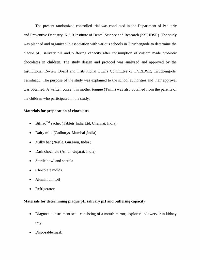
The present randomized controlled trial was conducted in the Department of Pediatric
and Preventive Dentistry, K S R Institute of Dental Science and Research (KSRIDSR). The study
was planned and organized in association with various schools in Tiruchengode to determine the
plaque pH, salivary pH and buffering capacity after consumption of custom made probiotic
chocolates in children. The study design and protocol was analyzed and approved by the
Institutional Review Board and Institutional Ethics Committee of KSRIDSR, Tiruchengode,
Tamilnadu. The purpose of the study was explained to the school authorities and their approval
was obtained. A written consent in mother tongue (Tamil) was also obtained from the parents of
the children who participated in the study.
Materials for preparation of chocolates
BifilacTM sachet (Tablets India Ltd, Chennai, India)
Dairy milk (Cadburys, Mumbai ,India)
Milky bar (Nestle, Gurgaon, India )
Dark chocolate (Amul, Gujarat, India)
Sterile bowl and spatula
Chocolate molds
Aluminium foil
Refrigerator
Materials for determining plaque pH salivary pH and buffering capacity
Diagnostic instrument set – consisting of a mouth mirror, explorer and tweezer in kidney
tray.
Disposable mask
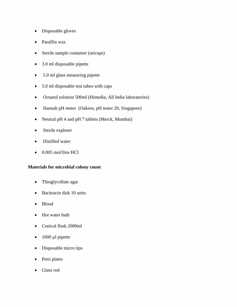
Disposable gloves
Paraffin wax
Sterile sample container (uricups)
3.0 ml disposable pipette
5.0 ml glass measuring pipette
5.0 ml disposable test tubes with caps
Octanol solution 500ml (Himedia, All India laboratories)
Hannah pH meter (Oakton, pH tester 20, Singapore)
Neutral pH 4 and pH 7 tablets (Merck, Mumbai)
Sterile explorer
Distilled water
0.005 mol/litre HCl
Materials for microbial colony count
Thioglycollate agar
Bacitracin disk 10 units
Blood
Hot water bath
Conical flask 2000ml
1000 µl pipette
Disposable micro tips
Petri plates
Glass rod
Ethanol
Plate rotator
Laminar flow chamber
Incubator
Automatic colony counter
Inclusion criteria
School children of 8-12 years.
def/DMFT score ≤ 3
The exclusion criteria includes children
On antibiotics
Under probiotics
Using xylitol chewing gums
Allergic to dairy products
Medically compromised like autoimmune disorders.
Invitro study
Determining the minimum inhibitory concentration
. The probiotic chosen for the present study was BifilacTM sachet which contains:
Prebiotics: Streptococcus faecalis T-110 (30 million)
Clostridium butyricum TO-A (2 million)
Bacillus mesentricus TO-A (1 million)
Probiotic: Lactobacillus sporogenes (50 million)
It was used for antimicrobial tests against S.mutans. The minimum inhibitory
concentration (MIC) of the probiotic against S.mutans was determined to be 0.5µg/mL.
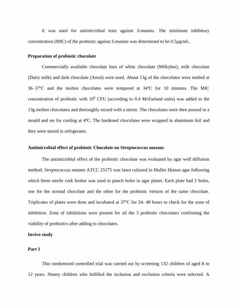
Preparation of probiotic chocolate
Commercially available chocolate bars of white chocolate (Milkybar), milk chocolate
(Dairy milk) and dark chocolate (Amul) were used. About 13g of the chocolates were melted at
36–37ºC and the molten chocolates were tempered at 34ºC for 10 minutes. The MIC
concentration of probiotic with 108 CFU (according to 0.4 McFarland units) was added to the
13g molten chocolates and thoroughly mixed with a stirrer. The chocolates were then poured in a
mould and set for cooling at 4ºC. The hardened chocolates were wrapped in aluminum foil and
they were stored in refrigerator.
Antimicrobial effect of probiotic Chocolate on Streptococcus mutans
The antimicrobial effect of the probiotic chocolate was evaluated by agar well diffusion
method. Streptococcus mutans ATCC 25175 was lawn cultured in Muller Hinton agar following
which 6mm sterile cork broker was used to punch holes in agar plates. Each plate had 2 holes,
one for the normal chocolate and the other for the probiotic version of the same chocolate.
Triplicates of plates were done and incubated at 37ºC for 24- 48 hours to check for the zone of
inhibition. Zone of inhibitions were present for all the 3 probiotic chocolates confirming the
viability of probiotics after adding to chocolates.
Invivo study
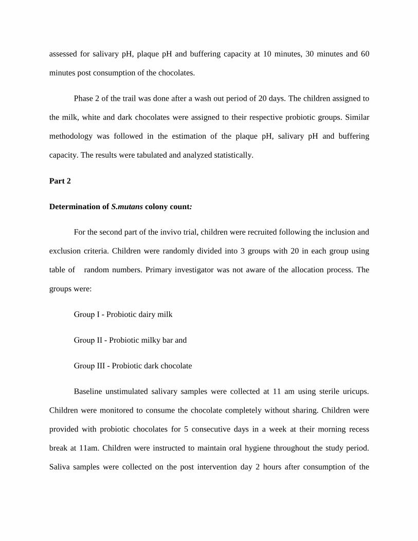
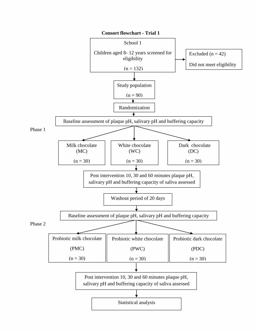
Part 1
This randomized controlled trial was carried out by screening 132 children of aged 8 to
12 years. Ninety children who fulfilled the inclusion and exclusion criteria were selected. A
blinded investigator randomly divided the children into 3 groups with 30 children in each group
using table of random numbers. Primary investigator was not aware of the allocation process.
The study was conducted in 2 phases. For phase 1 of the trial the groups were:
Milk chocolate (MC)
White chocolate (WC)
Dark chocolate (DC)
For phase 2 of the trial the groups were:
Probiotic Milk chocolate (PMC)
Probiotic White chocolate (PWC)
Probiotic Dark chocolate (PDC)
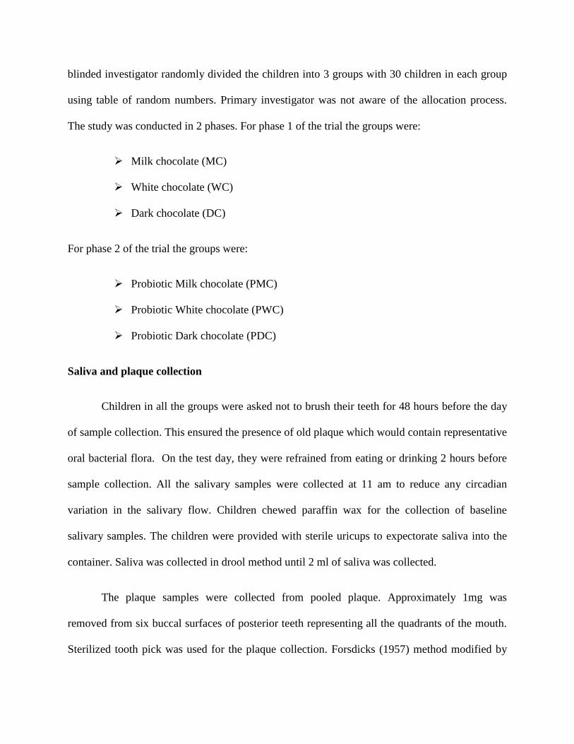
Saliva and plaque collection
Children in all the groups were asked not to brush their teeth for 48 hours before the day
of sample collection. This ensured the presence of old plaque which would contain representative
oral bacterial flora. On the test day, they were refrained from eating or drinking 2 hours before
sample collection. All the salivary samples were collected at 11 am to reduce any circadian
variation in the salivary flow. Children chewed paraffin wax for the collection of baseline
salivary samples. The children were provided with sterile uricups to expectorate saliva into the
container. Saliva was collected in drool method until 2 ml of saliva was collected.
The plaque samples were collected from pooled plaque. Approximately 1mg was
removed from six buccal surfaces of posterior teeth representing all the quadrants of the mouth.
Sterilized tooth pick was used for the plaque collection. Forsdicks (1957) method modified by
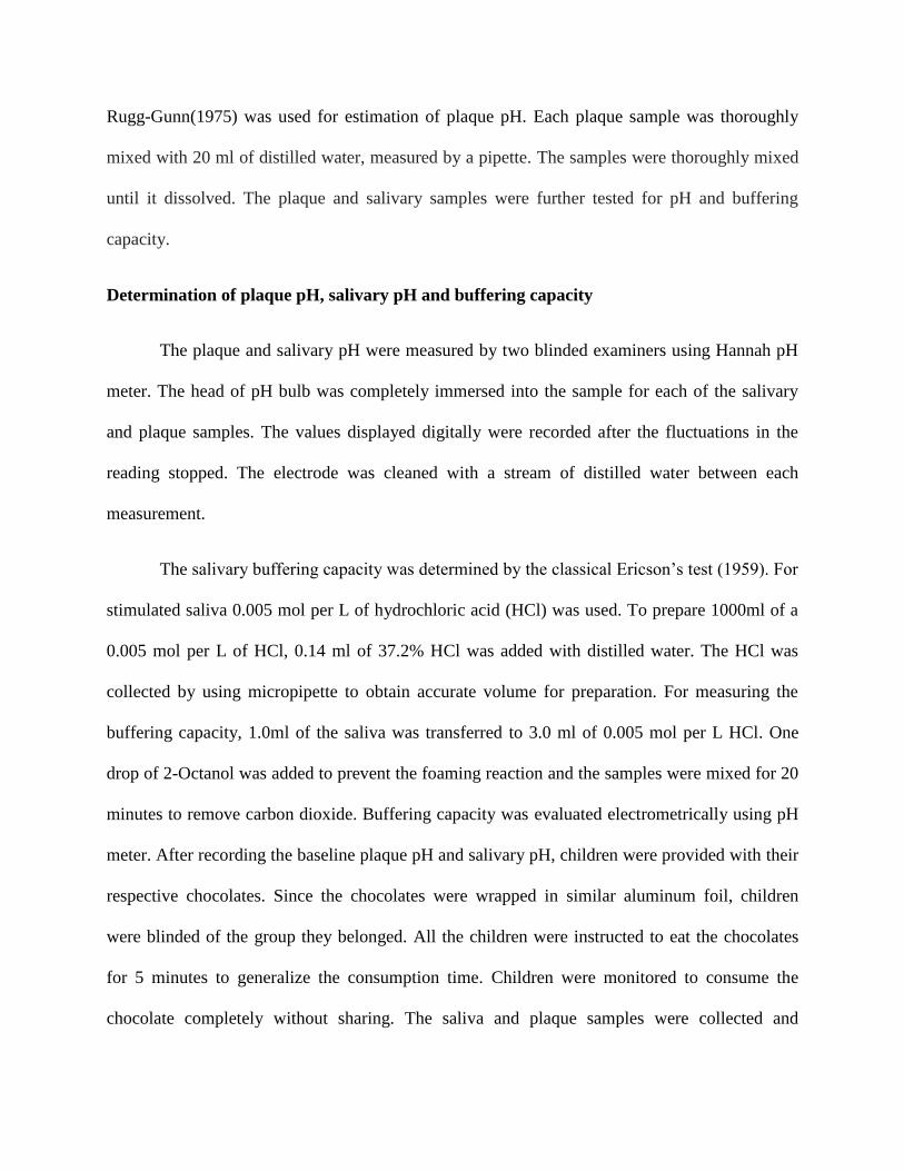
Rugg-Gunn(1975) was used for estimation of plaque pH. Each plaque sample was thoroughly
mixed with 20 ml of distilled water, measured by a pipette. The samples were thoroughly mixed
until it dissolved. The plaque and salivary samples were further tested for pH and buffering
capacity.
Determination of plaque pH, salivary pH and buffering capacity
The plaque and salivary pH were measured by two blinded examiners using Hannah pH
meter. The head of pH bulb was completely immersed into the sample for each of the salivary
and plaque samples. The values displayed digitally were recorded after the fluctuations in the
reading stopped. The electrode was cleaned with a stream of distilled water between each
measurement.
The salivary buffering capacity was determined by the classical Ericson’s test (1959). For
stimulated saliva 0.005 mol per L of hydrochloric acid (HCl) was used. To prepare 1000ml of a
0.005 mol per L of HCl, 0.14 ml of 37.2% HCl was added with distilled water. The HCl was
collected by using micropipette to obtain accurate volume for preparation. For measuring the
buffering capacity, 1.0ml of the saliva was transferred to 3.0 ml of 0.005 mol per L HCl. One
drop of 2-Octanol was added to prevent the foaming reaction and the samples were mixed for 20
minutes to remove carbon dioxide. Buffering capacity was evaluated electrometrically using pH
meter. After recording the baseline plaque pH and salivary pH, children were provided with their
respective chocolates. Since the chocolates were wrapped in similar aluminum foil, children
were blinded of the group they belonged. All the children were instructed to eat the chocolates
for 5 minutes to generalize the consumption time. Children were monitored to consume the
chocolate completely without sharing. The saliva and plaque samples were collected and
assessed for salivary pH, plaque pH and buffering capacity at 10 minutes, 30 minutes and 60
minutes post consumption of the chocolates.
Phase 2 of the trail was done after a wash out period of 20 days. The children assigned to
the milk, white and dark chocolates were assigned to their respective probiotic groups. Similar
methodology was followed in the estimation of the plaque pH, salivary pH and buffering
capacity. The results were tabulated and analyzed statistically.
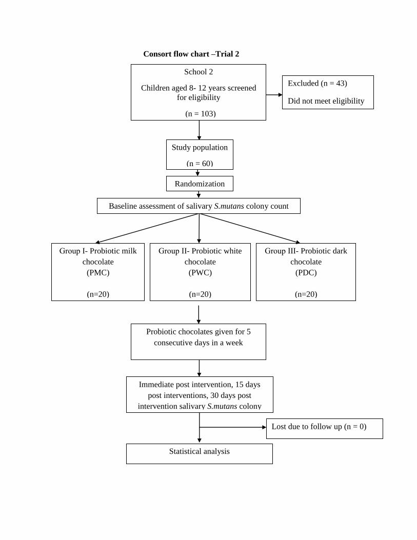
Part 2
Determination of S.mutans colony count:
For the second part of the invivo trial, children were recruited following the inclusion and
exclusion criteria. Children were randomly divided into 3 groups with 20 in each group using
table of random numbers. Primary investigator was not aware of the allocation process. The
groups were:
Group І - Probiotic dairy milk
Group ІІ - Probiotic milky bar and
Group ІІІ - Probiotic dark chocolate
Baseline unstimulated salivary samples were collected at 11 am using sterile uricups.
Children were monitored to consume the chocolate completely without sharing. Children were
provided with probiotic chocolates for 5 consecutive days in a week at their morning recess
break at 11am. Children were instructed to maintain oral hygiene throughout the study period.
Saliva samples were collected on the post intervention day 2 hours after consumption of the
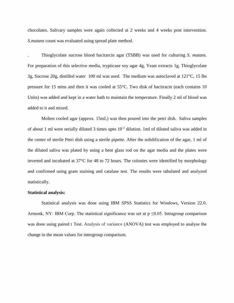
chocolates. Salivary samples were again collected at 2 weeks and 4 weeks post intervention.
S.mutans count was evaluated using spread plate method.
. Thioglycolate sucrose blood bacitarcin agar (TSBB) was used for culturing S. mutans.
For preparation of this selective media, trypticase soy agar 4g, Yeast extracts 1g, Thioglycolate
3g, Sucrose 20g, distilled water 100 ml was used. The medium was autoclaved at 121°C, 15 lbs
pressure for 15 mins and then it was cooled at 55°C. Two disk of bacitracin (each contains 10
Units) was added and kept in a water bath to maintain the temperature. Finally 2 ml of blood was
added to it and mixed.
Molten cooled agar (approx. 15mL) was then poured into the petri dish. Saliva samples
of about 1 ml were serially diluted 3 times upto 10-3 dilution. 1ml of diluted saliva was added in
the center of sterile Petri dish using a sterile pipette. After the solidification of the agar, 1 ml of
the diluted saliva was plated by using a bent glass rod on the agar media and the plates were
inverted and incubated at 37°C for 48 to 72 hours. The colonies were identified by morphology
and confirmed using gram staining and catalase test. The results were tabulated and analyzed
statistically.
Statistical analysis:
Statistical analysis was done using IBM SPSS Statistics for Windows, Version 22.0.
Armonk, NY: IBM Corp. The statistical significance was set at p ≤0.05. Intragroup comparison
was done using paired t Test. Analysis of variance (ANOVA) test was employed to analyse the
change in the mean values for intergroup comparison.
Consort flowchart - Trial 1
Phase 1
Phase 2
Study population
(n = 90)
Milk chocolate
(MC)
(n = 30)
White chocolate
(WC)
(n = 30)
Dark chocolate
(DC)
(n = 30)
Post intervention 10, 30 and 60 minutes plaque pH,
salivary pH and buffering capacity of saliva assessed
Probiotic milk chocolate
(PMC)
(n = 30)
Probiotic white chocolate
(PWC)
(n = 30)
Probiotic dark chocolate
(PDC)
(n = 30)
Post intervention 10, 30 and 60 minutes plaque pH,
salivary pH and buffering capacity of saliva assessed
School 1
Children aged 8- 12 years screened for
eligibility
(n = 132)
Statistical analysis
Excluded (n = 42)
Did not meet eligibility
criteria
Randomization
Baseline assessment of plaque pH, salivary pH and buffering capacity
Washout period of 20 days
Baseline assessment of plaque pH, salivary pH and buffering capacity
Consort flow chart –Trial 2
Group І- Probiotic milk
chocolate
(PMC)
(n=20)
(n = 20)
Group ІІ- Probiotic white
chocolate
(PWC)
(n=20)
(n = 20)
Group ІІІ- Probiotic dark
chocolate
(PDC)
(n=20)
(n = 20)
School 2
Children aged 8- 12 years screened
for eligibility
(n = 103)
Probiotic chocolates given for 5
consecutive days in a week
Immediate post intervention, 15 days
post interventions, 30 days post
intervention salivary S.mutans colony
count assessed
Statistical analysis
Lost due to follow up (n = 0)
Excluded (n = 43)
Did not meet eligibility
criteria
Study population
(n = 60)
Randomization
Baseline assessment of salivary S.mutans colony count
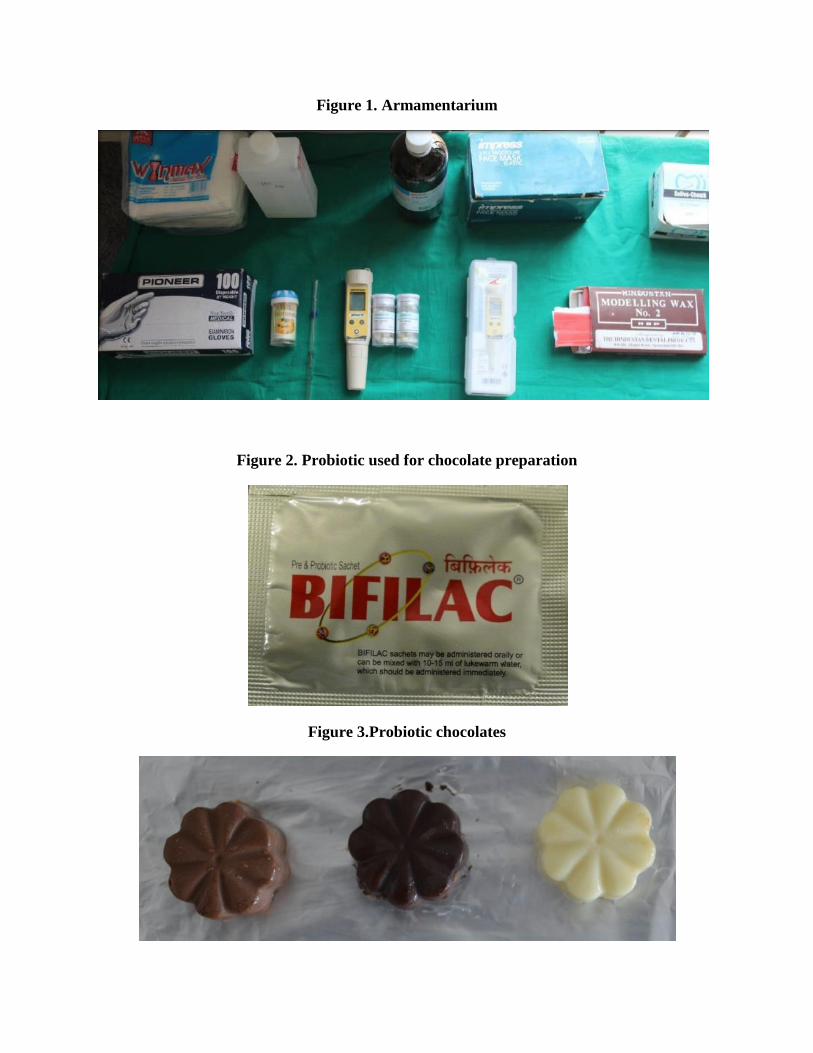
Figure 1. Armamentarium
Figure 2. Probiotic used for chocolate preparation
Figure 3.Probiotic chocolates
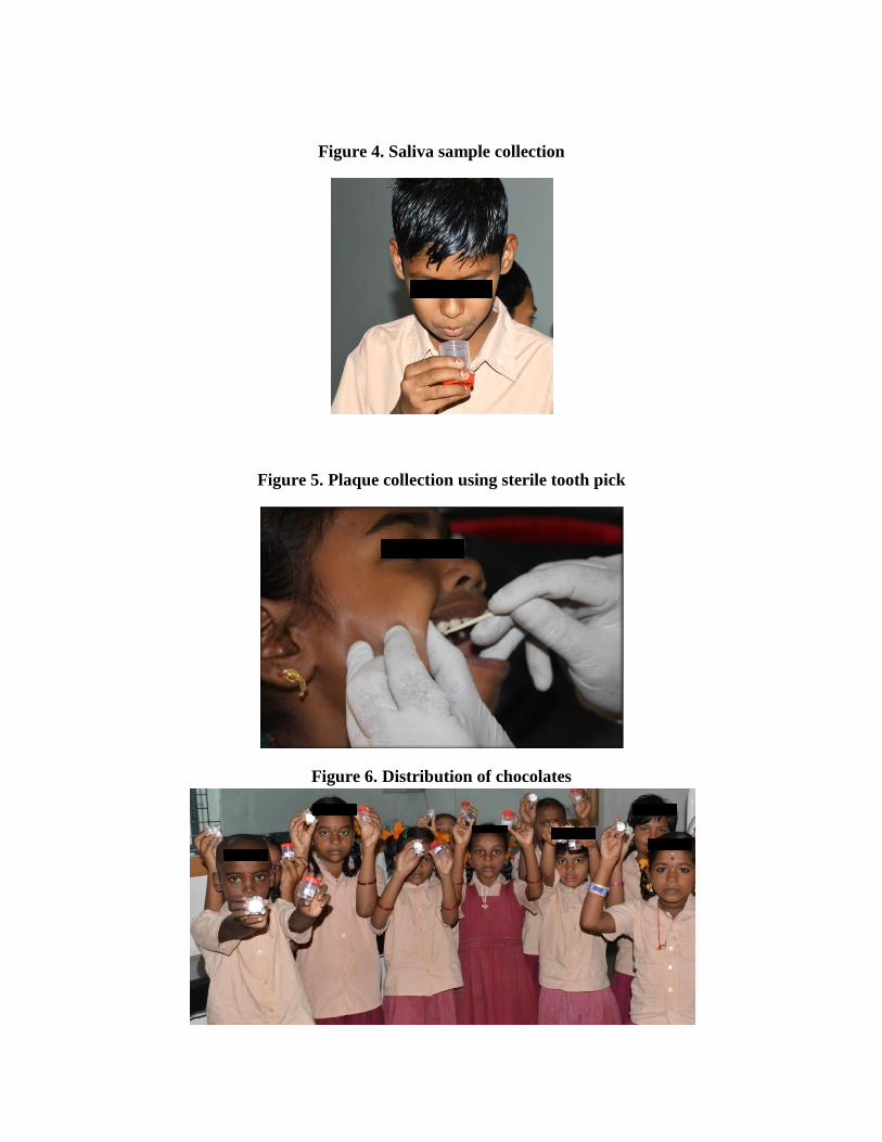
Figure 4. Saliva sample collection
Figure 5. Plaque collection using sterile tooth pick
Figure 6. Distribution of chocolates
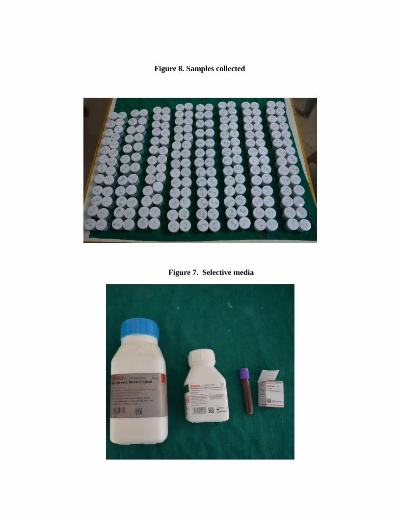
Figure 8. Samples collected
Figure 7. Selective media
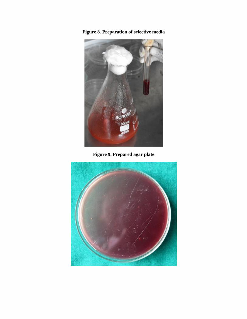
Figure 8. Preparation of selective media
Figure 9. Prepared agar plate
RESULTS
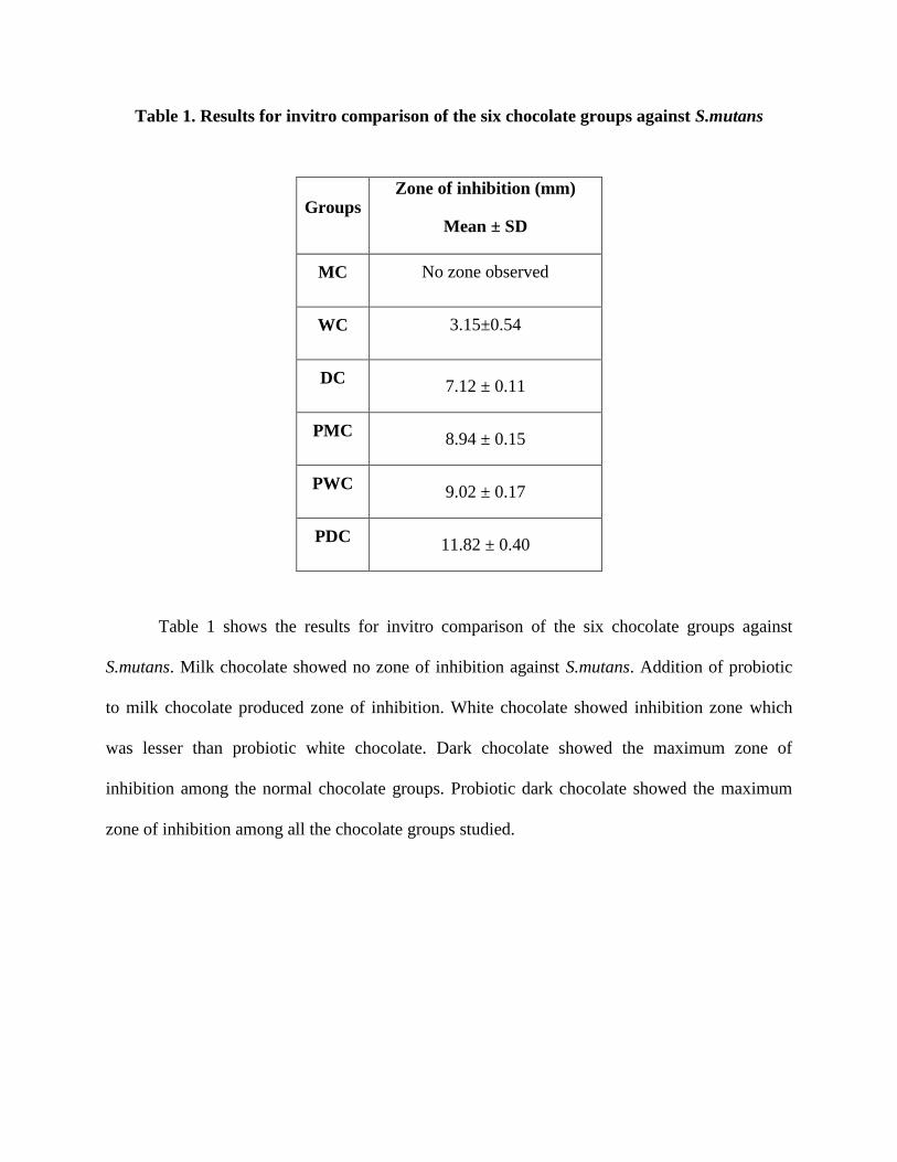
Table 1. Results for invitro comparison of the six chocolate groups against S.mutans
Table 1 shows the results for invitro comparison of the six chocolate groups against
S.mutans. Milk chocolate showed no zone of inhibition against S.mutans. Addition of probiotic
to milk chocolate produced zone of inhibition. White chocolate showed inhibition zone which
was lesser than probiotic white chocolate. Dark chocolate showed the maximum zone of
inhibition among the normal chocolate groups. Probiotic dark chocolate showed the maximum
zone of inhibition among all the chocolate groups studied.
Groups Zone of inhibition (mm)
Mean ± SD
MC No zone observed
WC 3.15±0.54
DC 7.12 ± 0.11
PMC 8.94 ± 0.15
PWC 9.02 ± 0.17
PDC 11.82 ± 0.40

Table 2. Mean distribution of plaque pH in six chocolate groups
Table 2 shows the descriptive statistics of plaque pH in all the six chocolate groups. All
the groups showed the minimum pH level at 10 minutes. At the end of 60 minutes PWC showed
an increase in plaque pH more than the baseline (6.57±0.36 to 6.77±0.27).
0
1
2
3
4
5
6
7
8
BASELINE 10 MINS 30 MINS 60 MINS
pH
Graph 1. Mean distribution of plaque pH
MC
WC
DC
PMC
PWC
PDC
Time
MC WC DC PMC PWC PDC
Mean
±SD
Mean
±SD
Mean
±SD
Mean
±SD
Mean
±SD
Mean
±SD
Baseline 6.17±0.27 6.51±0.19 6.52±0.27 6.63±0.20 6.57±0.36 6.57±0.26
10
minutes 5.28±0.45 5.76±0.48 6.06±0.47 5.87±0.46 6.15±0.31 5.82±0.54
30
minutes 5.86±0.23 6.25±0.21 6.31±0.18 6.36±0.15 6.53±0.26 6.28±0.22
60
minutes
6.08±0.
16 6.41±0.21 6.43±0.17 6.49±0.18 6.77±0.27 6.39±0.22

Table 3. Mean distribution of salivary pH in six chocolate groups
Table 3 shows the descriptive statistics of salivary pH in all the six chocolate groups. All
the groups showed the minimum pH level at 10 minutes.
0
1
2
3
4
5
6
7
8
9
BASELINE 15 MINS 30 MINS 60 MINS
pH
Graph 2. Descriptive Statistics of salivary pH
MC
WC
DC
PMC
PWC
PDC
Time
MC WC DC PMC PWC PDC
Mean
±SD
Mean
±SD
Mean
±SD
Mean
±SD
Mean
±SD
Mean
±SD
Baseline 7.32±0.31 7.53±0.27 7.43±0.35 7.65±0.19 7.41±0.21 7.59±0.18
10
minutes 5.45±0.72 6.02±1.05 6.41±0.76 5.65±0.92 7.04±0.40 6.93±0.44
30
minutes 6.61±0.49 7.31±0.35 7.23±0.42 7.14±0.28 7.23±0.33 7.36±0.24
60
minutes 7.23±0.28 7.33±0.24 7.42±0.29 7.36±0.25 7.27±0.23 7.51±0.18
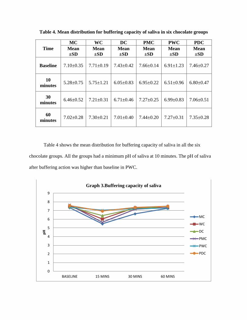
Table 4. Mean distribution for buffering capacity of saliva in six chocolate groups
Time
MC WC DC PMC PWC PDC
Mean
±SD
Mean
±SD
Mean
±SD
Mean
±SD
Mean
±SD
Mean
±SD
Baseline 7.10±0.35 7.71±0.19 7.43±0.42 7.66±0.14 6.91±1.23 7.46±0.27
10
minutes 5.28±0.75 5.75±1.21 6.05±0.83 6.95±0.22 6.51±0.96 6.80±0.47
30
minutes 6.46±0.52 7.21±0.31 6.71±0.46 7.27±0.25 6.99±0.83 7.06±0.51
60
minutes 7.02±0.28 7.30±0.21 7.01±0.40 7.44±0.20 7.27±0.31 7.35±0.28
Table 4 shows the mean distribution for buffering capacity of saliva in all the six
chocolate groups. All the groups had a minimum pH of saliva at 10 minutes. The pH of saliva
after buffering action was higher than baseline in PWC.
0
1
2
3
4
5
6
7
8
9
BASELINE 15 MINS 30 MINS 60 MINS
pH
Graph 3.Buffering capacity of saliva
MC
WC
DC
PMC
PWC
PDC
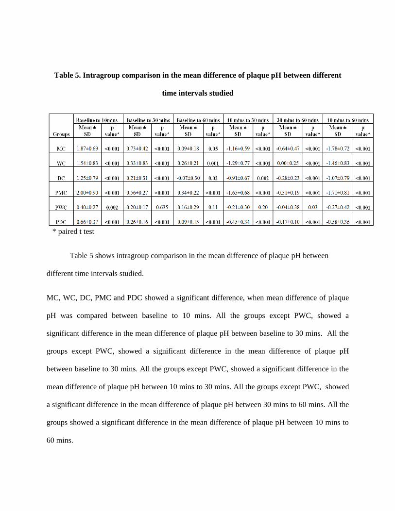
Table 5. Intragroup comparison in the mean difference of plaque pH between different
time intervals studied
* paired t test
Table 5 shows intragroup comparison in the mean difference of plaque pH between
different time intervals studied.
MC, WC, DC, PMC and PDC showed a significant difference, when mean difference of plaque
pH was compared between baseline to 10 mins. All the groups except PWC, showed a
significant difference in the mean difference of plaque pH between baseline to 30 mins. All the
groups except PWC, showed a significant difference in the mean difference of plaque pH
between baseline to 30 mins. All the groups except PWC, showed a significant difference in the
mean difference of plaque pH between 10 mins to 30 mins. All the groups except PWC, showed
a significant difference in the mean difference of plaque pH between 30 mins to 60 mins. All the
groups showed a significant difference in the mean difference of plaque pH between 10 mins to
60 mins.
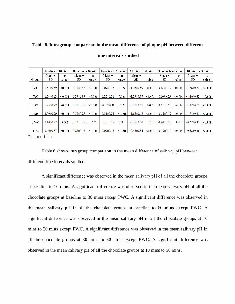
Table 6. Intragroup comparison in the mean difference of plaque pH between different
time intervals studied
* paired t test
Table 6 shows intragroup comparison in the mean difference of salivary pH between
different time intervals studied.
A significant difference was observed in the mean salivary pH of all the chocolate groups
at baseline to 10 mins. A significant difference was observed in the mean salivary pH of all the
chocolate groups at baseline to 30 mins except PWC. A significant difference was observed in
the mean salivary pH in all the chocolate groups at baseline to 60 mins except PWC. A
significant difference was observed in the mean salivary pH in all the chocolate groups at 10
mins to 30 mins except PWC. A significant difference was observed in the mean salivary pH in
all the chocolate groups at 30 mins to 60 mins except PWC. A significant difference was
observed in the mean salivary pH of all the chocolate groups at 10 mins to 60 mins.
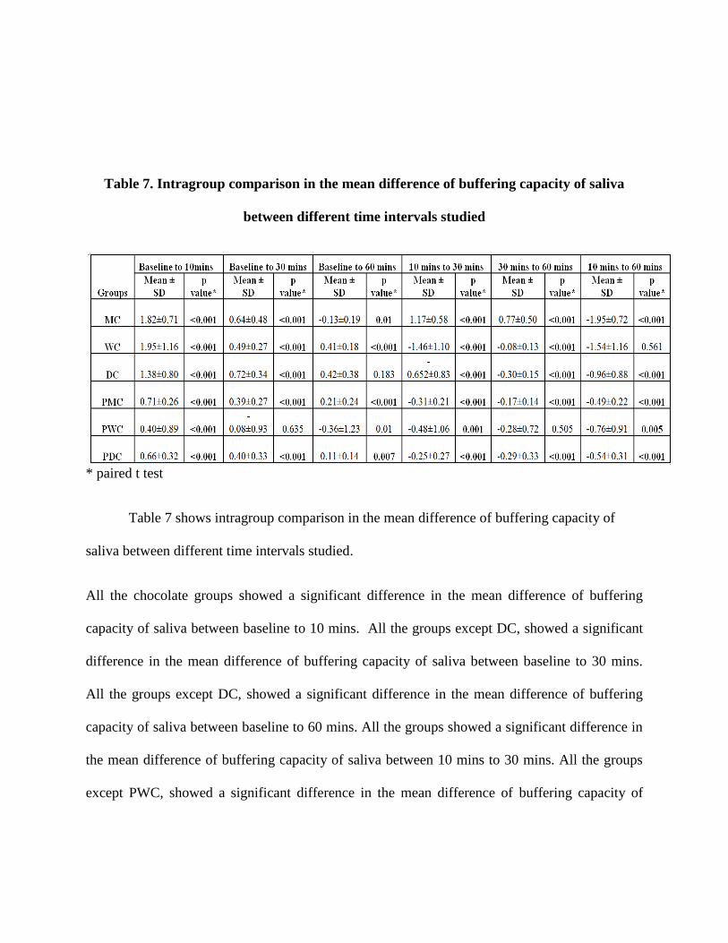
Table 7. Intragroup comparison in the mean difference of buffering capacity of saliva
between different time intervals studied
* paired t test
Table 7 shows intragroup comparison in the mean difference of buffering capacity of
saliva between different time intervals studied.
All the chocolate groups showed a significant difference in the mean difference of buffering
capacity of saliva between baseline to 10 mins. All the groups except DC, showed a significant
difference in the mean difference of buffering capacity of saliva between baseline to 30 mins.
All the groups except DC, showed a significant difference in the mean difference of buffering
capacity of saliva between baseline to 60 mins. All the groups showed a significant difference in
the mean difference of buffering capacity of saliva between 10 mins to 30 mins. All the groups
except PWC, showed a significant difference in the mean difference of buffering capacity of
saliva between 30 mins to 60 mins. All the groups showed a significant difference in the mean
difference of buffering capacity of saliva between 10 mins to 60 mins.
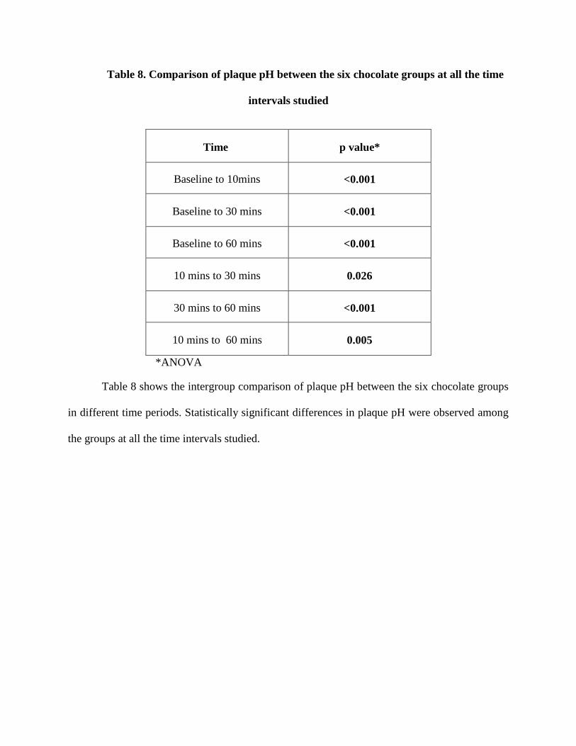
Table 8. Comparison of plaque pH between the six chocolate groups at all the time
intervals studied
Time p value*
Baseline to 10mins <0.001
Baseline to 30 mins <0.001
Baseline to 60 mins <0.001
10 mins to 30 mins 0.026
30 mins to 60 mins <0.001
10 mins to 60 mins 0.005
*ANOVA
Table 8 shows the intergroup comparison of plaque pH between the six chocolate groups
in different time periods. Statistically significant differences in plaque pH were observed among
the groups at all the time intervals studied.
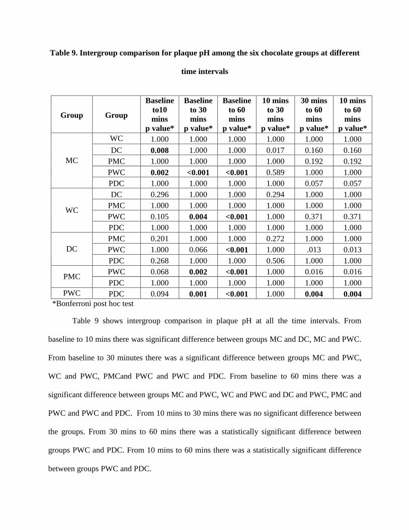
Table 9. Intergroup comparison for plaque pH among the six chocolate groups at different
time intervals
*Bonferroni post hoc test
Table 9 shows intergroup comparison in plaque pH at all the time intervals. From
baseline to 10 mins there was significant difference between groups MC and DC, MC and PWC.
From baseline to 30 minutes there was a significant difference between groups MC and PWC,
WC and PWC, PMCand PWC and PWC and PDC. From baseline to 60 mins there was a
significant difference between groups MC and PWC, WC and PWC and DC and PWC, PMC and
PWC and PWC and PDC. From 10 mins to 30 mins there was no significant difference between
the groups. From 30 mins to 60 mins there was a statistically significant difference between
groups PWC and PDC. From 10 mins to 60 mins there was a statistically significant difference
between groups PWC and PDC.
Group Group
Baseline
to10
mins
p value*
Baseline
to 30
mins
p value*
Baseline
to 60
mins
p value*
10 mins
to 30
mins
p value*
30 mins
to 60
mins
p value*
10 mins
to 60
mins
p value*
MC
WC 1.000 1.000 1.000 1.000 1.000 1.000
DC 0.008 1.000 1.000 0.017 0.160 0.160
PMC 1.000 1.000 1.000 1.000 0.192 0.192
PWC 0.002 <0.001 <0.001 0.589 1.000 1.000
PDC 1.000 1.000 1.000 1.000 0.057 0.057
WC
DC 0.296 1.000 1.000 0.294 1.000 1.000
PMC 1.000 1.000 1.000 1.000 1.000 1.000
PWC 0.105 0.004 <0.001 1.000 0.371 0.371
PDC 1.000 1.000 1.000 1.000 1.000 1.000
DC
PMC 0.201 1.000 1.000 0.272 1.000 1.000
PWC 1.000 0.066 <0.001 1.000 .013 0.013
PDC 0.268 1.000 1.000 0.506 1.000 1.000
PMC PWC 0.068 0.002 <0.001 1.000 0.016 0.016
PDC 1.000 1.000 1.000 1.000 1.000 1.000
PWC PDC 0.094 0.001 <0.001 1.000 0.004 0.004
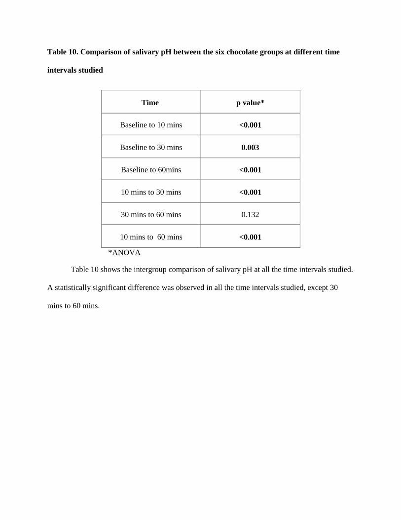
Table 10. Comparison of salivary pH between the six chocolate groups at different time
intervals studied
Time p value*
Baseline to 10 mins <0.001
Baseline to 30 mins 0.003
Baseline to 60mins <0.001
10 mins to 30 mins <0.001
30 mins to 60 mins 0.132
10 mins to 60 mins <0.001
*ANOVA
Table 10 shows the intergroup comparison of salivary pH at all the time intervals studied.
A statistically significant difference was observed in all the time intervals studied, except 30
mins to 60 mins.
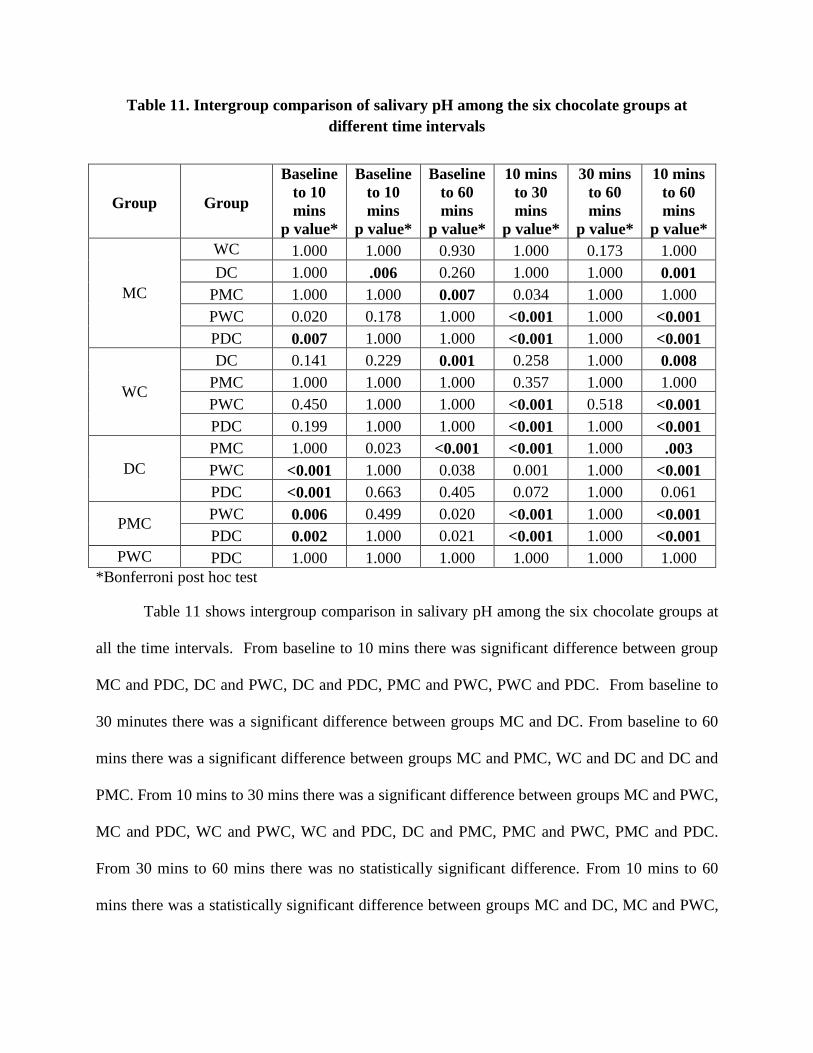
Table 11. Intergroup comparison of salivary pH among the six chocolate groups at
different time intervals
*Bonferroni post hoc test
Table 11 shows intergroup comparison in salivary pH among the six chocolate groups at
all the time intervals. From baseline to 10 mins there was significant difference between group
MC and PDC, DC and PWC, DC and PDC, PMC and PWC, PWC and PDC. From baseline to
30 minutes there was a significant difference between groups MC and DC. From baseline to 60
mins there was a significant difference between groups MC and PMC, WC and DC and DC and
PMC. From 10 mins to 30 mins there was a significant difference between groups MC and PWC,
MC and PDC, WC and PWC, WC and PDC, DC and PMC, PMC and PWC, PMC and PDC.
From 30 mins to 60 mins there was no statistically significant difference. From 10 mins to 60
mins there was a statistically significant difference between groups MC and DC, MC and PWC,
Group Group
Baseline
to 10
mins
p value*
Baseline
to 10
mins
p value*
Baseline
to 60
mins
p value*
10 mins
to 30
mins
p value*
30 mins
to 60
mins
p value*
10 mins
to 60
mins
p value*
MC
WC 1.000 1.000 0.930 1.000 0.173 1.000
DC 1.000 .006 0.260 1.000 1.000 0.001
PMC 1.000 1.000 0.007 0.034 1.000 1.000
PWC 0.020 0.178 1.000 <0.001 1.000 <0.001
PDC 0.007 1.000 1.000 <0.001 1.000 <0.001
WC
DC 0.141 0.229 0.001 0.258 1.000 0.008
PMC 1.000 1.000 1.000 0.357 1.000 1.000
PWC 0.450 1.000 1.000 <0.001 0.518 <0.001
PDC 0.199 1.000 1.000 <0.001 1.000 <0.001
DC
PMC 1.000 0.023 <0.001 <0.001 1.000 .003
PWC <0.001 1.000 0.038 0.001 1.000 <0.001
PDC <0.001 0.663 0.405 0.072 1.000 0.061
PMC PWC 0.006 0.499 0.020 <0.001 1.000 <0.001
PDC 0.002 1.000 0.021 <0.001 1.000 <0.001
PWC PDC 1.000 1.000 1.000 1.000 1.000 1.000

MC and PDC, WC and DC,WC and PDC, DC and PMC, DC and PDC, PMC and PWC, PMC
and PDC.
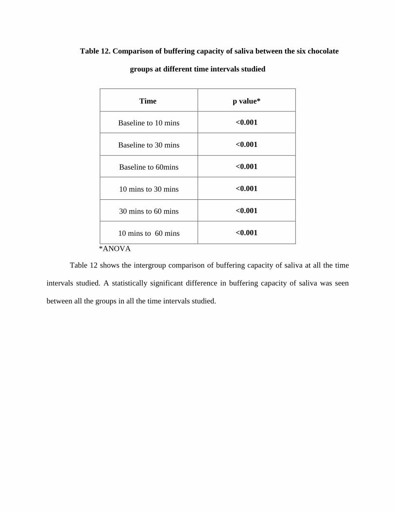
Table 12. Comparison of buffering capacity of saliva between the six chocolate
groups at different time intervals studied
Time p value*
Baseline to 10 mins <0.001
Baseline to 30 mins <0.001
Baseline to 60mins <0.001
10 mins to 30 mins <0.001
30 mins to 60 mins <0.001
10 mins to 60 mins <0.001
*ANOVA
Table 12 shows the intergroup comparison of buffering capacity of saliva at all the time
intervals studied. A statistically significant difference in buffering capacity of saliva was seen
between all the groups in all the time intervals studied.
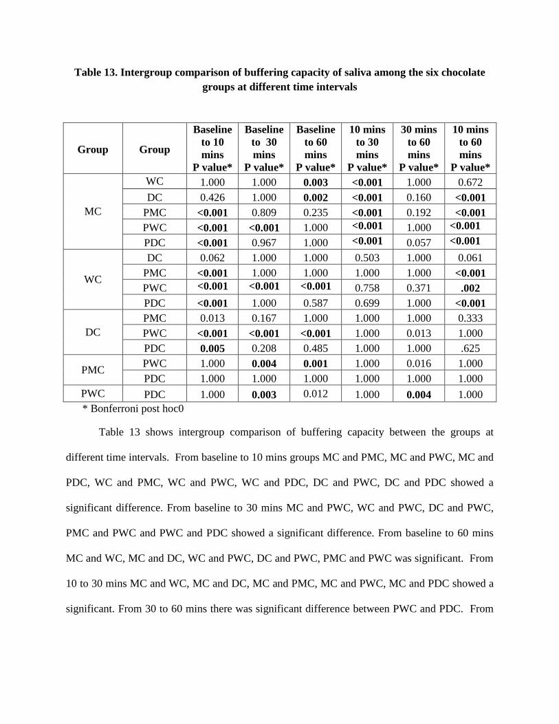
Table 13. Intergroup comparison of buffering capacity of saliva among the six chocolate
groups at different time intervals
* Bonferroni post hoc0
Table 13 shows intergroup comparison of buffering capacity between the groups at
different time intervals. From baseline to 10 mins groups MC and PMC, MC and PWC, MC and
PDC, WC and PMC, WC and PWC, WC and PDC, DC and PWC, DC and PDC showed a
significant difference. From baseline to 30 mins MC and PWC, WC and PWC, DC and PWC,
PMC and PWC and PWC and PDC showed a significant difference. From baseline to 60 mins
MC and WC, MC and DC, WC and PWC, DC and PWC, PMC and PWC was significant. From
10 to 30 mins MC and WC, MC and DC, MC and PMC, MC and PWC, MC and PDC showed a
significant. From 30 to 60 mins there was significant difference between PWC and PDC. From
Group Group
Baseline
to 10
mins
P value*
Baseline
to 30
mins
P value*
Baseline
to 60
mins
P value*
10 mins
to 30
mins
P value*
30 mins
to 60
mins
P value*
10 mins
to 60
mins
P value*
MC
WC 1.000 1.000 0.003 <0.001 1.000 0.672
DC 0.426 1.000 0.002 <0.001 0.160 <0.001
PMC <0.001 0.809 0.235 <0.001 0.192 <0.001
PWC <0.001 <0.001 1.000 <0.001 1.000 <0.001
PDC <0.001 0.967 1.000 <0.001 0.057 <0.001
WC
DC 0.062 1.000 1.000 0.503 1.000 0.061
PMC <0.001 1.000 1.000 1.000 1.000 <0.001
PWC <0.001 <0.001 <0.001 0.758 0.371 .002
PDC <0.001 1.000 0.587 0.699 1.000 <0.001
DC
PMC 0.013 0.167 1.000 1.000 1.000 0.333
PWC <0.001 <0.001 <0.001 1.000 0.013 1.000
PDC 0.005 0.208 0.485 1.000 1.000 .625
PMC PWC 1.000 0.004 0.001 1.000 0.016 1.000
PDC 1.000 1.000 1.000 1.000 1.000 1.000
PWC PDC 1.000 0.003 0.012 1.000 0.004 1.000
10 to 60 mins MC and DC, MC and PMC, MC and PWC, MC and PDC, WC and PMC, WC and
PWC along with WC and PDC showed a significant difference.
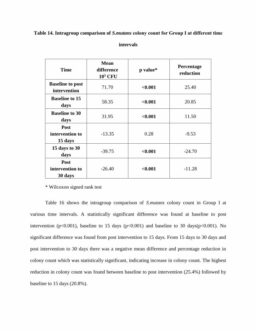
Table 14. Intragroup comparison of S.mutans colony count for Group І at different time
intervals
* Wilcoxon signed rank test
Table 16 shows the intragroup comparison of S.mutans colony count in Group І at
various time intervals. A statistically significant difference was found at baseline to post
intervention (p<0.001), baseline to 15 days (p<0.001) and baseline to 30 days(p<0.001). No
significant difference was found from post intervention to 15 days. From 15 days to 30 days and
post intervention to 30 days there was a negative mean difference and percentage reduction in
colony count which was statistically significant, indicating increase in colony count. The highest
reduction in colony count was found between baseline to post intervention (25.4%) followed by
baseline to 15 days (20.8%).
Time
Mean
difference
103 CFU
p value* Percentage
reduction
Baseline to post
intervention 71.70 <0.001 25.40
Baseline to 15
days 58.35 <0.001 20.85
Baseline to 30
days 31.95 <0.001 11.50
Post
intervention to
15 days
-13.35 0.28 -9.53
15 days to 30
days -39.75 <0.001 -24.70
Post
intervention to
30 days
-26.40 <0.001 -11.28
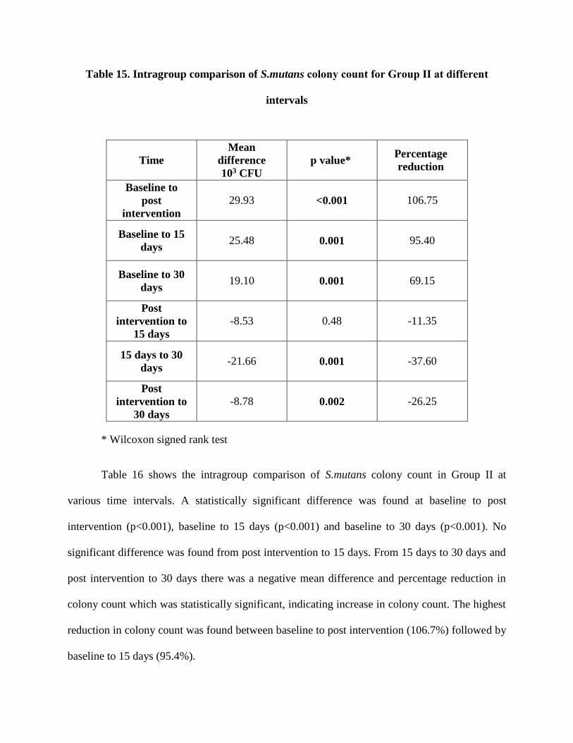
Table 15. Intragroup comparison of S.mutans colony count for Group ІІ at different
intervals
* Wilcoxon signed rank test
Table 16 shows the intragroup comparison of S.mutans colony count in Group ІІ at
various time intervals. A statistically significant difference was found at baseline to post
intervention (p<0.001), baseline to 15 days (p<0.001) and baseline to 30 days (p<0.001). No
significant difference was found from post intervention to 15 days. From 15 days to 30 days and
post intervention to 30 days there was a negative mean difference and percentage reduction in
colony count which was statistically significant, indicating increase in colony count. The highest
reduction in colony count was found between baseline to post intervention (106.7%) followed by
baseline to 15 days (95.4%).
Time
Mean
difference
103 CFU
p value* Percentage
reduction
Baseline to
post
intervention
29.93 <0.001 106.75
Baseline to 15
days 25.48 0.001 95.40
Baseline to 30
days 19.10 0.001 69.15
Post
intervention to
15 days
-8.53 0.48 -11.35
15 days to 30
days -21.66 0.001 -37.60
Post
intervention to
30 days
-8.78 0.002 -26.25
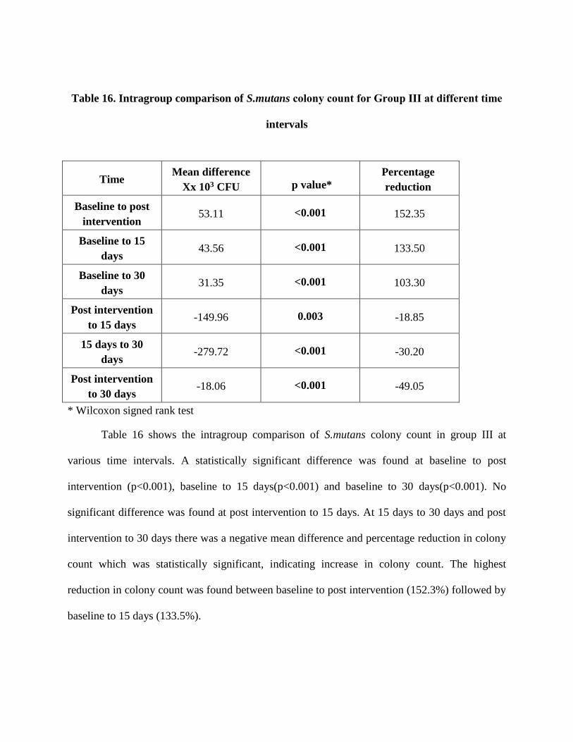
Table 16. Intragroup comparison of S.mutans colony count for Group ІІІ at different time
intervals
* Wilcoxon signed rank test
Table 16 shows the intragroup comparison of S.mutans colony count in group ІІІ at
various time intervals. A statistically significant difference was found at baseline to post
intervention (p<0.001), baseline to 15 days(p<0.001) and baseline to 30 days(p<0.001). No
significant difference was found at post intervention to 15 days. At 15 days to 30 days and post
intervention to 30 days there was a negative mean difference and percentage reduction in colony
count which was statistically significant, indicating increase in colony count. The highest
reduction in colony count was found between baseline to post intervention (152.3%) followed by
baseline to 15 days (133.5%).
Time Mean difference
Xx 103 CFU
p value*
Percentage
reduction
Baseline to post
intervention 53.11 <0.001 152.35
Baseline to 15
days 43.56 <0.001 133.50
Baseline to 30
days 31.35 <0.001 103.30
Post intervention
to 15 days -149.96 0.003 -18.85
15 days to 30
days -279.72 <0.001 -30.20
Post intervention
to 30 days -18.06 <0.001 -49.05
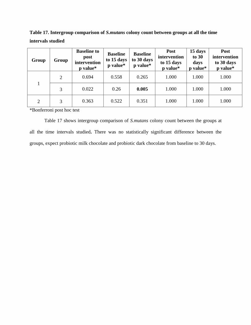
Table 17. Intergroup comparison of S.mutans colony count between groups at all the time
intervals studied
*Bonferroni post hoc test
Table 17 shows intergroup comparison of S.mutans colony count between the groups at
all the time intervals studied. There was no statistically significant difference between the
groups, expect probiotic milk chocolate and probiotic dark chocolate from baseline to 30 days.
Group Group
Baseline to
post
intervention
p value*
Baseline
to 15 days
p value*
Baseline
to 30 days
p value*
Post
intervention
to 15 days
p value*
15 days
to 30
days
p value*
Post
intervention
to 30 days
p value*
1
2 0.694 0.558 0.265 1.000 1.000 1.000
3 0.022 0.26 0.005 1.000 1.000 1.000
2 3 0.363 0.522 0.351 1.000 1.000 1.000
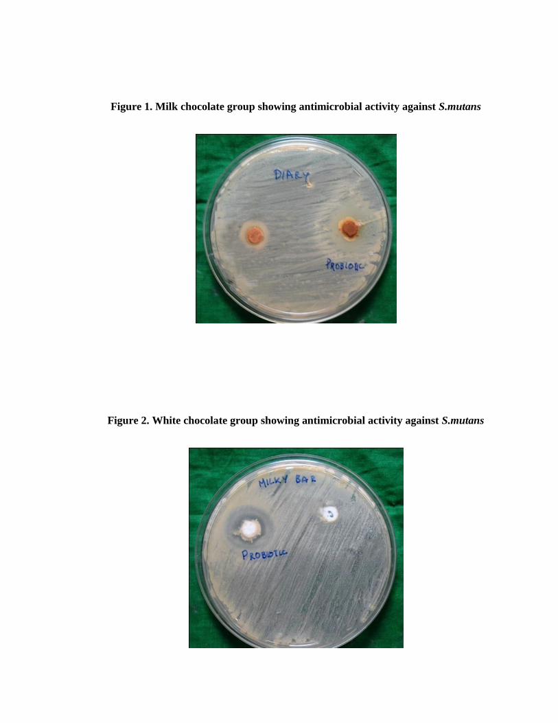
Figure 1. Milk chocolate group showing antimicrobial activity against S.mutans
Figure 2. White chocolate group showing antimicrobial activity against S.mutans
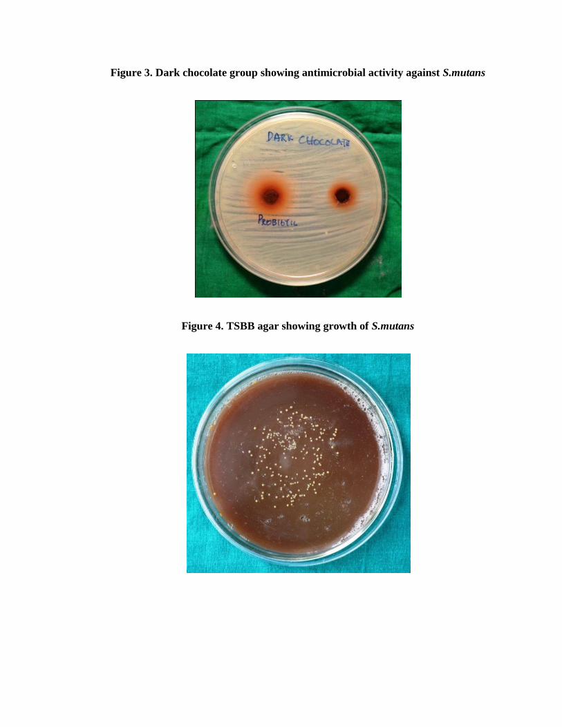
Figure 3. Dark chocolate group showing antimicrobial activity against S.mutans
Figure 4. TSBB agar showing growth of S.mutans

DISCUSSION
Rugg-Gunn et al75 conducted a study in England and reported that each child consumed a
mean of 35 g of chocolate per day. Children’s affinity to chocolates is very strong, and trying to
break the bond often results in failure. The age group of 8-12 years was selected because the
consumption of chocolates among school going children was observed to be higher than in
adolescence.58 Incorporation of probiotics into chocolate could be beneficial to children of this
age group who are known to be fond of chocolates. These children belong to the concrete
operational stage of Jean Piaget’s cognitive theory. They abide the rules, which make them
follow the instructions given to them.68
Selection of chocolates
Chocolates chosen for the study were the representatives of milk, white and dark
chocolate variants. Verakaki and Duggal94 compared various chocolates in the European market
and found milk chocolate with hazelnut and plain milk chocolate had low acidogenic potential.
Nirmala et al compared various filled and unfilled chocolates and found that filled chocolates
were more acidogenic. Unfilled plain chocolates were chosen for the present study to avoid
effects of other ingredients which could alter the original action of the chocolate. Chocolates
used in the present study contained various ingredients. Milk chocolate contained cocoa mass,
cocoa butter, milk, vegetable fat, emulsifiers and sugar. Dark chocolate contained 50% cocoa
solids, cocoa butter and sugar and white chocolate contained milk solids, hydrogenated vegetable
fat, soya lecithin and sugar.
Selection of probiotic
A healthy oral cavity is colonized by a large number of diverse bacteria. The relationship
between the oral microbiota and the host is generally harmonious and symbiotic. Changes in the
oral environment bring about deleterious shifts in this microbiome, leading to dysbiosis or

bacterial imbalance.47 The use of replacement therapies would help to maintain the equilibrium
of the biofilm and thereby foster health.91 The commonly studied probiotic in the oral cavity are
L.reuteri,57,13 L. rhamnosus,55,2,83,72 and L. acidophilus.78,85
The probiotic used for the present study was L.sporogenes which is a spore forming
bacteria. Spores are more resistant to adverse environmental conditions thus allowing it to
undergo industrial processing.76 This enables the viability even in extreme temperature ranges,
reduced cost of production and extended shelf life.42 The probiotic used in this study was the
commercially available BifilacTM sachet. It contains probiotic along with prebiotic bacteria
which act synergistically in symbiosis. Literature search showed its proven effect in minimal
hepatic encephalopathy,92 acute viral diarrhea54,4 efficacy against porphyromonas gingivalis23
and S.mutans.21
The preparation of chocolate was done in accordance to the FDA guidelines which
recommends at least 108 CFU of bacteria to attain its benefits. Kanafari et al 39 and posseimers et
al69 used similar concentrations of probiotic for chocolate preparation. Kanafari et al39 in an
invitro trial showed the inhibitory potential of L.plantarum containing probiotic dark chocolate
on S. mutans colony count. It was in accordance with the results of the present invitro trial which
showed that all types of chocolates added with probiotics had inhibitory effect on S.mutans. The
maximum zone of inhibition was obtained for probiotic dark chocolate. There was no zone of
inhibition for plain milk chocolate. However literature search did not reveal any studies which
dealt with acidogenic and cariogenic properties of probiotic chocolates. Hence this trial was
planned to include 2 parts. Part 1evaluated the effectiveness of chocolates on plaque ph, salivary
pH and buffering capacity of saliva. Part 2 of the trial evaluated its effectiveness against
S.mutans by administering chocolates for 5 consecutive days.
Culture media
The most commonly used selective media for S.mutans is Mitis Salivarius Bacitracin
agar, which was modified by Kimmel and Tinanoff40 as Mitis salivarius agar with kanamycin
sulfate, sorbitol, potasium tellurite and bacitracin (MSKB). Llena et al45 added 5% potassium
tellurite and 64 µg/ml fluconazol to MSKB. Many other medias such as Trypticase soy with
sucrose and bacitracin (TSY 20B), Staat82 and glucose-sucrose-tellurite-bacitracin (GSTB)
Tanzer88 are available. The culture media used for this study was Thioglycolate Sucrose Blood
Bacitracin agar (TSBB). Singh et al79 formulated the selective medium TSBB and compared it
with the other selective medias. Results indicated that TSBB medium inhibits the
all Streptococcus species other than S.mutans present in the oral cavity. Sodium thioglycolate
present in the medium consumes oxygen, making it an anaerobic medium which does not require
special anaerobic incubator for the growth of S.mutans.
Plain chocolates
Considering the plaque pH from baseline to 10 mins, 30 mins and 60 mins , DC
showed the least fall in pH. The acidogenicity of DC was the least followed by WC and MC.
Immediate fall in pH after 10 mins of consumption of chocolates was high in MC and this was
significantly different from DC. On observing the salivary pH from 10 mins, 30 mins and 60
mins, DC showed the least fall in pH. The acidogenicity of DC was the least followed by WC
and MC. The fall in pH after 30 mins of consumption of chocolates was high in MC and was
significantly different from DC. The fall in pH after 60 mins of consumption of chocolates was
low in DC and was significantly different from MC. On comparing the buffering capacity of
saliva from baseline to 10 mins, DC had the least fall in pH followed by MC and WC. From
baseline to 30 mins, WC had the least fall followed by MC and DC. From baseline to 60 mins,
MC had the maximum raise in salivary pH which was significantly different from WC and MC.
Results favor DC to be less acidogenic followed by WC and MC.
The probable reason for less acidogenicity of dark chocolates is that it contains flavanols.
They are a subclass of flavonoids which are, in turn, a subclass of polyphenols. Flavanols
appear as monomers and those found in cocoa beans are epicatechin and catechin. They
can exist as dimmers or polymers, commonly referred as polyphenols. In a study by
Percival et al66 cocoa polyphenols were tested against the growth of S.mutans. It was found
that acid production of S.mutans from sucrose was significantly inhibited and the rate of pH
change was markedly reduced. Further study by Oswaka et al63 found two types of
cariostatic substances in dark chocolate, higher molecular- weight polyphenolic
compounds which showed anti-Glucosyltransferase activities and unsaturated free fatty
acids, such as linoleic and oleic acids which had antibacterial activity against S. mutans.
Our results were in accordance with Frostell27 who carried out experiment with
chocolates containing different concentrations of sucrose and cocoa. They showed that
chocolates with high cocoa content and low sugar had a increase in plaque pH. Amith et al also
found that dark chocolate to have the least pH drop on comparison with milk, white and caramel
chocolate. Edgar et al22 and Rugg-Gunn et al74 showed both milk and dark chocolates to be less
cariogenic compared with other food stuffs. However Grenby and Mistry30 and Nirmala et al58
identified dark chocolate to be more cariogenic than milk chocolate, which was contradictory to
our results.
Probiotic chocolates
Considering the plaque pH from baseline to 10 mins, 30 mins and 60 mins, PWC showed
the least fall in pH. The acidogenicity of PWC was the least followed by PDC and PMC. From
baseline to 30 mins, the least fall in pH was found for PWC which was significantly different
from PDC and PMC. In terms of salivary pH from baseline to 10 mins and 30 mins, PWC
showed the least fall in pH. From baseline to 30 mins, the maximum rise in pH was found for
PWC which was significantly different from PDC and PMC. The acidogenicity of PWC was the
least followed by PDC and PMC. The buffering capacity of saliva was higher for PWC from
baseline to 10 mins, 30 mins and 60 mins. The acidogenecity of PWC was least followed by
PDC and PMC.
The higher pH raise in PWC could be attributable to its milk content. Milk components
such as casein phosphopeptides (CPP) and glycomacropeptide (GMP) get incorporated into the
salivary pellicle and reduce the adherence of S. sobrinus and S. mutan. By selectively inhibiting
adhesion, these dairy bioactives could modulate the microbial composition of dental plaque. This
could in turn control acid formation in dental plaque, thus reducing hydroxyapatite dissolution
from tooth enamel.56 Jenkins and Ferguson35 conducted invitro comparisons of 4 % lactose
solutions with cow's milk. They found a negligible fall in plaque pH which was due to milk's
high buffering capacity. High levels of calcium and phosphate could have contributed to the
lesser dissolution of enamel.
In the present study addition of probiotics to white chocolate was less acidogenic than
dark chocolate. Composition of chocolates plays a major role in the retention of chocolates on
the tooth surface and their oral clearance. Dark chocolates require relatively greater chewing
phase and oral processing time whereas milk chocolate have a lower solid fat index. Milk
chocolate is ready to swallow than dark chocolate and requires lesser chewing time for a swallow
able state.28 So WC which contains more of milk, will be swallowed readily and hence contribute
less to decrease in pH drop.
Plain chocolate vs probiotic chocolate
Estimation of acidogenecity of probiotics were studied by Sudhir et al85 and Srivatsav et
al81 following consumption of probiotic curd. The former found a decrease in salivary pH,
whereas the later found an increase in salivary pH. In the present study, irrespective of the type
of chocolate used, probiotic chocolates were better in salivary, plaque pH and buffering capacity
of saliva than their normal counterparts.
DC Vs PDC
In terms of plaque pH, salivary pH and buffering capacity of saliva, PDC showed the
least fall in plaque pH compared to DC at all the time intervals studied. Salivary pH and
buffering capacity of saliva from baseline to 10 mins showed significant increase in pH for PDC
than DC.
MC Vs PMC
PMC showed least fall in plaque pH, salivary pH and buffering capacity of saliva at all
the time intervals studied. Salivary pH from baseline to 60 mins showed a significant difference
between PMC and MC. Buffering capacity of saliva from baseline to 10 mins showed a
significant increase in buffering for PMC compared to MC.
WC Vs PWC
PWC showed least fall in plaque pH, salivary pH and buffering capacity of saliva at all
the time intervals studied. PWC showed a significant increase in plaque pH from baseline to 30
mins. Buffering capacity of saliva showed a significant increase in pH from baseline to 10mins,
30 mins as well as 60 mins.
One of the proposed mechanisms of action of probiotics is its production of lactic acid,
acetic acid, hydrogen peroxide and bacteriocins which in turn reduce the plaque pH. In the
present study, the probioitic used reduced the pH drop in plaque and salivary pH which were
either similar or lesser than the normal chocolates. Production of lactic acid by bacteria depends
on the type of sugar and bacterial strain.38 Hedgber33 showed that L. paracasei and L. reuteri did
not ferment any kind of sugar. L. rhamnosus exhibited negative reaction for sucrose and L.
plantarum fermented sucrose. Literature search did not reveal any proof for the production of
acids by the probiotic bacteria used in the present study. Accordingly the results of this study
also indicated that there was no significant acid production by probiotic bacteria, as the ph drop
in plaque and saliva were minimal in probiotic chocolate groups compared to their counterparts.
Effect on S.mutans colony count
The effect of probiotic against S.mutans was studied by different vehicles. Nase et al55
was the first to conduct a long term randomized controlled trial using probiotic milk. The
children in their experimental group showed a reduction in the S.mutans counts after 7 months of
intervention.
The present study was of a short duration with probiotic intake for 5 consecutive days.
Similar studies with short term administration of probiotics for 7 days have been carried out by
various researchers and proved the efficacy of probiotics,20,6,81,3,46 Chinnappa et al16 assessed the
S.mutans colony count with probiotic ice cream and probiotic yoghurt after 7 days
administration. They found a significant reduction in the S.mutans colony count at 1 hour and 7
days post intervention in both the probiotic groups.
Intra group comparison of bacterial colony count showed a significant reduction in the
number of colonies after 30 days. All the probiotic chocolate groups showed maximum decrease
in bacterial colony count at the post intervention period. However from post intervention to 15
days the colony count showed a steady increase but did not reach the baseline. Results indicated
the short term action and easy wash out of probiotics on cessation of its intake.
The results were in accordance with Ghasehmi et al28 who investigated the effect of
probiotic yoghurt and xylitol chewing gum on salivary S.mutans colony count after 3 weeks
intervention. The children were followed up for 4 weeks. Results showed a reduction in colony
count upto 2 weeks, followed by an increase in colony count in the 4th week which was however
lesser than the baseline. Chuang et al17 showed similar results after administering probiotic
tablets for 2 weeks. Ashwin et al3 conducted a similar study by administering probiotic ice cream
for 7 consecutive days and were followed up for a period of 6 months. There was a decrease in
the colony count till 30 days. Results of the present study were in contrast to Mahantesha et al46
who conducted a randomized controlled trail by administering probiotic milk and ice cream for 7
consecutive days. Their results showed a reduction in colony count in both the groups at 1
month, however the probiotic ice cream group showed a reduced colony count even after 3
months.
The probable mechanism of action of probiotics include; competitive inhibition,
production of bacteriocins, organic acids, hydrogen peroxide, modulating pH and modulating
redox potential.7 In this study the probiotics might have competitively inhibited S.mutans.77
However from the results of the present study and previous literature, it is questionable or not
they have any residual effect after discontinuation of intake. Systematic review by Cagetti et al12
evaluated the effect of probiotics administration on caries risk factors and showed that in two-
thirds of the selected papers, probiotics have demonstrated the capacity to reduce Mutans
sterptococci counts in saliva and/or plaque in short-term. Probiotic bacteria can only transiently
colonize the oral cavity during the use of probiotic products. 52,41,96 The retention of probiotic is
an important factor to be taken into account. Retention observed depends on the products, strains
and also host individuals. 11,67,41,96 Probiotics are given even to newborns because of their
beneficial health effects but information on their oral colonisation in children is not available.70,87
Results of the present study were contradictory to the findings of Lexner et al44 who
compared normal milk and milk supplemented with bacterium L. rhamnosus LB21 for 2 weeks.
They did not observe any significant difference in the salivary colony counts.
At baseline to 30 days probiotic dark chocolate (103%) showed a significant reduction in
colony count than probiotic dairy milk (11%). The mechanism for the observed inhibitory effect
of the cocoa polyphenols on biofilm formation and acid production is not clear. It has been
proposed that polyphenols could inhibit enzymes necessary for the production of energy in cells
or may cause changes in cell membrane permeability.1 Evidence has shown that cocoa extracts
can act against mutans streptococci by inhibiting sucrose-dependent adherence,
glucosyltransferase activity and insoluble glucan synthesis.62,64,63 Cocoa butter is one of the basic
ingredients of a cocoa bean. It consists mainly of a triglyceride ester form of palmitic, stearic,
oleic, and linoleic acids which were found to be antimicrobial agents.63
Probiotics have shown beneficial health impact in humans, leading to several new
recommendations for its use. Chocolates can serve as a vehicle for delivering probiotics in
children making it beneficial for health. Probiotic chocolates have the added advantage of
reducing the chocolates acidogenicity and hence making them tooth friendly.
Limitations
Evaluating the adherence of the probiotic bacteria in dental plaque and rate of its acid
production has not been studied. This could have given a clear insight on its mechanism
of action.
Larger sample size of children with different caries status could have been assessed, to
get a better picture of the anticariogenic properties of probiotic chocolates.
Accepted markers for caries such as caries increment or incidence can been assessed with
long term studies.

SUMMARY AND CONCLUSION
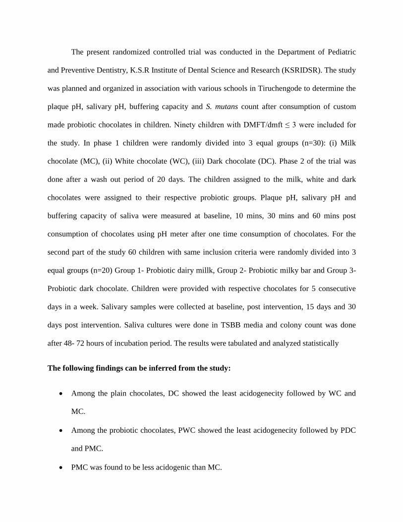
The present randomized controlled trial was conducted in the Department of Pediatric
and Preventive Dentistry, K.S.R Institute of Dental Science and Research (KSRIDSR). The study
was planned and organized in association with various schools in Tiruchengode to determine the
plaque pH, salivary pH, buffering capacity and S. mutans count after consumption of custom
made probiotic chocolates in children. Ninety children with DMFT/dmft ≤ 3 were included for
the study. In phase 1 children were randomly divided into 3 equal groups (n=30): (i) Milk
chocolate (MC), (ii) White chocolate (WC), (iii) Dark chocolate (DC). Phase 2 of the trial was
done after a wash out period of 20 days. The children assigned to the milk, white and dark
chocolates were assigned to their respective probiotic groups. Plaque pH, salivary pH and
buffering capacity of saliva were measured at baseline, 10 mins, 30 mins and 60 mins post
consumption of chocolates using pH meter after one time consumption of chocolates. For the
second part of the study 60 children with same inclusion criteria were randomly divided into 3
equal groups (n=20) Group 1- Probiotic dairy millk, Group 2- Probiotic milky bar and Group 3-
Probiotic dark chocolate. Children were provided with respective chocolates for 5 consecutive
days in a week. Salivary samples were collected at baseline, post intervention, 15 days and 30
days post intervention. Saliva cultures were done in TSBB media and colony count was done
after 48- 72 hours of incubation period. The results were tabulated and analyzed statistically
The following findings can be inferred from the study:
Among the plain chocolates, DC showed the least acidogenecity followed by WC and
MC.
Among the probiotic chocolates, PWC showed the least acidogenecity followed by PDC
and PMC.
PMC was found to be less acidogenic than MC.
PWC was found to be less acidogenic than WC.
PDC was found to be less acidogenic than MC.
S.mutans colony count reduced in all the three probiotic chocolate groups.
Maximum reduction in colony count was observed from baseline to immediate post
intervention period in all the three probiotic chocolate groups.
The colony count increased after 15days post intervention but did not raise above
baseline colony count in all the three chocolate groups.
PDC showed the maximum reduction in the S.mutans colony count among the probiotic
chocolates.
Invitro evaluation of chocolates against S.mutans showed inhibitory zones for all the
probiotic chocolates. The maximum zone of inhibition was observed for probiotic dark
chocolate. Plain dark chocolate and white chocolate also showed inhibitory effect against
S.mutans.
CONCLUSION
PWC was found to be least acidogenic among all the six chocolate groups studied.
DC was found to be least acidogenic among normal chocolates.
Probiotic chocolates were less acidogenic than their counterparts
All probiotic chocolates were effective in reducing the S.mutans colony count.
Probiotic dark chocolate showed the highest percentage reduction in S.mutans colony
count.
REFERENCES
1. Abram V, Donko M. Tentative identification of polyphenols in Sempervivum tectorum
and assessment of the antimicrobial activity of Sempervivum L. J Agric Food Chem
1999;47(2):485-89.
2. Ahola AJ, Yli-Knuuttila H, Suomalainen T, Poussa T, Ahlstrom A, Meurman JH et al.
Shortterm consumption of probiotic-containing cheese and its effect on dental caries risk
factors. Arch Oral Biol 2002;47(11):799-804.
3. Ashwin D, Vijayaprasad KE , Taranath M, Ramagoni NK, Naras A, Sarpangala M.
Effect of probiotic containing ice-cream on salivary mutans streptococci (sms) levels in
children of 6-12 years of age: a randomized controlled double blind study with six-
months follow up. J Clin Diagn Res 2015;9(2):ZC06–ZC09.
4. Bastola R, Bastola BS, Gurung R, Ghimire JJ, Shrestha SK. A Randomized Open Label
Comparative Clinical Study of a Probiotic against a Symbiotic in the Treatment of Acute
Diarrhoea in Children. Int J Med Res Health Sci 2017;6(9):96-100.
5. Beckett ST. Chocolate as a drink. The Science of Chocolate. 2nd edition. Cambridge:The
royal society of chemistry:2008:1-3.
6. Bhalla M, Ingle NA, Kaur N, Yadav P. Mutans streptococci estimation in saliva before
and after consumption of probiotic curd among school children. J Int Soc Prev
Community Dent 2015;5(1):31-34.
7. Bhushan J, Chachra S. Probiotics-their role in prevention of dental caries. J Oral Health
Comm Dent 2010;4(3):78-82
8. Bibby BG, Mundorff SA, Zero DT, Almekinder KJ. Oral food clearance and the pH of
plaque and saliva. J Am Dent Assoc 1986;112(3):333-37.

9. Burton JP, Drummond BK, Chilcott CN, Tagg JR, Thomson WM et al. Influence of
the probiotic Streptococcus salivarius strain M18 on indices of dental health in children: a
randomized double-blind, placebo-controlled trial. J Med Microbiol 2013;62(6):875-84.
10. Business news. India among world's fastest growing chocolate markets: Research. The
Times of India. April 30 2017. Available from https://timesofindia.indiatimes.com/
business/india-business/india-among-worlds-fastest-growing-chocolate-markets-
research/articleshow/58444087.cms. Assessed on December 12 2017.
11. Busscher HJ, Mulder AF, van der Mei HC. In vitro adhesion to enamel and in vivo
colonization of tooth surfaces by Lactobacilli from a bio-yoghurt. Caries Res
1999;33(5):403-14.
12. Cagetti MG, Mastroberardino S, Milia E, Cocco F, Lingstrom P, Campus G. The Use of
Probiotic Strains in Caries Prevention: A Systematic Review. Nutrients 2013;5(7):2530-
50.
13. Caglar E, Cildir SK, Ergeneli , Sandalli N, Twetman S. Salivary mutans streptococci and
lactobacilli levels after ingestion of the probiotic bacterium Lactobacillus reuteri ATCC
55730 by straws or tablets. Acta Odontol Scand 2006;64(5):314-18.
14. Caglar E, Kavaloglu SC, Kuscu OO, Sandalli N, Holgerson PL, Twetman S. Effect of
chewing gums containing xylitol or probiotic bacteria on salivary mutans streptococci
and lactobacilli. Clin Oral Invest 2007;11(4):425-29.
15. Caglar E, Kuscu OO, Cildir SK, Kuvvetli SS, Sandalli N. A probiotic lozenge
administered medical device and its effect on salivary mutans streptococci and
lactobacilli. Int J Paed Dent 2008;18(1):35-39.
16. Chinnappa A, Konde H, Konde S, Raj S, Beena JP. Probiotics for future caries control: A
short-term clinical study. Indian J Dent Res 2013;24(5):547-49.
17. Chuang LC, Huang CS, Ou-Yang LW, Lin SY. Probiotic Lactobacillus paracasei effect
on cariogenic bacterial flora. Clin Oral Investig 2011;15(4):471-76
18. Commane D, Hughes R, Shortt C, Rowland I. The potential mechanisms involved in the
anti-carcinogenic action of probiotics. Mutat Res 2005;591(1):276-89.
19. Cooper KA, Campos-Giménez E, Jimenez Alvarez D, Rytz A, Nagy K, Williamson G.
Predictive relationship between polyphenol and nonfat cocoa solids content of chocolate.
J Agric Food Chem 2008;56(1):260-65.
20. Deo PN, Deshmukh R. Evaluation of salivary levels of Streptococcus mutans pre- and
post-probiotics use. J Adv Clin Res Insights 2015;2(3):1-4.
21. Dhawan R, Dhawan S. Role of probiotics on oral health: A randomized, double-blind,
placebo-controlled study. J Interdiscip Dent 2013;3(2):71-78.
22. Edgar WM, Bibby BG, Mundorff S, Rowley J. Acid production in plaques after eating
snacks: modifying factors in foods. J Am Dent Assoc. 1975;90(2):418-25.
23. Elavarasu S, Suthanthiran T, Thangavelu A, Kanagaraj SS, Mohandas L, Sekar S.
Evaluation of efficacy of probiotic (BIFILAC) on Porphyromonas gingivalis: In vitro
study. J Pharm Bioallied Sci 2016;8(Suppl 1):S45-S47.
24. Eliasson L, Carlen A, Almstahl A, Wikstrom M, Lingstrom P. Dental plaque pH and
micro-organisms during hyposalivation. J Dent Res 2006;85(4):334-8.
25. Englander HR, Shklair IL, Fosdick LS. The Effects of Saliva on the pH and Lactate
Concentration of Plaque. I. Caries-rampant Individuals. J Dent Res 1959;38:848-53
26. Ericsson Y. Clinical Investigations of the Salivary Buffering Action. Acta Odontologica
Scandinavica 1959;17(2);131-63.
27. Frostell G. A method for evaluation of acid potentialities of foods. Acta Odontol
Scand 1970;28(5):599-622.
28. Gaikawad V. Oral processing of milk and dark chocolate. Unpublished thesis. The Riddet
Institute, Massey University, Palmerston North. New Zealand 2012.
29. Ghasemi E, Mazaheri R, Tahmourespour A. Effect of probiotic yogurt and xylitol-
containing chewing gums on salivary S.mutans count. J Clin Pediatr Dent
2017;41(4):257-63.
30. Grenby TH, Mistry M. Precise control of the frequency and amount of food provided for
small laboratory animals by a new electronic metering technique, used to evaluate the
cariogenic potential of chocolate. Caries Res 1995;29(5):418-23.
31. Hatakka K, Ahola AJ, Yli-Knuuttila H, Richardson M, Poussa T, Meurman JH, Korpela
R. Probiotics reduce the prevalence of oral Candida in the elderly- a randomized
controlled trial. J Dent Res 2007;86(2):125-30.
32. Hedayati-Hajikand T, Lundberg U, Eldh C, Twetman S. Effect of probiotic chewing
tablets on early childhood caries–a randomized controlled trial. BMC Oral
Health 2015;15(1):112.
33. Hedberg M, Hasslof P, Sjostrom I, Twetman S, Stecksen-Blicks C. Sugar fermentation in
probiotic bacteria–an in vitro study. Oral Microbiol Immunol 2008;23(6):482-485.
34. Hegde AM, Shetty R, Sequeira AR. The acidogenicity of various chocolates available in
Indian market: A comparative study. Int J Clin Pediatr Dent 2009;2(2):20-24.
35. Jenkins GN, Ferguson DB. Milk and dental caries. Br dent J 1966;120:472-77.
36. Jindal G, Pandey R.K, Agarwal J, Singh M. A comparative evaluation of probiotics on
salivary mutans streptococci counts in Indian children. Eur Arch Paediatr Dent
2011;12(4):211-15
37. Juneja, A, Kakade A. Evaluating the effect of probiotic containing milk on salivary
mutans streptococci levels. J Clin Pediatr Dent 2012;37(1):9-14.
38. KellerMK, TwetmanS.Acid production in dental plaque after exposure to probiotic bacter
ia. BMC Oral Health 2018;12:44
39. Khanafari A, Hosseini Porgham S, Tajabadi Ebrahimi M. Investigation of probiotic
chocolate effect on Streptococcus mutans Growth Inhibition. Jundishapur J Microbiol
2012;5(4):590-97.
40. Kimmel L, Tinanoff N. A modified mitis salivarius medium for a caries diagnostic test.
Mol Oral Microbiol 1991;6(5):275-79.
41. Krasse P, Carlsson B, Dahl C, Paulsson A, Nilsson A, Sinkiewicz G. Decreased gum
bleeding and reduced gingivitis by the probiotic Lactobacillus reuteri. Swed Dent J
2006;30(2):55-60.
42. La Rosa M, Bottaro G, Gulino N, Gambuzza F, Di Forti F, Ini G et al. Prevention of
antibiotic-associated diarrhea with Lactobacillus sporogens and fructo-oligosaccharides
in children. A multicentric double-blind vs placebo study. Minerva Pediatr
2003;55(5):447-52.
43. Laleman I, Detailleur V, Slot DE, Slomka V, Quirynen M, Teughels W. Probiotics
reduce mutans streptococci counts in humans: a systematic review and meta-analysis.
Clin Oral Investig 2014;18(6):1539-52.
44. Lexner MO, Blomqvist S, Dahlen G, Twetman S. Microbiological profiles in saliva and
supragingival plaque from caries-active adolescents before and after a short-term daily
intake of milk supplemented with probiotic bacteria - a pilot study. Oral Health Prev Dent
2010;8(4):383-88.
45. Llena MC, Perez-Gracia MT, Fernandez-Barredo S, Galiana C: New culture media for
the isolation of Streptococcus mutans and Lactobacillus in the saliva of head- and neck-
irradiated. Oral Health Prev Dent 2007;5(4):285-89.
46. Mahantesha T, Parveen KM, Praveen NH, Asha N, Ashwin D, Vinutna B. Comparative
study of probiotic ice cream and probiotic drink on salivary Streptococcus mutans levels
in 6-12 years age group children. J Int Oral Health 2015;7(9):47-50.
47. Marsh P D, Moter A and Devine D A. Dental plaque biofilms: communities conflict and
control. Periodontology 2000 2011;55(1):16-35.
48. Marsh P D. Dental plaque as a biofilm and a microbial community – implications for
health and disease. BMC Oral Health 2006;6(Suppl 1):S14.
49. Marteau P, Flourie B, Pochart P, Chastang C, Desjeux JF, Rambaud JC. Effect of the
microbial lactase (EC 3.2. 1.23) activity in yoghurt on the intestinal absorption of lactose:
an in vivo study in lactase-deficient humans. Br J Nutr 1990;64(1):71-79.
50. Marteau PR, de Vrese M, Cellier CJ, Schrezenmeir J. Protection from gastrointestinal
diseases with the use of probiotics. Am J Clin Nutr 2001;73(2):430s-6s.
51. McFarland LV. Beneficial microbes: health or hazard. Eur J Gastroenterol Hepatol
2000;12:1069–1071.

52. Montalto M, Vastola M, Marigo L, Covino M, Graziosetto R, Curigliano V et al.
Probiotic treatment increases salivary counts of lactobacilli: a double-blind, randomized,
controlled study. Digestion 2004;69(1):53-56.
53. Nakamura Y, Yamamoto N, Sakai K, Takano T. Antihypertensive effect of sour milk and
peptides isolated from it that are inhibitors to angiotensin I-converting enzyme. J Dairy
Sci 1995;78(6):1253-7.
54. Narayanappa D. Randomized double blinded controlled trial to evaluate the efficacy and
safety of Bifilac in patients with acute viral diarrhea. Indian J Pediatr 2008;75(7):709-13.
55. Nase L, Hatakka K, Savilahti E, Saxelin M, Ponka A, Puossa T et al. Effects of long-term
consumption of a probiotic bacterium, Lactobacillus rhamnosus GG, in milk on dental
caries and caries risk in children. Caries Res 2001;35(6):412-20.
56. Neeser JR, Golliard M, Woltz A, Rouvet M, Dillmann ML, Guggenheim B. In vitro
modulation of oral bacterial adhesion to saliva‐coated hydroxyapatite beads by milk
casein derivatives. Molecular Oral Microbiology 1994;9(4):193-201.
57. Nikawa H, Makihira S, Fukushima H, Nishimura H, Ozaki Y, Ishida K et al.
Lactobacillus reuteri in bovine milk fermented decreases the oral carriage of mutan
streptococci. Int J Food Microbiol 2004; 95(2):219-23.
58. Nirmala S, Quadar MA, Veluru S. pH modulation and salivary sugar clearance of
different chocolates in children: A randomized clinical trial. J Indian Soc Pedod Prev
Dent 2016;34(1):10-16.
59. Nishihara T, Suzuki N, Yoneda M, Hirofuji T. Effects of Lactobacillus salivarius-
containing tablets on caries risk factors: A randomized open-label clinical trial. BMC
Oral Health 2014;14(1):110.
60. Nozari A, Motamedifar M, Seifi N, Hatamizargaran Z, Ranjbar MA. The effect of iranian
customary used probiotic yogurt on the children’s salivary cariogenic microflora. J Dent
(Shiraz) 2015;16(2):81-86.
61. Ooi LG, Liong MT. Cholesterol-lowering effects of probiotics and prebiotics: a review of
in vivo and in vitro findings. Int J Mol Sci 2010;11(6):2499-522.
62. Ooshima T, Osaka Y, Sasaki H, Osawa K, Yasuda H, Matsumura M et al. Caries
inhibitory activity of cacao bean husk extract in in-vitro and animal experiments. Arch
Oral Biol 2000;45(8):639-45.
63. Osawa K, Miyazaki K, Shimura S, Okuda J, Matsumoto M, Ooshima T. Identification of
cariostatic substances in the cacao bean husk: their anti-glucosyltransferase and
antibacterial activities. J Dent Res 2001;80(11):2000-04.
64. Paolino VJ, Kashket S. Inhibition by cocoa extracts of biosynthesis of extracellular
polysaccharide by human oral bacteria. Arch Oral Biol 1985;30(4):359-63.
65. Parvez S, Malik KA, Ah Kang S, Kim HY. Probiotics and their fermented food products
are beneficial for health. J Appl Microbiol 2006;100(6):1171-85.
66. Percival RS, Devine DA, Duggal MS, Chartron S, Marsh PD. The effect
of cocoa polyphenols on the growth, metabolism, and biofilm formation by Streptococcus
mutans and Streptococcus sanguinis. Eur J Oral Sci 2006;114(4):343-48.
67. Petti S, Tarsitani G, D'Arca AS. A randomized clinical trial of the effect of yoghurt on
the human salivary microflora. Arch Oral Biol 2001;46(8):705-12.
68. Piaget J. Piaget and his school. Piaget’s theory. Springer Berlin Heidelberg 1976. pp. 11-
23.
69. Possemiers S, Marzorati M, Verstraete W, Van de Wiele T. Bacteria and chocolate: a
successful combination for probiotic delivery. Int J Food Microbiol 2010;141(1-2):97-
103.
70. Rautava S, Arvilommi H, Isolauri E. Specific probiotics in enhancing maturation of IgA
responses in formula-fed infants. Pediatr Res 2006;60(2):221-24.
71. Reid G, Beuerman D, Heinemann C, Bruce AW. Probiotic Lactobacillus dose required to
restore and maintain a normal vaginal flora. FEMS Immunol Med Microbiol
2001;32(1):37-41.
72. Rodriguez G, Ruiz B, Faleiros S, Vistoso A, Marro ML, Sanchez J et al. Probiotic
compared with standard milk for high-caries children: a cluster randomized trial. J Dent
Res 2016;95(4):402-07.
73. Romani Vestman N, Chen T, Lif Holgerson P, Ohman C, Johansson I. Oral Microbiota
Shift after 12-Week Supplementation with Lactobacillus reuteri DSM 17938 and PTA
5289; A Randomized Control Trial. PLoS One 2015;10(5):1-18.
74. Rugg-Gunn AJ, Edgar WM, Geddes DA, Jenkins GN. The effect of different meal
patterns upon plaque pH in human subjects. Br Dent J 1975;139(9):351-6.
75. Rugg-Gunn AJ, Hackett AF, Appleton DR, Jenkins GN, Eastoe JE. Relationship between
dietary habits and caries increment assessed over two years in 405 English adolescent
school children. Arch Oral Biol 1984; 29(12):983-92.
76. Sanders TA. Food safety and risk assessment: naturally occurring potential toxicants and
anti-nutritive compounds in plant foods. Forum Nutr 2003;56:407-09.
77. Silva M, Jacobus NV, Deneke C, Gorbach SL. Antimicrobial substance from a human
Lactobacillus strain. Antimicrob Agents Chemother 1987;31(8):1231-33.
78. Singh RP, Damle SG, Chawla A. Salivary mutans streptococci and lactobacilli
modulations in young children on consumption of probiotic ice-cream containing
Bifidobacterium lactis Bb12 and Lactobacillus acidophilus La5. Acta Odontol Scand
2011;69(6):389-94.
79. Singh S, Vishnoi N, Dwivedi D, Khare M and Singh V: Modified TSBB Culture Media
Enhance Faster Growth of Streptococci Mutans as Compared to Existing Culture Media.
Int J Pharm Sci Res 2016;7(9):3689-94.
80. Solga SF. Probiotics can treat hepatic encephalopathy. Med Hypotheses 2003;61(2):307-
13.
81. Srivastava S, Saha S, Kumari M, Mohd S. Effect of probiotic curd on salivary pH
and streptococcus mutans: a double blind parallel randomized controlled trial. J Clin
Diagn Res 2016;10(2):ZC13–ZC16.
82. Staat RH. Inhibition of Streptococcus mutans strains by different mitis-salivarius agar
preparations. J Clin Microbiol 1976;3(3):378-80.
83. Stecksen-Blicks C, Sjostrom I, Twetman S. Effect of long-term consumption of milk
supplemented with probiotic lactobacilli and fluoride on dental caries and general health
in preschool children: a cluster-randomized study. Caries Res 2009;43(5):374-81.
84. Stephan RM. Intra-oral hydrogen-ion concentrations associated with dental caries
activity. J Dent Res 1944;23:257-266.
85. Sudhir R, Praveen P, Anantharaj A, Venkataraghavan K. Assessment of the effect of
probiotic curd consumption on salivary pH and Streptococcus mutans counts. Niger Med
J 2011;53(3):135-39.
86. Taipale T, Pienihakkinen K, Alanen P, Jokela J, Soderling E. Administration of
Bifidobacterium animalis subsp. lactis BB-12 in early childhood: a post-trial effect on
caries occurrence at four years of age. Caries Res 2013;47(5):364-72.
87. Taipale T, Pienihakkinen K, Alanen P, Jokela J, Soderling E. Dissolution of xylitol from
a food supplement administered with a novel slow-release pacifier: preliminary
results. Eur Arch Paediatr Dent 2007;8(2):123-26.
88. Tanzer JM, Borjesson AC, Laskowski L, Kurasz AB, Testa M. Glucose-sucrose-
potassium tellurite-bacitracin agar, an alternative to mitis salivarius-bacitracin agar for
enumeration of Streptococcus mutans. J Clin Microbiol 1984;20(4):653-59.
89. Tomas MS, Claudia Otero M, Ocana V, Elena Nader-Macias M.. Production of
antimicrobial substances by lactic acid bacteria I: determination of hydrogen peroxide.
Methods Mol Biol 268, 337–346.
90. Trinchieri V, Di Carlo S, Bossu M, Polimeni A. Use of lozenges containing Lactobacillus
brevis CD2 in recurrent aphthous stomatitis: a double-blind placebo-controlled trial.
Ulcers 2011;24:1-6.
91. Twetman S, Stecksén-Blicks C. Probiotics and oral health effects in children. Int J
Paediatr Dent 2008;18(1):3-10.
92. Unnikrishnan D, Tharani CB, Samy N, Rajagopal A, Muthiah M, Kumari U. Role of
Synbiotic [Bifilac™ Hp] in the Management of Minimal Hepatic Encephalopathy.
Journal of Pharmaceutical Sciences and Pharmacology 2014;1(3):182-86.
93. Vasanthakumar H, Sharan J, D.Cruz AM. Plaque pH and Dental Retention after
Consumption of Different Types of Chocolates. Int J Clin Prev Dent 2016;12(2):97-102.
94. Verakaki E. Duggal M.S. A comparison of different kinds of European chocolates on
human plaque pH. Eur J Paediatr Dent 2003;4(4):203-10.
95. Verdu EF, Collins SM. Irritable bowel syndrome. Best Pract Res Clin Gastroenterol
2004;18(2):315-21.
96. Yli-Knuuttila H, Snall J, Kari K, Meurman JH. Colonization of Lactobacillus rhamnosus
GG in the oral cavity. Oral Microbiol Immunol 2006;21(2):129-31.
97. Yousuf A, Nagaraj A, Ganta S, Sidiq M, Pareek S, Vishnani P et al. Comparative
evaluation of commercially available freeze dried powdered probiotics on mutans
streptococci count: a randomized, double blind, clinical study. J Dent (Tehran) 2015;
12(10):729-38.
98. Zhang M, Wang F, Jiang L, Liu RH, Zhang L, Lei X, Li J, Jiang J, Guo H, Fang B, Zhao
L. Lactobacillus salivarius REN inhibit rat oral cancer induced by 4-nitroquioline 1-
oxide. Cancer Prev Res (Phila) 2013;6(7):686-94.

APPENDIX
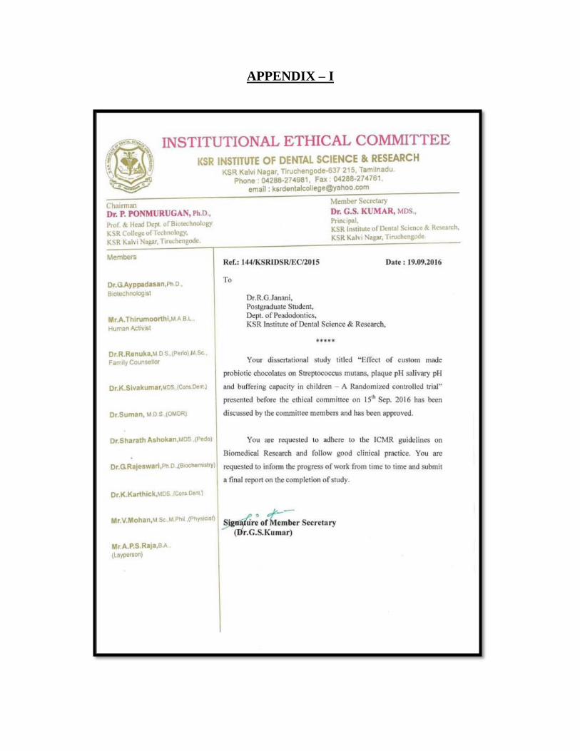
APPENDIX – I
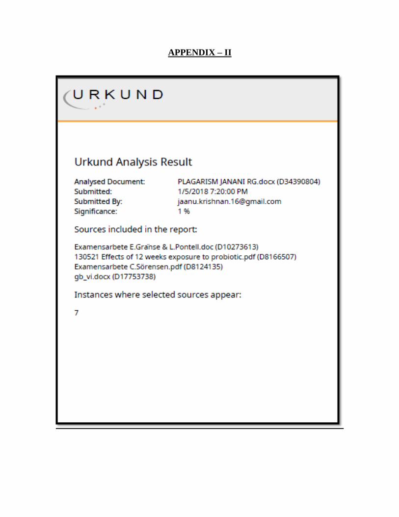
APPENDIX – II
APPENDIX III
CERTIFICATE - II
This is to certify that this dissertation work titled “Effect of custom-made
probiotic chocolates on Streptococcus mutans, plaque pH, salivary pH and
buffering capacity in children- A radomized controlled trial” of the candidate
Dr.Vijayasankari V with registration number for the award of
“Master of Dental Surgery” in the branch of Pedodontics and Preventive
Dentistry. I personally verified the urkund.com website for the purpose of
plagiarism Check. I found that the uploaded thesis file contains from introduction
to conclusion pages and result shows 7% percentage of plagiarism in the
dissertation.
Guide & Supervisor sign with Seal.
APPENDIX – IV
CONSENT FORM
I ..........., as legally responsible parent/guardian give my consent for the participation of my child
................................, standard........... In the study title “Effect of custom made probiotic
chocolates on Streptococcus mutans, plaque pH, salivary pH and buffering in children- A
randomised controlled trial’. Dr. Janani RG discussed with me to my satisfaction, the procedures,
possible discomforts, as well as possible benefits of the study. I have read this consent and have
clearly understood the procedures to be performed on my child.
Legally responsible parent/guardian: ...................................... Date: ......................
Address: ........................................................................
Contact number:
I certify that I explained the above information to the parent/guardian, before requesting his or
her signature.
Signature of the dentist: ........................................................... Date: .....................
K.S.R Institute of Dental Science and Research
Tiruchengode - 637215
APPENDIX – V
தகவலறிந்த ஒப்புதல் படிவம்
பபயர் __________________________ வயது _________ ஆண்/பபண் ஆகியநான்என்மகன்/மகள்
_________________________ ,வயது ___ அவர்களை மருத்துவர் அவர்கைின் ஆராய்ச்சிக்கு
உட்படுத்த அனுமதி ககாரப்பட்டுள்ைது. இவ்வாராய்ச்சிப்பற்றி விைக்கங்களும்,
முளறகளும் நான் படித்துப்பார்த்கதன்/படித்துக்காட்டப்பட்டது. எனது சந்கதகங்களுக்கு
பதைிவாகவிைக்கம்அைிக்கப்பட்டது .எனகவநான்எனதுமகளன /மகளை
இவ்வாராய்ச்சியில் பங்பகடுக்க அனுமதி அைிக்கிகறன்.
இடம்:
கததி : (பபற்கறார் ளகபயாப்பம்)
K.S.R பல் மருத்துவக் கல்லூாி மற்றும் ஆராய்ச்சி ளமயம்
திருச்பசங்ககாடு - 637215
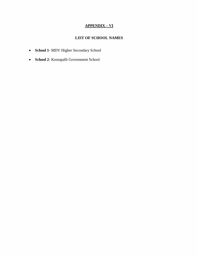
APPENDIX – VІ
LIST OF SCHOOL NAMES
School 1- MDV Higher Secondary School
School 2- Kootapalli Government School
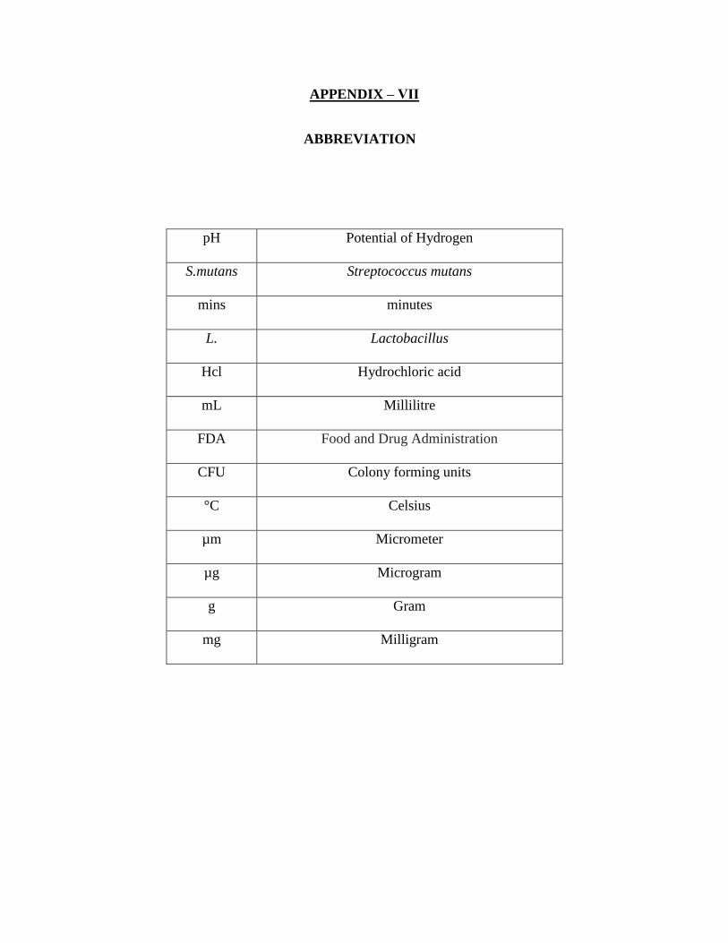
APPENDIX – VІІ
ABBREVIATION
pH Potential of Hydrogen
S.mutans Streptococcus mutans
mins minutes
L. Lactobacillus
Hcl Hydrochloric acid
mL Millilitre
FDA Food and Drug Administration
CFU Colony forming units
°C Celsius
µm Micrometer
µg Microgram
g Gram
mg Milligram
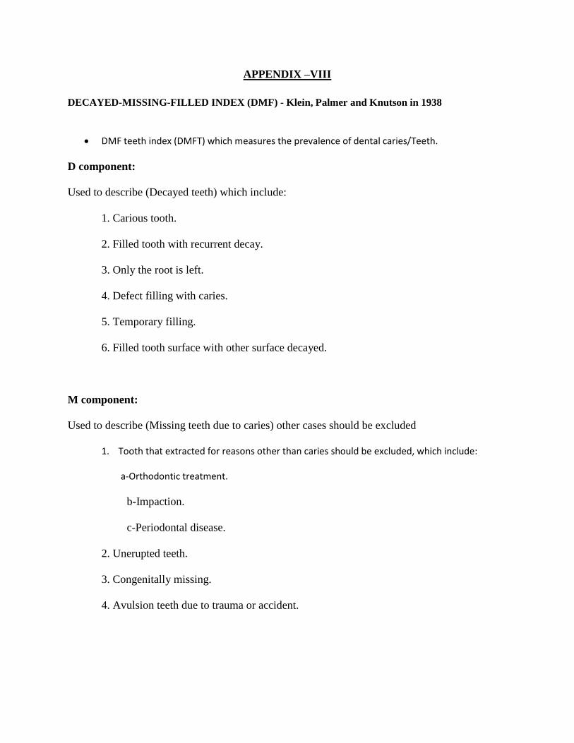
APPENDIX –VІІІ
DECAYED-MISSING-FILLED INDEX (DMF) - Klein, Palmer and Knutson in 1938
DMF teeth index (DMFT) which measures the prevalence of dental caries/Teeth.
D component:
Used to describe (Decayed teeth) which include:
1. Carious tooth.
2. Filled tooth with recurrent decay.
3. Only the root is left.
4. Defect filling with caries.
5. Temporary filling.
6. Filled tooth surface with other surface decayed.
M component:
Used to describe (Missing teeth due to caries) other cases should be excluded
1. Tooth that extracted for reasons other than caries should be excluded, which include:
a-Orthodontic treatment.
b-Impaction.
c-Periodontal disease.
2. Unerupted teeth.
3. Congenitally missing.
4. Avulsion teeth due to trauma or accident.
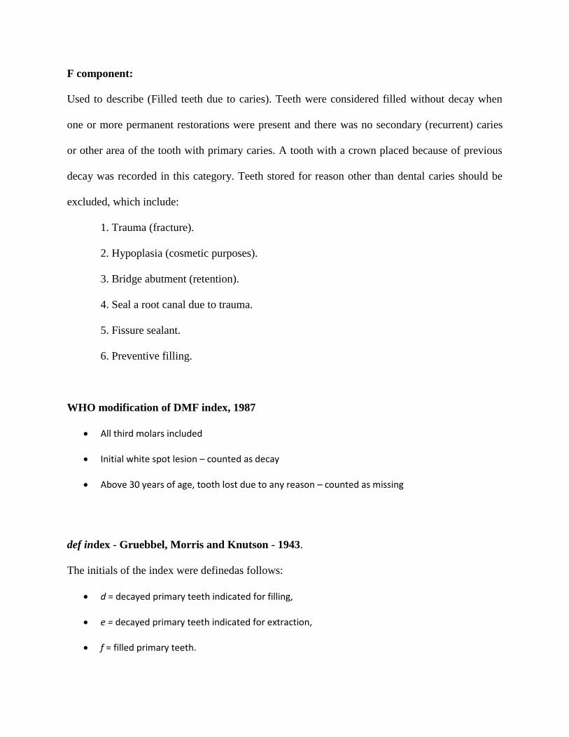
F component:
Used to describe (Filled teeth due to caries). Teeth were considered filled without decay when
one or more permanent restorations were present and there was no secondary (recurrent) caries
or other area of the tooth with primary caries. A tooth with a crown placed because of previous
decay was recorded in this category. Teeth stored for reason other than dental caries should be
excluded, which include:
1. Trauma (fracture).
2. Hypoplasia (cosmetic purposes).
3. Bridge abutment (retention).
4. Seal a root canal due to trauma.
5. Fissure sealant.
6. Preventive filling.
WHO modification of DMF index, 1987
All third molars included
Initial white spot lesion – counted as decay
Above 30 years of age, tooth lost due to any reason – counted as missing
def index - Gruebbel, Morris and Knutson - 1943.
The initials of the index were definedas follows:
d = decayed primary teeth indicated for filling,
e = decayed primary teeth indicated for extraction,
f = filled primary teeth.
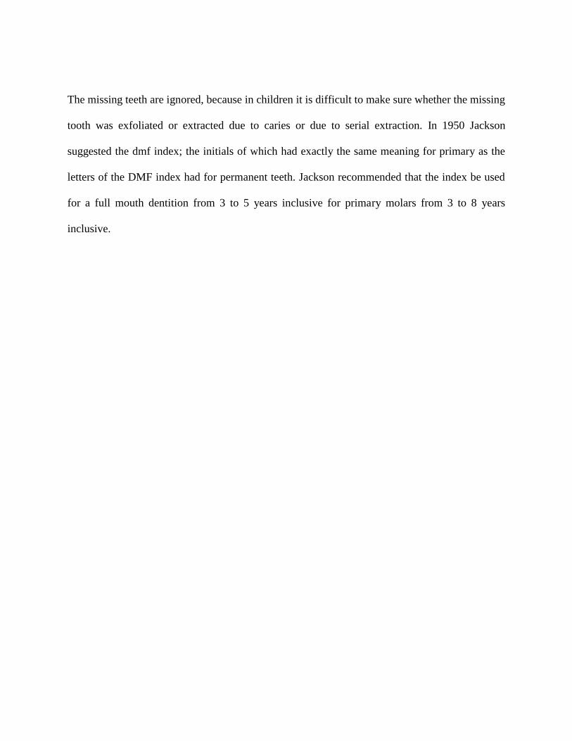
The missing teeth are ignored, because in children it is difficult to make sure whether the missing
tooth was exfoliated or extracted due to caries or due to serial extraction. In 1950 Jackson
suggested the dmf index; the initials of which had exactly the same meaning for primary as the
letters of the DMF index had for permanent teeth. Jackson recommended that the index be used
for a full mouth dentition from 3 to 5 years inclusive for primary molars from 3 to 8 years
inclusive.
Top Related