


Languages
Pages
Legal


Stents ou BVS Bioabsorbable Vascular Scaffolding
Serviço de Cardiologia I C.H.L.N.
Diogo Torres

First PTCA and 23-Year Follow Up
In patients who did not suffer sub-acute closure due to dissections, or restenosis due to negative
remodeling in the first few months, long term results following balloon angioplasty were very
encouraging and durable, with loss in MLD not seen until
17 years post procedure1
1. Hatrick, R., et al. EuroIntervention. 2009;5:121-126.
1977 2000

Balloon Angioplasty
(PTCA)
Bare Metal Stents (BMS)
Coronary Drug
Eluting Stents (DES)
Bioresorbable
Vascular
Scaffold
(BVS)
Advancements in PCI
1977 1986 2001 2006
Evolution of PCI Therapy Improving Patient Outcomes

What is Required of a Fully Biorebsorbable Scaffold to Fulfill the Desire for a ‘Vascular Restoration Therapy’?
Revasculascularization
0 to 3 months
Performance should mimic that of a standard DES
• Good deliverability
• Minimum of acute recoil
• Hight acute radial strength
• Controlled delivery of drug to abluminal tissue
Restoration
3 to 6-9 months +
Transition from scaffolding to discontinous structure
• Gradually lose radial strenght
• Struts must be incorporated into the vessel wall (strut coverage)
• Become structurally discontinuous
• Allow the vassel to respond naturally to physilogical stimuli
Resorption
9 months +
Implant is discontinous and inert
• Resorb in a benign fashion
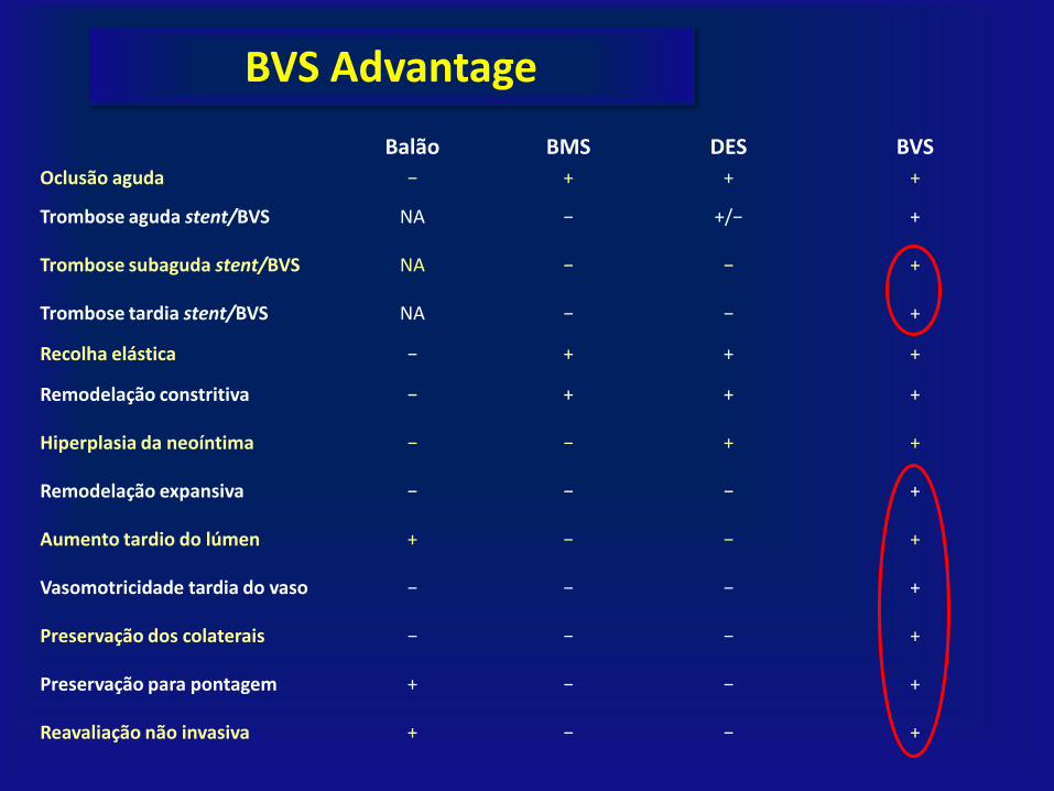
BVS Advantage
Balão BMS DES BVS
Oclusão aguda − + + +
Trombose aguda stent/BVS NA − +/− +
Trombose subaguda stent/BVS NA − − +
Trombose tardia stent/BVS NA − − +
Recolha elástica − + + +
Remodelação constritiva − + + +
Hiperplasia da neoíntima − − + +
Remodelação expansiva − − − +
Aumento tardio do lúmen + − − +
Vasomotricidade tardia do vaso − − − +
Preservação dos colaterais − − − +
Preservação para pontagem + − − +
Reavaliação não invasiva + − − +
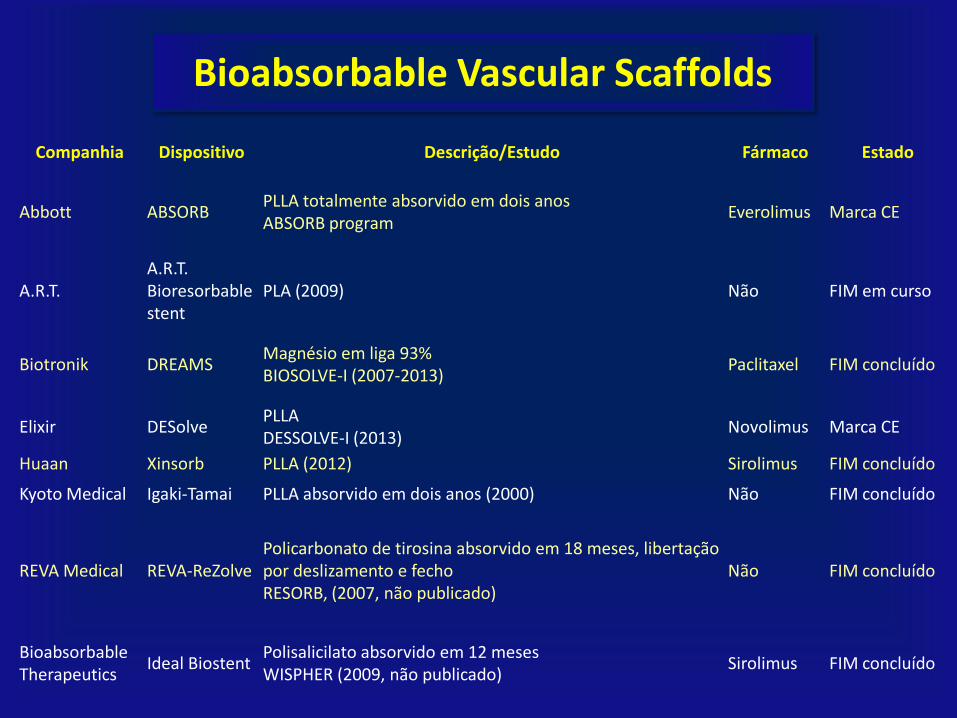
Bioabsorbable Vascular Scaffolds
Companhia Dispositivo Descrição/Estudo Fármaco Estado
Abbott ABSORB PLLA totalmente absorvido em dois anos ABSORB program
Everolimus Marca CE
A.R.T. A.R.T. Bioresorbable stent
PLA (2009) Não FIM em curso
Biotronik DREAMS Magnésio em liga 93% BIOSOLVE-I (2007-2013)
Paclitaxel FIM concluído
Elixir DESolve PLLA DESSOLVE-I (2013)
Novolimus Marca CE
Huaan Xinsorb PLLA (2012) Sirolimus FIM concluído
Kyoto Medical Igaki-Tamai PLLA absorvido em dois anos (2000) Não FIM concluído
REVA Medical REVA-ReZolve Policarbonato de tirosina absorvido em 18 meses, libertação por deslizamento e fecho RESORB, (2007, não publicado)
Não FIM concluído
Bioabsorbable Therapeutics
Ideal Biostent Polisalicilato absorvido em 12 meses WISPHER (2009, não publicado)
Sirolimus FIM concluído

ABSORB BVS
Polymer backbone
Drug/polymer matrix
Everolimus/PDLLA Matrix Coating
• Thin layer
• Amorphous (non-crystalline)
• 1:1 ratio of Everolimus/PDLLA matrix
• Conformal coating, 2-4 m thick
• Controlled drug release
PLLA Scaffold
• Semi-crystalline
• Provides device structure
• Processed for required radial strength

Absorb Comprehensive AV-Sponsored Clinical Trial Program
2011 2012 2013 2014 2015 2016
Each trial n reflects total patients. Data effective September 2013
*ABSORB IV trial is in the planning stage and subject to change.
Total Pts Studied n=~599 n~965 n~5,709 n~7,609 n~8,709 n~9,709
ABSORB China n = ~440, China Pivotal RCT
Enrollment & Follow-Up
ABSORB II n = ~501, International RCT
1 Y
Enrollment & Follow-Up
ABSORB EXTEND n = ~800, Registry
Enrollment & Follow-Up
ABSORB Cohort B n = 101; FIM
ABSORB Cohort A n = 30; FIM
ABSORB FIRST n = ~1,800, International Registry
Enrollment & Follow-Up
ABSORB IV* n = ~3,000, US RCT
UK Registry n = 1000, UK Registry
Enrollment & Follow-Up
1 Y 2 Y 3 Y 4 Y 5 Y
1 Y 2 Y
1 Y 2 Y
3 Y
3 Y
ABSORB Japan n = ~400, Japan Pivotal RCT
Enrollment & Follow-Up
ABSORB III n = ~2,250, US Pivotal RCT
Enrollment & Follow-Up
1 Y
1 Y
1 Y
1 Y
1 Y 2 Y
2 Y
2 Y
5 Y

Investing in a Comprehensive ABSORB Clinical Program – Investigator Sponsored Trials
Randomized Controlled Trials (2,764 Pts)
Study Title Design Number of
Patients
Primary Endpoint Patient FU
(Years)
AIDA All – comers RCT vs Xience 2194 2-Yr TVF 5
TROFI II STEMI RCT vs XIENCE 190 6-Mo neo-intimal healing score 3
PROSPECT II
ABSORB
RCT vs OMT in unstable
asymptomatic pts
300 2-Yr IVUS MLA 3
PROACTIVE RCT vs XIENCE 20 Peri-Proc Platelet Reactivity 1
VANISH RCT vs XIENCE 60 Evolution of myocardial blood flow
values over time
3
Registries (10,030 Pts)
BVS EXPAND All – comers Registry (excl STEMI) 300 1 – Yr MACE 5
ASSURE All – comers Registry 180 Safety and Efficacy 3
ABSORB CTO Feasibility in CTO 20 Safety and Performance 2
PABLOS Feasibililty in Bifurcations 30 Device, Procedural, Main and Side
Branch Success
2
IT-DISSAPEARS MVD and Long Lesion Registry 1000 Safety and Efficacy 5
GABI-R All – comers Registry 5000 Safety and Efficacy 5
REPARA All – comers Registry 1500 1- Yr MACE 1
POLAR ACS ACS Registry 100 Safety, clinical device, procedure
success and in-hospital MACE
1
France ABSORB Feasibility in de novo lesions 2000 1 – Yr MACE 1

Introduction ABSORB Cohort A
QCA, IVUS, OCT, IVUS VH
24 12 6
MSCT
18 36 48 60
Study Objective First In Man, Single Arm – safety/performance
Endpoints Typical PCI clinical and imaging endpoints
Treatment Single, de novo native coronary lesion in a vessel with a reference vessel diameter of 3.0 mm
Device Sizes 3.0 x 12 mm scaffolds (3.0 x 18 mm scaffolds available after enrolment start and used in 2 pts)
Follow-Up (Months)
Clinical
30 subjects (Non-randomized) 4 sites in Europe & New Zealand
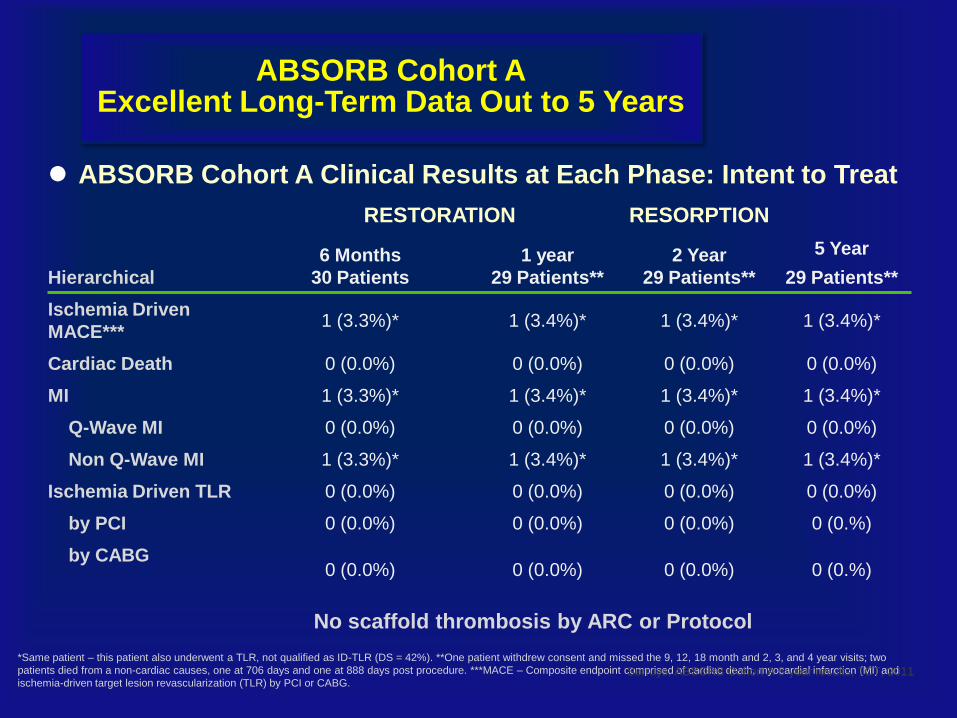
ABSORB Cohort A Excellent Long-Term Data Out to 5 Years
*Same patient – this patient also underwent a TLR, not qualified as ID-TLR (DS = 42%). **One patient withdrew consent and missed the 9, 12, 18 month and 2, 3, and 4 year visits; two
patients died from a non-cardiac causes, one at 706 days and one at 888 days post procedure. ***MACE – Composite endpoint comprised of cardiac death, myocardial infarction (MI) and
ischemia-driven target lesion revascularization (TLR) by PCI or CABG.
RESTORATION RESORPTION
Hierarchical
6 Months
30 Patients
1 year
29 Patients**
2 Year
29 Patients**
5 Year
29 Patients**
Ischemia Driven
MACE*** 1 (3.3%)* 1 (3.4%)* 1 (3.4%)* 1 (3.4%)*
Cardiac Death 0 (0.0%) 0 (0.0%) 0 (0.0%) 0 (0.0%)
MI 1 (3.3%)* 1 (3.4%)* 1 (3.4%)* 1 (3.4%)*
Q-Wave MI 0 (0.0%) 0 (0.0%) 0 (0.0%) 0 (0.0%)
Non Q-Wave MI 1 (3.3%)* 1 (3.4%)* 1 (3.4%)* 1 (3.4%)*
Ischemia Driven TLR 0 (0.0%) 0 (0.0%) 0 (0.0%) 0 (0.0%)
by PCI 0 (0.0%) 0 (0.0%) 0 (0.0%) 0 (0.%)
by CABG
0 (0.0%) 0 (0.0%) 0 (0.0%) 0 (0.%)
Serruys, ABSORB Cohort A 5-year results; TCT, 2011
No scaffold thrombosis by ARC or Protocol
ABSORB Cohort A Clinical Results at Each Phase: Intent to Treat

ABSORB Cohort A
Temporal Lumen Dimensional Changes, Per Treatment
Late lumen loss at 6 months mainly due to reduction in scaffold area
Very late lumen gain noted from 6 months to 2 years
*Adapted from Serruys, PW, ABSORB Cohort A 2-year IVUS and OCT results; ACC 2009.
ABSORB Cohort A
Unpaired Analysis*
Lumen Area
6.04 mm2 5.19 mm2 5.46 mm2
n = 25 n = 25 n = 18
Scaffold
Area
11.8%
Lumen
Area
10,85%
Post-PCI 6 Months 2 Years
EEL
unchanged

Restoration and Resorption Late Lumen Enlargement
6 month follow up
5 year follow up
OCT
MLA 6.35 mm2
MLA 8.77 mm2
IVUS
∆ 1.42mm
Late lumen enlargement/gain and ‘characteristic ‘final golden tube’ on OCT illustrating functional reparation of the vessel
ABSORB A 5 Yr

ABSORB Cohort A Vasomotor Function Testing at 2 Years
The reappearance of vasomotion in the proximal, distal, as well as treated segments in
response to methergine or acetylcholine suggests that vessel vasoreactivity has been
restored and that a physiological response to vasoactive stimulus might occur anew.
Serruys, PW, et al. Lancet 2009; 373: 897-910.
Acetylcholine
(Vasodilator)
Methergine
(Vasoconstrictor)
ABSORB Cohort A at 2 years
Vasodilation
Vasoconstriction
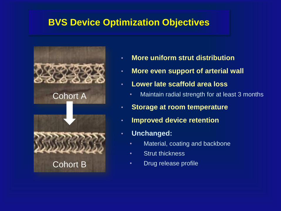
BVS Device Optimization Objectives
Cohort A
Cohort B
• More uniform strut distribution
• More even support of arterial wall
• Lower late scaffold area loss
• Maintain radial strength for at least 3 months
• Storage at room temperature
• Improved device retention
• Unchanged:
• Material, coating and backbone
• Strut thickness
• Drug release profile
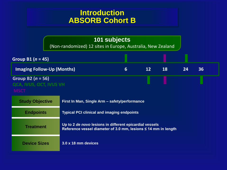
Introduction ABSORB Cohort B
Study Objective First In Man, Single Arm – safety/performance
Endpoints Typical PCI clinical and imaging endpoints
Treatment Up to 2 de novo lesions in different epicardial vessels Reference vessel diameter of 3.0 mm, lesions ≤ 14 mm in length
Device Sizes 3.0 x 18 mm devices
Imaging Follow-Up (Months)
101 subjects (Non-randomized) 12 sites in Europe, Australia, New Zealand
24 12 6 18 36
Group B1 (n = 45)
Group B2 (n = 56) QCA, IVUS, OCT, IVUS VH MSCT

ABSORB Cohort B Groups 1&2 Clinical Results – Intent to Treat
Serruys, PW, ABSORB Cohort B 3-year results; ACC 2013 / *One patient missed the 2 year FUP
Non Hierarchical
Cardiac Death %
Myocardial Infarction % (n)
Q - wave MI
Non Q - wave MI
Ischemia driven TLR % (n)
CABG
PCI
Hierarchical MACE % (n) 2.0 (2)
0
0
0
2.0 (2)
0
2.0 (2)
0
30 Days
n = 101
6.9 (7)
4.0 (4)
0
4.0 (4)
3.0 (3)
0
3.0 (3)
0
1 Year
n = 101 -
-
-
2 Years
0
3.0 (3)
0
3.0 (3)
6.0 (6)
0
6.0 (6)
9.0 (9)
n = 100*
5.0 (5)
2.0 (2)
0
2.0 (2)
3.0 (3)
0
3.0 (3)
0
6 Months
n = 101
MACE: Cardiac death, MI, ischemia-driven TLR
TVF: Cardiac death, MI, ischemia-driven TLR, ischemia-driven TVR
No scaffold thrombosis by ARC or Protocol out to 3 Years only 3 additional TLR
events between 1 and 3 years
3 Years
0
3.0 (3)
0
3.0 (3)
7.0 (7)
0
7.0 (7)
10.0 (10)
n = 100*
Hierarchical TVF % (n) 2.0 (2) 6.9 (7) 11.0 (11) 5.0 (5) 13.0 (13)

ABSORB Cohort B Clinical Results – MACE
Numerically Lower Long-Term Event Rates versus a Best-in-Class DES
Serruys PW, ABSORB Cohort B 3Year Data, Rotterdam EuroPCR Focus on BVS 2013

ABSORB Cohort B Temporal Lumen Dimensional Changes
ABSORB Cohort B1 Serial Analysis*
Lumen Area
6.53 mm2 6.36 mm2 6.85 mm2
n = 33 n = 33 n = 33
Lumen
Area
7.2%1
Late Loss = 0.19 mm
Post-PCI 6 Months 2 Years
ABSORB Cohort B2
Paired Analysis**
Lumen Area
6.29 mm2 6.35 mm2 6.81 mm2
n = 56 n = 56 n = 56
Lumen
Area
7.2%2
Late Loss = 0.27 mm
Post-PCI 12 Months 3 Years
Late Loss = 0.27 mm
Late Loss = 0.29 mm
*Serruys, PW., ABSORB Cohort B 2-year results; TCT 2011
**Serruys, PW., ABSORB Cohort B 3-year results; ACC 2013 1. Patient-level serial analysis
2. Calculated from overall mean values
Scaffold
Area
1.7%
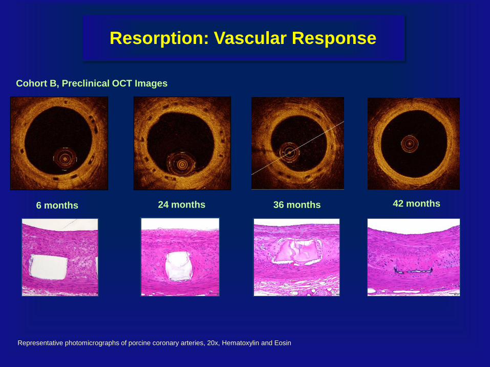
Resorption: Vascular Response
42 months 6 months 24 months
Cohort B, Preclinical OCT Images
36 months
Representative photomicrographs of porcine coronary arteries, 20x, Hematoxylin and Eosin

ABSORB EXTEND Non-Randomized, Single-Arm, Continued Access Trial
Study Objective Continued Access trial. FPI: Jan 11, 2011
Endpoints Typical PCI clinical endpoints
Treatment Up to 2 de novo lesions in different epicardial vessels Planned overlapping allowed in lesions >22 and ≤ 28 mm
Device Sizes Scaffold diameters: 2.5, 3.0, 3.5 mm Scaffold lengths: 12*, 18, 28 mm
Clinical Follow-up (months)
~1,000 subjects Up to 100 global sites (non-US)
Clinical Follow-Up
MSCT follow up (n=100)
OCT follow up (n=50)
24 12 6 18 36

ABSORB EXTEND Clinical Results – Intent to Treat; Interim Snapshot
MACE: cardiac death, MI, ischemia-driven TLR
*Reflects an interim snapshot with only cleaned data as of the cut-off date of 03 December 2012.
**No Absorb BVS was implanted in the target lesion
Non-Hierarchical
Cardiac Death % (n)
Myocardial Infarction % (n)
Q-wave MI
Non Q-wave MI
Ischemia Driven TLR % (n)
PCI
CABG
Hierarchical MACE % (n)
6 Months*
n = 450
0.2 (1)**
0.7 (3)
2.0 (9)
2.7 (12)
0.4 (2)
0.4 (2)
0.0 (0)
2.9 (13)
12 Months*
n = 450
0.2 (1)**
0.9 (4)
2.0 (9)
2.9 (13)
1.8 (8)
1.6 (7)
0.2 (1)
4.2 (19)
Scaffold Thrombosis
(ARC Def/Prob) % (n) 0.7 (3) 0.9 (4)
Chevalier, ABSORB EXTEND 12-month outcomes in the first 450 patient enrolled, Rotterdam EuroPCR Focus on BVS 2013
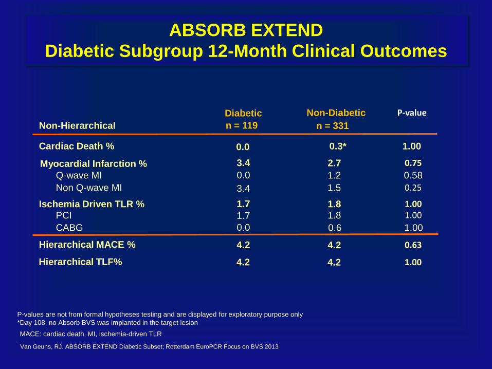
ABSORB EXTEND Diabetic Subgroup 12-Month Clinical Outcomes
MACE: cardiac death, MI, ischemia-driven TLR
P-values are not from formal hypotheses testing and are displayed for exploratory purpose only
*Day 108, no Absorb BVS was implanted in the target lesion
Non-Hierarchical
Cardiac Death %
Myocardial Infarction %
Q-wave MI
Non Q-wave MI
Ischemia Driven TLR %
PCI
CABG
Hierarchical MACE %
Diabetic
n = 119
0.0
0.0
3.4
3.4
1.7
1.7
0.0
4.2
Non-Diabetic
n = 331
0.3*
1.2
1.5
2.7
1.8
1.8
0.6
4.2
Van Geuns, RJ. ABSORB EXTEND Diabetic Subset; Rotterdam EuroPCR Focus on BVS 2013
Hierarchical TLF% 4.2 4.2
P-value
1.00
0.58
0.25
0.75
1.00 1.00
1.00
0.63
1.00

Absorb BVS (B+EXTEND) vs. XIENCE V (SPIRIT I, II, III) Diabetic Subgroup – Unadjusted 1Y Clinical Outcomes
P-values are descriptive and are displayed for exploratory purpose only. Study funded by Abbott Vascular. C. Naber, Patients I Select to Treat with BVS, EuroPCR 2013
Non-Hierarchical Absorb BVS
(N=170)
XIENCE V
(N=252)
Unadjusted
P-value
Cardiac Death % 0.0 1.2 0.55
Myocardial Infarction % 2.9 4.4 0.59
Q-wave MI 0.0 0.8 0.54
Non Q-wave MI 2.9 3.6 1.00
Ischemia driven TLR % 1.5 4.4 0.15
CABG 0.0 0.4 1.00
PCI 1.5 4.0 0.23
Hierarchical MACE % 3.7 8.4 0.09
Hierarchical TVF % 3.7 10.8 0.02
Scaffold Thrombosis (def/prob) % 0.7 1.6 0.66
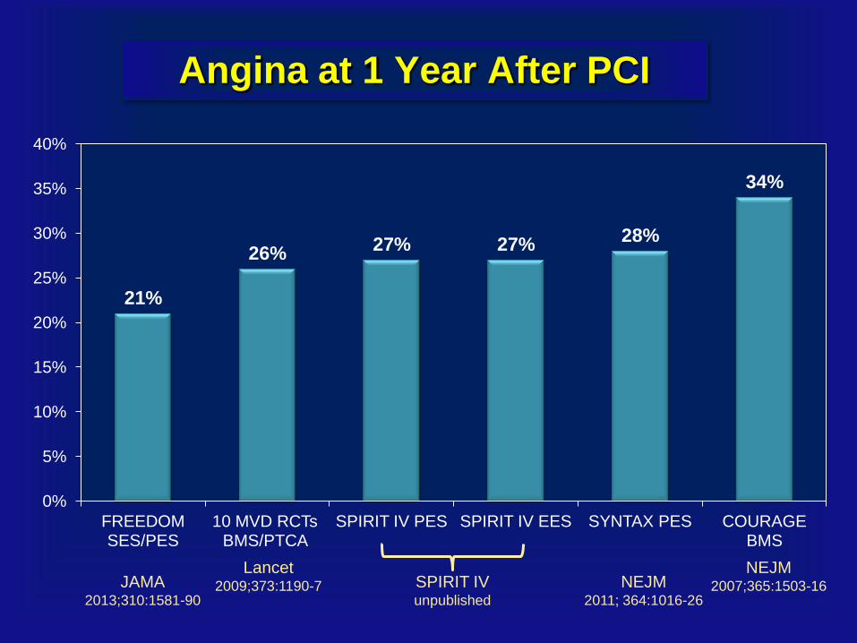
Angina at 1 Year After PCI
21%
26% 27% 27% 28%
34%
0%
5%
10%
15%
20%
25%
30%
35%
40%
FREEDOM SES/PES
10 MVD RCTs BMS/PTCA
SPIRIT IV PES SPIRIT IV EES SYNTAX PES COURAGE BMS
Lancet 2009;373:1190-7
NEJM 2007;365:1503-16 NEJM
2011; 364:1016-26
JAMA 2013;310:1581-90
SPIRIT IV unpublished

Angina Status: EXTEND* vs. SPIRIT IV** Propensity matched cohorts
Days post procedure 0 37 194 393
Absorb pts at risk: 287 267 250 240
Xience pts at risk: 602 535 478 429
An
gin
a o
r
an
gin
a e
qu
iva
len
t (%
)
0%
5%
10%
15%
20%
25%
30%
35%
40%
45%
50%
Days post procedure
0 60 120 180 240 300 360 420
7.0%
11.3% 16.0%
28.1%
Δ=4.3%
Δ=12.1%
12.9%
20.6%
HR [95%CI] =
0.53 [0.39,0.74]
P=0.0001
ABSORB (n=287)
XIENCE (n=602)
Δ=7.7%
*Excludes non-Japanese Asian pts because of low event reporting rates; **Excludes complex pts and lesions (3 vessel PCI; PCI of 2 lesions per vessel;
RCA aorto-ostial lesions; bifurcation lesions)
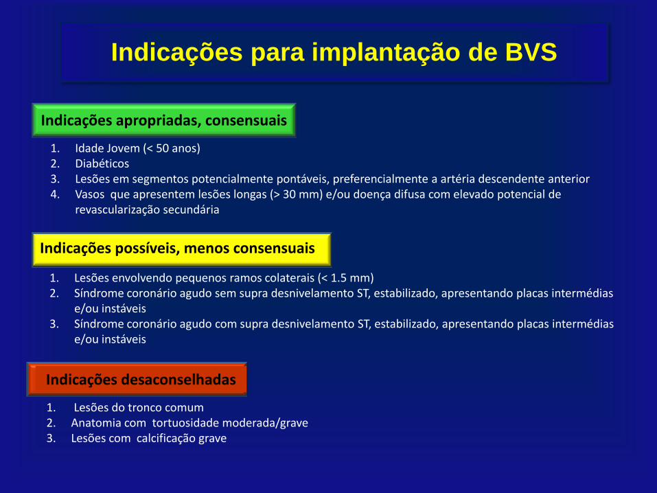
Indicações para implantação de BVS
Indicações apropriadas, consensuais
1. Idade Jovem (< 50 anos) 2. Diabéticos 3. Lesões em segmentos potencialmente pontáveis, preferencialmente a artéria descendente anterior 4. Vasos que apresentem lesões longas (> 30 mm) e/ou doença difusa com elevado potencial de
revascularização secundária
Indicações possíveis, menos consensuais
Indicações desaconselhadas
1. Lesões envolvendo pequenos ramos colaterais (< 1.5 mm) 2. Síndrome coronário agudo sem supra desnivelamento ST, estabilizado, apresentando placas intermédias
e/ou instáveis 3. Síndrome coronário agudo com supra desnivelamento ST, estabilizado, apresentando placas intermédias
e/ou instáveis
1. Lesões do tronco comum 2. Anatomia com tortuosidade moderada/grave 3. Lesões com calcificação grave

Caso clinico
55 anos, sexo masculino DMNID, HTA, Dislipidémia ECG de esforço positivo para isquémia Coronariografia: lesão única na CD
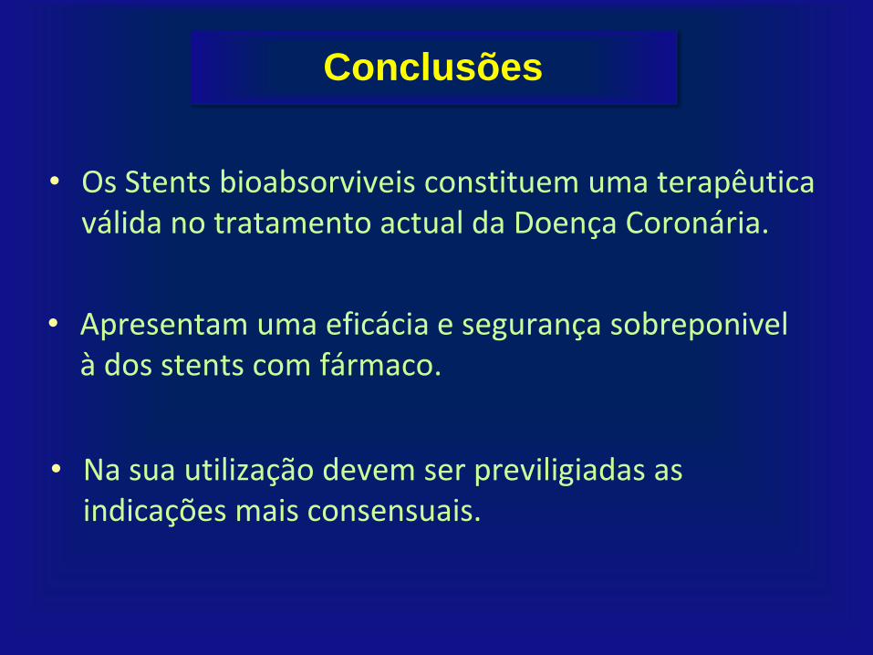
Conclusões
• Os Stents bioabsorviveis constituem uma terapêutica válida no tratamento actual da Doença Coronária.
• Apresentam uma eficácia e segurança sobreponivel à dos stents com fármaco.
• Na sua utilização devem ser previligiadas as indicações mais consensuais.
Top Related