


Languages
Pages
Legal

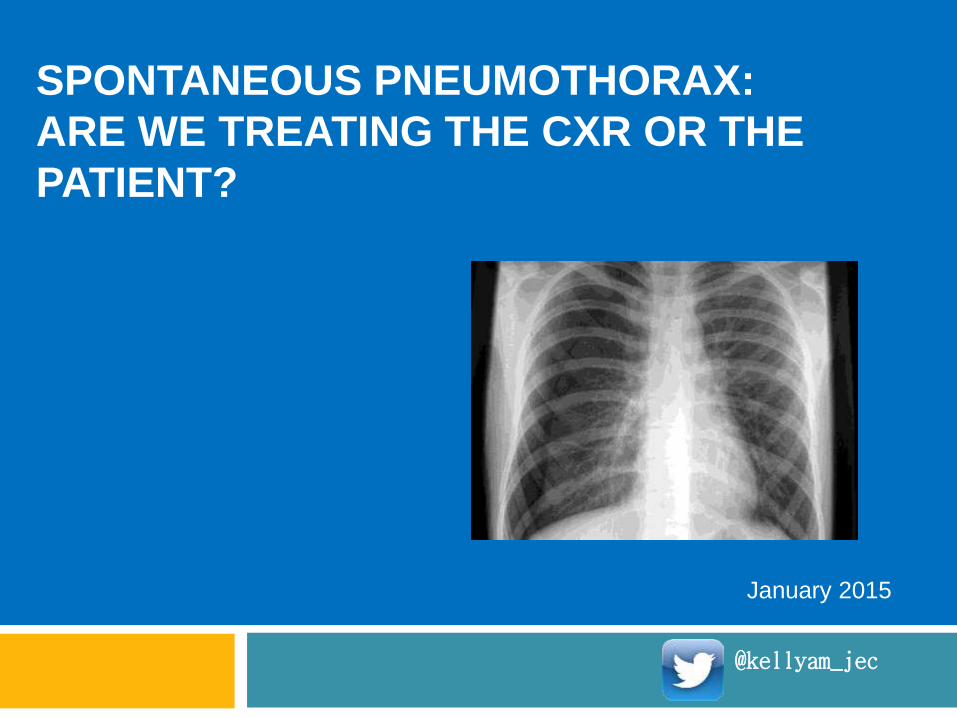
SPONTANEOUS PNEUMOTHORAX:
ARE WE TREATING THE CXR OR THE
PATIENT?
January 2015
@kellyam_jec
Permission
This presentation may be used freely for
educational purposes
It is the responsibility of the user to ensure that
the content is up to date and relevant to their
practice environment

Conflicts of interest
None to declare

Objectives
To review current evidence-based
guidelines for management of
spontaneous pneumothorax
To discuss the practical challenges in
managing spontaneous
pneumothorax

Before we start ...
According to recent guidelines, which
of the following should be the main
determinant of ED therapeutic
intervention in primary spontaneous
pneumothorax?
A. Pneumothorax size
B. Presence or absence of significant
breathlessness
C. Previous spontaneous
pneumothorax
D. Occupation

Mike
Aged 19
Onset of pleuritic chest pain yesterday
Mildly SOB on exertion only
Smoker, no other past history
At rest, pulse 60, O2
sat 98% (on room air), other vital signs normal

What would you do?
A. Large bore intercostal catheter
(ICC) and underwater-seal drain
(UWSD)
B. Small bore ICC and Heimlich
valve/ UWSD
C. Aspirate
D. Nothing –conservative
management with outpatient followup
E. Admit to ED observation ward for
oxygen therapy

Would this xray change your
mind?
Same history,
symptoms and
vital signs

Epidemiology
Primary spontaneous pneumothorax
is a disease of the young
Peak incidence late teens/ twenties
Male> Female; about 4:1
Smoking is a major risk factor

Clinical features
Chest pain: 90%
Sharp, dull
Dyspnoea- but can be transient
Presentation delayed > 24 hours in >50% of patients
Signs
Resonant chest
Reduced breath sounds
May be subtle depending on size, chest wall thickness, etc.

Imaging
Chest xray Erect CXR is highly sensitive for clinically
relevant pneumothorax
Expiratory films add little and should be avoided
Supine films are of little use
CT Highly sensitive and can identify other
pathology
Concern that overcalls clinically irrelevant pneumothorax
Ultrasound Used in trauma and increasing accepted in
non-trauma

A question of size?
No international agreement on size
definitions!
Australia and UK
Small: <2 cm rim around lung (measured at
hilum)
US
Small: <3cm inter-pleural distance at apex
US
method
UK and Australian
method

International variation in
guidelines
Guideline Year Definition of
‘large’
pneumothora
x
Management
recommendation for
‘large’ pneumothorax
in stable patient
British Thoracic
Society
2010 >2cm
interpleural
distance at
hilum
Conservative if minimal
symptoms, otherwise
aspiration
American
College of
Chest
Physicians
2001 >3cm apical
interpleural
distance
Pleural catheter insertion
(small bore or ICC) with
UWSD
Belgian Society
for
Pulmonology
2005 Interpleural gap
along entire
length of lateral
chest wall
Aspiration or pleural
catheter insertion with
USWD/valve
Therapeutic
guidelines
(Australia)
2014 >2cm
interpleural
distance at
hilum
Aspiration

International variation in
opinionsRegion/count
ry
Year
of
repo
rt
Comment
Wales 2000 Small PT not breathless only 50% would
treat conservatively.
Initial treatment of moderate/large: 75%
would aspirate
USA 1997 20% PSP in well young patient:
conservative 52%; aspirate 14%, pleural
catheter 31%
Czechoslovaki
a
2008 First PSP: 69% pleural catheter, 6%
aspiration
Switzerland 2006 Stable first large PSP: High consensus
for insertion pleural catheter; no support
for conservativeWales: Yeoh et al, Post Grad Med J; USA: Baumann et al, Chest;
Czech: Stolz et al, Rozhl Chir; Switz: Kuester et al, Interact
Cardiovas Thorac Surg

International variation in
practice
Country/
Region
Size Year
of
repor
t
Conservati
ve
Aspiratio
nPleural
catheter
drainage
Hong
Kong
(N=12)
Large 2009 2% 18% 80%
Australia
(N=19)
Large 2008 10% 17% 72%
UK (N=1) Large 2002 0% 75% 25%
Singapore
(N=1,
SGH)
All 2004 35% 18% 47%
HK: Chan et al, HK Med J; Aus: Kelly et al, Int Med J; UK: Mendis et al,
Post Grad Med J; Sing: Ong et al, Eur JEM.
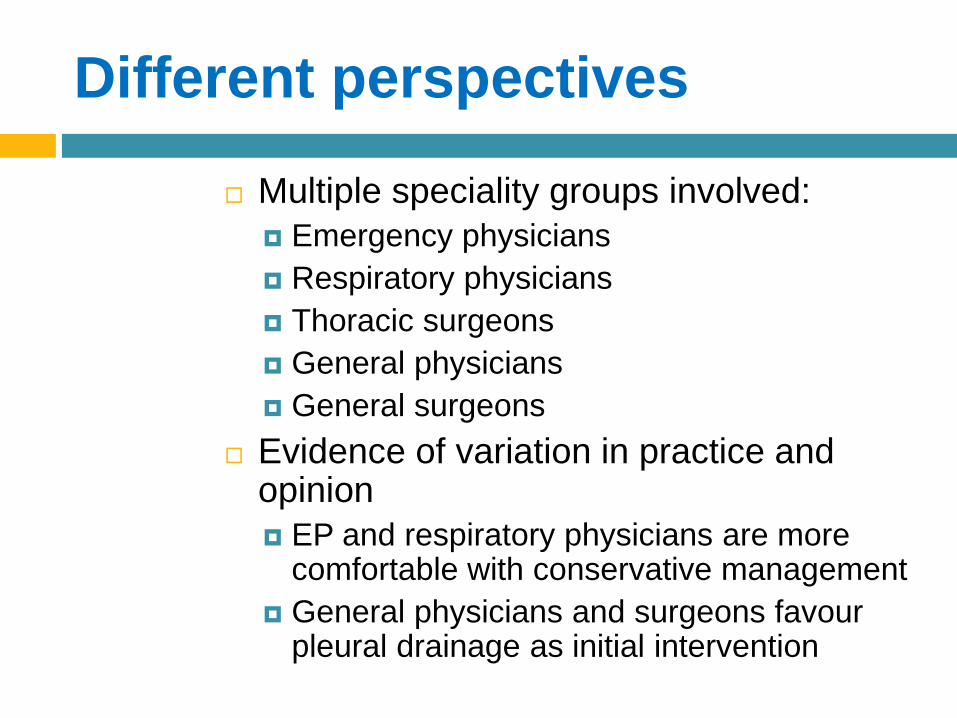
Different perspectives
Multiple speciality groups involved:
Emergency physicians
Respiratory physicians
Thoracic surgeons
General physicians
General surgeons
Evidence of variation in practice and opinion
EP and respiratory physicians are more comfortable with conservative management
General physicians and surgeons favour pleural drainage as initial intervention

What does this tell us?
There is international and inter-disciplinary lack of consensus regarding management strategies
The various national guidelines are not being followed
There is variation in how the available evidence is being interpreted Same evidence, different recommendations

The evidence
Evidence base is NOT strong
Factors to consider:
Type of pneumothorax: primary or secondary
Clinical evidence of respiratory compromise, in particular significant breathlessness
Size: Pneumothoraces resolve at a rate of approximately 1.25 to 2.2% of the volume of hemithorax per day.
Age: Evidence suggests that aspiration is less successful in patients aged over 50.
Availability of followup

Emergent drainage
Who?
Patients with severe respiratory compromise
Patients with shock
How?
14G IV catheter
Small bore catheter (e.g. Cook’s) via Seldinger technique
Definitive treatment required

Problems with IV catheter emergent
drainage
Traditional recommendation has been
14G, 5cm needle in second intercostal
space
Radiological studies:
24-42% of people have chest wall
thickness at 2ICS >5cm.
Chest wall thickness increases steadily
with BMI
Cadaver study:
Average chest wall thickness >4cm
Only 58% of 14G, 5cm needles entered
pleural space

Solutions
Use a longer needle
Go straight for a
Seldinger-type kit
Quick
Has long needle
Connects directly to
drainage so no
secondary procedure
required

Minimal symptoms
Evidence supports conservative treatment irrespective of size on xray
Re-absorb at rate of 1.5-2.3% hemithorax/ day
Can be managed at home!
Follow-up
Weekly is safe (some use 2-4 weekly)
Caveat: for early presenters (<24 hours), may be prudent to check in 4-6 hours and next day
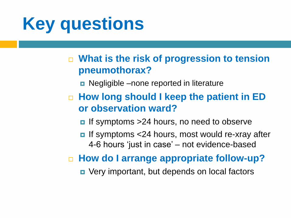
Key questions
What is the risk of progression to tension
pneumothorax?
Negligible –none reported in literature
How long should I keep the patient in ED
or observation ward?
If symptoms >24 hours, no need to observe
If symptoms <24 hours, most would re-xray after
4-6 hours ‘just in case’ – not evidence-based
How do I arrange appropriate follow-up?
Very important, but depends on local factors

Symptomatic
Main indication for intervention is
presence of significant
breathlessness
Options
Aspiration
Catheter drainage
Small bore
Large bore
Pigtail catheters / similar

Aspiration
Usually performed using a small catheter inserted by Seldinger technique
Aim is to convert a large pneumothorax to a small one
Success = rim <2cm and resolution of breathlessness without re-accumulation over 4-6 hours
Success rate 50-80%

Predictors of failure
Much less likely to be successful if
patient aged >50 years
If you have aspirated >3 litres,
success unlikely
Connect to Heimlich valve or UWSD

Key questions
What site should be used?
Second ICS mid-clavicular line or 4 ICS
mid axillary line
How long should I observe the
patient after successful drainage?
4-6 hours is probably enough
Need to check no re-accumulation

Catheter drainage
Small bore catheters (5-16F) are as effective as large catheters Many inserted by Seldinger-type approach
Success rate 65-95%
Suction does not improve outcome and should be avoided
Trocars should not be used
Complication rate of ‘traditional’ open catheter insertion 5-10% (excludes persistent air leak).

Key questions
Where should I insert a pleural
catheter?
If using small catheter, either anterior or
lateral is fine
If using large catheter, lateral side
preferred for aesthetic and comfort
reasons
Are large bore catheters really
‘out’?
Yes!
No evidence of benefit, more
uncomfortable, high complication rate

Surgery
About 10% of patients require surgical intervention
Indications:
persistent air leak after 2-7 days
recurrent pneumothoraces
airline pilots, frequent plane travelers and divers
contralateral or bilateral pneumothoraces and
pregnancy

Ambulatory management
optionsCountry/
region
Year
of
report
Study
design
Findings
Singapore 2011 RCT Aspiration vs mini chest tube. No
difference in outcome; ~50%
discharge from ED
France 1 2014 Case
series
Pigtail catheter + valve. 78%
success rate
France 2 2014 Case
series
Pigtail catheter + valve, 83%
success rate; 60% treated as
outpatients
Canada 2009 Case
series
Small bore catheter + valve; 81%
discharged from ED; 60%
managed totally as outpatients
Japan 2014 Case
series
Small bore catheter + valve; 94%
success rateSingapore: Ho et al, Am J Emerg Med 2011; France 1: Voisin et al, Ann Emerg med 2014;
France 2: Massongo et al, Eur Respir J, 2014; Canada: Hassani et al, Acad Emerg Med 2009;
Japan: Karaskai et al, Thorac Cardiovasc Surg
All
concluded
outpatient
management
was safe.
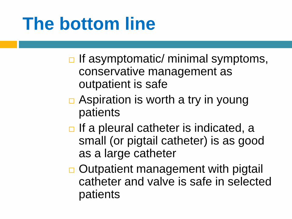
The bottom line
If asymptomatic/ minimal symptoms, conservative management as outpatient is safe
Aspiration is worth a try in young patients
If a pleural catheter is indicated, a small (or pigtail catheter) is as good as a large catheter
Outpatient management with pigtail catheter and valve is safe in selected patients

Recurrence
Up to 50% after first pneumothorax
Greatest risk in first year
Up to 70% after subsequent
pneumothorax
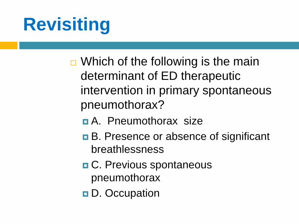
Revisiting
Which of the following is the main
determinant of ED therapeutic
intervention in primary spontaneous
pneumothorax?
A. Pneumothorax size
B. Presence or absence of significant
breathlessness
C. Previous spontaneous
pneumothorax
D. Occupation
Revisiting
Which of the following is the main
determinant of ED therapeutic
intervention in primary spontaneous
pneumothorax?
A. Pneumothorax size
B. Presence or absence of
breathlessness
C. Previous spontaneous
pneumothorax
D. Occupation

Did you change your mind?
Aged 19
Onset of pleuritic
chest pain yesterday
Mildly SOB on
exertion
Smoker, no past
history
At rest, pulse 60, O2
sat 98% on room air
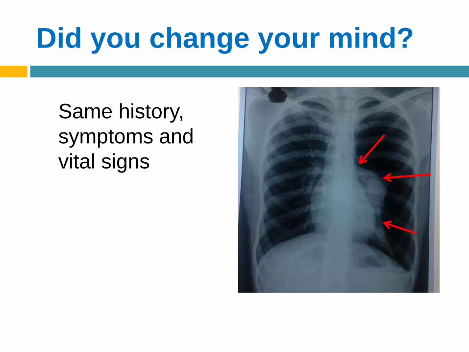
Did you change your mind?
Same history,
symptoms and
vital signs

Did you change your mind?
Same history, symptoms
and vital signs
I would treat both of
these conservatively
as outpatients
Question #1
23 year old man
50% pneumothorax with minimal
symptoms
Past history of ipsilateral PSP a year
ago
What is the preferred treatment option?
Question #1
No intervention needed in ED
Refer to thoracic surgery team for
definitive procedure (can be as
outpatient)

Question #1a
Same history
Pneumothorax ~70% and significant
breathlessness
What should be done?

Question #1a
This is an evidence free zone
Definitive procedure recommended as likelihood of further recurrence is very high (>70%)
Options:
Aspirate – if successful, home for semi-elective thoracic surgical procedure
Small bore/pigtail catheter – as outpatient/ inpatient
Primary thoracoscopy without ED intervention
Question #2
Can I send a patient home with a
pigtail (similar) catheter and one
way valve in situ?

Question #3
Is it safe to observe (treat
conservatively) a young patient
with large first PSP who has
minimal symptoms?
Question #3
The evidence says ‘YES’
Require:
Sensible patient
Access to appropriate followup
Means of return to hospital if gets worse

Question #4
What advice should I give about
work, flying and return to sport?
Question #4
Evidence-free zone
Return to work is OK as long as
symptoms are resolved
Air travel should be avoided until full
resolution confirmed by xray
Most airlines have a rule requiring 1 week
after full resolution
Sports are OK
Caveat: reasonable to avoid sports with
extreme exertion or contact until full
resolution

Question #5
Are there trials comparing
aspiration / small bore catheter
management with traditional large
bore pleural catheter management
for clinically significant outcomes?
Question #5
Clinically significant outcomes
Requirement for hospital admission
Requirement for secondary procedure
Requirement for surgery
Recurrence

Question #5
Aspiration vs. chest tube Cochrane review (CD 004479)
No difference in immediate success rate, early failure rate, duration of hospitalisation, one year success rate and number of patients requiring pleurodesis at one year.
Aspiration had lower admission rate
Small vs. Large pleural catheters Several RCT comparing small and large
catheters show similar outcomes (methodological issues)
Large bore catheters inserted by ‘open’ technique may have higher complication rate

Thankyou @kellyam_jec
Research opportunity
Do defined high risk features identify patients
who require definitive surgical intervention for
radial fracture?
Instability markers: age >60, intra-articular,
osteoporosis, dorsal angulation >20%, ulna
fracture, dorsal comminution, radial shortening
>3 instability markers, outcome was better with
surgery

What is involved?
Reviewing medical records for demographics,
mechanism of injury and instability markers
Reviewing xrays/ reports for instability markers
Participating in interpretation of data

What is in it for me?
Taste of research
Opportunity to be published
Opportunity to present at local / national
conferences
Top Related