


Languages
Pages
Legal


Accepted Manuscript
Title: Spironolactone loaded nanostructured lipid carrier gelfor effective treatment of mild and moderate acne vulgaris: Arandomized, double-blind, prospective trial
Author: Hamid Reza Kelidari Majid Saeedi ZohrehHajheydari Jafar Akbari Katayoun Morteza-Semnani JavadAkhtari Hadi Valizadeh Kofi Asare-Addo Ali Nokhodchi
PII: S0927-7765(16)30367-8DOI: http://dx.doi.org/doi:10.1016/j.colsurfb.2016.05.042Reference: COLSUB 7891
To appear in: Colloids and Surfaces B: Biointerfaces
Received date: 5-2-2016Revised date: 11-4-2016Accepted date: 16-5-2016
Please cite this article as: Hamid Reza Kelidari, Majid Saeedi, ZohrehHajheydari, Jafar Akbari, Katayoun Morteza-Semnani, Javad Akhtari, Hadi Valizadeh,Kofi Asare-Addo, Ali Nokhodchi, Spironolactone loaded nanostructured lipidcarrier gel for effective treatment of mild and moderate acne vulgaris: Arandomized, double-blind, prospective trial, Colloids and Surfaces B: Biointerfaceshttp://dx.doi.org/10.1016/j.colsurfb.2016.05.042
This is a PDF file of an unedited manuscript that has been accepted for publication.As a service to our customers we are providing this early version of the manuscript.The manuscript will undergo copyediting, typesetting, and review of the resulting proofbefore it is published in its final form. Please note that during the production processerrors may be discovered which could affect the content, and all legal disclaimers thatapply to the journal pertain.

1
Spironolactone loaded nanostructured lipid carrier gel for effective treatment of mild and moderate acne vulgaris: a randomized, double-blind, prospective trial
Hamid Reza Kelidaria, Majid Saeedib,*, Zohreh Hajheydaric, Jafar Akbarib, Katayoun Morteza-Semnanib, Javad Akhtarid Hadi Valizadehe, Kofi Asare-Addof, Ali Nokhodchie,g,* aPharmaceutical Sciences Research Center, Mazandaran University of Medical Sciences, Sari, Iran; bFaculty of Pharmacy, Mazandaran University of Medical Sciences, Sari, Iran; cDepartment of Dermatology, Boo Ali Sina (Avicenna) Hospital, Faculty of Medicine, Mazandaran University of Medical Sciences, Sari, Iran; dDepartment of Nanobiomedicine, School of Medicine, Mazandaran University of Medical Sciences, Sari, Iran; eDrug Applied Research center and Faculty of Pharmacy, Tabriz University of Medical Sciences, Tabriz, Iran. fPharmacy Department, University of Huddersfield, Huddersfield, HD1 3DH, UK; gPharmaceutics Research Laboratory, School of Life Sciences, University of Sussex, Brighton, BN1 9QJ, UK Total number of words: 4455 (including the title and references) Total number of tables: 2 Total number of figures: 5 *Corresponding authors: Ali Nokhodchi, PharmD, PHD, e-mail: [email protected]; tel: +44 1273 872811 and Majid Saeedi, PharmD, PhD; e-mail: [email protected])

2
Graphical abstract
Highlights
Spironolactone‐SLN (SP‐SLN) particles can be formulated in carbopol gel
SP‐SLN gel formulations showed non‐Newtonian independent pseudoplastic behaviour
Water content of the skin increased after using SP‐SLN gels
SP‐SLN gels are more effective than Spironolactone alcoholic gels to treat acne
Abstract
Spironolactone (SP) known as an anti-androgen drug, has been proven to be
effective in treatment of acne. The quest to minimize the unnecessary systemic side
effects associated with the oral drug administration of spironolactone, has led to a
growing interest of loading SP on lipid nanoparticles to deliver the drug in a topical
formulation. The aim of the current investigation was to prepare and compare the

3
performance of SP loaded nanostructured lipid carrier (SP-NLC) and SP alcoholic
gels (SP-ALC) on two groups of respective patient populations, group A and group
B in the treatment of mild to moderate acne vulgaris. The results showed that SP-
NLCs were spherical in shape with an average diameter of ~240 nm. The
polydispersity index (PI) and zeta potential of these nanoparticles were 0.286 and
-21.4 respectively. The gels showed non-Newtonian independent pseudoplastic and
shear thinning behavior. The SP-NLCs was not toxic to fibroblast cell strains at the
24 and 48 h periods. Results showed that the mean number of total lesions (37.66 ±
9.27) and non-inflammatory lesions (29.26 ± 7.99) in group A significantly
decreased to 20.31 ± 6.58 (p<0.05) and to 13.95 ± 5.22 (p<0.05) respectively. A
similar pattern was observed for group B where the mean number of total lesions
and non-inflammatory lesions reduced from 33.73 ± 9.40 to 19.13 ± 5.53 (p<0.05)
and from 25.65±8.12 to 13.45 ± 4.48 (p<0.05) respectively. The total lesion count
(TLC) was significantly decreased from 37.16 ± 9.28 to 19.63 ± 6.36 (for group A;
p<0.071) and 32.60 ± 9.32 to 18.33 ± 5.55 (for group B; p<0.05) respectively. After
treatment with SP-NLC for 8 weeks, the water content of the skin significantly
(p<0.05) increased from 37.44 ± 8.85 to 45.69 ± 19.34 instrumental units.
Therefore, the SP-NLC gel may help in controlling acne vulgaris with skin care
benefits.
Keywords: Acne, Nanostructured lipid carriers (NLCs), Spironolactone, Topical,
Skin condition

4
1. Introduction
Acne vulgaris is a disease of the pilosebaceous glands that usually occurs in
adolescence following a sharp increase in androgen [1]. Spironolactone (SP) is
classified as a BCS class II drug (high permeability and poor solubility). The BCS
is a tool which is used to differentiate the drugs on the basis of their solubility
and permeability as follows: Class I covers drugs with high permeability and
solubility; Class II covers drugs with high permeability but poor solubility;
Class III drugs have low permeability but high solubility and Class IV drugs
have low permeability and solubility. As an anti-androgen drug, it has proved
effective in reducing the sebum secretion rate in various clinical reports [2, 3].
However, following oral administration, SP is poorly absorbed from the
gastrointestinal (GI) tract and observed endocrine side effects restricted its clinical
application due to its variable oral bioavailability [4]. It has been shown that the
topical delivery of SP can allow high drug levels at the site of action which in turn
can lessen the systemic side effects and also improve patient compliance [5, 6].
Nanostructured lipid carriers (NLCs) represent a relatively new type of colloidal
drug delivery system that consists of solid-lipid and liquid-lipid, and offers the
advantage of improved drug loading capacity and release properties. These
nanoparticles contain non-irritative and non-toxic lipids and as such well suited for
use on inflamed and damaged skin [7]. The small particle size of these nanoparticles
ensures close contact with the stratum corneum and also increases the amount of
encapsulated compounds penetrating into the skin due to the formation of an intact
film on the skin surface upon drying. This lipid nanocarrier have been used to
improve the skin/dermal uptake of several drugs such as cyproterone acetate [8],

5
tretinoin [9], isotretinoin [10] and adapalene [8] which supports the notion that these
nanocarriers can be employed for the topical delivery of SP. Clinical studies with
alcoholic topical formulation of SP have been previously reported and the results
demonstrate beneficial effects in patients with acne without any systemic hormonal
changes [3, 12, 13]. Shamma and Aburahma used spironolactone loaded NLC for
follicular targeting of drug molecules for the management of alopecia and they
successfully showed the presence of SP in the scalp hair follicles and decreasing
androgen production within sebaceous glands and blocking the androgen receptor in
dermal papillae [14]. The efficacy of SP gel 5% in the treatment of facial acne also
showed that total lesion count and acne severity index reduced significantly however,
efficacy on non-inflammatory lesion (comedones) was more effective than on
inflammatory ones (papules and pustules) because of poor ability of SP penetration to
specific micro environmental conditions in the inflammatory acne lesions [13]. The
aim of this current research was to develop novel NLC formulations in gel
containing SP with a suitable rheology (spreadability), pH and better efficacy
and tolerability (SP-NLC; 1%) versus alcoholic SP gel (SP-ALC; 5%) in the
treatment of facial mild to moderate acne vulgaris.
2. Material and Methods
2.1 Materials
Spironolactone (SP) was supplied by Behdashtkar Co. (Tehran, Iran). Stearic acid
(SA), Oleic acid (OA), Tween 80, Span 80, hydroxyethyl cellulose, propylene
glycol, Methyl paraben and triethanolamine were purchased from Merck Co.
(Germany). Carbopol 934P was obtained from BF Goodrich (Cleveland, Ohio,

6
USA). Carbopol gels are approved for pharmaceutical use in several different
administration routes. The cutaneous use of these gels is advantageous as they
possess good rheological properties resulting in long residue times at the site of
administration [15, 16], therefore Carbopol was selected as the gelling agent in
the present study. Deionized water was purified using a Milli-Q system (Millipore,
Direct-Q). Dimethyl sulfoxide (DMSO, solvent) was purchased from Merck and
Tetrazolium salt (MTT) was supplied from Sigma-Aldrich. Human Caucasian
foetal foreskin fibroblast (HFFF2) was purchased from the National Cell Bank of
Iran (Pasteur Institute, Tehran, Iran).
2.2. Preparation of the formulations
SP loaded NLC was prepared using the probe-ultrasonication method as described
elsewhere [17]. Briefly, solid lipid (stearic acid 2.8 g) in combination with liquid
lipid (oleic acid 1.2 g), lipophilic surfactant (Span 80 2.5 g) and SP (1 g), were
melted at 85 °C using a hot plate. The hot lipid phase produced was dispersed in a
1/3 (28.77 g) of the aqueous solution of hydrophilic surfactant (Tween 80) prepared
by weighing out 1.67 g Tween 80 heated at the same temperature and sonicated by
using a probe sonicator (Bandelin sonopuls, Berlin, Germany) for 5 min (Model HD
3200, Prob TT25, 50% power and 19.82 KJ) to form a coarse pre-emulsion. At the
end of the sonication, the mixture was dispersed into the remaining 2/3 of the
hydrophilic surfactant solution (containing 3.33 g Tween 80) maintained in an ice
bath. The final mixture was sonicated again for 10 min (50 % power and 51.77 KJ)
whilst still immersed in the ice-bath. This cooling step promoted the formation of
the lipid nanoparticles. SP-NLC dispersion was incorporated into 1 % w/v Carbopol

7
gel containing 0.2 g methyl paraben as preservative. The obtained gel was allowed
to hydrate for 24 h. The resulting mixture was then stirred followed by
neutralization with tri-ethanolamine (approximately 10 drops) to obtain an adequate
semisolid carbopol gel matrix (for a full composition of each formulation refer to
Table 1S in the supplementary material).
Alcoholic SP (SP-ALC) was composed of SP, hydroxyethyl cellulose, propylene
glycol and methyl paraben in a base of deionized water. Briefly, 5 g of SP was
dissolved in hydroxyethyl cellulose (5 g) and propylene glycol (10 g) for 10 min
under stirring (300 rpm). 78.8 g of water was then added to the solution under
stirring (300 rpm) for an additional 5 min stirring. This was followed by the
addition of the methyl paraben (0.2 g) and carbopol (1 g). This solution was then
stirred for a further 10 min. The final solution was then neutralized by
triethanolamine (about 10 drops) under stirring condition (500 rpm) to obtain the
SP-ALC gel.
2.3. Characterization of the gels
In order to determine the shape of SP-NLC, the transmission electron microscopy
(TEM, CM 30, Phillips, Netherlands) was utilized. Briefly, the SLN samples were
first diluted two times with distilled water. One drop of the diluted sample was
placed on a 200-mesh carbon-coated copper grid, stained with 2 %
phosphotungstic acid solution and dried at room temperature. Representative
images of each sample were reported.

8
Photon correlation spectroscopy (PCS) with a Malvern zetasizer ZS (Malvern
Instruments, UK) was used to determine the particle size, profile the size
distribution (polydispersity index, PI) and zeta potential of the nanoparticles.
Briefly, the Zeta potential and the poly dispersity index (PDI) of the nanoparticle
formulations were determined using the Zetasizer (Nano ZA, Malvern Instruments,
UK). In this method the sample was measured at 25 °C with an angle detection of 90°.
The concentration of the samples for analysis on the Zeta Sizer was 20-400 kilo counts
per second (KCPS) and the intensity of diffraction was 100000 counts per second.
For spreadability 500 mg of the formulated gel was placed within a circle of 1 cm
diameter pre-marked on a flat glass plate over which a second glass plate was
placed. A weight of 500 g was allowed to rest on the upper glass plate for 5 min.
The increase in the diameter due to spreading of the formulated gel was noted.
Finally, 1 g of the formulated gel was dissolved in 100 mL of distilled water and
stored for 2 h. The pH of aqueous dispersion of the formulated gel was determined
using Jenway Digital pH meter Model 3510, standardized using pH 4.0 and 7.0
standard buffers before use. The rheology of the SP-NLC and SP-ALC gels was
obtained with a Brookfield Viscometer (Model DV-II+, Brookfield Engineering
Laboratories, Inc., USA) using spindle TD. Viscosity was measured by increasing
the shear rate from 0.5 rpm to 100 rpm at 25 ± 1 ˚C.

9
2.4. Cytotoxicity assays:
Cell lines and culture
Human Caucasian foetal foreskin fibroblast (HFFF2) was cultured at 37 °C in a 5 %
CO2/95 % air humidified atmosphere in a RPMI 1640 medium containing 2 mM l-
glutamine supplemented with 10 % (v/v) heat-inactivated FBS (fetal bovine serum),
100 IU/mL penicillin and 100 mg/mL streptomycin (Gibco).
2.5. Treatment of cells with nanoparticles
The HFFF2 cells (1 × 10⁴) were seeded in each well containing 100 µl of the RPMI
medium supplemented with 10 % FBS in a 96-well plate. After 24 h of adhesion, a
serial of doubling dilution of the SP-NLC was added to four replicate wells. Cell
viability was assessed by the 3-(4, 5-dimethylthiazol-2-yl)-2, 5-diphenyl tetrazolium
bromide (MTT) reduction assay described below.
2.6. MTT reduction assay
After 1 and 2 days, 10 µl of 3-(4, 5-dimethylthiazol-2-yl)-2, 5-diphenyl tetrazolium
bromide (MTT) (5 mg/ml stock solution) was added to each well and the plates
were incubated at 37 °C for an additional 4h. The medium was discarded and the
formazan blue, which formed in the cells, was dissolved using 200 µl dimethyl
sulfoxide (DMSO). The absorbance was measured at 570 nm in a microplate reader
(ELx800, BioTek Instruments, Inc., Winooski, VT). Cell viability data were
expressed as the mean ± standard deviation (SD) of two independent experiments
run in four replicates. The percentage of viability compared with the control wells

10
(the means optical density of untreated cells was set to 100 % viability) was
calculated from the concentration–response curves by linear regression analysis.
The gels were kept in 4, 25 and 40 ˚C for physical stability evaluation (viscosity,
syneresis, swelling and color change) for 2 weeks [18]. The selected formulation for
clinical trial was prepared freshly and controlled microbiologically according to
USP 30 (United States Pharmacopoeia).
2.7. Clinical study
In selecting volunteers for the clinical study, patients received a systemic or topical
anti acne therapy 3 months before or during the study, pregnant patients, patients
planning to become pregnant, lactating patients, patients with skin diseases that
might interfere with the diagnosis or evaluation of their hyper pigmentation were
excluded from the study. Seventy-six patients with mild to moderate acne, 8 years
or older, defined as a score of 1–30 on the global acne grading system (GAGS)
scale [17], who were not satisfied with their previous acne therapies participated in
the double-blind clinical trial study after giving written informed consent.
Permission was obtained from the Mazandaran University of Medical Sciences
ethical committee before starting the study. At the first visit, a detailed
questionnaire including data of demographic status, acne duration and medical
history was completed for each patient. Thereafter, the patients were randomly
assigned to one of the two treatment groups, namely group A (where SP-NLC gel
was used) and group B (where SP-ALC gel was used). Both the physicians and
patients were blinded to the type of treatment. Every morning and evening, patients
washed their face with non-medicated soap, then thoroughly rinsed and dried it.

11
Over the 8-week course, each patient applied 2 tubes (each tube contained 30 g gel
with the patient instructed to apply about 1 g of the gel containing 10 mg SP
for SP-NLC gel (group A) or 50 mg drug of the SP-ALC (group B) during the 8
weeks) of the formulated gel and was asked to apply about 2 cm (knuckle) of the
gel each morning and evening to the area and massage it for about 2 minutes. They
were left on individual acne lesions for 2–3 h, after which they were washed off.
Non-medicated cosmetics were permitted during the study. The patients were asked
about adherence to the protocol and to report any side effects. During the trial the
patients were prohibited from using any drugs or other skin care treatments for
acne.
2.8. Clinical assessments
The patients were assessed for any changes in the facial lesion counts (non-
inflammatory lesions: open and closed comedones; inflammatory lesions: papules,
pustules, and nodules) at each clinic visit (0, 2, 4 and 8 Wk). At every session, the
acne lesions were assessed based on their numbers, type and distribution. For the
final assessment and to determine the efficacy of the treatment, the following two
formulas were used:
Total Lesion Count (TLC) = comedones + papules+ pustules
Acne Severity Index (ASI) = papules + (2 pustules) + (comedones/4).
The hydratation, sebum, elasticity, melanin and redness of the skin were measured
at each visiting time using the multi skin test instrument (MC 900; Enviro derm,

12
Gloucestershire, UK). The signs and symptoms were evaluated every 2 weeks from
the baseline.
2.9. Statistical analysis
Statistical analysis was performed using SPSS for Windows (version 15; SPSS Inc.,
Chicago, IL, USA). Alterations in skin-related parameters were statistically
examined using the Student's t-test. Differences were considered to be significant at
P < 0.05.
3. Results
3.1. Characterization of the gels
The TEM micrographs of the SP-NLC gel (Figure 1) revealed that the SP-NLC
particles were spherical in nature. The results obtained from the zetasizer (Table
2S in supplementary materials) showed smaller particle sizes and higher zeta
potential for the SP-NLC gel (239.7±99.48 nm and -21.39±0.051 mV) compared to
that of the SP-ALC gel formulation (8093.5±722.83 nm and 5.13±0.05 mV;
P<0.001). SP-NLC particles showed lower polydispersity index of 0.286±0.051
compared to the SP-ALC gel which was 0.951±0.068. The diameters for both gels
are an indication of good spreadability (between 5-6.5 cm) and also pH was found
to be within acceptable limits (pH 4-6). Regarding the rheology, the gels exhibited
non-Newtonian independent pseudoplastic and shear thinning behaviour meaning
these systems have different viscosities at different shear rates (different rpm;
P>0.05). For more details on these parameters please refer to supplementary data in
Table 2S.

13
3.2. Cytotoxicity
The results of the cytotoxicity assays performed with the nanoparticles containing
SP are presented in Figure 2. The results showed that SP-NLC was not toxic to the
cell strain at 24 and 48h.
3.3. Demographic characteristics
A total of seventy six patients with mild to moderate acne were recruited in the
present study. Among these patients, 36 (47.37 %) were in the group treated with
SP-NLC gel (group A) and 40 (52.63 %) were in the group treated with SP-ALC gel
(group B). In group A, thirty three (91.66 %) were female and three (8.34 %) were
male; in group B, there were thirty seven (92.5 %) female and three (7.5 %) male
participants. Among the 36 patients in group A, six (16.66 %) patients were
excluded from the study for reasons cited as “personal reasons”. Personal reasons
were cited for four patients (10 %) in group B. There were also six (15 %) patients
excluded for allergy reasons in group B. Hence, 30 patients in group A and 30
patients in group B were assessed for up to 8 weeks of treatment. The minimum and
maximum age in group A were 11 and 38 years, respectively; and in group B these
values were 8 and 34 years, respectively. There were no significant differences
between patients’ age in the two groups (p = 0.232). At baseline, no significant
differences were noted between groups in terms of mean total lesion score (P =
0.596). This similarity was observed between groups in the mean inflammatory
lesion score (P = 0.988) and the mean non-inflammatory score (P = 0.811) as well
(Table 1).

14
3.4. Efficacy on Total Lesion Score (TLS)
The percentage reductions in the total lesion score are summarized in Figure 3(I).
From the baseline to weeks 2, 4 and 8 the percentage reduction of total lesion scores
in the group A compared to group B were not significant (P=0.596; P=987),
however in both groups, the reduction in week 8 compared to the baseline was
significantly higher (P < 0.004 and P = 0.074, respectively).
3.5. Efficacy on non-Inflammatory and inflammatory lesion scores
Decreases in non-inflammatory and inflammatory lesions over time are summarized
in Figures 3(II) and 3(III), respectively. The mean number of non-inflammatory
lesion significantly reduced (P <0.01) in both groups when week 8 was compared to
the baseline. In group A, the mean number of non-inflammatory and inflammatory
lesions decreased from 25.65 ± 8.12 to 13.45 ± 4.49 (a reduction rate of 47.6 %)
and 8.08 ± 2.80 to 5.68 ± 1.94 (reduction rate 29.7 %), respectively. In group B,
these values dropped from 29.26 ± 7.99 to 13.95 ± 5.22 (a reduction rate of 52.3 %)
and 8.23 ± 2.66 to 6.36 ± 2.43 (a reduction rate of 22.7 %), respectively (P>0.05).
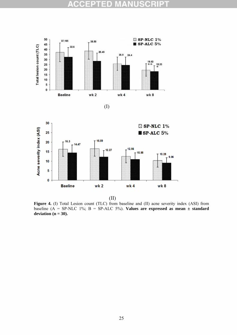
3.6. Efficacy on TLC and ASI
Figure 4(I) showed a statistically significant difference (week 8 compared to the
baseline) in both groups A and B towards a decrease of the total lesion count
(P<0.001). The TLC in group A was 37.165 ± 9.28 (baseline) and 19.63 ± 6.36 (Wk
8), (P = 0.003), and 32.60 ± 9.32 (baseline) and 18.33 ± 5.56 (Wk 8) for group B (P
=0.022).

15
We report a representative case of a 15-year-old female patient, whose acne lesions
remarkably improved after topical application of SP-NLC gel for 8 weeks (Figure
5).
3.7. Efficacy on skin condition
The data of patients in both groups' subjects for skin conditions are listed in Table 2.
There were no differences in skin hydration, sebum, elasticity, melanin and redness
between groups A and B during the study period (P>0.05). The skin elasticity,
melanin and redness of both groups remained unchanged during the study period
(P>0.05). Notably, skin hydration increased in weeks 4 and 8 in group A and the
increase was more significant in week 8 (P =0.002). Also skin sebum decreased
significantly (P =0.013) in the same time (Wk 8) in group B. There were no serious
adverse experiences that were related to the treatment. Dryness and itching were the
most frequent symptoms reported in group A (2.7 %) and B (15 %).
4. Discussion
4.1 Characterization of the gels
A reduction in the size of nanoparticles and PI in this study maybe as a result of the
higher interfacial tension between nanoparticles and the aqueous phase which leads
to a more homogenized nanoparticles in the aqueous phase. The phenomenon for
zeta potential can be explained by the fact that oleic acid has negatively charged
carboxylic groups. NLC revealed the highest zeta potential values, possibly due to
the accumulation of oleic acid at the surface of the nanoparticles [20].

16
4.2. Clinical assessments
The purpose of this double-blind, randomized study was to compare the efficacy
and tolerability of SP nanogel 1 % (SP-NLC) to an alcoholic SP gel 5 % (SP-ALC)
formulation which had previously been reported to be beneficial in acne by Afzali et
al, in the treatment of facial mild to moderate acne vulgaris [13]. In this present
study we have demonstrated for the first time that daily application of the SP-NLC
gel for up to 8 weeks has a therapeutic effect on mild to moderate acne vulgaris.
Compared with the baseline, non-inflammatory lesions and TLC decreased
significantly in groups A and B at week 8. In contrast, inflammatory lesion score
and ASI did not change after 8 weeks in both groups. The dermatologic
improvement of acne vulgaris in groups A and B maybe accompanied by a
significant decrease in sebum content, which is over produced in acne [21]. The
efficacy of 5 % topical SP cream acts as an anti-androgen in human sebaceous
glands, competing with dihydrotestosterone (DHT) receptors and producing a
decrease of labelled DHT [13]. Califano et al. reported that the patient’s treatment
with 5 % SP cream revealed complete regression of acne in 30 % and an
improvement in 65 % of the patients [22]. Recently, the efficacy of a 5 % SP gel in
the treatment of facial acne was studied in a randomized placebo-controlled trial by
Afzali et al. [13]. They showed that TLC and ASI in a placebo and SP groups
reduced significantly and emphasized that perhaps the alcoholic content of the gel
caused this phenomenon to occur in both groups. This could be the reason here as
well as for group B as around 65 % of non-inflammatory lesions reduced after 8
weeks treatment. But in group A however, SP-NLC may have achieved the

17
targeting to skin appendages, especially to the pilosebaceous units (hair follicles
with their associated sebaceous glands). This is an alternative pathway for the
dermal absorption of NLC, since it contributes significantly to the absorption of
small molecules within the lag time after application. Walton et al. showed that
when SP was applied topically in concentration of 3 and 5 % in humans there was
no effect on sebum excretion. They suggested that, the lack of anti-androgen effect
from topical SP cream maybe related to the vehicle which is used and has to be
adequate to deliver the drugs to the target sebaceous glands [23]. Interest in
pilosebaceous units is directed towards their utilization as reservoirs for localized
therapy and also as a transport pathway for systemic drug delivery. Moreover, for
some hair follicle related diseases such as acne, the hair follicle itself is the target
site [24]. Some researchers hypothesize that lipid coating or lipophilic material
properties may favour higher uptake into hair follicles, because the hair follicles are
filled with sebum and provide relatively lipophilic environment [25]. Follicular
deliveries play a key role in-vivo for the penetration of substances topically applied
since the pilosebaceous unit is more permeable than corneocytes. Moreover, it has
been demonstrated in reports that, colloidal particles larger than 10 μm can remain
on the skin surface, those in the range of 3–10 μm accumulate in the follicle, when
smaller than 3 μm, they can penetrate into follicles [26]. Furthermore, in this study
the particle size of SP-NLC was 239 nm while SP-ALC was around 8 μm meaning
SP-NLC may penetrate into the follicles whilst the SP-ALC may remain on the
skin surface and that the alcoholic effect is what is paramount in the reduction of
lesions. The lipid structures of NLC in contrast to an alcoholic formulation may
affect the interaction with skin. Lipid-based carriers could attach themselves onto

18
the skin surface, making close contact with superficial junction of corneocyte
clusters and channels between corneocyte islands. Finally it makes drug permeation
easier, since the lipid cover could reduce corneocyte packing and widen the inter
corneocytes gaps [27]. NLC’s have occlusive properties, i.e. they can be used in
order to increase the water content of the skin [28]. Occlusive compounds affect the
skin hydration and penetration of compounds into the skin. The increase in skin
moisture results in group A may be attributed to the retention of water content of
the skin. Nanoparticles have been found to be 15-folds more occlusive than
microparticles [27]. Wissing and Müller performed an in-vivo study investigating
the skin hydration effect after repetitive application of an o/w cream containing
lipid nanoparticles and a conventional o/w cream for 28 days. The lipid
nanoparticles containing o/w cream increased the skin hydration significantly more
than the conventional o/w cream [27].
5. Conclusion
The results of this randomized, double-blind trial demonstrated that the therapy of
SP-NLC and SP-ALC were well tolerated and resulted in significantly greater
improvement in mild to moderate acne vulgaris after 8 weeks treatment in
comparison to the baseline. This therapy effectively treated non-inflammatory
lesions, and showed high skin hydratation in the SP-NLC group (group A).
However, the role of SP-NLC gel developed in the present investigation still
requires clinical evaluation in a larger number of human subjects at different
locations.

19
Acknowledgment
This work was supported by a grant from the research council of the Mazandaran
University of Medical Sciences.
Declaration of interest
The authors report no conflict of interest. The authors alone are responsible for the
content and writing of the paper.
References [1] J.S. Archer, R.J. Chang, Best. Pract. Res. Cl. Ob. 18 (2004) 737-754.
[2] K. Sato, D. Matsumoto, F. Iizuka, E. Aiba-Kojima, A. Watanabe-Ono, H. Suga, K. Inoue, K. Gonda, K. Yoshimura, Aesthet Surg J. 30 (2006) 689-694.
[3] H.-Y. Chiu, T.-F. Tsai, J Am Acad Dermatol. 65 (2011) 1048. e1041-1048. e1022.
[4] Y. Dong, W.K. Ng, S. Shen, S. Kim, R.B.H. Tan, Int. J. Pharm. 375 (2009) 84-88.
[5] M. Schäfer-Korting, W. Mehnert, H.-C. Korting, Adv. Drug. Deliv. Rev. 59 (2007) 427-443.
[6] M. Gupta, S.P. Vyas, Chem. Phys. Lipids 165 (2012) 454-461.
[7] R.H. Müller, R.D. Petersen, A. Hommoss, J. Pardeike, Adv. Drug. Deliv. Rev. 59 (2007) 522-530.
[8] J. Štecová, W. Mehnert, T. Blaschke, B. Kleuser, R. Sivaramakrishnan, C.C. Zouboulis, H. Seltmann, H.C. Korting, K.D. Kramer, M. Schäfer-Korting, Pharm. Res. 24 (2007) 991-1000.
[9] K.A. Shah, A.A. Date, M.D. Joshi, V.B. Patravale, Int. J. Pharm. 345 (2007) 163-171.

20
[10] J. Liu, W. Hu, H. Chen, Q. Ni, H. Xu, X. Yang, Int. J. Pharm. 328 (2007) 191-195.
[11] A.K. Jain, A. Jain, N.K. Garg, A. Agarwal, A. Jain, S.A. Jain, R.K. Tyagi, R.K. Jain, H. Agrawal, G.P. Agrawal, Colloids Surf. B: Biointerfaces 121 (2014) 222-229.
[12] J.C. Shaw, J. Am. Acad. Dermatol. 24 (1991) 236-243.
[13] B.M. Afzali, E. Yaghoobi, R. Yaghoobi, N. Bagherani, M.A. Dabbagh, J Dermatol Treat. 23 (2012) 21-25.
[14] R.N. Shamma, M.H. Aburahma, Int J Nanomedicine, 9 (2014) 5449.
[15] M. Joshi, V. Patravale, Int. J. Pharm. 346 (2008) 124-132. [16] W. Liu, M. Hu, W. Liu, C. Xue, H. Xu, X. Yang, Int. J. Pharm., 364 (2008) 135-141. [17] S. Bose, Y. Du, P. Takhistov, B. Michniak-Kohn, Int. J. Pharm. 441 (2013) 56-66.
[18] Z. Hajheydari, M. Saeedi, K. Morteza-Semnani, A. Soltani, J Dermatol Treat. 25 (2014) 123-129.
[19] A. Doshi, A. Zaheer, M.J. Stiller, Int J derm.36 (1997) 416-418.
[20] F. Marquele-Oliveira, D.C. de Almeida Santana, S.F. Taveira, D.M. Vermeulen, A.R. Moraes de Oliveira, R.S. da Silva, R.F.V. Lopez, J Pharm Biomed Anal. 53 (2010) 843-851.
[21] D.S. Berson, A.R. Shalita, J Am Acad Dermatol, 32 (1995) S31-S41.
[22] L. Califano, S. Cannavo, M. Siragusa, R. Girardi, Clin Ter. 135 (1990) 193-199.
[23] S. Walton, W. Cunliffe, P. Lookingbill, K. Keczkes, Br. J. Dermatol. 114 (1986) 261-264. [24] Y. Zhai, G. Zhai, J. Con. Rel. 193 (2014) 90-99.
[25] U. Münster, C. NakamuraNachname, A. Haberland, K. Jores, W. Mehnert, S. Rummel, M. Schaller, H. Korting, C.C. Zouboulis, U. Blume-Peytavi, Die. Pharmazie. Int. J. Pharm.60 (2005) 8-12. [26] H. Wosicka, K. Cal, J. Dermatol.Sci. 57 (2010) 83-89.

21
[27] J. Pardeike, A. Hommoss, R.H. Müller, Int. J. Pharm. 366 (2009) 170-184.
[28] S. Wissing, O. Kayser, R. Müller, Adv. Drug. Deliv. Rev. 56 (2004) 1257-1272.

22
Figure 1. Transmission electron micrograph (TEM) of SP-NLC.

23
Figure 2. Cytotoxicity of SP-NLC in HFFF2 fibroblast. Data shown as mean ± standard deviation, n = 5. The nanoparticle concentration is presented in units of NLC concentration.

24
Figure 3. Mean percentage changes in: (I) total lesion scores from baseline; (II) inflammatory lesion scores from baseline and (III) non-inflammatory lesion scores from baseline (A = SP-NLC 1 % ; B = SP-ALC 5 %). Values are expressed as mean ± standard deviation (n = 30).
(I)
(II)
(III)
25
(I)
(II) Figure 4. (I) Total Lesion count (TLC) from baseline and (II) acne severity index (ASI) from baseline (A = SP-NLC 1%; B = SP-ALC 5%). Values are expressed as mean ± standard deviation (n = 30).

26
Figure 5. Fifteen year old female patient at baseline (A) and after treatment with SP-NLC gel at week 8 (B)

27
Table 1. Baseline characteristics of the intent-to-treat population. Values are expressed as mean ± standard deviation (n = 30). Demographic parameter SP-NLC 1 % SP-ALC 5 % p-value Mean age, y (±SD) 21.80 ± 5.97 20.86 ± 5.36 0.232 Sex, No. (%) Female Male
27 (% 87.5) 3 (% 12.5)
28 (% 93.3) 2 (% 6.67)
Mean acne duration, m (±SD) 3.72 ± 2.67 3.54± 2.88 0.712 Patients who used medication, No. 36 40 Patient who completed study, No. 30 30 Reason for discontinuation Allergy to medication Personal reason
0 6
6 4
Acne lesion count (mean±SD) Total Non-inflammatory Inflammatory TLC (± SD) ASI (± SD)
37.66 ±9.27 29.26 ± 7.99 8.23 ± 2.66 37.16 ± 9.28 16.3 ± 3.85
33.73 ±9.40 25.65 ± 8.12 8.08 ± 2.80 32.6 ± 9.32 14.47 ±4.14
0.596 0.988 0.811 0.564 0.121
TLC = Total Lesion Count; ASI = Acne Severity Index

28
Table 2. Changes of skin conditions from baseline to week 8 (%).Values are expressed as mean ± standard deviation (n = 30). BASELINE WEEK 2 WEEK 4 WEEK 8
Hydratation (± SD)
SP-NLC 1 %
SP-ALC 5 %
37.44±8.85
37.61±9.38
40.46±10.09
38.87±14.30
41.25±12.80*
38.24±18.13
45.69±19.34**
37.74±10.92
Sebum (± SD)
SP-NLC 1 %
SP-ALC 5 %
18.24±10.28
21.93±12.66
15.25±7.85
16.78±10.09
14.64±9.71
15.64±8.49
15.10±9.03
13.87±7.15*
Elasticity (± SD)
SP-NLC 1 %
SP-ALC 5 %
73.92±7.61
71.20±11.89
69.35± 12.38
68.69± 10.56
71.58±10.24
71.58±10.24
71.52±13.35
70.29±9.73
Melanin (± SD)
SP-NLC 1 %
SP-ALC 5 %
31.91±5.90
29.47±5.99
31.32± 6.45
31.47±11.21
30.33±5.89
29.84±6.62
30.25±6.16
28.78±6.87
Redness (± SD)
SP-NLC 1 %
SP-ALC 5 %
37.26±6.09
36.45±6.74
37.03±5.52
36.67± 5.70
36.15±6.05
36.01±5.73
35.86±5.95
35.89±6.11
* Values differ significantly (P < 0.05). ** Values differ significantly (P < 0.001).
Top Related