


Languages
Pages
Legal


Happy New Year, everyone! Welcome to all those who have joined the SMRT this year and thank you to those who have renewed their membership. I hope you take full advantage of all the excellent educational and collegial opportunities that the SMRT provides. If you are new to the SMRT you may be interested to learn about how it operates and how you can be an active participant in the society.
About the SMRTThe SMRT was founded in 1990 and is an affiliated section of the International Society for Magnetic Resonance in Medicine (ISMRM). The SMRT is governed by its Bylaws, as well as Bylaws of the ISMRM where applicable, and by additional policies approved by the ISMRM or SMRT Policy Board. It is also governed by the laws of Pennsylvania, in which the ISMRM is incorporated. Individuals participate in the operation of the SMRT only in their capacities as Members, Officers, or Trustees of the ISMRM. The ISMRM/SMRT’s Central Office is located in Berkeley, California, USA. The Society employs 16 staff in its Central Office.
Mission of the SMRTThe objectives of the SMRT are:
• To advance the education, training and quality of magnetic resonance technologists;
• To promote world-wide communication of information in the field of magnetic resonance, and establish a forum for its dissemination, and;
• To work, with the approval and support of the ISMRM, with local, regional, and federal governments and governmental and private agencies, organizations, firms and institutions in efforts to accomplish one or more of the above purposes.
Strategic DirectionThe SMRT uses a Strategic Plan to provide ongoing direction and focus for its leadership and members. The plan is currently being updated.
SMRT Officers and Policy Board
The officers of the SMRT are the President,
President-Elect, Immediate Past-President, Secretary and Treasurer. The officers are members of the Policy Board
which also consists of 15 other at-large
members of the SMRT.
NewSleTTeR Of The SecTiON fOR MAgNeTic ReSONANce TechNOlOgiSTS
Continued on page 2 ➠
Number 68
2009 Issue 1
3 Editor’s Letter
Membership News
3 Election Results
6 Annual Meeting Update
7 Educational Committee Update
Joint Forum
8 Educational Seminars Update
10 External Relations
local, Regional, global News12 Chapter Chat
South Carolina Chapter Meeting
14 Australia Chapter and President’s Regional
16 New England Regional
MRi Around the globe17 Australia
New Zealand
18 Canada
20 Malta
Safety information
22 Frank Shellock, Ph.D.
Upcoming events
24 Calendar
in this issue
SMRT President’s MessageWendy Strugnell, B.App.Sc.(MIT) “The SMRT leadership is committed to the
continued delivery of high-quality education”
Number 68 2009 Issue 1 S i g n a l s 2
Policy Board members are elected by the membership for a three-year term. To ensure continuity, board terms are staggered, with five new members elected each year. All these positions are honorary and can require significant time commitment; however there is considerable professional reward in representing your peers on the board of an international society.
SMRT committeesThe SMRT’s functions are accomplished through the efforts of the SMRT’s committees. There are 12 standing committees which report directly to the Policy Board: Awards, By-Laws, Education, Executive, External Relations, Finance, Local Chapters, Membership, Nominations, Program, Publications and Regionals. Three of these committees have subcommittees: Global Relations (External Relations), Student Scope (Education), Home Studies, Signals and Electronic Submissions (Publications). There are also two ad-hoc committees: RCEEM and Advisory. A full list of committees and their members is available at:http://www.ismrm.org/smrt/comm.htm.
SMRT committee MembershipThe chair of each standing committee is appointed by the President and must be an elected member of the Policy Board. Committee membership is determined by the committee chair in conjunction with the President and is for a term of one year, starting at the SMRT Annual Meeting. Members are often under the misunderstanding that only elected Policy Board members are able to serve on SMRT committees. This is not the case —SMRT committee membership is open to all SMRT members. If you are interested in serving on any committee please contact me
([email protected]) or the President-Elect Pam Vincent([email protected]). Committee membership is an extremely rewarding way to serve your profession and participate in the strategic direction of our society. With an increasingly global membership it is also a great way to interact with international colleagues and broaden your understanding of different practices around the world.
Membership categoriesThere are three membership categories for the SMRT: Voting Member, Non-voting Member and Student Member. I have often been told by members that they were unaware of their eligibility to be voting members of the SMRT. Full voting membership is available to all those who fulfill either one of the following criteria:
EITHER (A): Practiced as a Technologist/Radiographer in the field of magnetic resonance for a minimum of one year WITH appropriate professional certification (as approved by the SMRT).
OR (B): Practiced as a Technologist/Radiographer in the field of magnetic resonance for a minimum of two years AND have appropriate equivalent professional competence in radiologic practice or in work in support of biochemical, biophysical or biological programs.
If you fulfill the above criteria but are listed as a Non-voting Member please contact Kristina King, Director of Membership ([email protected]) and request your status be changed to Voting Member. It is important for the ongoing integrity of our society that all eligible members are voting members and contribute to the direction and future leadership of the SMRT.
A Value for Money MembershipIn this increasingly volatile time in world economics, we appreciate that you have made a choice to either maintain your membership of the SMRT or join for the first time. The SMRT leadership is committed to the continued delivery of high-quality education. We believe that our membership fees are very competitive in today’s world and provide extremely good value for money. For most countries in the world, our educational activities provide more than the yearly requirement of continuing education credits. We are continuing to work on faster delivery of credits with on-line activities and electronic submission and return of credits.
We are also committed to providing opportunities for you to interact with your colleagues both locally and internationally. There are currently ten local chapters of the SMRT. Visit http://www.ismrm.org/smrt/chapters.htm to locate your nearest chapter or email the Local Chapter Committee Chair, Ashok Saraswat ([email protected]) if you would like help establishing a chapter in your region. The number of Regional meetings held each year continues to grow with very large meetings now held yearly in Europe and Australasia. Please visit http://www.ismrm.org/smrt/CEopp.htm for information about meetings and educational opportunities available.
The highlight of the year continues to be the Annual Meeting held in conjunction with the ISMRM Annual Scientific Meeting and Exhibition. I hope you are able to join us this year in Hawaii for what promises to be a very inspiring and educational program in a fun and enjoyable setting.
President’s Message continued from page 1
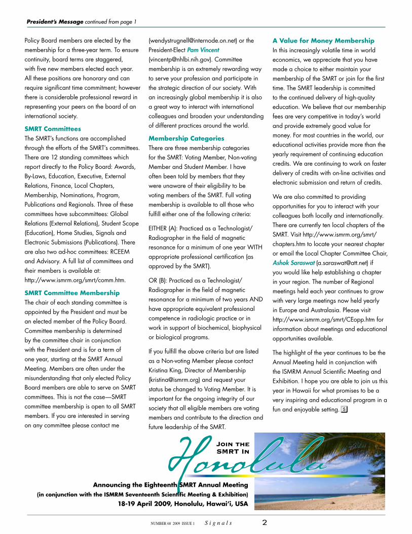
HonoluluJoin the SMRT In
Announcing the eighteenth SMRT Annual Meeting(in conjunction with the iSMRM Seventeenth Scientific Meeting & exhibition)
18-19 April 2009, honolulu, hawai’i, USA
Number 68 2009 Issue 1 S i g n a l s 3
Julie Strandt-Peay, B.S.M., R.T., (R)(MR), FSMRT
Editor’s Letter
Greetings in thisNew Year!
The SMRT continues
to evolve and offer
professional activities
for MR Technologists
and Radiographers in all corners of our world.
President Wendy Strugnell explains the intricacies
of the SMRT and the expanding programs for
members. We congratulate those individuals
newly elected to the position of President-elect,
SMRT Policy Board and the recipient of the Crues-
Kressel award as reported by Nominations and
Awards Chair, Carolyn Bonaceto.
The Annual Meeting promises a quality
experience for all who attend thanks to the efforts
of Program Chair, Ben Kennedy. Sonja Robb-
Belville Chair, Education Committee, presents an
update of the activities leading up to the Annual
Meeting.
Anne Sawyer explains the latest in the
Educational Seminars home study series as well
as additional on-line opportunities for continuing
education. Connecting with related professional
organizations is the reflected in the work by the
External Relations committee Co-chaired by Gina
Greenwood.
Chapter Chat returns with Local Chapter Chair,
Ashok Saraswat. The South Carolina Chapter
hosted another successful meeting which is
described by Carol Lee. A combination of a
local chapter meeting and a regional education
seminar was held in Sydney, Australia according
to Michael Macilquham. An SMRT regional
educational seminar was hosted by the members
in New England as related by Maryann Blaine.
A new feature in this issue of Signals is how MRI
is performed around the world. That is, what
is the process from symptom to scanning and
diagnosis. My appreciation is expressed to those
who took the time to share the MRI experience
in their respective countries. In this issue we
here about MRI in Canada from Caron Murray,
in Australia from Michael Macilquham, in New
Zealand from Anna-Marie Lydon, and from the
island nation of Malta from Joseph Castillo. If
YOU are willing to share your experience and
how the MRI process is conducted in your area
of the world, please feel free to contact me at
[email protected] or through the SMRT office.
We thank MRI safety expert Frank Shellock for his
contribution addressing Epicardial Pacing Wires
and Intracardiac Pacing Wires. As usual you are
reminded to check the calendar of up coming
educational opportunities and visit the SMRT web-
site for new information and updates.
Happy Reading!
This fall the SMRT Membership was presented
with a very difficult task in choosing our new
President Elect and Policy Board members
from an exceptional field of candidates. The
successful candidates for Policy Board are Titti
Owman from Sweden, Michael Macilquham
from Australia, Muriel Cockburn from the United
Kingdom, and Carol Lee and Colleen Hammond,
from the United States. Each of these individuals
will bring experience and new ideas to the
table when they begin their three year terms at
the annual meeting this April in Honolulu. We
are very fortunate to have them as part of our
leadership. Congratulations to each of you!
Our new President-Elect is Julia Lowe from the
United States. Julia has a wealth of experience
as a technologist and as a member of the SMRT
Policy Board. She has served as a member of the
SMRT Executive Committee as External Relations
Committee Chair, representing our organization
to the entire health care community. She brings
much leadership presence to the role and will
serve us well as President. Congratulations
Julia, we are with you 100% and are looking
forward to working with you as you guide our
organization into the future.
Your New Representatives:
President-elect
Julia Lowe, B.S., R.T., (R)
(MR), received an A.S.
degree in Radiologic
Technology from
Indiana University
School of Medicine,
Indianapolis in 1988.
While working part time
at Indiana University Hospital as an inpatient,
surgery x-ray technologist, she pursued her B.S.
degree in Medical Imaging. She graduated from
Indiana University School of Medicine in May of
1989 with a B.S. in Medical Imaging. In 1997
the Indiana University Department of Radiology
purchased a 1.5 Tesla MRI scanner dedicated
to biomedical research. Julie was hired to start
up the facility as the first MRI technologist. As
the research facility expanded she became
lead technologist and assisted in setting up a
3T MRI research site at IU. After five years of
research and clinical work at IU, Julie accepted
a clinical position at St. Vincent ‘s Heart Center
of Indiana, which was the first hospital dedicated
to cardiac and vascular patients in the country.
Julie’s interest in SMRT activities began in 1999
when she joined the organization and attended
the 8th Annual SMRT Meeting in Philadelphia.
She recognized the importance of such an
organization and was very impressed with the
mission of the SMRT to provide educational
opportunities to MRI technologists. Julie was
Election Results
Carolyn Bonaceto, B.S., R.T., (R)(MR) Chair, Nominations and Awards Committee
Continued on page 4 ➠
Number 68 2009 Issue 1 S i g n a l s 4
elected to serve as a Policy Board Member from
2001-2004 and was a member of the Education
Committee until 2002. Subsequently, she served
as chair of the Education Committee and helped
extensively to organize the 2003 SMRT Annual
Meeting in Toronto. Julie helped implement the
first Oral Poster Presentation session, which took
place at the 2003 Poster Walking Tour. Julie also
worked with the Program Committee to have
the proffered paper presentations at the annual
meeting approved for continuing education
credits; this was accomplished for the first time at
the 2003 meeting. She has also been involved
in the Home Studies, Student Scope projects,
and hosted a Regional in 2004. Julie served
as Chair of the External Relations Committee
(ERC) from 2004-2007. Julie has been very
involved in supporting the RadCARE bill, serving
the Associated Sciences Consortium of RSNA,
communicating and attending meetings with the
Health Professions Network and also supporting
the Global Relations Committee. Julie was a
part of the Educational Standards Committee at
the Summit in Seattle, Washington in 2006 and
helped to author the educational guidelines which
will provide a mechanism for U.S. MRI schools
to become accredited. She was presented with
the Distinguished Service Award by the SMRT
in 2007 and was made Fellow of the SMRT in
2008, awards of which she is very proud and
honored. Clinical MRI technologists form the
core membership of the SMRT and Julie’s twenty
years of work in MRI, primarily as a clinical
technologist, is the foundation of her commitment
to the SMRT as a service organization. She is
committed to the SMRT as an organization that
provides a means for technologists to continue
education, communicate with other technologists,
and participate in a society dedicated to the
service of their profession. Julie is proud to be a
part of the SMRT.
Policy Board
Titti Owman has been
involved MRI since the first
NMR imaging attempts in
Scandinavia in 1983 at the
Department of Radiophysics
at Lund University. Currently
Titti is a Research coordinator/lecturer at the
Center for Imaging and Physiology at Lund
University Hospital, Sweden. Since 1986, when
the first clinical MRI-scanner was installed in
Lund, she has been working in clinical practice;
research related work as well as MRI-safety.
She has held the position of Course Director for
MRI education for technologists at the University
since 1995. Titti has also been engaged by the
Swedish Society of Neuroradiology to plan and
organize three courses in Neuroradiology in
Cyprus in 2002, 2003 and 2006. In the spring of
last year she was co-organizer of the SMRT’S first
meeting in Scandinavia held in Aarhus, Denmark.
She has been co-author on scientific papers and
an invited speaker on many occasions and in
2003, 2004 and 2009 invited lecturer at the
European Congress of Radiology in Vienna,
Austria.
Muriel Cockburn, D.C.R.,
B.Sc.Hons., Superintendent
Radiographer Raigmore
Hospital Inverness. Muriel
qualified as a diagnostic
radiographer in 1980 and
has been a superintendent
in MRI since 1993, Muriel has served on the
policy board of the British Association of MR
Radiographers and became president. She
was honored to Co-Chair the SMRT meeting in
Glasgow 2001. The exposure to the SMRT has
given her great access to MR peers across the
world and allowed her to develop and share her
interest and love of MRI clinical service. Muriel has
always been keen to learn and share her interest
in MRI; as a result of this she has presented many
talks and lectures. She is an external lecturer on
an undergraduate university course, and a mentor
to University M.Sc. students in MRI. Her most
recent personal achievement has been to become
a reporting MRI radiographer.
Colleen A. Hammond, R.T.,
(R)(MR), is a Registered
Radiologic Technologist
and began her career
in MRI in 1987. Her MR
career has included
operating and overseeing
MRI programs at two area hospitals in Michigan
prior to her working her current position; MRI
Service Manager for Michigan State University.
Colleen has worked on several field strengths
and vendors in her career, allowing her to work
with different manufacturers to advance the field
of MRI. Colleen is very interested in research
and clinical MRI. Over the years she has trained
numerous technologists and helped them attain
their advanced certification in MRI. Her love of
teaching led her to her latest project of bringing
MRI imaging into sub-Sahara Africa. Colleen
has been working on this project for two years
at Queen Elizabeth Central Hospital in Blantyre,
Malawi, and the 3T magnet was delivered and
installed in June 2008. Colleen is working closely
with the only radiologist within Malawi, Dr.
Sam Kampondeni, to establish a cross sectional
training program and enhance their Radiological
Technologist training program to provide better
patient care. Colleen has coordinated several
research studies in the fields of alzheimer’s,
DTI and carotid plaque imaging and has had
presentations accepted at RSNA and ISMRM.
Carol Lee, B.S., R.T., (R) (CT)(MR), graduated from
Greenville Technical
College with an A.S. in
Radiologic Technology
in 1982 and received
her Bachelor of Science
degree in Radiologic
Health Sciences from
the Medical University
of South Carolina in 1984. She is a Registered
Radiologic Technologist and received both CT
and MR advanced certification from the ARRT in
1995. Carol started her career in MRI in 1990
as the Chief Technologist of a CT/MRI Imaging
Center in Northern California. She is currently
the MRI Supervisor of two magnet sites at
Piedmont Orthopedic Associates in Greenville,
South Carolina, where she has worked for over
seven years. Carol has been instrumental in the
formation of the South Carolina Chapter of the
SMRT (SC SMRT) and helps to organize two
chapter meetings a year and currently serves as
the President. Carol believes the SMRT provides
excellent continuing educational opportunities
Election Results continued from page 3
Continued on page 5 ➠
Number 68 2009 Issue 1 S i g n a l s 5
and she is honored to be a member of the Policy
Board of the SMRT.
Michael D. Macilquham,
B.App.Sc., M.H.Sc. (MRI),
MSMRT, Michael graduated
as a radiographer in
Sydney, Australia in 1995,
and began work in MRI in
1999. He completed the
University of Sydney post-
graduate Masters of Health Science degree in
MRI in 2002. In 2004 Michael is currently the
MRI Supervisor at John Fawkner Hospital. He is
also employed in a concurrent role as the MRI
tutor for MIA Victoria, in a role encompassing
10 sites with a variety of vendor’s equipment, at
field strengths of 1.5 to 3.0T. Michael has been
a member of the SMRT since 2003 and was
also first author of the article entitled “The Role
of MRI and Spectroscopy in Assessing Prostate
Cancer” published in the SMRT Home Studies
Seminar “MR Imaging and Spectroscopy of the
Prostate” (Volume 7, Issue 4, 2004). Michael
was the Co-Chair of the 2007 Australia – New
Zealand (ANZ) SMRT Chapter Meeting held in
Melbourne, and is now serving as the President
of the ANZ Chapter. Michael has been inspired
by the enthusiasm and professionalism of the
members of the SMRT and strongly believes that
the continual expansion of the global focus of the
SMRT, and the increasing interaction between
international members will help promote and
improve the profession.
cindy hipps to Receive Award
In addition to selecting our future representation,
the ballots included nominees for the Crues
Kressel Award for Outstanding Contributions
to the Education of Magnetic Resonance
Technologists. Again the competition was stiff and
each candidate was very deserving. This year’s
successful candidate is Cindy Hipps. Cindy
exemplifies what this award is about. She has set
the bar high and continually hurdles it by hosting
many regional meetings and chapter meetings
in the Carolinas. Congratulations Cindy! You are
truly an SMRT star.
crues Kressel Award Recipient
Cindy T. Hipps, B.H.S.,
R.T., (R)(MR), became a
Registered Radiologic
Technologist in 1981 after
completion of an Associate
of Science degree in
Radiologic Technology
from Greenville Technical
College in Greenville, South Carolina. She also
pursued her Bachelors of Health Sciences degree
while working full time, and, in 1988, received
a BHS from the Medical University of South
Carolina. Presently, she is employed as the MRI
Coordinator for Greenville Radiology, PA, which
is a professional practice of 40-plus Radiologists.
There, she manages the day-to-day operations of
Easley MRI, LLC, which is a joint venture between
Greenville Radiology, PA, and Palmetto Health
Baptist Easley. She joined the SMRT in 1993
and attended her first annual meeting in New
York. She supported the Local SMRT Chapter of
Atlanta, too. The chapter was and is instrumental
in helping her connect the dots between local
SMRT and International SMRT. She was elected
by the membership in 1999 to the SMRT Policy
Board. As Policy Board Member, Cindy chaired
the Annual Meeting Committee and the Education
Committee. She also continued to serve the
SMRT with appointments to the RCEEM, Finance,
Publications, Annual Meeting and Education
Committees after rotating off of the Policy Board.
In 2003, Cindy was chosen as President-Elect of
the SMRT. During her tenure as President, and
with the help of the Executive Committee and
Policy Board, she was instrumental in mapping
out the first strategic plan that was submitted
by President Maureen Ainslie. After much work
with the RCEEM committee, the RCEEM became
a reality during her tenure, and the RCEEM
committee is now one of the standing ad hoc
committees that report to the Executive Committee
and attend all Policy Board Meetings. She is
very proud of the work that was done during
her tenure with the Alliance and the CARE Bill.
She also was instrumental in starting the first
Advisory Committee of the SMRT, as well as
establishing the first Educational Standards Ad
Hoc Committee. Cindy was diligent in planning
the first MR Primary Pathway Summit meeting
in Seattle to discuss and revise the current MR
Curriculum in order to reflect the changes in the
field of MR where one could pursue the field
as a primary pathway. She was instrumental in
getting representatives from the ARRT, AEIRS,
ASRT, and JRCERT to attend this meeting. With
the leadership of Luann Culbreth and much work
from a host of MR professionals, this project
became a reality this year and the newly revised
MR Curriculum has been published and can be
found on the SMRT website. Cindy has chaired
many SMRT Regional Meetings in South Carolina.
Her President’s Regional Meeting was hosted in
Charleston, SC, and other meetings have been
held in conjunction with the South Carolina
SMRT Chapter. The SC Chapter’s mission is
to promote the SMRT while also providing
continuing education for area technologists. She
is proud of her appointment to The Institute for
Magnetic Resonance Safety, Education, and
Research (IMRSER) as a Medical, Scientific, and
Technology Advisory Board member. Cindy
Hipps is extremely honored that she is to be
awarded the prestigious Crues Kressel Award of
the Section for Magnetic Resonance Technologists
of the International Society of Magnetic
Resonance in Medicine. She will always be an
advocate for technologists being all that they
can be and belonging to an organization that
promotes the education of MR technologists, such
as the SMRT. She is very proud to be a member
of the SMRT.
Election Results continued from page 4
Number 68 2009 Issue 1 S i g n a l s 6
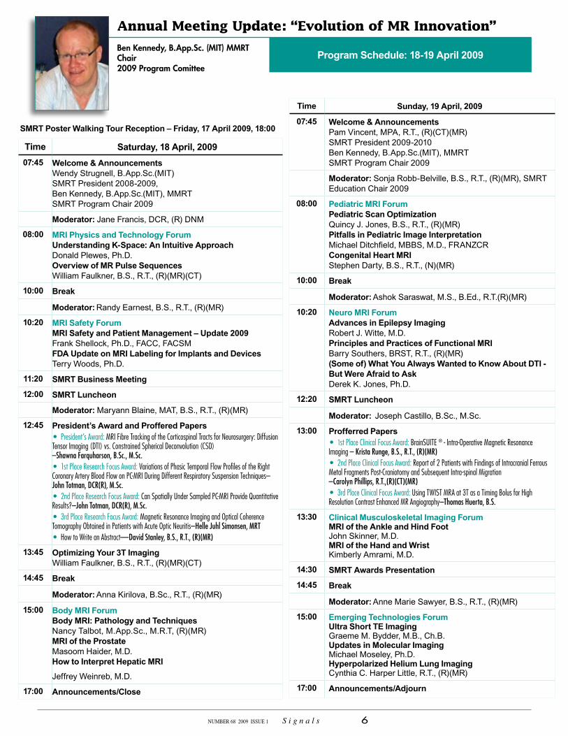
Annual Meeting Update: “Evolution of MR Innovation”
Ben Kennedy, B.App.Sc. (MIT) MMRTChair2009 Program Comittee
Program Schedule: 18-19 April 2009
SMRT Poster Walking Tour Reception – Friday, 17 April 2009, 18:00
Time Saturday, 18 April, 2009
07:45 Welcome & AnnouncementsWendy Strugnell, B.App.Sc.(MIT)SMRT President 2008-2009,Ben Kennedy, B.App.Sc.(MIT), MMRTSMRT Program Chair 2009
Moderator: Jane Francis, DCR, (R) DNM
08:00 MRI Physics and Technology ForumUnderstanding K-Space: An Intuitive ApproachDonald Plewes, Ph.D.Overview of MR Pulse SequencesWilliam Faulkner, B.S., R.T., (R)(MR)(CT)
10:00 Break
Moderator: Randy Earnest, B.S., R.T., (R)(MR)
10:20 MRI Safety ForumMRI Safety and Patient Management – Update 2009Frank Shellock, Ph.D., FACC, FACSMFDA Update on MRI Labeling for Implants and DevicesTerry Woods, Ph.D.
11:20 SMRT Business Meeting
12:00 SMRT Luncheon
Moderator: Maryann Blaine, MAT, B.S., R.T., (R)(MR)
12:45 President’s Award and Proffered Papers• President’s Award: MRI Fibre Tracking of the Corticospinal Tracts for Neurosurgery: Diffusion Tensor Imaging (DTI) vs. Constrained Spherical Deconvolution (CSD) –Shawna Farquharson, B.Sc., M.Sc.• 1st Place Research Focus Award: Variations of Phasic Temporal Flow Profiles of the Right Coronary Artery Blood Flow on PC-MRI During Different Respiratory Suspension Techniques–John Totman, DCR(R), M.Sc.• 2nd Place Research Focus Award: Can Spatially Under Sampled PC-MRI Provide Quantitative Results?–John Totman, DCR(R), M.Sc.• 3rd Place Research Focus Award: Magnetic Resonance Imaging and Optical Coherence Tomography Obtained in Patients with Acute Optic Neuritis–Helle Juhl Simonsen, MRT• How to Write an Abstract—David Stanley, B.S., R.T., (R)(MR)
13:45 Optimizing Your 3T ImagingWilliam Faulkner, B.S., R.T., (R)(MR)(CT)
14:45 Break
Moderator: Anna Kirilova, B.Sc., R.T., (R)(MR)
15:00 Body MRI Forum Body MRI: Pathology and TechniquesNancy Talbot, M.App.Sc., M.R.T, (R)(MR)MRI of the ProstateMasoom Haider, M.D.How to Interpret Hepatic MRI
Jeffrey Weinreb, M.D.
17:00 Announcements/Close
Time Sunday, 19 April, 2009
07:45 Welcome & AnnouncementsPam Vincent, MPA, R.T., (R)(CT)(MR)SMRT President 2009-2010Ben Kennedy, B.App.Sc.(MIT), MMRTSMRT Program Chair 2009
Moderator: Sonja Robb-Belville, B.S., R.T., (R)(MR), SMRT Education Chair 2009
08:00 Pediatric MRI ForumPediatric Scan OptimizationQuincy J. Jones, B.S., R.T., (R)(MR)Pitfalls in Pediatric Image InterpretationMichael Ditchfield, MBBS, M.D., FRANZCRCongenital Heart MRIStephen Darty, B.S., R.T., (N)(MR)
10:00 Break
Moderator: Ashok Saraswat, M.S., B.Ed., R.T.(R)(MR)
10:20 Neuro MRI ForumAdvances in Epilepsy ImagingRobert J. Witte, M.D.Principles and Practices of Functional MRIBarry Southers, BRST, R.T., (R)(MR)(Some of) What You Always Wanted to Know About DTI - But Were Afraid to AskDerek K. Jones, Ph.D.
12:20 SMRT Luncheon
Moderator: Joseph Castillo, B.Sc., M.Sc.
13:00 Profferred Papers• 1st Place Clinical Focus Award: BrainSUITE ® - Intra-Operative Magnetic Resonance Imaging – Krista Runge, B.S., R.T., (R)(MR)• 2nd Place Clinical Focus Award: Report of 2 Patients with Findings of Intracranial Ferrous Metal Fragments Post-Craniotomy and Subsequent Intra-spinal Migration–Carolyn Phillips, R.T.,(R)(CT)(MR)• 3rd Place Clinical Focus Award: Using TWIST MRA at 3T as a Timing Bolus for High Resolution Contrast Enhanced MR Angiography–Thomas Huerta, B.S.
13:30 Clinical Musculoskeletal Imaging ForumMRI of the Ankle and Hind FootJohn Skinner, M.D.MRI of the Hand and WristKimberly Amrami, M.D.
14:30 SMRT Awards Presentation
14:45 Break
Moderator: Anne Marie Sawyer, B.S., R.T., (R)(MR)
15:00 Emerging Technologies ForumUltra Short TE ImagingGraeme M. Bydder, M.B., Ch.B.Updates in Molecular ImagingMichael Moseley, Ph.D. Hyperpolarized Helium Lung ImagingCynthia C. Harper Little, R.T., (R)(MR)
17:00 Announcements/Adjourn

Number 68 2009 Issue 1 S i g n a l s 7
We would like to invite all SMRT members to attend the
ISMRM/SMRT Joint Forum on “How To Do a Multisite Neuroimaging Study.” This forum will be held on Monday 20 April 2009 at 14:00 hours, following the weekend SMRT Annual Meeting. Your registration for the SMRT Annual Meeting allows you to attend this ISMRM/SMRT Joint Forum Presentation.
This year the ISMRM/SMRT Joint Forum is organized by Bryon A. Mueller, Ph.D., Gary H. Glover, Ph.D., Caron Murray, M.R.T., (R) AC, (CT)(MR), and Douglas C. Noll, Ph.D. The two-hour forum will present the process introducing issues common to all types of multi-center MRI (MC-MRI) studies. These studies are an important tool to validate and establish MRI methodology, particularly for their use as a biomarker. MC-MRI studies
are widely used in clinical trials to evaluate pharmaceuticals, MRI-compatible devices, and related technology.
The forum by design is a collaboration of knowledge and talent between the ISMRM and the SMRT. This relationship continuously promotes the highest quality of education in the MR field.
Education Committee Update
The Abstract submission scoring for the 18th Annual SMRT Meeting “Evolution of
MR Innovation” in Honolulu, Hawai’i 18 and 19 April 2009 is underway by volunteers from the Education Committee. Twenty reviewers are evaluating the content of a record setting 80 oral and poster abstract submissions. The submissions demonstrate the global nature of the SMRT as they come from 16 different nations, including Australia (5), Canada (9), Denmark (3), Germany (5), Israel (1), Italy (1), Japan (6), the Netherlands (2), Norway (1), Poland (2), Russia (1), South Korea (5), Sweden (1), Switzerland (1), the United Kingdom (8), and the United States of America (29).
The first round of scoring will determine which of the oral and poster abstracts will be accepted, as well as which of the oral abstracts will be presented as Proffered Papers at the Annual Meeting in Honolulu. The President’s Award will be given to the best overall oral abstract, in addition to 1st, 2nd, and 3rd place awards for oral abstracts in the clinical and research focuses. Authors of oral abstracts not selected for presentation as a Proffered Paper are invited to submit their abstract as a poster. A second round of scoring is conducted by the reviewers to select the 1st, 2nd, and 3rd place posters in the clinical and research focuses. The award-receiving posters will be presented during the SMRT Reception and Poster Walking Tour
beginning at 18:00 on 17 April. You don’t want to miss this excellent educational and networking opportunity!
Additional continuing education credits Available in honolulu!Several courses throughout the scientific portion of the 17th ISMRM Scientific Meeting will award Category A Continuing Education Credit to MR Technologists/Radiographers. The low registration cost for SMRT members to attend the ISMRM Scientific Meeting makes this an excellent value. Please consider extending your stay in Honolulu to take advantage of the excellent opportunity and stay tuned for future announcements on which courses will qualify!
ISMRM/SMRT Joint Forum
Sonja K Robb-Belville B.S., R.T., (R)(MR) Chair, 2009 Education Committee
How To Do a Multi-Site Neuroimaging Study
Time Presentation Presenter
2:00-2:18 Foundations for Performing Any Multi-Center Neuroimaging Study Gary Glover, Ph.D., Stanford University
2:18-2:36 How To Do a STRUCTURAL Multi-Center Neuroimaging Study Matt Bernstein, Ph.D., Mayo Clinic
2:36-2:54 How To Do a DTI Multi-Center Neuroimaging Study Carlo Pierpaoli, M.D., Ph.D., National Institutes of Health
2:54-3:12 How To Do a FUNCTIONAL Multi-Center Neuroimaging Study Bryon Mueller, Ph.D., University of Minnesota
3:12-3:30 How To Do an ASL Multi-Center Neuroimaging Study Xavier Golay, Ph.D., Singapore BioImaging Consortium
3:30-3:48 How To Do a Multi-Center Neuroimaging Study:A Technologist’s Perspective Maureen Ainslie, M.S., R.T., (R)(MR), DIAL, Duke
3:48-4:00 Panel Discussion Panel
“The forum by design is a collaboration of knowledge and talent between the ISMRM and the SMRT.”
Caron Murray, M.R.T., (R) AC,(CT)(MR)Co-ChairSMRT/ISMRM Joint Forum

Number 68 2009 Issue 1 S i g n a l s 8
We are pleased to present the SMRT Educational Seminars, Volume 12, Number 1: “Contrast-Enhanced Musculoskeletal MR Imaging.” This is the forty-third home study developed by the SMRT, exclusively for the SMRT members.
For this issue, we have selected six articles that discuss direct and indirect methods in the use of gadolinium contrast agent to improve the diagnostic capabilities of MR imaging of the musculoskeletal system. As stated by John D. MacKenzie and collaborators in our first article, “The rationale for MR arthrography is to distend the joint in order to reveal fine anatomic detail and joint derangement.” Despite the increased cost and risks such as NSF, MR contrast agents continue to drive increased applications throughout the body and brain.
Our second article focuses on the indirect method of MR arthrography in which contrast is injected intravenously and imaging occurs after a delay. This type of MR arthrography is based on the principle that IV contrast material will diffuse into the joint space over time, producing an arthrographic effect. According to Renata La Rocca Vieira and colleagues, “The indirect technique allows for greater signal-to-noise ratio on small field-of-view images and has higher sensitivity to subtle cartilage defects and tendon disorders.”
The third article is a comprehensive discussion of contrast-enhanced MR imaging of the shoulder joint, specifically glenoid cartilage lesions. The MR scan protocol includes ABER (abduction and external rotation) views of the shoulder
joint. According to the authors, Hamidreza Torshizy and collaborators, the ABER views “allow greater sensitivity than does conventional MRI in detecting anterior labral lesions, as well as other LLC (labral-ligamentous complex) abnormalities.”
Our fourth article reviews the use of contrast media in MRI of musculoskeletal neoplasms. The authors, Daniel Vanel et al, take us through a detailed discussion comparing bone tumors to soft tissue sarcomas from diagnosis, through staging, treatment evaluation, and finally, the detection of recurrences. The authors make a valid point for serious consideration, “Even if the diagnostic value of dynamic MRI has been shown in soft tissue sarcomas (but not in bone tumors), the main problem is, in fact, suggesting the diagnosis and correctly referring the patient.”
Enhanced MR Imaging in Musculoskeletal Infection by Maryam Golshan Momeni and collaborators is our fifth article. Enhanced MR imaging of several conditions including cellulitis, fasciitis, pyomyositis, infectious arthritis, and osteomyelitis, are described. “Comparisons of enhanced MRI with other imaging modalities have documented its greater sensitivity and specificity for detecting infection” which is especially important as the “differentiation of soft tissue infection from bone involvement is a difficult clinical and imaging problem.”
Our final article is an expanded discussion
of contrast-enhanced MRI of inflammatory and
degenerative arthritis that follows nicely after the previous
article. Oganes Ashikyan and colleagues conclude “Dynamic contrast-enhanced MRI can determine the extent of the vascularity in the inflamed synovium and can differentiate active from fibrotic (inactive) synovial tissue.”
We would like to express our grateful appreciation to Michael R. Terk, M.D., Professor of Radiology, Emory School of Medicine, Director of Musculoskeletal Imaging, Atlanta, Georgia, USA, for acting as our expert reviewer.
The accompanying quiz is long; however as a RCEEM (Recognized Continuing Education Evaluation Mechanism) of the ARRT, there are a required number of questions per article that is required to obtain Continuing Education Credits.
Thanks also to Paul McElvogue, SMRT Publications Chair and in the Berkeley, California, USA office of the ISMRM/SMRT, Jennifer Olson, Associate Executive Director, Mary Keydash, Publications Director, and the staff for their insight and long hours supporting these educational symposia.
We would especially like to thank John Wilkie and all of the people at Invivo Corporation who generously support our home studies program, the SMRT Educational Seminars. Their continuing investment advancing technologist
Contrast-Enhanced Musculoskeletal
MR Imaging
SMRTEducational Seminars Volume 12, Number 1
SMRT Educational Seminars Home Study Update
Anne Marie Sawyer, B.S., R.T., (R)(MR) EditorSMRT Educational Seminars Home Study Program
Continued on page 9 ➠

Number 68 2009 Issue 1 S i g n a l s 9
and radiographer knowledge brings quality continuing education to the SMRT membership worldwide.
Finally, in selecting articles for this home study as well as others including our new electronic home studies, I am constantly asking myself (and others, driving them to
distraction) “What do technologists and radiographers need to know?” Possibly of greater importance, “What do technologists and radiographers want to know?” I am sure there are many things that we need to know but without the desire to learn, it is practically impossible to absorb the information and to retain it for later
utilization. With that in mind, I am once again requesting all SMRT members to send me an email and tell me what topics you would like to see in these home studies, paper and/or electronic ([email protected]).
Please note that the SMRT Educational Seminars Home Study
publications are renewed continuously by the SMRT RCEEM
Committee for Category A CE accreditation. You may have some of
the earlier published editions of the printed Educational Seminars
Home Studies in which you are planning to complete the Quiz for CE
credits.
IMPORTANT: Please be aware that the earlier published Home
Study CE credit amounts have been adjusted due to changes in the
guidelines made by American Registry of Radiologic Technologists
(ARRT) in the CE approval process. Self-learning activities such
as directed readings and home studies that have previously been
approved and/or renewed after 1 January 2006 are now evaluated
on content only. CE credits are awarded based on the time spent
reading the text. The time spent completing the Quiz will no longer
be awarded CE credit. All RCEEMs are required to follow this
ARRT guideline change. The ARRT also requires that the number of
questions that compose the Quiz, is determined by the guidelines
established by the ARRT.
Visit the website http://www.ismrm.org/smrt/homestudy/index.htm
to verify your credit values.
Home Studies continued from page 8

Number 68 2009 Issue 1 S i g n a l s 10
External Relations Committee Update
While Committee names generally indicate quite precisely what the charge to the Committee may be (e.g. Finance, Membership, Program), “External Relations” may be a bit nondescript to some. As a point of reference, the External Relations Committee is concerned as a liaison with governments at all levels, with relations in other professional and ethical societies, with industry (except for those immediate issues of seeking financial support) and with the public at large. Currently, there are three primary ways in which the duties of the External Relations Committee Chair manifest. One is to represent the SMRT as a participating member of the Alliance for Quality Medical Imaging and Radiation Therapy through attendance at all meetings of the Alliance. Another is to represent the SMRT as a member of the Health Professions Network by attending all meetings of the HPN. Also, the External Relations Committee Chair serves as the SMRT representative to the Radiological Society of North America (RSNA) Associated Sciences Consortium and participates in planning the Associated Sciences workshop and refresher courses held each year at the RSNA Annual Meeting. As reported in the past, the amount of travel, participation and involvement that is required of the External Relations Committee Chair has precipitated the role evolving into that of “Co-Chair” in effort to ensure adequate representation and fulfillment of duties.
health Professions NetworkThe Health Professions Network (HPN) represents health care provider organizations, educators, regulators, and government agencies. The aim of the HPN is to provide a forum for collaboration among health care professions on issues of
common interest. They hope to accomplish this mission through: identifying issues of common interest, communicating these issues to all participants, seeking consensus and facilitating responses and advocating on behalf of health care professionals to the public, professional associations, and federal and state policy makers. Gina Greenwood, MBA, R.T., (R)(MR) attended the HPN 2008 fall meeting in St. Louis, Missouri, USA, 22–24 October 2008. Attendees of the meeting heard presentations that covered varied topics.
The keynote presentation was delivered by Dr. Ed O’Neil of the UCSF Center for Health Professions. Dr. O’Neil drew on Greek mythology to illustrate the many health care challenges the nation faces, sighting that we are like Sisyphus, and the current health care paradigm is the rock—we’re in love with the rock, but we need to let it go. Dr. O’Neil went on to explain that our health care technology is marvelous, but costly. A full 16.5% of the nation’s GDP is spent on health care—that’s $2.4 trillion. Further, the fourth leading cause of death is admission to the hospital: “We have a health system that’s incredibly sloppy and inefficient compared to those of other countries,” noted O’Neil. All is not bleak, however, and Dr. O’Neil focused the rest of his presentation on the drivers of our health care future and opportunities improvements. In short, Dr. O’Neil feels that healthcare needs to focus on chronic condition prevention and management, becoming price competitive, being consumer responsive and implementing evidence-based practice.
Centers for Medicare and Medicaid Services (CMS) officials Arnold Z. Balanoff, M.D. FAAP, and Robert Epps,
MPA collaborated to deliver a presentation entitled “Future Medicare Reimbursement Trends: A CMS Perspective.” According to Epps and Balanoff, the Medicare program is “unsustainable” in its current form, in terms of both its programmatic and its financial structure. He explained that the CMS had traditionally focused on paying bills, efficiently and effectively, without regard to the quality of services that was provided. He further stated that this disconnect between cost and value simply is no longer acceptable, given the financial status of the entire program. CMS is moving to a ‘prudent purchaser’ focus within the agency that is called value-based purchasing. The cornerstones of value-based healthcare are interoperable health information technology, quality standards, price standards, and the promotion of quality and efficiency-of-care standards through a better system of incentives.
Another theme of the meeting was scope of practice. Jayme Matchinski, J.D. discussed the many changes in health care and scope of practice. “Collaboration should be the professional norm,” she said; “changes in scope of practice are inherent in health care as it continues to change and grow. No one profession owns a skill or activity, and one activity should not define a profession. Professions seeking expanded licensure have to be organized at the grassroots level—and it takes a lot of money, and reaching out to other professions to get buy-in. First and foremost, however, public protection should be the highest priority.”
Currently, the major undertaking of the HPN is the launch of the Health Professions Awareness Campaign (HPAC). HPAC will seek to increase the awareness of health
Gina Greenwood, MBA, R.T., (R)(MR) Co-ChairExternal Relations Commitee
“Healthcare needs to focus on chronic condition prevention and management, becoming price competitive, being consumer responsive and implementing evidence-based practice.”
Continued on page 11 ➠
Number 68 2009 Issue 1 S i g n a l s 11
professions among the public and policy makers. As this is not a small task, the HPN is seeking major investors with which to partner. The goal is to raise between $7 to $10 million dollars to support media campaigns, print campaigns, website development, Public Service Announcements, artwork, etc. Thus far, the HPN has developed a message to potential investors, drafted a business plan and completed Phase I of prospect research. They have identified 15 potential high-level investors and narrowed that list to a “Top 5” to begin focusing efforts.
The Alliance for Quality Medical imaging and Radiation TherapyGina Greenwood and Charles Stanley both attended the Alliance for Quality Medical Imaging and Radiation Therapy (Alliance) meeting in Albuquerque, New Mexico, USA on 9-11 November 2008. A good portion of the meeting was focused on the recently passed Medicare Bill H.R. 6331. This Bill, passed in July, is also known as the Medicare Improvements for Patients and Providers Act (MIPPA). As it relates to the Consistency, Accuracy, Responsibility and Excellence in Medical Imaging and Radiation Therapy (CARE) Bill, MIPPA requires accreditation of providers of the technical component for advanced diagnostic imaging services (MRI, CT, and nuclear medicine/PET) by an entity identified by the Secretary of Health and Human Services prior to 1 January 2012 in order to be eligible for the technical component payment. This applies only to outpatient imaging centers. The Secretary of
Health and Human Services must designate accrediting organizations by 1 January 2010. The accreditation organizations must have criteria to evaluate medical personnel, medical directors, supervising physicians, equipment, safety procedures, and quality assurance programs.
To put it more simply, by requiring accreditation, the Bill includes provisions establishing standards for medical personnel performing CT, MRI, Nuclear Medicine and PET exams. Unfortunately, this bill excluded x-ray, fluoroscopy, ultrasound and radiation therapy, which falls short of establishment of minimum education and credentialing standards for personnel who perform all medical imaging and radiation therapy procedures.
The main concern at this point is members of Congress may believe that MIPPA has addressed all of the points of CARE Bill. The Alliance needs to spend some time developing the best strategy to move forward. Questions and issues facing the
Alliance at this time include:
• Should we consider revising the CARE Bill?• Change in direction of the new
administration• Need to arrange meetings with transition team and/or new players• There will be some changes in committee chairmanships• Philosophically new administration should be ‘on our side’• MIPPA supports and supplements what CARE bill wants to do• Even if revised, the principals behind the CARE Bill will not change• Question of how to position it for the new Congress in light of MIPPA• Question of how to put together a bill which contains the CARE provisions but has a better chance of passage?
The next meeting of the Alliance will take place at the end of March 2009. Hopefully more will be known regarding the enforcement of MIPPA and potential strategy changes that need to be taken by the Alliance in effort to pass the CARE Bill.
Associated Sciences consortiumCharles Stanley participated in the Associated Sciences Consortium Planning Committee for the 2008 RSNA meeting. Charles will be moderating an Associated Sciences Consortium session entitled “Imaging in the Operating Room.”
Best wishes to all for a healthy happy 2009! Stay tuned for more updates from the External Relations Committee at the Annual Meeting.
External Relations continued from page 10
The accreditation organizations
must have criteria to evaluate
medical personnel, medical
directors, supervising physicians,
equipment, safety procedures, and
quality assurance programs.
Number 68 2009 Issue 1 S i g n a l s 12
Chapter Chat
Local chapters represent the mission of SMRT. Our local
chapter leaders and their teams have been working diligently to promote the mission of SMRT in different geographical areas globally. Recent educational seminars hosted by most local chapters are clear examples of work being done by our chapter leaders and their teams of dedicated volunteers. All active chapters have either recently hosted an educational seminar or are planning to do so before the annual meeting in Hawaii.
On behalf of the SMRT local chapter committee, I congratulate chapter committee members and organizers from the Australian & New Zealand Local Chapter for their
highly successful 3rd Aannual seminar in November 2008. Chapter president Michael Macilquhamn reported a great turnout, excellent speakers and a great interest by attendees in this ever popular and growing annual event every November. To see the complete list and the brains behind this most successful local chapter, please visit ANZ chapter website (www.ismrm.org/smrt/anz.htm). ANZ chapter recently elected Kristen Moffat as their president-elect. Please welcome Kristen in her new role. ANZ chapter, under the leadership of Greg Brown has formed an MRI Safety Committee. As far I recall this is a first MRI safety committee by any local chapter. Congratulations to all ANZ chapter members for taking new initiatives and promoting SMRT membership
in your region.
The New England local chapter, Atlanta, Rocky Mountain, South Carolina, BeNeLux, Central Virginia, Northeast Ohio and the newly formed New York/New Jersey chapter have either recently offered or are planning an educational event soon. On behalf of the SMRT local chapter committee we congratulate all active chapters and their teams on their outstanding efforts. We will share their experience with the SMRT members on a routine basis.
If you would like to start a new local chapter please contact Ashok Saraswat, e-mail [email protected] or any local chapter committee member.
Ashok Saraswat, M.S., B.Ed., R.T., (R)(MR) Chair, Local Chapter Committee
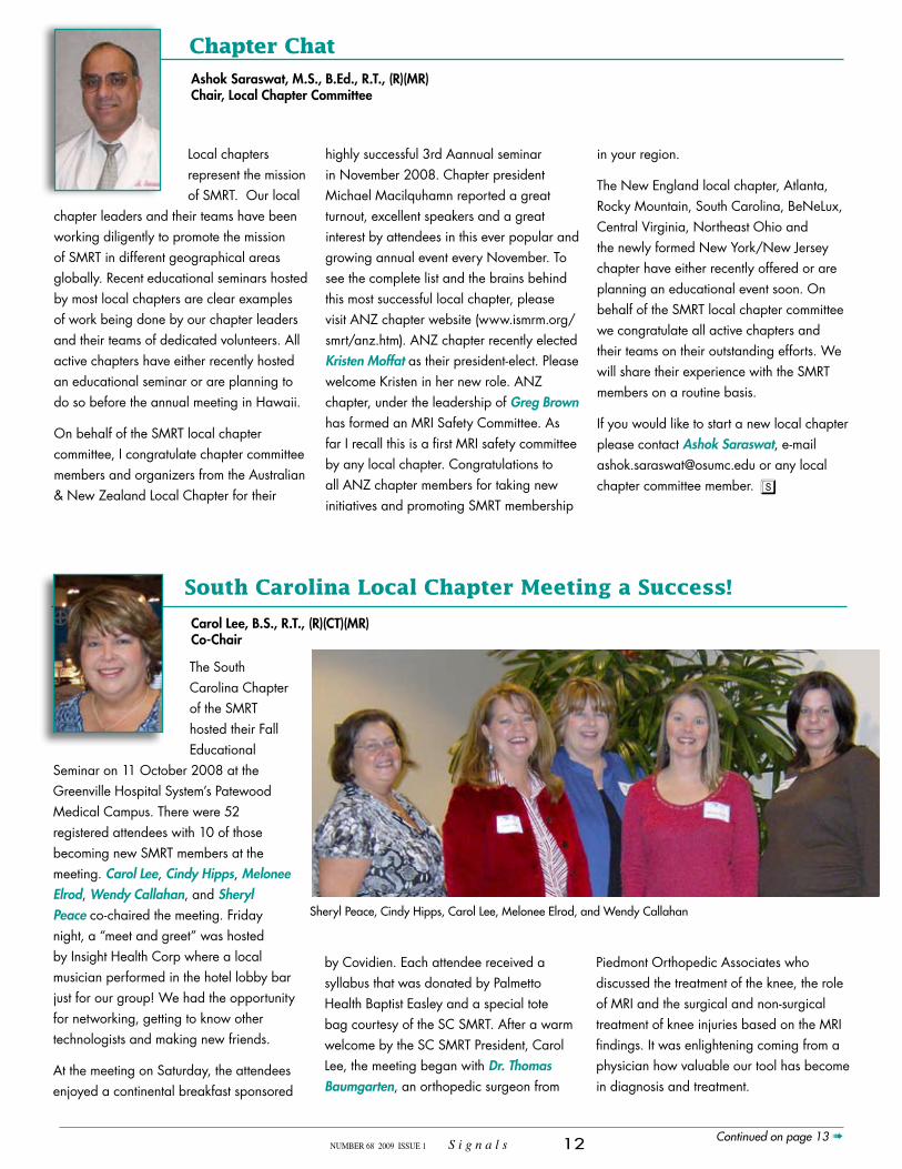
South Carolina Local Chapter Meeting a Success!
Carol Lee, B.S., R.T., (R)(CT)(MR)Co-Chair
The South Carolina Chapter of the SMRT hosted their Fall Educational
Seminar on 11 October 2008 at the Greenville Hospital System’s Patewood Medical Campus. There were 52 registered attendees with 10 of those becoming new SMRT members at the meeting. Carol Lee, Cindy Hipps, Melonee Elrod, Wendy Callahan, and Sheryl Peace co-chaired the meeting. Friday night, a “meet and greet” was hosted by Insight Health Corp where a local musician performed in the hotel lobby bar just for our group! We had the opportunity for networking, getting to know other technologists and making new friends.
At the meeting on Saturday, the attendees enjoyed a continental breakfast sponsored
by Covidien. Each attendee received a syllabus that was donated by Palmetto Health Baptist Easley and a special tote bag courtesy of the SC SMRT. After a warm welcome by the SC SMRT President, Carol Lee, the meeting began with Dr. Thomas Baumgarten, an orthopedic surgeon from
Piedmont Orthopedic Associates who discussed the treatment of the knee, the role of MRI and the surgical and non-surgical treatment of knee injuries based on the MRI findings. It was enlightening coming from a physician how valuable our tool has become in diagnosis and treatment.
Sheryl Peace, Cindy Hipps, Carol Lee, Melonee Elrod, and Wendy Callahan
Continued on page 13 ➠
Number 68 2009 Issue 1 S i g n a l s 13
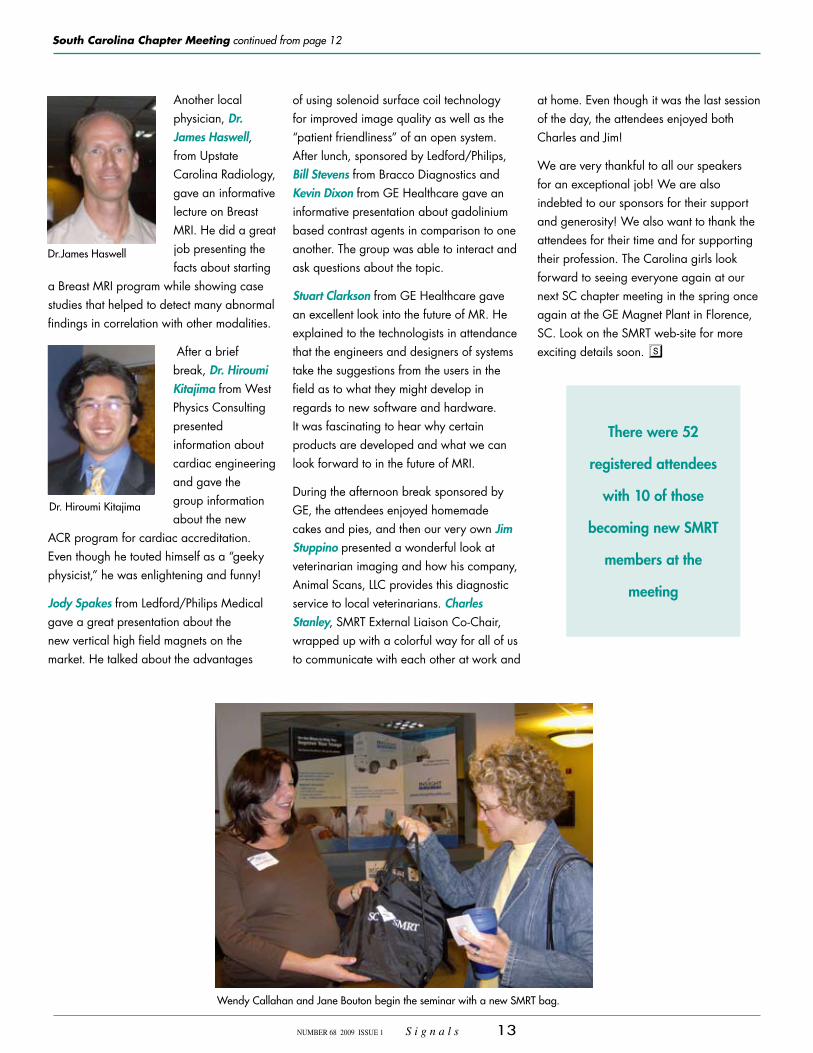
Another local physician, Dr. James Haswell, from Upstate Carolina Radiology, gave an informative lecture on Breast MRI. He did a great job presenting the facts about starting
a Breast MRI program while showing case studies that helped to detect many abnormal findings in correlation with other modalities.
After a brief break, Dr. Hiroumi Kitajima from West Physics Consulting presented information about cardiac engineering and gave the group information about the new
ACR program for cardiac accreditation. Even though he touted himself as a “geeky physicist,” he was enlightening and funny!
Jody Spakes from Ledford/Philips Medical gave a great presentation about the new vertical high field magnets on the market. He talked about the advantages
of using solenoid surface coil technology for improved image quality as well as the “patient friendliness” of an open system. After lunch, sponsored by Ledford/Philips, Bill Stevens from Bracco Diagnostics and Kevin Dixon from GE Healthcare gave an informative presentation about gadolinium based contrast agents in comparison to one another. The group was able to interact and ask questions about the topic.
Stuart Clarkson from GE Healthcare gave an excellent look into the future of MR. He explained to the technologists in attendance that the engineers and designers of systems take the suggestions from the users in the field as to what they might develop in regards to new software and hardware. It was fascinating to hear why certain products are developed and what we can look forward to in the future of MRI.
During the afternoon break sponsored by GE, the attendees enjoyed homemade cakes and pies, and then our very own Jim Stuppino presented a wonderful look at veterinarian imaging and how his company, Animal Scans, LLC provides this diagnostic service to local veterinarians. Charles Stanley, SMRT External Liaison Co-Chair, wrapped up with a colorful way for all of us to communicate with each other at work and
at home. Even though it was the last session of the day, the attendees enjoyed both Charles and Jim!
We are very thankful to all our speakers for an exceptional job! We are also indebted to our sponsors for their support and generosity! We also want to thank the attendees for their time and for supporting their profession. The Carolina girls look forward to seeing everyone again at our next SC chapter meeting in the spring once again at the GE Magnet Plant in Florence, SC. Look on the SMRT web-site for more exciting details soon.
Dr.James Haswell
Wendy Callahan and Jane Bouton begin the seminar with a new SMRT bag.
Dr. Hiroumi Kitajima
South Carolina Chapter Meeting continued from page 12
There were 52
registered attendees
with 10 of those
becoming new SMRT
members at the
meeting
Number 68 2009 Issue 1 S i g n a l s 14
ANZ Chapter Provides Venue for President’s Regional
3rd Annual Local Chapter Meeting, Sydney, Australia, 15-16 November 2008
and the 2008 SMRT President’s Regional Meeting
Due to a combination of late-Spring Sydney storms, not all delegates (and speakers!) made it to the 3rd Annual ANZ Chapter Meeting at the Sydney Convention and Exhibition Centre on time, but by the conclusion of the meeting, most of the 260 in attendance agreed this was the best ANZ Chapter Meeting yet. The meeting format was again a solid two-day programme interspersed with a vibrant Saturday night social function. Delegates enjoyed listening to a variety of speakers talk on clinical, theoretical and advanced topics. Keynote speakers included the ever-popular Dr. Mike Moseley from Stanford, Dr. Elizabeth Moore from Philips in the Netherlands, and the always-relevant Dr. Frank Shellock from the IMRSER. There was also a strong local faculty, who confirmed via an excellent series of presentations that the knowledge base of MRI radiographers from Australia and New Zealand is second to none.
For the second time at the ANZ Meeting, proffered papers were included in the program. Three excellent presentations were given from clinical and research-oriented
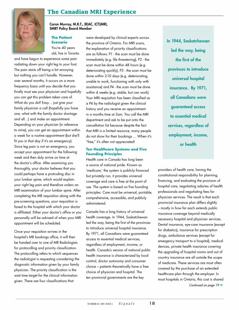
MR radiographers, demonstrating the high standard of work being carried out locally. The First Prize Award for Best Proffered Paper went to Ali Zailaa for his work on fMRI in Acupuncture. Congratulations to all of our presenters, and thank you for making the effort to present your work at our meeting. The ANZ Executive firmly believes that this format provides the most relevant format for MR radiographers to present their work, and we look forward to receiving next year’s submissions, in either of the two newly-formed sub-sections of Clinical or Research MRI.
Boxed picnic lunches gave delegates a
chance to soak up the fabulous Saturday Sydney sunshine whilst socializing and networking with colleagues from throughout the region. The Sunday lunch included a BBQ boasting local king prawns, salads and meats. The Saturday night social function was held by the world-famous Sydney Harbour at the Cruise Bar in Circular Quay.
The continued growth of the ANZ Chapter has allowed the SMRT to announce that registration for future meetings will be in Australian dollars – particularly relevant this year, with local currency fluctuations affecting affordability. Rest assured, the ANZ Executive is working diligently to
Michael Macilquham, B.App.Sc., M.H.Sc.(MRI), MSMRT, ANZ Chapter President
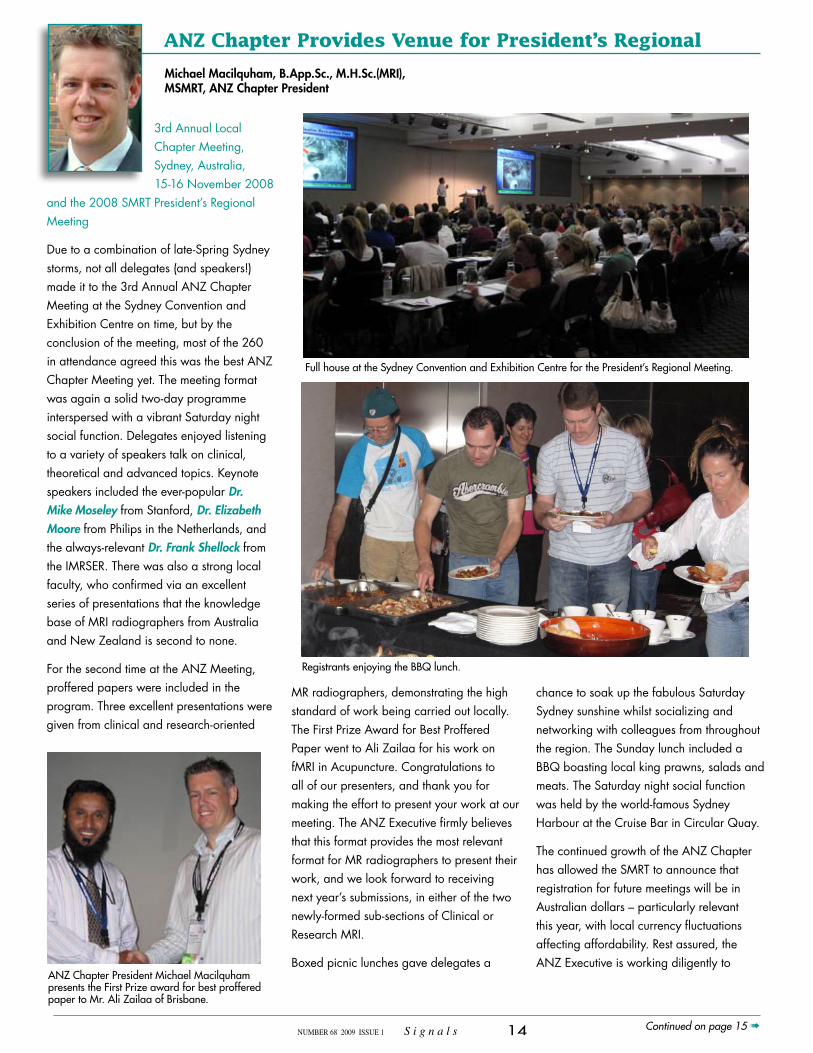
Full house at the Sydney Convention and Exhibition Centre for the President’s Regional Meeting.
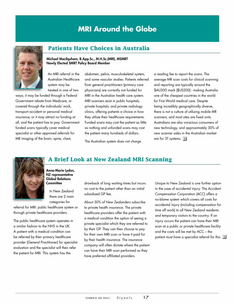
Registrants enjoying the BBQ lunch.
ANZ Chapter President Michael Macilquham presents the First Prize award for best proffered paper to Mr. Ali Zailaa of Brisbane.
Continued on page 15 ➠

Number 68 2009 Issue 1 S i g n a l s 15
ANZ Chapter Provides Venue for President’s Regional
ensure that the Annual ANZ Chapter Meeting remains the most affordable AND premier educational meeting and organisation for your ongoing MR educational needs. To this end, the ANZ Chapter also announced the formation of an MR Safety Committee, represented by experienced radiographers and industry professionals keen to ensure the safe promotion of the modality in the region. More details can be found at http://www.ismrm.org/smrt/anz.htm Planning for the Adelaide 2009 Meeting is already underway. Keep abreast of updates at the ANZ Chapter website, and if you are
interested in assisting with the Meeting, please contact your local ANZ Chapter Representative, whose details are also listed on this website.
Meetings of this magnitude do not organize themselves, so it is most appropriate to acknowledge the contributions of local Chairs Kirsten Moffat and Gloria Olivieri, as well as the ANZ SMRT Executive committee of Ben Kennedy, Glenn Cahoon, Wendy Strugnell and Michael Macilquham. There were also contributions from local MR radiographers, who helped staff the registration desk, man the door at the
function, fill delegates’ satchels, and help with local administrative tasks. Of course, the seminar would not have been possible without the generous support of our corporate sponsors, particularly Platinum sponsors Philips Medical Systems and Siemens Healthcare.
Overall, this meeting was a fantastic opportunity to catch up with colleagues from Australia, New Zealand, Asia, the UK and USA, to learn from experts in the field, and to be inspired to continue striving for excellence in our work.
SMRT President Wendy Strugnell (left) and ANZ Chapter President Michael Macilquham with local organizers and co-chairs, Gloria Olivieri and Kirsten Moffat.
The valuable local organizing committee who enabled the success of the 3rd Annual ANZ Chapter Meeting.
ANZ Chapter Meeting continued from page 14

Number 68 2009 Issue 1 S i g n a l s 16
The New England Regional Committee would like to thank the SMRT for their
support in providing another successful conference here in New England. The Seminar was held on 1 November 2008 in Boston, Massachusetts, USA at the Brigham and Women’s Hospital with over 70 attendees. The day began with registration, coffee, and plenty of delicious food, and included a variety of informative presentation, a great lunch, lots of door prizes, and a chance to chat with fellow professionals.
We really enjoyed having Cindy Hipps from Greenville, North Carolina up here in New England as our first presenter. Cindy provided her personal perspective on patient satisfaction, an insight to surveys, and some helpful methods to improve our
patient satisfaction in our own practice. This presentation was very useful, and the video clip, “It’s A Dog’s World,” seemed to be an all too realistic portrayal of the human experience in the health care system today. Hopefully with the tips Cindy gave us we can make it just a little better for our patients.
There was a mix up on speaker times so our own Vera Miller stepped up to the plate to begin a presentation on Breast
MRI. She did a fantastic job, and we all realized that it is a great idea to carry your presentations with you at all times. The speaker did arrive shortly after Vera began
her talk, and all was well. As a continuation of improving patient care, Angela Franceschi spoke to us about the “Essence of Patient Care.” Angela is a Child Life Specialist working at Children’s Hospital in Boston, MA, and she shared with us the importance of effective communication and providing a connection between the hospital and home for children and all patients. We were left with the thought that we all leave a mark on the life of each patient we care for, and Angela helped us to be able to leave a positive mark.
Our next two speakers took us to the technical side of our practice. Dr. Hiroshi from the Brigham and Women’s Hospital talked to us about MRI imaging of the knee. This presentation included a list of typical sequences used to image three structures in the knee; the menisci, the ligaments, and cartilage. Dr. Hiroshi provided some helpful hints regarding anatomic structure, the appearance of common injuries, and finished with some remedies for artifacts and pitfalls common to MR imaging of
the knee. It was interesting to see the advances being made and the new applications in this area. Dr. Panych then provided a “back to basics” MR physics
presentation. It is always good to bring ourselves back to where the images come from, and Dr. Panych did a great job of this with the help of some slide animation and descriptions. The best part was that Dr. Panych offered his slides to anyone who e-mails him and a good website for MR physics. After all that thinking, it was finally time for lunch.
After lunch we all gathered in the conference room once again, to listen to
Boston is Site for New England Regional Seminar
Maryann Blaine, R.T., (R)(MR) Organizer
Cindy Hipps
Maryanne Blaine and Angela Franceschi
Continued on page 23 ➠
Janice Fairhurst
Number 68 2009 Issue 1 S i g n a l s 17
An MRI referral in the Australian Healthcare system may be treated in one of two
ways; it may be funded through a Federal Government rebate from Medicare, or covered through the individuals’ work, transport accident or personal medical insurance; or it may attract no funding at all, and the patient has to pay. Government funded scans typically cover medical specialist or other approved referrals for MR imaging of the brain, spine, chest,
abdomen, pelvis, musculoskeletal system, and some vascular studies. Patients referred from general practitioners (primary care physicians) are currently not funded for MRI in the Australian health care system. MRI scanners exist in public hospitals, private hospitals, and private radiology clinics, offering patients a choice in how they utilize their healthcare requirements. Funded scans may cost the patient as little as nothing and unfunded scans may cost the patient many hundreds of dollars.
The Australian system does not charge
a reading fee to report the scans. The average MR scan costs for clinical scanning and reporting are typically around the $AU300 mark ($US200) - making Australia one of the cheapest countries in the world for First World medical care. Despite being incredibly geographically diverse, there is not a culture of utilizing mobile MR scanners, and most sites are fixed units. Australians are also voracious consumers of new technology, and approximately 30% of new scanner sales in the Australian market are for 3T systems.
Michael Macilquham, B.App.Sc., M.H.Sc.(MRI), MSMRTNewly Elected SMRT Policy Board Member
A Brief Look at New Zealand MRI Scanning
Patients Have Choices in Australia
In New Zealand there are 2 main categories for
referral for MRI: public healthcare system or through private healthcare providers.
The public healthcare system operates in a similar fashion to the NHS in the UK. A patient with a medical condition can be referred by their primary healthcare provider (General Practitioner) for specialist evaluation and the specialist will then refer the patient for MRI. This system has the
drawback of long waiting times but incurs no cost to the patient other than an initial subsidized GP fee.
About 30% of New Zealanders subscribe to private health insurance. The private healthcare providers offer the patient with a medical condition the option of seeing a private specialist which they are referred to by their GP. They can then choose to pay for their own MRI scan or have it paid for by their health insurance. The insurance company will often dictate where the patient can have their MRI scan performed as they have preferred affiliated providers.
Unique to New Zealand is one further option in the case of accidental injury. The Accident Compensation Corporation (ACC) offers a no-blame system which covers all costs for accidental injury (including compensation for time off work) to all New Zealand residents and temporary visitors to the country. If an injury occurs the patient can have their MRI scan at a public or private healthcare facility and the costs will be met by ACC – the patient must have a specialist referral for this.
Anna-Marie Lydon,NZ representativeGlobal RelationsCommittee
MRi Around the globe
Number 68 2009 Issue 1 S i g n a l s 18
Continued on page 19 ➠
The Canadian MRI Experience
Caron Murray, M.R.T., (R)AC, (CT)(MR), SMRT Policy Board Member
The Patient ScenarioYou’re 40 years old, live in Toronto
and have begun to experience some pain radiating down your right leg to your foot. The pain starts off being a bit annoying but nothing you can’t handle. However, over several months, it occurs on a more frequency basis until you decide that you finally must see your physician and hopefully you can get this problem taken care of. What do you do? Easy… just give your family physician a call (hopefully you have one, what with the family doctor shortage and all…) and make an appointment. Depending on your physician (in reference to mine), you can get an appointment within a week for a routine appointment (but she’ll fit you in that day if it’s an emergency). Since leg pain is not an emergency, you accept your appointment for the following week and then duly arrive on time at the doctor’s office. After examining you thoroughly, your doctor believes that you could perhaps have a protruding disc in your lumbar spine, which would explain your right leg pain and therefore orders an MRI examination of your lumbar spine. After completing the MR requisition along with the pre-screening questions, your requisition is faxed to the hospital with which your doctor is affiliated. Either your doctor’s office or you personally will be advised of when your MRI appointment will be scheduled.
Once your requisition arrives in the hospital’s MR bookings office, it will then be handed over to one of MR Radiologists for protocolling and priority classification. The protocolling refers to which sequences the radiologist is requesting considering the diagnostic information given by your family physician. The priority classification is the wait time target for the clinical information given. There are four classifications that
were developed by clinical experts across the province of Ontario. For MRI scans, the explanation of priority classifications are as follows: P1 - the scan must be done immediately (e.g. life threatening), P2 - the scan must be done within 48 hours (e.g. deteriorating quickly), P3 - the scan must be done within 2-10 days (e.g. deteriorating, unable to work, functioning with only with assistance) and P4 - the scan must be done within 4 weeks (e.g. stable, but can work). Your MRI requisition has been classified as a P4 by the radiologist given the clinical history and you receive an appointment in a months time at 3am. You call the MRI department and ask to be put onto the cancellation list because despite the fact that MRI is a limited resource, many people do not show for their bookings…. When it’s “free,” it’s often not appreciated!
Ten healthcare Systems and five founding PrinciplesHealth care in Canada has long been a source of national pride. Known as ‘medicare,’ the system is publicly financed but privately run, it provides universal coverage and care is free at the point of use. The system is based on five founding principles. Care must be universal, portable, comprehensive, accessible, and publicly administered.
Canada has a long history of universal health coverage. In 1944, Saskatchewan led the way, being the first of the provinces to introduce universal hospital insurance. By 1971, all Canadians were guaranteed access to essential medical services, regardless of employment, income, or health. Canada’s version of national public health insurance is characterized by local control, doctor autonomy and consumer choice – patients theoretically have a free choice of physician and hospital. The ten provincial governments are the key
providers of health care, having the constitutional responsibility for planning, financing, and evaluating the provision of hospital care, negotiating salaries of health professionals and negotiating fees for physician services. The result is that each provincial insurance plan differs slightly – mostly in how far each extends public insurance coverage beyond medically necessary hospital and physician services. Dental insurance, eye-care insurance (except for diabetics), insurance for prescription drugs, ambulance services (except for emergency transport to a hospital), medical devices, private health insurance covering the upgrading of hospital rooms and out of country insurance are all outside the scope of medicare. These services are most often covered by the purchase of an extended healthcare plan through the employer. In most hospitals in Ontario, this cost is shared
In 1944, Saskatchewan
led the way, being
the first of the
provinces to introduce
universal hospital
insurance. By 1971,
all Canadians were
guaranteed access
to essential medical
services, regardless of
employment, income,
or health
Number 68 2009 Issue 1 S i g n a l s 19
by the employee and employer and can range in cost depending on the services covered. Ballpark cost for dental premiums for family coverage would be $29 per month and for extended health coverage (chiropractic, physiotherapy, eye glasses, prescription medication, etc.) for families, $82 per month.
health care without hindrance The Canadian Health Act of 1984 defines and solidifies the principles of medicare, including: comprehensiveness (provinces must provide medically necessary hospital and physician services), universality (100 per cent of provincial residents are entitled to the plan), accessibility (there should be reasonable access to services, not impeded by user charges or extra billing), portability (protection for Canadians traveling outside of their home province), and public administration (provinces must administer and operate the health plan on a non-profit basis). These principles aim to provide a one-tiered service.
healthcare expenditure The Canadian healthcare system is funded primarily by tax dollars. The federal government makes cash transfers to the provinces, but the provinces may levy their own taxes to help defray the costs. Alberta and British Columbia require a health insurance premium, and other provinces have instituted employer payroll taxes. If you work in a hospital in Ontario, this health tax is paid for by your employer and would be amount to approximately $800 per year.
healthcare Providers Healthcare providers are predominantly private, but are funded by public monies via provincial budgets. Hospital systems are largely private non-profit organizations with their own governance structures
(usually supervised by a community board or trustees) that receive an annual global operating budget from the provinces. Physicians are mostly in private practice and remunerated on a fee-for-service basis (with an imposed cap to prevent excessive utilization and costs) by the provincial health plan.
Rationing Like other nations experiencing limitless demand, an ageing population and the costly advance of medical technology, Canada has faced pressure to control health expenditure. It has done so through explicit rationing. For example, in the case of new cancer treatment, the latest pharmaceuticals, and high-tech diagnostic tests (MRI, PET), Canadian governments simply reduce their expenses by limiting the service. In 2005 Canadians waited 12.3 weeks for an MRI scan, 5.5 weeks for a CT-scan and 3.4 weeks for an ultrasound. It can be argued that Canadian health care is inefficient in that financing (lack of direct payment) does not encourage users and providers of health care to be accountable for the economic benefits and costs of services.
Waiting times (owing to rationing by queuing) are a serious concern to Canadians. There are relatively short waits for general X-rays but waiting times for some other examinations such as routine MRI are excessive. To address this issue, the Ontario government has implemented a wait list strategy for 6 specific services: cancer surgery, cardiac procedures, ophthalmic surgery, orthopedic surgery, diagnostic scans – CT and MRI, and general surgery. For instance in MRI, the wait time data is tracked from when the scan is ordered to when it is completed. Each facility must report on their median wait time and
average wait time along with their efficiency rate. The Ontario Ministry of Health has provided additional funding to hospitals to perform additional examinations. As a condition of the funding, the hospitals are required to report wait times and efficiency for these services. If the appropriate efficiency is not achieved, funding may be withdrawn and must be paid back to the Ministry. The goal is to have every MRI department achieve 100% efficiency to better utilize their magnets instead of just having more inefficiently-run magnets. So if a certain department running 16 hours a day performs say 4500 examinations a year, the expectation is that every other hospital running 16 hours a day also performs 4500 examinations a year.
Despite poor availability in Canada of advanced medical technology, international comparison reveals pretty good healthcare outcomes – generally better than those in the USA and the UK and more akin to those associated with high spending European social insurance systems such as France and Switzerland. Life expectancy is high, cancer survival rates are good and deaths from IHD and stroke are average. So why does Canada perform relatively well? Studies have shown that a number of non-health system related factors affect health outcomes. Perhaps the high level of expenditure is important, the push towards preventative medicine plays a significant role; and perhaps the fact that Canada also benefits from lower levels of income inequality than the US and UK.
Canadian MRI Experience continued from page 18

Number 68 2009 Issue 1 S i g n a l s 20
Health Care and MRI Services in Malta
The Maltese archipelago is made up of three main islands: Malta, Gozo
and Comino. It is located in the centre of the Mediterranean Sea with Sicily 93Km to the north and African continent 288 Km to the south. The total land area is 315Km2 and the total population is approximately 400,000 giving a population density of close to 1200 persons per Km2. This is one of the highest country population densities in the world.
Malta scores high on the human development index with a life expectancy of 75 years for males and 80 years for females. The infant mortality is 6.4/1000 live births.
Coronary heart disease and stroke are the major cause of mortality and morbidity. Cancers account for 25% of deaths. Accidents are an important cause of death in those under the age of 65 years. Diabetes is a significant national health problem with a prevalence of 10% in adults over the age of 35 years old.
health care System in MaltaHealth care in Malta is provided through two systems: Statutory and private. The statutory system is funded from general taxation and is free at the point of delivery. Private health care also has a significant role in Malta.
The parliament is responsible for enacting health care legislation and for approving the health care budget. In the past the Department of Health (DoH) was responsible for the management of all the Government Health Centres and Hospitals. Now through reforms, the DoH functions as a regulatory body responsible for coordination and implementation of quality standards or other health service policies. Each public hospital has its own autonomous management committee.
A highly comprehensive package of health services is offered by the state health care system. At primary care level the services include GP consultations, home visits, minor treatment, community nursing, and preventive programmes such as immunization and screening. Within the hospital setting diagnostic and therapeutic interventions are carried out both at specialist clinics and inpatient hospital wards. Treatment abroad, especially in the UK, is also paid for by the government for those requiring highly specialized care which is not available in Malta. The state also provides visiting consultant clinics, where specialists are brought over to Malta where together with Maltese consultants provide specialized treatment.
Persons with low income are ‘means tested’ by the Department of Social Security. If they qualify for assistance they receive a card which entitles them to free pharmaceuticals. Moreover, a person who suffers from one or more of a specified list of chronic diseases (e.g. Rheumatoid arthritis, Asthma) is also entitled to receive free treatment for his/her ailment irrespective of financial means.
Although the National Health Service provides such universal and specialized health coverage, a number of private hospitals have opened during the last
decade, as profit-making organizations. Generally people tend to use the private sector when they perceive that their condition require extra attention by a particular consultant, or are not willing to wait for health care services in the public hospitals. Private hospitals are subject to licensing by the Ministry of Health, which regulates facility and care standards. Around 8% of all hospital beds are in the private sector.
The method of payment for all private health care services varies from direct out of pocket or through a private health insurance. Insured patients may either pay first and then claim reimbursement or pay through direct settlement. In this case, the hospital checks with the insurance whether the patient is covered for diagnostic or therapeutic service. It is not known precisely how many people take out private voluntary insurance, but the trend is increasing and is expected to continue to gain in popularity.
MRi services in MaltaIn Malta there are 3 MRIs. There is a 1.5T (GE Signa Twin Speed) in the Public General Hospital – Mater Dei, a 1.5T (Philips Intera) at St. James Hospital and an open 0.35T (Somatom Siemens) St. Philip Hospital. These
Joseph Castillo, B.Sc., M.Sc., Senior principal radiographer at Mater Dei Hospital, Malta and SMRT Policy Board Member
The Maltese archipelago
1.5T Magnet at Mater Dei Hospital
Continued on page 21 ➠

Number 68 2009 Issue 1 S i g n a l s 21
last two hospitals are private. Whereas there is no waiting list in the private health sector, in the public sector there is now a waiting list of almost seven months for elective cases. In the National Health Services patients are referred for an MRI by their consultant after being seen at the respective outpatient clinics in Malta and Gozo. All referrals are vetted by Consultant Radiologists for justification and priority. The MRI unit at the General Hospital is a radiographer led unit and all decisions during scanning regarding
protocol optimization and administration of contrast is taken by radiographers.
In the private sector, patients are referred by any doctor from GPs to Surgeons, and are scanned on demand as there is no waiting list. From the number of scanners on the Island to the amount of MR exams (6700 per year) in the public hospital, one could reason out that the number of MR scans done in private hospitals is low. The general impression is that patients are not ready to use insurance for a diagnostic test
and mainly prefer to use their insurance coverage for the eventuality of elective surgery, hospital care and medical treatment overseas.
Mater Dei Hospital – the new public hospital in Malta
NEW Member Benefit: Electronic Home Studies
The SMRT introduces a new way for its members to earn Category A CE Credits fast! Earn your Category A Continuing Education Credits on-line at http://www.ismrm.org/smrt/ehs.
The SMRT now has electronic versions of their Educational Seminars home studies available at the SMRT website. These electronic home studies will be available ONLY on-line and ONLY to SMRT mem-bers. At the SMRT website you can read the articles and take the quiz on-line!
These electronic home studies are being made available in addition to the quarterly issues that are mailed to SMRT members.
The “Use of Contrast Agents in MR Imaging of the Spine” is the second SMRT electronic home study available now on-line. Simply read the article, answer the short quiz and earn 1.0 (one) Category A CE credit.
All future SMRT Educational Seminars Home Studies will now have the quiz and answer form on line at http://www.ismrm.org/smrt/ehs. Pending reprint permissions, we plan to post the actual article(s) from the home study on-line as well. The quiz and answer forms are now on-line for the SMRT Educational Seminars Home Studies, Volume 12, Number 1. Read the home study, take the quiz, and earn Category A CE credits – Simple!! Your certificate of completion will be e-mailed to you.
Home Study Quiz On-l ine for Future Hardcopy Issues
Paul McElvogue, R.T., (R)(MR)SMRT Publications Chair
Anne Marie Sawyer, B.S., R.T., (R)(MR)Educational Seminars Editor
Electronic Versions of New Home Studies
Section for Magnetic Resonance Technologists
Healthcare and MRI Services in Malta continued from page 20
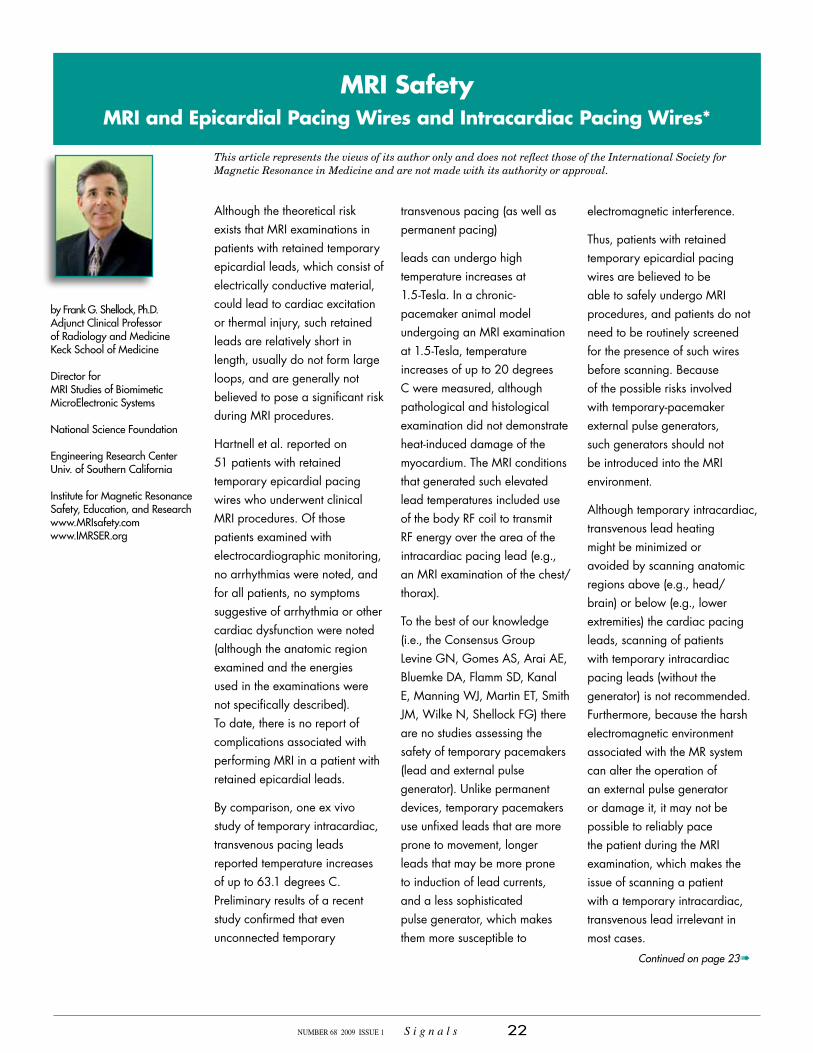
Number 68 2009 Issue 1 S i g n a l s 22
Although the theoretical risk exists that MRI examinations in patients with retained temporary epicardial leads, which consist of electrically conductive material, could lead to cardiac excitation or thermal injury, such retained leads are relatively short in length, usually do not form large loops, and are generally not believed to pose a significant risk during MRI procedures.
Hartnell et al. reported on 51 patients with retained temporary epicardial pacing wires who underwent clinical MRI procedures. Of those patients examined with electrocardiographic monitoring, no arrhythmias were noted, and for all patients, no symptoms suggestive of arrhythmia or other cardiac dysfunction were noted (although the anatomic region examined and the energies used in the examinations were not specifically described). To date, there is no report of complications associated with performing MRI in a patient with retained epicardial leads.
By comparison, one ex vivo study of temporary intracardiac, transvenous pacing leads reported temperature increases of up to 63.1 degrees C. Preliminary results of a recent study confirmed that even unconnected temporary
transvenous pacing (as well as permanent pacing)
leads can undergo high temperature increases at 1.5-Tesla. In a chronic-pacemaker animal model undergoing an MRI examination at 1.5-Tesla, temperature increases of up to 20 degrees C were measured, although pathological and histological examination did not demonstrate heat-induced damage of the myocardium. The MRI conditions that generated such elevated lead temperatures included use of the body RF coil to transmit RF energy over the area of the intracardiac pacing lead (e.g., an MRI examination of the chest/thorax).
To the best of our knowledge (i.e., the Consensus Group Levine GN, Gomes AS, Arai AE, Bluemke DA, Flamm SD, Kanal E, Manning WJ, Martin ET, Smith JM, Wilke N, Shellock FG) there are no studies assessing the safety of temporary pacemakers (lead and external pulse generator). Unlike permanent devices, temporary pacemakers use unfixed leads that are more prone to movement, longer leads that may be more prone to induction of lead currents, and a less sophisticated pulse generator, which makes them more susceptible to
electromagnetic interference.
Thus, patients with retained temporary epicardial pacing wires are believed to be able to safely undergo MRI procedures, and patients do not need to be routinely screened for the presence of such wires before scanning. Because of the possible risks involved with temporary-pacemaker external pulse generators, such generators should not be introduced into the MRI environment.
Although temporary intracardiac, transvenous lead heating might be minimized or avoided by scanning anatomic regions above (e.g., head/brain) or below (e.g., lower extremities) the cardiac pacing leads, scanning of patients with temporary intracardiac pacing leads (without the generator) is not recommended. Furthermore, because the harsh electromagnetic environment associated with the MR system can alter the operation of an external pulse generator or damage it, it may not be possible to reliably pace the patient during the MRI examination, which makes the issue of scanning a patient with a temporary intracardiac, transvenous lead irrelevant in most cases.
MRi SafetyMRi and epicardial Pacing wires and intracardiac Pacing wires*
by Frank G. Shellock, Ph.D.Adjunct Clinical Professorof Radiology and MedicineKeck School of Medicine
Director forMRI Studies of Biomimetic MicroElectronic Systems
National Science Foundation
Engineering Research CenterUniv. of Southern California
Institute for Magnetic Resonance Safety, Education, and Researchwww.MRIsafety.comwww.IMRSER.org
This article represents the views of its author only and does not reflect those of the International Society for Magnetic Resonance in Medicine and are not made with its authority or approval.
Continued on page 23➠

Number 68 2009 Issue 1 S i g n a l s 23
[Excerpted with permission from Levine GN, Gomes AS, Arai AE, Bluemke DA, Flamm SD, Kanal E, Manning WJ, Martin ET, Smith JM, Wilke N, Shellock FG. Safety of magnetic resonance imaging in patients with cardiovascular devices: an American Heart Association scientific statement from the Committee on Diagnostic and Interventional Cardiac Catheterization. Circulation 2007;116:2878-2891.]
REFERENCES
Achenbach S, Moshage W, Diem B, Bieberle T, Schibgilla V, Bachmann K. Effects of magnetic resonance imaging on cardiac pacemakers and electrodes. Am Heart J 1997;134:467-473.
Dempsey MF, Condon B, Hadley DM. Investigation of the factors responsible for burns during MRI. J Magn Reson Imaging. 2001;13:627–631.
Hartnell GG, Spence L, Hughes LA, Cohen MC, Saouaf R, Buff B. Safety of MR imaging in patients who have retained metallic materials after cardiac surgery. AJR Am J Roentgenol. 1997;168:1157–1159.
Levine GN, Gomes AS, Arai AE, Bluemke DA, Flamm SD, Kanal E, Manning WJ, Martin ET, Smith JM, Wilke N, Shellock FG. Safety of magnetic resonance imaging in patients with cardiovascular devices: an American Heart Association scientific statement from the Committee on Diagnostic and Interventional Cardiac Catheterization. Circulation 2007;116:2878-2891.
Luechinger R, Zeijlemaker VA, Pedersen EM, Mortensen P, Falk E, Duru F, Candinas R, Boesiger P. In vivo heating of pacemaker leads during magnetic resonance imaging. Eur Heart J. 2005;26:376-383.
Shellock FG, Valencerina S, Fischer L. MRI-related heating of pacemaker at 1.5- and 3-Tesla: Evaluation with and without pulse generator attached to leads. Circulation 2005;112;Supplement II:561.
Dr. Kim Lindsey from McLean Hospital Brain Imaging Center speak about her interesting use of fMRI. Dr. Lindsey explained how she studies the pharmacologic effects of smoked tobacco through direct pharmacologic fMRI. She explained that she is actually looking at the drugs effect on the physiological process, and explained how difficult it was to devise her own mechanism for having the research subjects smoke in the scanner while being scanned. Dr. Lindsey is looking forward to many more challenging research studies, and we were so impressed by her innovative approach to research as well as her ability to hold the crowds attention immediately after lunch!
After discussing smoking and nicotine, we moved on to vascular and cardiac imaging for some information on how we might image the results of smoking on circulation and the heart. Dr. Hale Ersoy demonstrated an amazing ability to make vascular imaging simple and straightforward. She began with the basics of pulse sequences and K-space, and then demonstrated the usefulness of specific pulse sequences and K-space acceleration techniques in vascular imaging. That was followed by a comparison of contrast and non-contrast vascular imaging, with a brief discussion of common artifacts and remedies to complete this well done presentation.
The final presentation was an excellent complement to Dr. Ersoy’s presentation. Dr. Sanjeev Francis provided an up-to-date comparison of cardiac MR imaging to echocardiography and nuclear imaging. Dr. Francis began with a concise description of the various cardiac MR imaging techniques currently used, followed by some of the common clinical indicators for cardiac imaging. He told us how MR has become the “Gold Standard” for determining left ventricular volume dimensions, and how MR has advantages over other modalities due to its ability to answer multiple questions with one study with regard to valve disease, and the use of late enhancement imaging to look for scar tissue. Dr. Francis completed his presentation with a look to the future, and what might be possible with the development of better gating, 3D acquisition, new clinical trials, and advances in device safety.
From the morning coffee, to the focus on patient care, to the imaging of the knee and the physics behind it all, to the tasty lunch, to the discussion about how to see what smoking does to our brain, to how to image our vessels and our hearts, this was a fantastic day.
The New England Regional organizers would like to thank all the attendees,
speakers, and most of all our donors for their generous support of this seminar. We thank Vera Miller for taking the photographs. We are all looking forward to the next New England Regional thanks to all of you.
M.R. Students
New England Regional Seminar continued from page 16
Attendees networking.
MRI Safety continued from page 22
Number 68 2009 Issue 1 S i g n a l s 24
is published by the International Societyfor Magnetic Resonance in Medicine, andproduced quarterly for the benefit of theSMRT membership.
SIgNAlS NewSleTTeR cOMMiTTee: Julie Strandt-Peay, B.S.M., R.T., (R)(MR), EditorAnne Dorte Blankhom, M.Sc., R.T.Carolyn Bonaceto, B.S., R.T., (R)(MR)Cindy R. Comeau, B.S., R.T., (N)(MR)Janice Fairhurst, B.S., R.T., (R)(MR)Caron Murray, M.R.T., (R) AC, (CT)(MR)Anne Marie Sawyer, B.S., R.T., (R)(MR) Educational Seminars EditorFrank Shellock, Ph.D.Charles Stanley, R.T., (R)(CT)(MR)Nancy Talbot, M.R.T., (MR)(R)Pam Vincent, MPA, R.T., (R)(M)(CT)(MR)
PUBlicATiONS cOMMiTTee: Paul McElvogue, R.T., (R)(MR), ChairCarolyn Bonaceto, B.S., R.T., (R)(MR)Greg Brown, R.T.Cindy R. Comeau, B.S., R.T., (N)(MR)Ellen Condon, R.T.Cindy T. Hipps, B.H.S., R.T., (R)(MR)Bobbi Lewis, B.A., ARMRITAnne M. Sawyer, B.S., R.T., (R)(MR)Mark Spooner, B.P.S., R.T., (R)(MR)(CT)Julie Strandt-Peay, B.S.M., R.T., (R)(MR)Pamela Vincent, MPA, R.T., (R)(M)(CT)(MR)
elecTRONic SUBMiSSiONS cOMMiTTee:Pam Vincent, MPA, R.T., (R)(M)(CT)(MR), ChairCarolyn Bonaceto, B.S., R.T., (R)(MR)Cindy R. Comeau, B.S., R.T., (N)(MR) Gina Greenwood, MBA, R.T., (R)(MR)Vera Miller, B.S., R.T., (R)(MR)Steven P. Shannon, R.T., (R)(MR)Charles Stanley, R.T., (R)(CT)(MR)David Stanley, B.S., R.T., (R)(MR)
OfficeRS: President: Wendy Strugnell, BAppSc(MIT) President-elect: Pamela Vincent, MPA, R.T., (R)(M)(CT)(MR)
Past-President: Carolyn Bonaceto, B.S., R.T., (R)(MR)
Secretary: Vera Miller, B.S., R.T., (R)(MR)
Treasurer:Steven P. Shannon, R.T., (R)(MR)
ISMRM Executive Director: Roberta A. KravitzISMRM Associate Executive Director: Jennifer OlsonISMRM Publications Director: Mary Keydash© 2007 by International Society for Magnetic Resonance in MedicinePhone: +1 510 841 1899 Fax: +1 510 841 2340 E-mail: [email protected] Web Page: http://www.ismrm.org/smrt
UPCOMING Calendar of Eventsvisit http://www.ismrm.org/smrt for current calendar and activities
SMRT Rocky Mountain ChapterEducational Seminar Denver, Colorado, USA
SMRT South Carolina ChapterRegistry Review Seminar Greenville, South Carolina
2nd Regional BeNeLux SMRT Chapter Symposium: MR Cardio Amsterdam, The Netherlands
28 February
28 March
31 March
Announcing the eighteenth SMRT Annual Meeting(in conjunction with the iSMRM Seventeenth Scientific Meeting
& exhibition) 18-19 April 2009, honolulu, hawai’i, USA
Visit: www.ismrm.org/smrt for more information
HonoluluJoin the SMRT In
ISMRM Premier Accredited Scientific Workshopsand Body MRI Clinical Courses
ISMRM ThirdInternational Workshop onParallel MRI
23-26 October 2009
Santa Cruz, California, USAWorkshop
Los Angeles, California, USACase-Based Educational Program
12-13 September 2009Body MRI Weekend
Miami, Florida, USACase-Based Educational Program
6-7 February 2010Body MRI Weekend
6-7 June 2009Body MRI Weekend
New York, New York, USACase-Based Educational Program
Top Related