


Languages
Pages
Legal


5/23/2015
1
Small Glandular ProliferativeLesions of the Breast
Yunn-Yi Chen, MD, PhDProfessor
Director of Immunohistochemistry LaboratoryDirector of Breast Pathology Services
UCSF
Small Glandular Lesions of Breast
Complex sclerosing lesion Benign lobules in fat Micro glandular adenosis
Sclerosing adenosis
Radial scar
Tubular carcinoma
Invasive ductal ca
Biopsy-related changes LG adenosquamous ca Adenoid cy stic ca, tubular
� Distribution--� Lobulocentric vs diffuse pattern; organized vs hapha zard
� Stromal appearance
� Glandular architecture, cytologic features
� Luminal content
� IHC markers� Myoepithelial cell (MEC) markers: p63, SMM,
Calponin, (SMA, CK5/6)� ER� S100� Cytokeratins: CK5/6, others
Approach for Small Glandular LesionsBenign Breast Lobules in Fat: “Respect” the Fat
5/23/2015
2
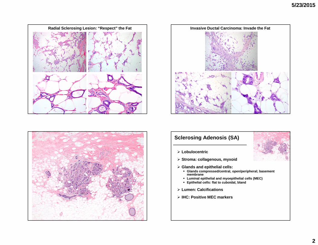
Radial Sclerosing Lesion: “Respect” the Fat Invasive Ductal Carcinoma: Invade the Fat
Sclerosing Adenosis (SA)
� Lobulocentric
� Stroma: collagenous, myxoid
� Glands and epithelial cells:� Glands compressed/central, open/peripheral; basemen t
membrane� Luminal epithelial and myoepithelial cells (MEC)� Epithelial cells: flat to cuboidal, bland
� Lumen: Calcifications
� IHC: Positive MEC markers

5/23/2015
3
Sclerosing Adenosis
� Incidental or mammographic calcifications
� Mimic invasion
� Nodular adenosis
� Involved by lobular neoplasia or DCIS
� Apocrine cytology
� Perineural invasion
Nodular Adenosis
� Florid sclerosing adenosis, nodular contour
� Mammographic mass or palpable lesion
� Also “adenosis tumor” (connotation of neoplasm)

5/23/2015
4
Biopsy for Mammographic Mass with Calcifications
Nodular adenosis
Sclerosing Adenosis and Nodular Adenosisp63
CK5/6SMM
Lobular Neoplasia Involving SA
� Mimic invasive carcinoma
� Lobulocentric
� MEC markers
Apocrine Adenosis

5/23/2015
5
Apocrine Adenosis Apocrine Adenosis
� SA with apocrine cytology� Eosinophic granular or foamy cytoplasm
� Mimic carcinoma� Lobulocentric, MEC markers
� Atypical apocrine adenosis
Invasive Apocrine CA Mimicking Apocrine Adenosis
SMM
Atypical Apocrine Adenosis--3x nuclear enlargement with prominent pleomorphic n ucleoli
(O’Malley FP and Bane AL. Adv Anat Pathol 2004)

5/23/2015
6
Apocrine Adenosis
� SA with apocrine cytology� Eosinophic granular or foamy cytoplasm
� Mimic carcinoma
� Atypical apocrine adenosis� 3x nuclear enlargement, prominent pleomorphic nucle oli� Long-term breast cancer risk: not well-defined� On CNB: recommend excision to exclude DCIS� On excision: regular follow-up
(Carter D et al: Mod Pathol 1991; Seidman J et al: Cancer 1996; Fuehrer N et al: Arch Pathol Lab Med 2 012)
DCIS with Apocrine Features
p63
Sclerosing Adenosis with Perineural “Invasion”
CNB for a Palpable Lesion

5/23/2015
7
p63SMM
Sclerosing Lesion with Perineural “Invasion”
Peri- and Intraneural “Invasion” in Benign Breast Lesions
� Ackerman: 1 st description in 1957
� Taylor and Norris (AFIP): series of 20 patients in 1967
� Incidence: ~2%
� Also reported in benign lesions of other anatomic sites
� In breast: SA, radial scar, sclerosing papilloma
� Pathogenesis unclear : post-traumatic, involvement by the proliferative process
Radial Scar
5/23/2015
8
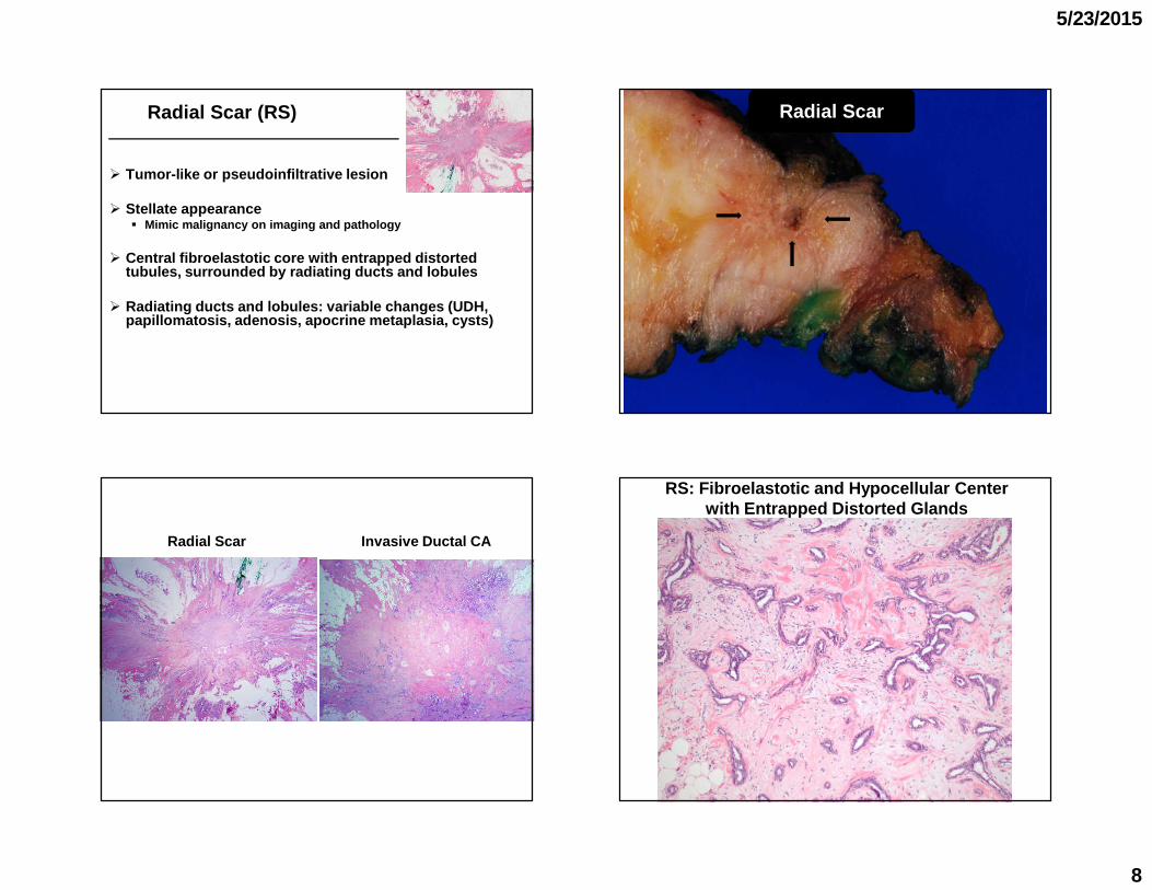
Radial Scar (RS)
� Tumor-like or pseudoinfiltrative lesion
� Stellate appearance� Mimic malignancy on imaging and pathology
� Central fibroelastotic core with entrapped distorted tubules, surrounded by radiating ducts and lobules
� Radiating ducts and lobules: variable changes (UDH, papillomatosis, adenosis, apocrine metaplasia, cyst s)
Radial Scar
Radial Scar Invasive Ductal CA
RS: Fibroelastotic and Hypocellular Centerwith Entrapped Distorted Glands
5/23/2015
9
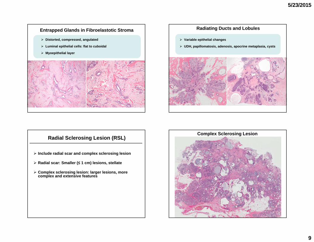
Entrapped Glands in Fibroelastotic Stroma
� Distorted, compressed, angulated
� Luminal epithelial cells: flat to cuboidal
� Myoepithelial layer
Radiating Ducts and Lobules
� Variable epithelial changes
� UDH, papillomatosis, adenosis, apocrine metaplasia, cysts
Radial Sclerosing Lesion (RSL)
� Include radial scar and complex sclerosing lesion
� Radial scar: Smaller ( ≤ 1 cm) lesions, stellate
� Complex sclerosing lesion: larger lesions, more complex and extensive features
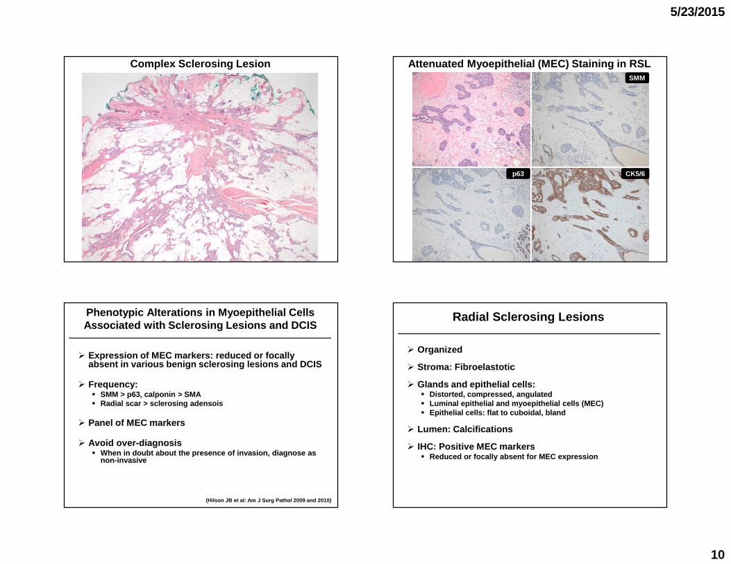
Complex Sclerosing Lesion
5/23/2015
10
Complex Sclerosing Lesion Attenuated Myoepithelial (MEC) Staining in RSLSMM
CK5/6p63
Phenotypic Alterations in Myoepithelial Cells Associated with Sclerosing Lesions and DCIS
� Expression of MEC markers: reduced or focally absent in various benign sclerosing lesions and DCIS
� Frequency: � SMM > p63, calponin > SMA� Radial scar > sclerosing adensois
� Panel of MEC markers
� Avoid over-diagnosis� When in doubt about the presence of invasion, diagn ose as
non-invasive
(Hilson JB et al: Am J Surg Pathol 2009 and 2010)
Radial Sclerosing Lesions
� Organized
� Stroma: Fibroelastotic
� Glands and epithelial cells:� Distorted, compressed, angulated� Luminal epithelial and myoepithelial cells (MEC)� Epithelial cells: flat to cuboidal, bland
� Lumen: Calcifications
� IHC: Positive MEC markers� Reduced or focally absent for MEC expression

5/23/2015
11
Tubular Carcinoma--Diffuse/infiltrative growth
Tubular Carcinoma--Desmoplastic or elastotic stroma
Tubular Carcinoma with FEA

5/23/2015
12
� Infiltrative
� Desmoplastic cellular stroma, ± elastosis
� Open round, oval, or angulated tubules
� Cytology� Single layer, non-stratified, cuboidal to columnar cells,
prominent cytoplasmic apical snouts� Minimal pleomorphism, basally located round to oval nuclei� Mitosis rare
� Lack all MEC markers
� Diffusely and strongly positive for ER
Tubular Carcinoma
� > 90% with tubular morphology
� Incompatible features--� Complex architecture� Multiple layers of cells� Significant nuclear pleomorphism� Frequent mitoses
Diagnosing Tubular Carcinoma
� 10-year survival: ~100%
� LN metastasis: rare, 1 node, no significant impact on survival
� Luminal A� ER/PR +, HER2 -, low Ki-67
Tubular Carcinoma-- PrognosisDiagnosis of TC on CNB?--
� IDC may have focal tubularmorphology
� Dx: Invasive ductal ca with tubular features with a comment
5/23/2015
13
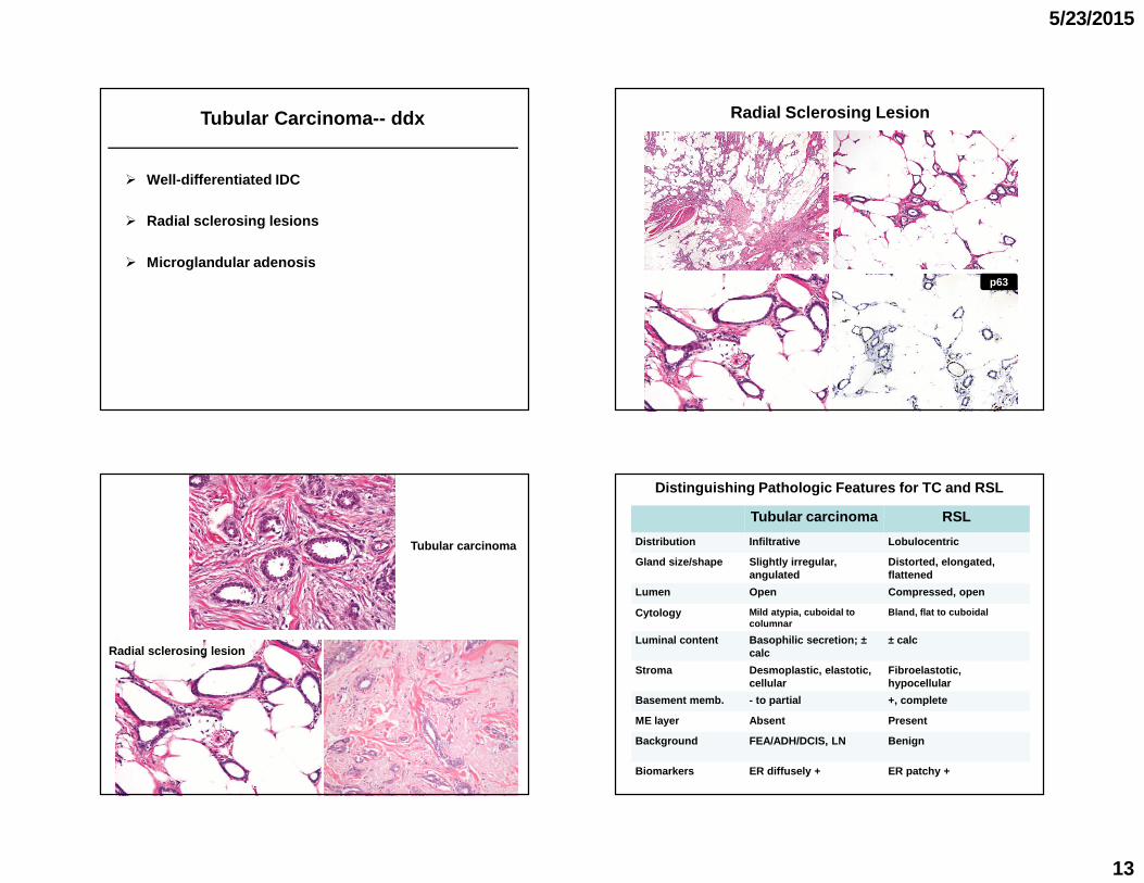
� Well-differentiated IDC
� Radial sclerosing lesions
� Microglandular adenosis
Tubular Carcinoma-- ddx Radial Sclerosing Lesion
p63
Tubular carcinoma
Radial sclerosing lesion
Distinguishing Pathologic Features for TC and RSL
Tubular carcinoma RSL
Distribution Infiltrative Lobulocentric
Gland size/shape Slightly irregular, angulated
Distorted, elongated, flattened
Lumen Open Compressed, open
Cytology Mild atypia, cuboidal to columnar
Bland, flat to cuboidal
Luminal content Basophilic secretion; ±calc
± calc
Stroma Desmoplastic, elastotic, cellular
Fibroelastotic,hypocellular
Basement memb. - to partial +, complete
ME layer Absent Present
Background FEA/ADH/DCIS, LN Benign
Biomarkers ER diffusely + ER patchy +

5/23/2015
14
Well-differentiated IDC ? Well-differentiated IDC ?
Well-diff IDC Tubular carcinoma
Irregular glands Slightly irregular, angulated glands
± Trabeculae and ribbons Open glands
Branching and anastomosis No branching or anastomosi s (may have cribriform glands)
Mild to moderate pleomorphism Minimal pleomorphism
Stratified cells, loss of polarity Single layer of c ells, basal nuclei
Sclerotic to desmoplastic stroma Desmoplastic/elasto tic stroma
� Randomly distributed
� Hypocellular dense collagenous stroma or fat
� Uniform small round glands, eosinophilic secretion
� Cytology� Single layer, flat to cuboidal cells, clear to amph ophilic
cytoplasm, bland round nuclei
� Immunophenotype� MEC markers (p63, SMM, calponin, SMA) –; S100 diffu sely +� ER -� Laminin and type IV collagen +
Microglandular Adenosis (MGA)

5/23/2015
15
Microglandular Adenosis--
Haphazard distribution
Microglandular Adenosis--Hypocellular collagenous stroma
PAS stain
Microglandular Adenosis--Uniform small glands, open lumen, eosinophilic secr etion
Microglandular AdenosisSMMcalponin
Lamininp63

5/23/2015
16
Microglandular Adenosis
S100ER
Distinguishing Pathologic Features for MGA and TC
MGA Tubular carcinoma
Distribution Random Infiltrative
Gland size/shape Uniform, small, round Slightly irregul ar, angulated
Lumen Open Open
Cytology Bland, flat to cuboidal Mild atypia, cuboidal to columnar
Luminal content Eosinophilic secretion Basophilic sec retion; ± calc
Stroma Collagenous to fatty Desmoplastic, elastotic, cellular
Basement memb. +, complete - to partial
ME layer Absent Absent
Background Benign FEA/ADH/DCIS, LN
Biomarkers ER -, S100 + ER diffusely +
(Courtesy of Dr. Timothy Jacobs)
CNB for a Palpable Mass
Microglandular Adenosis
Follow-up Lumpectomy
Regular MGA
Atypical MGA
Metaplastic ca

5/23/2015
17
Regular MGA Atypical MGA Atypical MGA Metaplastic Carcinoma
Metaplastic CA Arising in MGA and Atypical MGA--
Atypical MGA Metaplastic CAMGA
S100 Stain � Presentation: mass, mammographic calcifications or an incidental microscopic finding
� Spectrum of MGA, atypical MGA, invasive carcinoma� Share immunophenotype and genetic alterations
� Non-obligate precursor for triple negative carcinom a� IDC, metaplastic ca (chondroid diff), adenoid cysti c ca
� Management� CNB: excision� Excision: negative margin, careful clinical follow -up
Microglandular Adenosis
(Wen YH et al: Histol Histopathol 2013; Shin SJ et al: AJSP 2009; Khlifeh IM et al. AJSP 2008)

5/23/2015
18
Low-grade Adenosquamous Carcinoma (LGASC)
� Infiltrative (may resemble RS in some cases)
� Spindle cellular stroma, prominent lymphoid reactio n
� Glands (long, irregular) and solid squamous nests (comma shaped extension), ± squamous cysts
� Cytology: bland� Glands: some with epithelial and myoepithelial cells ; variable
squamous diff.� Solid nests: squamous cells
Low-grade Adenosquamous CA (LGASC)
LGASC-- Infiltrative Growth ? LGASC– Cellular Stroma and Prominent Lymphoid Reacti on

5/23/2015
19
LGASC-- Glands and Squamous Nests ? LGASC-- Squmous Cysts ?

5/23/2015
20
LGASC-- Squamous Cyst/Nests with Comma-like Extensio n? LGASC-- Infiltrating Between and Into Lobules
� MEC markers (p63, SMM, calponin, SMA): consistently variable pattern� Continuous, discontinuous or absent staining around
glands/epithelial nests in the same lesion� No tumor shows complete absence of staining by any of the
MEC markers
� Squamous cells: p63 and CK5/6 +
� ER/PR/HER2 negative
LGASC-- Immunophenotype
(Kawaguchi and Shin: AJSP 2012; Boecher W et al: Hi stopathology 2014)
LGASC– Immunophenotype ?p63
CalponinSMM

5/23/2015
21
p63 SMM
LGASC-- Immunophenotype ?
CNB for a Palpable Mass ?
Squamous Nests into LobulesSolid Tubules and NestsCellular Stroma
Prominent Perineural Invasion, Squamous Atypia
5/23/2015
22
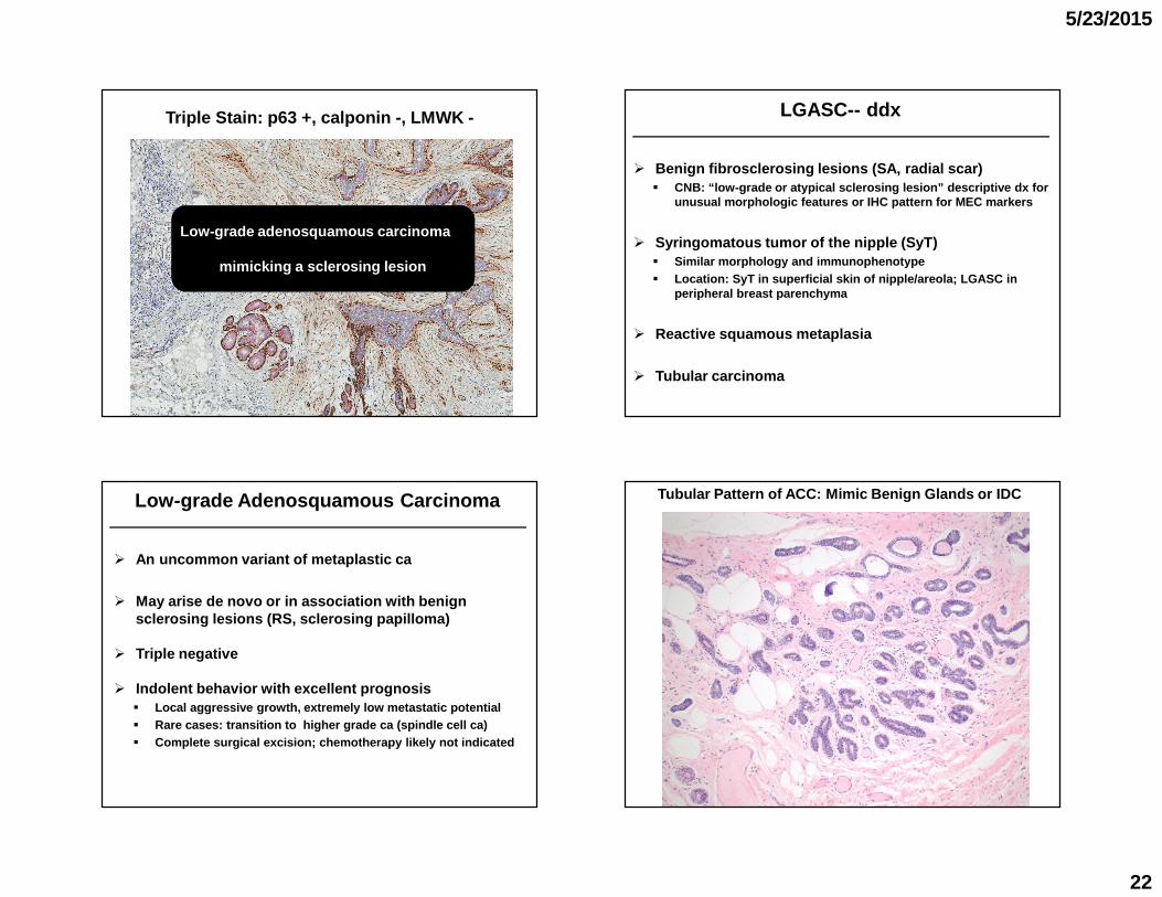
Triple Stain: p63 +, calponin -, LMWK -
Low-grade adenosquamous carcinoma
mimicking a sclerosing lesion
� Benign fibrosclerosing lesions (SA, radial scar)� CNB: “low-grade or atypical sclerosing lesion” descr iptive dx for
unusual morphologic features or IHC pattern for MEC markers
� Syringomatous tumor of the nipple (SyT)� Similar morphology and immunophenotype� Location: SyT in superficial skin of nipple/areola; LGASC in
peripheral breast parenchyma
� Reactive squamous metaplasia
� Tubular carcinoma
LGASC-- ddx
� An uncommon variant of metaplastic ca
� May arise de novo or in association with benign sclerosing lesions (RS, sclerosing papilloma)
� Triple negative
� Indolent behavior with excellent prognosis� Local aggressive growth, extremely low metastatic p otential� Rare cases: transition to higher grade ca (spindle cell ca)� Complete surgical excision; chemotherapy likely not indicated
Low-grade Adenosquamous Carcinoma Tubular Pattern of ACC: Mimic Benign Glands or IDC

5/23/2015
23
Adenoid Cystic Carcinoma (ACC)
Cribriform Pattern Tubular Pattern
Tubular ACC-- Biphasic Epi-Myoepithelial Diff.
ACC– Aberrant MEC Expression and Negative ERp63
ERCalponin
� Diffuse pattern
� Dual cell types--� Myoepithelial-like/basaloid cells� Epithelial cells
� Immunophenotype
� MEC markers: p63/SMA + & SMM/calponin - in basaloid cells
� LMW CK (CK7) + in epithelial cells
� ER/PR/HER2 -
Adenoid Cystic Carcinoma
(Rabban Mod Pathol 2006;19:1351; Foschini Semin Diagn Pathol 2010;27:77)

5/23/2015
24
� Problem with IHC stains
� Special types of breast tumors� Microglandular adenosis� Low-grade adenosquamous carcinoma� Adenoid cystic carcinoma
� Metastatic carcinoma
When a Low-grade “Infiltrative” Epithelial Lesion is ER Negative--
� Morphologic alterations secondary to procedures� Prior needling (CNB, FNA)� Current procedure (injection for SLN, tissue proces sing)
� Various changes� Mimic stromal invasion� Mimic LVI� Mimic LN metastasis
� Factors� Time interval� Lesion type: papillary lesion
� MEC markers often not helpful
Iatrogenic Small Glandular Lesions
(Phelan S et al: J Clin Pathol 2007)
Mechanical Displacement of DCIS Cells--
Bx tracts
Mechanical Displacement of DCIS Cells--Tumor cells associated with biopsy site changes

5/23/2015
25
Mechanical Displacement of DCIS Cells--Tumor cells associated with biopsy site changes
Epithelial Displacement s/p FNA--Epithelial cells in stoma and vascular space
Epithelial Displacement s/p CNB Re-excision for Extensive HG DCIS with + Margin

5/23/2015
26
SMM
Squamous Metaplasia at Biopsy SiteSquamous Metaplasia at Biopsy Site
SMM p63
Squamous Metaplasia at Biopsy Site Squamous Metaplasia at Biopsy Site
Prior DCIS Current Epithelial Lesion

5/23/2015
27
CK5/6
Re-excision for DCIS
Squamous Metaplasiaat the Biopsy Site
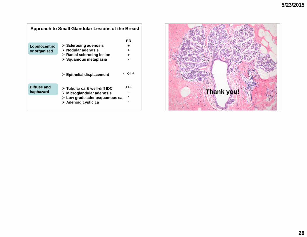
Lobulocentric or organized
Diffuse and haphazard
� Sclerosing adenosis� Nodular adenosis� Radial sclerosing lesion� Squamous metaplasia
� Epithelial displacement
� Tubular ca & well-diff IDC� Microglandular adenosis� Low grade adenosquamous ca� Adenoid cystic ca
Approach to Small Glandular Lesions of the Breast
� Benign sclerosing lesions may exhibit reduced or absent expression in one 1 or more MEC markers
� MGA lacks expression of multiple MEC markers
� Some invasive carcinomas (LGASC, ACC) express 1 or more MEC markers
Myoepithelial cell (MEC) markers
� It cannot necessarily be concluded that lack of one MEC marker indicates invasion or that expression of one MEC marker supports a benign lesion.
� A panel of MEC markers should be used.
Lobulocentric or organized
Diffuse and haphazard
� Sclerosing adenosis� Nodular adenosis� Radial sclerosing lesion� Squamous metaplasia
� Epithelial displacement
� Tubular ca & well-diff IDC� Microglandular adenosis� Low grade adenosquamous ca� Adenoid cystic ca
ME markers+++
+ & -
- or +
--
+ & -+ & -
Approach to Small Glandular Lesions of the Breast
5/23/2015
28
Lobulocentric or organized
Diffuse and haphazard
� Sclerosing adenosis� Nodular adenosis� Radial sclerosing lesion� Squamous metaplasia
� Epithelial displacement
� Tubular ca & well-diff IDC� Microglandular adenosis� Low grade adenosquamous ca� Adenoid cystic ca
ER+++-
- or +
+++---
Approach to Small Glandular Lesions of the Breast
Thank you!
Top Related