


Languages
Pages
Legal


2
dR = separated by a distance d, and one mirror is a
partially refractive mirror 2M [13]. We consider the
middle of the resonator in the point 022
=+−=dd
z .
Fig. 1 A schematic of the image capture system
After certain calculus [12], the modes in the middle
of the resonator can be express as
( )
+−==
00
20
22
0
22
exp)0,,(
w
yH
w
xH
w
yxEzyx
nm
ψ
(1)
where 0w is the waist of the beam, mH is the
Hermite Gaussian polynomials
( ) ( )22
1 x
m
mxm
m edx
dexH −−= . (2)
a b c
Fig. 2 The Hermite Gaussian intensity distribution in
transverse plane a) 00TEM , b) 11TEM , c) 12TEM
Each set (m,n) corresponds to a particular transverse
electromagnetic mode of the resonator as the electric
(and magnetic) field of the electromagnetic wave is
orthogonal in the middle of the resonator in point
0=z . The lowest-order Hermite polynomial 0H is
equal to unity; hence the mode corresponding to the
set (0,0) is called the 00TEM mode and has a
Gaussian radial profile. The laser output comprises a
small fraction of the energy in the resonator that is
coupled out through a partially refractive mirror.
The width of the Gaussian beam is a monotonically
increasing function of propagation on direction z,
and reaches 2 times its original width at Rayleigh
range. For a circular beam, this means that the mode area is doubled at this point [12,13].
In this paper we consider that the laser generates
a pulse with a Gaussian radial profile ( 00TEM ). In
order not to spread too much its width, in the
Rayleigh range at 20mm, we focus the pulse in to a
graded index fiber using a lens.
2.2 The optical system analysis When we work with optical components, the most
important problem is that it is impossible to image a point object as a perfect point image. An optical
system is made by a set of components (surfaces)
through which the light passes. The optical sensor is analyzed in space by the PSF (point spread function) and in the spatial frequency by the MTF
(modulation transfer function), which are the most
important integrative criterions of imaging evaluation for the optical system [3,4,9,10,16]. The
PSF gives the 2D intensity distribution about the image of a point source. PSF gives the physically correct light distribution in the image plane
including the effects of aberrations and diffraction.
Errors are introduced by design (geometrical
aberrations), optical and mechanical fabrication or alignment. MTF characterize the optical system functionality in spatial frequencies. Most optical
systems are expected to perform a predetermined level of image integrity. A method to measure this
quality level is the ability of the optical system to
transfer various levels of details from the object to
the image. This performance is measured in terms of contrast or modulation, and is related to the degradation of the image of a perfect source
produced by a lens. MTF describe the image structure as a function of spatial frequency and is
specified in lines per millimeter. It is obtain by
Fourier transform in the image spatial distribution or spread function.
When an optical system process an image using
incoherent light, then the function which describe
the intensity in the image plane produced by a point in the object plane is called the impulse response
function [3,4,9,10,16]:
( ) ( )[ ]yxfHyxg ,, = (3)
H is an operator representing a linear, position (or space) invariant system. The input object intensity
pattern and the output image intensity pattern are related by a simple convolution equation:
( ) ( ) ( )( )[ ]∫ ∫+∞
∞−
−−= βαβαδβα ddyxHfyxg ,,,
WSEAS TRANSACTIONS on CIRCUITS and SYSTEMS Toadere Florin, Nikos E. Mastorakis
ISSN: 1109-2734 23 Issue 1, Volume 9, January 2010

( ) ( ) ( )∫ ∫+∞
∞−
−−= βαβαβα ddyxhfyxg ,,, (4)
( ) ( )[ ]βαδβα −−=−− yxHyxh ,, is the impulse
response of H; in optics, it is called the point spread
function (PSF) [3,4,9,10,16]. The image acquisition sensor’s PSF is a multiple
convolution of individual response from each optical
component trough which the light propagates: the
lens and the transfer function of the CMOS [14,15]
CMOSlens PSFPSFPSF ∗= . (5)
The PSF characterizes the image analyses in
space but also we can characterize the image in frequency using the OTF (optical transfer function). OTF is the normalized autocorrelation of the transfer
function and has the formula:
2
( , ) ( , )2 2 2 2( , )
( , )
P u v P u v dxdy
HP u v dxdy
α β α β
α β+ + − −
=∫∫
∫∫ (6)
The numerator represents the area of overlap of two pupil functions P (square or circle), one of which is
displaced by 2
α in direction u and by
2
βin direction
v; and the other in opposite direction by -2
α and -
2
β. The denominator represents the complete area
of the pupil function [3,4,9,16].
The change in contrast when an image passes
trough an optical system is expected to have a lot to do with the optical transfer function that specifies
the quality of the system.
The MTF (modulation transfer function) is defined as: the ratio of the contrast of the output
image to that of the input image
contrast of output imageMTF
contrast of input image= .
The OTF describe the response of the optical system
to a know input and the relation between OTF and
MTF is:
MTF OTF= . (7)
In conclusion, the modulation transfer function is
identical to the absolute value of the optical transfer
function. The net sensor’s MTF is a multiplication of individual transfer functions in a way similar to
equation 5.
CMOSlens MTFMTFMTF ⋅= (8)
In general, the contrast of any image which has
gone through an image capture system is worse that
the contrast of the input image.
The PSF afferent to the sensor’s optical part is a convolution of individual response from the lens and the optical part of the CMOS sensor [14,15].
We work with multiple convolutions, and we focus
our attention on space analysis using PSF specific to
each device from the optical sensor. The optical fiber is analyzed from the spatial resolution point of view.
2.3 The lens design Optical lens design refers to the calculation of lens construction parameters that will meet a set of performance requirements and constraints.
Construction parameters include surface profile
types and the parameters such as radius of curvature, thickness, semi diameter, glass type and
optionally tilt and decenter [4]. In our particularly
case we design a lens with power and coma x errors [10]. Before we proceed, we notice that the human eye can only distinguish aberrations up to the fourth
or fifth order. When we design the lens we have to take in consideration the: aberrations, the aberration
correction and the design consideration [18].
2.3.1. The monochromatic aberrations Aberrations are the failure of light rays emerging
from a point object to form a perfect point image after passing through an optical system. Aberrations
lead to blurring of the image, which is produced by
the image-forming optical system [4,10,18]. The wave front emerging from a real lens is complex because has error in the design, fabrication and lens
assembly. Nevertheless, well made and carefully
assembled lenses possess certain inherent aberrations. To describe the primary monochromatic
aberrations, of rotationally symmetrical optical systems, we specify the shape of the wave front emerging from the exit pupil. For each object point,
there will be a quasi-spherical wave front
converging toward the paraxial image point.
Fig. 3 The wavefront aberrations
WSEAS TRANSACTIONS on CIRCUITS and SYSTEMS Toadere Florin, Nikos E. Mastorakis
ISSN: 1109-2734 24 Issue 1, Volume 9, January 2010

The wave aberration function, W(x,y), is defined as the distance, in optical path length, from the reference sphere to the wavefront in the exit pupil
measured along the ray as a function of the
transverse coordinates (x,y) of the ray intersection
with a reference sphere centered on the ideal image point [4,10,18].
To specifies the aberrations we use the Siedel
field aberration formula:
( ) ( )
( )( )termsorderhigher
rhWrhW
hrWrWrWrW
++
+++=
θ
θθ
cos
cos,
3311
22220
3131
4040
2020
(9)
klmW are the wave aberration coefficients for the
various terms of modes, h is the height of the object
and ,r θ are the polar coordinate in the pupil plane,
2r Defocus, 4r Spherical Aberration,
( )θcos3
hr Coma,
( )θ222 cosrh Astigmatism,
22rh Field Curvature,
( )θcos3rh Distortion.
These Seidel aberrations formula represents orthogonal polynomials which have the next properties: field aberrations describe the wavefront
for a single object point as a function of pupil
coordinates (x,y) and field height h. The aberrations
are described functionally as a linear combination of polynomials. Point aberrations depend only on pupil coordinates and each polynomial term represents a
single aberration. The aberration polynomial may be extended to higher order; these are all the terms to
fourth order. Ray aberrations are described by a
polynomial one order lower than the corresponding wave aberrations [4,10,18].
The design variables are the two surface
curvatures. The defect functions [10] are power and
coma. The wavefront errors introduced are given by
power 020W and coma 131W
V
yW a
φ
λ2
0202
1= (10)
)(4
165
22131 MaBaLyW a −= φ
λ (11)
λ - the wave length,
ay - the aperture,
φ - the lens power,
V - the Abbe number,
ca ynuL −= - the Lagrange invariant,
21
21
cc
ccB
−
+= - the bending,
gn is the glass refraction indices,
m
mM
−
+=
1
1 is the magnification,
)1(
15
−
+=
gg
g
nn
na ,
g
g
n
na
126
+= .
2.3.2 Aberrations correction We have the mathematical relation that describes the optical design which implies Seidel aberrations [4,
10,18]. The defect vector f is a set of m functions i
f
that depend on a set on n variables. The function is
of the type:
ff t ⋅=2σ (12)
A is a )( mn× matrix of first derivatives:
j
iij
x
fA
∂
∂=
and s are changes in the variables from the current
design. The gradient g is a )1( ×n vector given by:
2
2
1σ∇=g (13)
its components are:
∂
∂+
∂
∂+
∂
∂=
∂
∂=
i
mm
iii
ix
ff
x
ff
x
ff
xg ...2 2
21
1
2σ
fAg t= . (14)
Method of Least Squares
)( 0 AsfAg t +=
AsAggt+= 0
AAC t=
00 =+ Csg
is a set of simultaneous linear equations known as the normal equations of least-squares. Providing that
the matrix C is not singular, these equations can always be solved, and the formal solution s may be
written:
01gCs −−=
. (15)
The basic idea of the damped least-squares is to start
with the basic equation for the least squares
condition. 0g is the gradient at the starting point and
augment the diagonal of the matrix C by the addition or factoring of a damping coefficient. Modifications
of the form pcii + for example, are called additive
damping [10]. In the case of additive damping, the equation for the damped least-squares solution
reduces to:
00 =++ Cspsg . (16)
As the damping factor p increases, the third term in
WSEAS TRANSACTIONS on CIRCUITS and SYSTEMS Toadere Florin, Nikos E. Mastorakis
ISSN: 1109-2734 25 Issue 1, Volume 9, January 2010

the equation above becomes small and the solution vector becomes parallel to the gradient vector.
0
1g
ps −= . (17)
2.3.3 Calculus example Make the lens focal length 20mm with an f/2
aperture (ay = 5mm). Let the half field angle
cu be
0.1 (5.73◦) and the wavelength be 0.55µm. Let the
glass index of refraction gn be 1.5 and we assume
the object is at infinity (M = 1). To solve this problem we must solve the equation system:
−=
−=
)(2
165
22
11
MaBaLf
f
φ
φφ
a b
Fig. 4 The lens design, a) the MTF on Cartesian
scale, b) the PSF on logarithmic scale
2.4 The graded index fiber During the radiation propagation trough the optical fiber it supports two types of dispersions: material
dispersion and modal dispersion. Happily, due to
type of fiber that we use the modal dispersion is not
important. A graded-index fiber is an optical fiber
whose core has a refractive index that decreases
with increasing radial distance from the fiber axis
[9,12,13]. The index profile is very nearly parabolic.
The advantage of the graded-index is the
considerable decrease in modal dispersion ensuring
a constant propagation velocity for all light rays
We are interested to see what happens to a pulse
that propagates trough a graded index fiber [9,12].
We use the beam propagation method and we
assume the graded index medium has a refractive
index variation of the form [9,12]:
( )[ ]yxnyxn ,1),( 0 ∆+= . (18)
0n is the intrinsic refractive index of the medium,
n(x,y) is the medium index of refraction in the
location (x,y),
( )yx,∆ is the variation of n(x,y).
In reference [12] is presented a beautiful
demonstration in which a plane wave propagates
trough a graded index fiber. After the plane wave is
substitute in the wave equation, the equation is
solved and the results are the Hermite Gaussian
polynomials. Since we have total mathematical
compatibility the only concern should be related to
propagation to the refractive index. Due to the
periodic focusing by the graded index distribution
the Gaussian pulse does not deform as it propagates
through the fiber. This means that the Gaussian
spatial confining of the light wave is preserved as
the light propagates through the fiber. So the fiber
preserves the spatial resolution of the original
Gaussian pulse. At the output of the fiber the light is
projected on the CMOS.
2.5 The CMOS MTF The sharpness of a photographic imaging system or
of a component of the system (the lens and the
optical part of CMOS) is characterized by the MTF,
also known as spatial frequency response. The
CMOS optical part is characterized by its afferent
MTF. The contrast in an image can be characterized
by the modulation [4,7,14,15]
minmax
minmax
ss
ssM
+
−= (19)
where maxs and mins are the maximum and
minimum pixel values over the image. Note that:
10 ≤≤ M .
Let the input signal to an image sensor be a 1D
sinusoidal monochromatic photon flux:
[ ])2cos(1),( 0 fxFfxF π+= (20)
for 0 Nyquistf f≤ ≤ .
The sensor modulation transfer function is defined
as:
( )( )( )fM
fMfMTF
in
out= (21)
from the definition of the input signal, 1=inM .
MTF is difficult to find analytically and is typically
determined experimentally. For the beginning we
made a 1D analysis for simplicity and at the end we
generalize the results to 2D model, which we will
use in our analyses.
By making several simplifying assumptions, the
sensor can be modeled as a 1D linear space-invariant
system with impulse response h(x) that is real,
nonnegative, and even. In this case the transfer
function:
( ) ( )[ ]xhFfH = (22)
is real and even, and the signal at x is:
WSEAS TRANSACTIONS on CIRCUITS and SYSTEMS Toadere Florin, Nikos E. Mastorakis
ISSN: 1109-2734 26 Issue 1, Volume 9, January 2010

( ) ( ) ( )xhfxFxS ∗= ,
( ) [ ] ( )xhfxFxS ∗+= )2cos(10 π
( ) ( ) ( )[ ])2cos(00 fxfHHFxS π+= (23)
therefore:
( ) ( )[ ]fHHFS += 00max
( ) ( )[ ]fHHFS −= 00min
and the sensor MTF is given by:
( )( )( )0H
fHfMTF = (24)
We consider a 1-D doubly infinite image sensor.
Fig. 5 The CMOS sensor model
L- quasi neutral region
dL - depletion depth
w- aperture length p- pixel size To model the sensor’s response as a linear space-
invariant system, we assume n+/p-sub photodiode
with very shallow junction depth, and therefore we
can neglect generation in the isolated n+ regions and only consider generation in the depletion and p-type quasi-neutral regions. We assume a uniform
depletion region (from −∞ to ∞ ) [4,7]. The monochromatic input photon flux F(x) to the pixel
current iph(x) can be represented by the linear space
invariant system (Fig. 6). iph(x) is sampled at regular intervals p to get the pixels photocurrents.
Fig. 6 The process of photogeneration and
integration
≤=
otherwise
wx
w
xr
02
1 (25)
d(x) is the (spatial) impulse response corresponding
to the conversion from photon flux to photocurrent
density, and we assume a square photodetector. The
impulse response of the system is thus given by its
Fourier transform (transfer function) [14,15]
( ) ( )
∗=
ωω
xrxdxh (26)
and its Fourier transform (transfer function) is given
by:
( ) ( ) ( )fcfDfH ωω sin2= (27)
note that:
( ) ( )λnD =0 ,
( )λn - spectral response.
By definition: the spectral response is a fraction of photon flux that contributes to photocurrents as a
function of wave length. So D(f) can be viewed as a
generalized spectral response (function of spatial
frequency as well as wavelength).
After some calculus we get D(f) as:
( )( )
( )( )
−
−
−+
−+=
f
f
L
L
LLf
f
L
f
L
LL
eeeqL
L
eLqfD
fd
d
sinh11
1
2α
α
α
α
αα
α
( ) ( ) ( )wfcwfDfH sin2= (28)
the modulation transfer functions for p
f2
1≤ is:
( )( )( )
( )wfcwD
D
H
fHfMTF
fsin
0
2
0
== (29)
0D
D f is called the diffusion MTF and ( )wfcsin is
called the geometric MTF.
Consequently, we have:
geometricdiffuCMOS MTFMTFMTF ⋅= sin (30)
But in our analyses we use 2D signals (image) so we
must generalize 1D case to 2D case. We know that
we have square aperture at each photodiode with
length w; so the analyses is made in Cartesian
coordinate and we must generalize in x-y coordinate
MTF(f) and we have:
( )( )
( )0
,,
H
ffHffMTF
yx
yx = (31)
( ) ( )( ) ( )yx
ff
yx wfcwfcwD
DffMTF
yx
sinsin, 2
0
,=
xf - spatial frequency on x direction
yf - spatial frequency on y direction
Spatial frequency (lines/mm) is defined as the rate of
repetition of a particular pattern in unit distance. It is
indispensable in quantitatively describing the
resolution power of a lens. The first level in a
CMOS image sensor is a lens which focuses the
light on each pixel photodiode.
WSEAS TRANSACTIONS on CIRCUITS and SYSTEMS Toadere Florin, Nikos E. Mastorakis
ISSN: 1109-2734 27 Issue 1, Volume 9, January 2010
a b
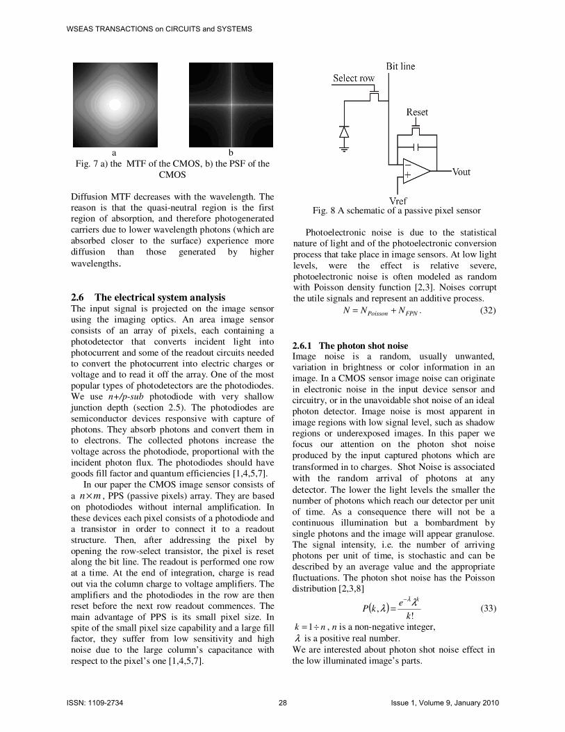
Fig. 7 a) the MTF of the CMOS, b) the PSF of the
CMOS
Diffusion MTF decreases with the wavelength. The
reason is that the quasi-neutral region is the first
region of absorption, and therefore photogenerated
carriers due to lower wavelength photons (which are
absorbed closer to the surface) experience more
diffusion than those generated by higher
wavelengths.
2.6 The electrical system analysis The input signal is projected on the image sensor using the imaging optics. An area image sensor
consists of an array of pixels, each containing a
photodetector that converts incident light into
photocurrent and some of the readout circuits needed
to convert the photocurrent into electric charges or
voltage and to read it off the array. One of the most
popular types of photodetectors are the photodiodes.
We use n+/p-sub photodiode with very shallow
junction depth (section 2.5). The photodiodes are
semiconductor devices responsive with capture of
photons. They absorb photons and convert them in
to electrons. The collected photons increase the
voltage across the photodiode, proportional with the
incident photon flux. The photodiodes should have
goods fill factor and quantum efficiencies [1,4,5,7].
In our paper the CMOS image sensor consists of
a n m× , PPS (passive pixels) array. They are based
on photodiodes without internal amplification. In
these devices each pixel consists of a photodiode and
a transistor in order to connect it to a readout
structure. Then, after addressing the pixel by
opening the row-select transistor, the pixel is reset along the bit line. The readout is performed one row
at a time. At the end of integration, charge is read
out via the column charge to voltage amplifiers. The
amplifiers and the photodiodes in the row are then
reset before the next row readout commences. The
main advantage of PPS is its small pixel size. In
spite of the small pixel size capability and a large fill
factor, they suffer from low sensitivity and high
noise due to the large column’s capacitance with
respect to the pixel’s one [1,4,5,7].
Fig. 8 A schematic of a passive pixel sensor
Photoelectronic noise is due to the statistical
nature of light and of the photoelectronic conversion
process that take place in image sensors. At low light
levels, were the effect is relative severe,
photoelectronic noise is often modeled as random with Poisson density function [2,3]. Noises corrupt
the utile signals and represent an additive process.
.FPNPoisson NNN += (32)
2.6.1 The photon shot noise Image noise is a random, usually unwanted,
variation in brightness or color information in an
image. In a CMOS sensor image noise can originate
in electronic noise in the input device sensor and
circuitry, or in the unavoidable shot noise of an ideal
photon detector. Image noise is most apparent in
image regions with low signal level, such as shadow
regions or underexposed images. In this paper we
focus our attention on the photon shot noise
produced by the input captured photons which are
transformed in to charges. Shot Noise is associated with the random arrival of photons at any
detector. The lower the light levels the smaller the
number of photons which reach our detector per unit
of time. As a consequence there will not be a
continuous illumination but a bombardment by
single photons and the image will appear granulose.
The signal intensity, i.e. the number of arriving
photons per unit of time, is stochastic and can be
described by an average value and the appropriate
fluctuations. The photon shot noise has the Poisson
distribution [2,3,8]
( )!
,k
ekP
kλλ
λ−
= (33)
nk ÷= 1 , n is a non-negative integer,
λ is a positive real number.
We are interested about photon shot noise effect in
the low illuminated image’s parts.
WSEAS TRANSACTIONS on CIRCUITS and SYSTEMS Toadere Florin, Nikos E. Mastorakis
ISSN: 1109-2734 28 Issue 1, Volume 9, January 2010

2.6.2 The fixed pattern noise In a CMOS image sensors the noise source can be divided into temporal noises and FPN (Fixed Pattern
Noise). In this paper we use only the FPN and do not
treat temporal noises. We analyze the FPN specific
to CMOS PPS (passive pixel sensor) [1,4,5,7]. In a perfect image sensor, each pixel should have the same output when the same input is applied, but in
current image sensors the output of each sensor is different. The FPN is defined as the pixel-to-pixel
output variation under uniform illumination due to
device and interconnect mismatches across the image sensor array. These variations cause two types of FPN: the offset FPN, which is independent of
pixel signal, and the gain FPN or photo response non
uniformity, which increases with signal level. Offset FPN is fixed from frame to frame but varies from
one sensor array to another. The most serious
additional source of FPN is the column FPN introduced by the column amplifiers [1,5,6]. In general PPS has FPN, because PPS has very large
operational amplifier offset at each column. Such FPN can cause visually objectionable streaks in the
image. Offset FPN caused by the readout devices
can be reduced by CDS (correlated double sampling). Each pixel output is readout twice, once right after reset and a second time at the end of the
integration. The sample after reset is then subtracted
from the one after integration. For a more detailed explanation, check out the
paper by Abbas El Gammal that is listed in the
reference section [6]. In this paper we focus our attention in FPN effects on image quality and we do not compute the FPN, we accept the noises as they
are presented in references [6].
a b Fig. 9 PPS FPN a) PPS FPN without CDS, b) PPS
FPN with CDS
2.6.3 The analog to digital conversion The analog to digital conversion is the last block of
the analog signal processing circuits in the CMOS image sensor. In order to convert the analog signal in
to digital signal we compute the: analog to digital
curve, the voltage swing and the number of bits. The quality of the converted image is good and the image seams to be unaffected by the conversion [1,4,5].
3 The image reconstruction At the output of the optical part the image is blurred as a result of its propagation trough the optical system and also present shape’s deformations due to
aberrations. In order to recover the image resolution we need to sharp the image [2,3,8], using a
Laplacian filter. At the output of the electrical part
the image is corrupted by the combined noise. In
order to reduce the FPN we use a frequencies amplitude filter to block the spikes spectrum of the
FPN, and also we use a bilateral filter in order to
reduce the photon shot noise [16,17].
3.1 The image sharpening In order to correct the blur and to preserve the
impression of depth, clarity and fine details we have
to sharp the image using a Laplacian filter [2,3,8]. A Laplace filter is a 3x3 pixel mask
−−−
−−
−−−
=
111
181
111
L (34)
In order to restore the blurred image we subtract the Laplacian image from the original image.
3.2 The amplitude filter The FPN is introduced by the sensor’s column amplifiers and consists of vertical stripes with different amplitudes and periods. Such type of noise
in the Fourier plane produces a set of spikes periodic
orientate. A procedure to remove this kind of noise is to make a transmittance mask in Fourier 2D
logarithm plane. The first step is to block the
principal components of the noise pattern. This block can be done by placing a band stop filter
H(u,v) in the location of each spike [8,12,13,16]. If
H(u,v) is constructed to block only components
associated with the noise pattern, it fallows that the Fourier transform of the pattern is given by the
relation [16]:
( ) ( ) ( )[ ]vuGvuHvuP ,log,, = (35)
where G(u,v) is Fourier transform of the corrupted
image g(x,y).
After a particular filter has been set, the
corresponding pattern in the spatial domain is obtained making the inverse Fourier transform:
p(x,y) =F{exp[P(u,v)]}. (36)
WSEAS TRANSACTIONS on CIRCUITS and SYSTEMS Toadere Florin, Nikos E. Mastorakis
ISSN: 1109-2734 29 Issue 1, Volume 9, January 2010

3.3 The bilateral filter In order to reduce the random noise effect we use a
bilateral filter. It extends the concept of Gaussian
smoothing by weighting the filter coefficients with
their corresponding relative pixel intensities. Pixels that are very different in intensity from the central
pixel are weighted less even though they may be in
close proximity to the central pixel. This is effectively a convolution with a non-linear Gaussian
filter, with weights based on pixel intensities. This is
applied as two Gaussian filters at a localized pixel neighborhood, one in the spatial domain, named the domain filter, and one in the intensity domain,
named the range filter [17].
4 The simulation results In this paper we imagine the 00TEM laser pulse
propagation trough the proposed image acquisition
system. We assume that we have a confocal
resonator which generates the Gaussian pulse. In order not to spread too much we focus the pulse (Fig. 10, a)), in to a graded index fiber using a lens
(Fig. 10, b)). Due to the fiber characteristics, the
Gaussian spatial confining of the light wave is preserved as the light propagates through the fiber.
Consequently the fiber preserves the spatial
resolution of the original Gaussian pulse. At the output of the fiber the radiation is projected on a
CMOS image acquisition sensor. The sensor has an
optical part which is characterized by its PSF; the
output image can be seen in Fig. 11 a). At the end of the optical part we use the Laplace sharpening filter
in order to correct the blur of the Gaussian pulse (Fig. 11, b)), which was produced during the radiation propagation trough the optical system. We
are interest to preserve the pulse shape during its
propagation trough the system and for our purpose a black and white analysis should be enough.
Consequently we can use a sensor that don’t Bayer
sample and interpolate the input signal and also the signal luminosity is considered to be good enough. Having those aspects set, we focus our attention to
the noises. We simulate the photon shot noise and
the FPN afferent to a CMOS PPS, and the noises combination represent an additive process (Fig. 12,
a)). Finally the analog signal is converted into digital signal. During the signal propagation through the electrical part of the CMOS sensor, its
characteristics are degraded by noises. In order to
recover the image characteristics we use an
amplitude filter and a bilateral filter (Fig. 12, b)). To better understand the simulation effect, in Fig. 13 we have a 3D spatial representation of the original
image and the recovered image. Due to the modest quality of the lens, we see that the final image (Fig. 13 b)) is degraded by the aberrations. As a
consequence of this fact the pulse is a little
attenuated in amplitude and widen at the base. The
noises can be rejected by the proposed filters’ combination.
a b
Fig. 10 a) the Gaussian pulse, b) the Gaussian pulse at the lens output
a b
Fig. 11 a) the Gaussian pulse at the output of the
CMOS optical part, b) the sharp image
a b
Fig. 12 a) the noisy image at the output of the electrical part, b) the recovered image
a b Fig. 13 a) the original pulse, b) the recovered pulse
WSEAS TRANSACTIONS on CIRCUITS and SYSTEMS Toadere Florin, Nikos E. Mastorakis
ISSN: 1109-2734 30 Issue 1, Volume 9, January 2010

Conclusions In this paper we simulate the 00TEM confocal laser
pulse propagation trough an image acquisition system. We simulated the image characteristics at
the output of each block from our system
configuration. The purpose of this paper was to put
to work together, in the same system, optical and electrical components and to recover the degraded
signal. The simulation algorithm works in real time; many other configurations can be done using other different optical and electrical components. Also we
can combine in different ways the aberrations and
noises obtaining other simulations which can be done using our proposed image capture system.
References:
[1] Bigas M., Cabruja E.,Forest J., Salvi J., “Review
of CMOS image sensors”, Microelectronics
Journal, vol. 37, 2006 [2] Bovik A. C., Handbook of Image and Video
Processing, Elseiver, 2005
[3] Castleman K., Digital image processing,
Prentice Hall, 1996 [4] www.optics.arizona.edu/detlab/
[5] El Gamal and H. Eltoukhy, “CMOS Image
Sensors” IEEE Circuits and Devices Magazine,
Vol. 21. Iss. 3, May-June 2005 [6] El Gamal A., Fowler B., Min H., Liu X.
“Modeling and Estimation of FPN Components in CMOS Image Sensor”, SPIE, vol. 3301, 1998
[7] Holst G., Lomheim, T, CMOS/CCD Sensors and
Camera Systems, SPIE, 2007
[8] Gonzales R., Wood R., Eddins S., Digital Image
Processing Using MATLAB, Gatesmark
Publising, 2009
[9] Okan E., Diffraction, Fourier Optics and
Imaging, Willey&Sons, 2007 [10]http://www.engr.udayton.edu/faculty/jloomis/co
urses.html [11] Mastorakis N., A continuous model for 2-
Dimensional (LSI) Systems, PLENARY
LECTURE in the International Colloquium
on Numerical Analysis and Applications, Plodviv, Boulgaria, August 13-17, 1998.
[12] Poon T., Banerjee P., Enginering optics with
Matlab, World Scientific, 2006 [13] Saleh B., Teich M., Fundamentals of photonics,
Wiley&Sons, 1991
[14] Toadere F., Mastorakis N., “Imaging the Optical Part of a Web Cam ”, Signal Processing,
Computational Geometry and Artificial Vision,
ISCGAV 2009, Moskow, Russia [15] Toadere F., Mastorakis N., “Imaging a laser
pulse propagation trough an image acquisition
system”, Circuits, Systems Electronics, Control
& Signal Processing CSECS 2009, Tenerife, Spain
[16] Yzuca K., Enginering Optics, Springher, 2008
[17] http://en.wikipedia.org/wiki/Bilateral_filter [18] Welford W., Aberration of optical systems,
Adam Hilger, 1986
WSEAS TRANSACTIONS on CIRCUITS and SYSTEMS Toadere Florin, Nikos E. Mastorakis
ISSN: 1109-2734 31 Issue 1, Volume 9, January 2010
Top Related