


Languages
Pages
Legal

SIALIC ACID IN LIPOPROTEINS
WITH A SPECIAL REFERENCE TOLOW DENSITY LIPOPROTEINS
NINA LINDBOHM
Department of MedicineUniversity of Helsinki
Finland
Academic dissertation
To be publicly discussed with the permission of the Medical Facultyof the University of Helsinki in the small auditorium, Haartman Institute,
on March 3rd, 2000, at 12 noon
Helsinki 2000

ISBN 951-45-9131-3 (PDF version)
Helsingin yliopiston verkkojulkaisutHelsinki 2000

to my family

Supervisors Tatu MiettinenProfessorDepartment of MedicineUniversity of HelsinkiHelsinki, Finland
and
Helena GyllingDocentDepartment of MedicineUniversity of HelsinkiHelsinki, Finland
Reviewers Jorma ViikariProfessorDepartment of MedicineUniversity of TurkuTurku, Finland
and
Matti JauhiainenDocentNational Public Health InstituteHelsinki, Finland
Opponent Jussi HuttunenDirector, ProfessorNational Public Health InstituteHelsinki, Finland
5
CONTENTS
LIST OF ORIGINAL PUBLICATIONS.............................................................. 7ABBREVIATIONS........................................................................................... 81 INTRODUCTION ..........................................................................................92 REVIEW OF THE LITERATURE ................................................................10
2.1 Overview of lipoprotein metabolism.....................................................102.2 Low density lipoproteins ......................................................................12
2.2.1 LDL particle structure....................................................................122.2.2 LDL metabolism............................................................................132.2.3 LDL subclasses ............................................................................142.2.4 LDL and atherosclerosis ...............................................................15
2.2.4.1 Arterial proteoglycans ............................................................162.2.4.2 Small dense LDL....................................................................162.2.4.3 Other risk factors of atherosclerosis.......................................18
2.3 Sialic acids...........................................................................................182.3.1 General.........................................................................................182.3.2 Serum sialic acid...........................................................................192.3.3 Sialic acid in vascular tissue .........................................................202.3.4 Sialic acid in lipoproteins...............................................................20
2.3.4.1 Chylomicrons, VLDL and IDL.................................................202.3.4.2 LDL ........................................................................................202.3.4.3 Cell culture studies.................................................................222.3.4.4 Coronary artery disease.........................................................232.3.4.5 LDL density classes ...............................................................252.3.4.6 Oxidation and electronegativity..............................................262.3.4.7 Lp(a) ......................................................................................26
2.4 Cholesterol metabolism .......................................................................272.4.1 Cholesterol absorption..................................................................272.4.2 Cholesterol synthesis....................................................................282.4.3 Cholesterol and lipoprotein metabolism........................................29
2.5 Statin treatment ...................................................................................303 AIMS OF THE STUDY................................................................................324 PATIENTS AND METHODS.......................................................................33
4.1 Patients and study designs..................................................................334.2 Methods...............................................................................................35
4.2.1 Separation of lipoproteins.............................................................354.2.2 Analyses of lipids, apolipoproteins and non-cholesterol sterols....354.2.3 Analysis of sialic acids ..................................................................364.2.4 Lipoprotein kinetic studies ............................................................374.2.5 Statistics .......................................................................................37
6
5 RESULTS...................................................................................................395.1 Type 2 diabetes mellitus (Study I)........................................................395.2 Type IIa and IIb hyperlipidemia (Study II).............................................415.3 Coronary artery disease (Study III) ......................................................445.4 Statin treatment (Study IV) ..................................................................47
6 DISCUSSION .............................................................................................516.1 General................................................................................................516.2 Clinical characteristics .........................................................................516.3 Measurement of sialic acid ratio ..........................................................526.4 Lipoproteins and sialic acids................................................................536.5 LDL sialic acid content in different conditions......................................56
6.5.1 Type 2 diabetes mellitus ...............................................................566.5.2 Type IIa and IIb hyperlipidemia .....................................................576.5.3 Coronary artery disease................................................................57
6.6 LDL sialic acid content and lipoprotein metabolism .............................596.6.1 LDL catabolism.............................................................................596.6.2 LDL production .............................................................................61
6.7 LDL sialic acid content and cholesterol metabolism ............................636.8 The effect of statin treatment on LDL sialic acid content .....................64
7 SUMMARY AND CONCLUSIONS..............................................................66ACKNOWLEDGEMENTS............................................................................. 68REFERENCES............................................................................................. 70
7
LIST OF ORIGINAL PUBLICATIONS
This thesis is based on the following original articles, which are referred to inthe text by their Roman numerals.
I Melajärvi (Lindbohm) N, Gylling H, Miettinen TA. Sialic acids and themetabolism of low density lipoprotein. J Lipid Res 1996; 37: 1625-1631
II Lindbohm N, Gylling H, Miettinen TE, Miettinen TA. Sialic acid content ofLDL and lipoprotein metabolism in combined hyperlipidemia and primarymoderate hypercholesterolemia. Clin Chim Acta 1999; 285: 69-84
III Lindbohm N, Gylling H, Miettinen TA. Sialic acid content of LDL and itsrelation to lipid concentrations and metabolism of LDL and cholesterol.Submitted.
IV Lindbohm N, Gylling H, Miettinen TE, Miettinen TA. Statin treatmentincreases the sialic acid content of LDL in hypercholesterolemic patients.Atherosclerosis. In press.
8
ABBREVIATIONS
AMI acute myocardial infarctionANOVA analysis of varianceapo apolipoproteinAsn asparagineBMI body mass indexBMDP BioMedical Data ProgramCAD coronary artery diseaseCE cholesterol esterCETP cholesterol ester transfer proteinCM chylomicronCMR chylomicron remnantd densityDM diabetes mellitusFCHL familial combined hyperlipidemiaFCR fractional catabolic rateFFA free fatty acidsFH familial hypercholesterolemiaHDL high density lipoproteinHL hepatic lipaseHMG-CoA 3-hydroxy-3-methylglutaryl coenzyme AIDL intermediate density lipoproteinLCAT lecithin:cholesterol acyltransferaseLDL low density lipoproteinLp(a) lipoprotein(a)LPL lipoprotein lipasePLTP phospholipid transfer proteinTG triglycerideTR transport rateTSA total sialic acidVLDL very low density lipoprotein
9
1 INTRODUCTION
Atherosclerosis is a major cause of morbidity and mortality all over the world,and its most common manifestation is coronary artery disease (CAD). Thepathology of atherosclerosis is due to deposition of lipids, especiallycholesterol, in the arterial wall intimal layer both intracellularly andextracellularly. Cholesterol in the atherosclerotic plaques is mainly derivedfrom circulating lipoproteins, especially low density lipoprotein (LDL), and highlevels of LDL cholesterol in blood are strongly associated with CAD (Keys1970, Martin et al 1986). Furthermore, increased atherogenicity is associatedwith small dense LDL particles (Austin et al 1988, Griffin et al 1994) andminimally modified LDL generated by various physical and chemicalmodifications (Steinberg et al 1989).
Sialic acids are aminosaccharides characteristically located in the terminalends of carbohydrate chains of glycoproteins, and their removal exposesgalactose molecules that can interact with receptors, which leads to theremoval of the proteins from the circulation (Stryer 1988). LDL has sialic acid-containg carbohydrate chains both in its protein and lipid parts(Swaminathan and Aladjem 1976). Because of the role sialic acid has in themetabolism of glycoproteins its content in LDL is an interesting factor withregard to the atherogenicity of this lipoprotein. Desialylation of LDL has beenshown to increase its uptake into cultured cells (Filipovic et al 1979, Orekhovet al 1992) and its interaction with extracellular matrix components (Camejoet al 1985) proportionately to the degree of desialylation. In addition, a lowsialic acid content has been found in LDL of CAD patients as compared withhealthy control subjects (Orekhov et al 1991b, Ruelland et al 1993), althoughall studies do not agree on this (Chappey et al 1998). Consequently,desialylation of LDL seems to take place in vivo, and thus it could be anatherogenic modification of the lipoprotein.
The level of LDL cholesterol in blood is regulated by the metabolism ofcholesterol and LDL. As the sialic acid content of LDL affects its catabolismat the cellular level, it could be assumed that the content is also associatedwith LDL metabolism in vivo. This work was conducted to investigate therelations of LDL sialic acid content with various factors connected withatherosclerosis, including the concentration, composition, and metabolism oflipids and lipoproteins, and with states where the risk of CAD is elevated,namely type 2 diabetes mellitus (DM) and type II hyperlipidemias. In addition,the effect of statin treatment on LDL sialic acid content was studied.
10
2 REVIEW OF THE LITERATURE
2.1 Overview of lipoprotein metabolism
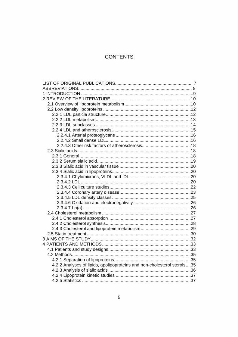
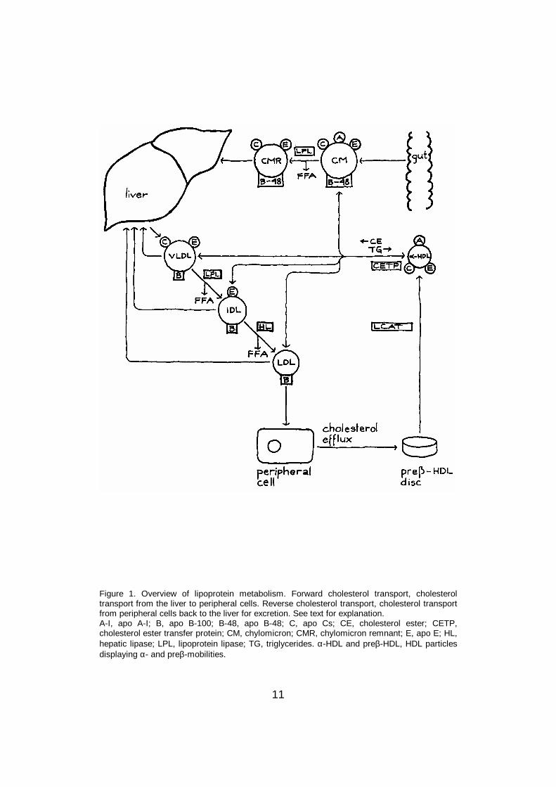
In human plasma, lipids are transported as lipoproteins which consist ofcholesterol esters and triglycerides in the hydrophobic core, and of freecholesterol, phospholipids and apolipoproteins on the hydrophilic surface.Plasma lipoproteins are separated into five major classes on the basis of theirdensity: chylomicrons (d < 0.94 g/ml), very low density lipoproteins (VLDL,d = 0.94-1.006 g/ml), intermediate density lipoproteins (IDL, d = 1.006-1.019g/ml), low density lipoproteins (LDL, d = 1.019-1.063 g/ml), and high densitylipoproteins (HDL, d = 1.063-1.210 g/ml) (Havel and Kane 1995). The mainfeatures of lipoprotein metabolism are presented in Figure 1.
Chylomicrons are very large lipoproteins that consist mainly of triglyceridesand of minor amounts of cholesterol and phospholipids. Their major protein isapolipoprotein (apo) B-48, and they also contain apolipoproteins A, C and E.Chylomicrons are formed postprandially from dietary fats in the intestinalepithelial cells, and they enter the circulation via lymph. In the blood,triglycerides are rapidly hydrolyzed into free fatty acids by lipoprotein lipase.Some cholesterol and phospholipids, and the A and C apolipoproteins aretransferred to HDL. The resulting particle, called a chylomicron remnant, istaken up by hepatic receptors.
VLDL is produced in hepatocytes, and consists of a large amount oftriglycerides and smaller amounts of cholesterol and phospholipids. Its majorprotein is apo B-100, and it contains also some E and C apolipoproteins.VLDL carries endogenously synthesized lipids from the liver. In the blood,lipoprotein lipase hydrolyzes triglycerides, and the size of the particlesdiminishes. Some of these particles, called VLDL remnants, are directlyremoved from the blood by LDL receptors on hepatocytes. VLDL surfaceremnants, especially phospholipids, are transported to HDL via the functionof phospholipid transfer protein (PLTP) (Jiang et al 1999). The remainingparticles are transformed into IDL, and, through loss of apolipoproteins E andC and further hydrolysis of triglycerides by hepatic lipase, further into LDL.
LDL consists of one single copy of apo B-100, and of large amounts ofcholesterol esters and smaller amounts of free cholesterol, triglycerides andphospholipids. LDL is the main carrier of cholesterol in blood, and it isresponsible for transporting cholesterol to peripheral cells. Apo B interacts
11
Figure 1. Overview of lipoprotein metabolism. Forward cholesterol transport, cholesteroltransport from the liver to peripheral cells. Reverse cholesterol transport, cholesterol transportfrom peripheral cells back to the liver for excretion. See text for explanation.A-I, apo A-I; B, apo B-100; B-48, apo B-48; C, apo Cs; CE, cholesterol ester; CETP,cholesterol ester transfer protein; CM, chylomicron; CMR, chylomicron remnant; E, apo E; HL,hepatic lipase; LPL, lipoprotein lipase; TG, triglycerides. α-HDL and preβ-HDL, HDL particlesdisplaying α- and preβ-mobilities.
12
with specific LDL receptors located on cell surfaces in many tissues, includingthe liver, and thus LDL particles are mainly removed from the circulation.
HDL is formed as a precursor in cells of the liver and intestine. Nascent HDLcontains mainly protein and phospholipids, but in the circulation it gainsmaterial, including phospholipids and free cholesterol via lipolysis oftriglyceride-rich particles and from cell membranes. The primaryapolipoproteins in mature HDL are apo A-I, A-II and A-IV. Free cholesterol inHDL is esterified by lecithin:cholesterol acyltransferase (LCAT), andcholesterol esters are transferred from HDL to apo B -containing lipoproteinsby cholesterol ester transfer protein (CETP) in exchange for triglycerides (Tall1986). Thus, HDL has an important role in reverse cholesterol transport: firstspecific HDL subclasses (preβ-HDL) function as primary cholesterolacceptors and are able to remove cholesterol from peripheral cells, and aftercholesterol esterification cholesterol esters are delivered from HDL to apo B-containing lipoproteins, which can be removed from the circulation byhepatic receptors.
A sixth lipoprotein class, lipoprotein(a) (Lp(a)), consists of one LDL particleassociated with one molecule of a glycoprotein called apo(a) (Utermann1989, Utermann 1995). Its density range is 1.04-1.125 g/ml, overlappingthose of LDL and HDL. Apo(a) is secreted by the liver but the site ofassembly and that of catabolism of Lp(a) are unclear. Its plasmaconcentration is genetically determined and there are large interindividualdifferences. A physiological role for Lp(a) has not been found, but apo(a) hasa close structural resemblance with plasminogen, and it has been shown tobe able to interfere with fibrinolysis (Utermann 1995).
2.2 Low density lipoproteins
2.2.1 LDL particle structure
Like all mature lipoproteins, LDL is a spherical particle. It has a density of1.019-1.063 g/ml and a diameter of 22-28 nm. It contains 35-45% ofcholesterol esters and 6-12% of triglycerides in the core, and 20-25% ofphospholipids, 6-10% of free cholesterol, and 20-25% of protein on thesurface (Deckelbaum 1987). The sole protein of LDL is apo B, and one LDLparticle always contains one molecule of apo B.
13
2.2.2 LDL metabolism
LDL is the end product of VLDL metabolism (Sigurdsson et al 1975, Reardonet al 1978, Fisher et al 1980, Thompson et al 1987, Demant et al 1996). It isproduced from small VLDL particles, larger VLDL particles being rapidlyremoved from the circulation (Packard et al 1984). The production ratedepends both on the rate at which VLDL is produced and the rate at whichVLDL remnants and IDL are removed from the circulation via LDL receptors,and also on the lipolytic activity in the transformation of VLDL and IDL intoLDL. In addition, some LDL can be directly secreted by hepatocytes,especially in hyperlipidemic states (Janus et al 1980, Kissebah et al 1984,Fisher et al 1994, Gaw et al 1995), but also in normolipidemia (Cohn et al1990).
LDL is cleared from the circulation mainly by the specific LDL receptor (Brownand Goldstein 1976, Shireman et al 1977, Brown and Goldstein 1986,Goldstein et al 1995), the rest being removed by unspecific pathways, forexample by the scavenger receptor pathway. The LDL receptor is located onthe surface of hepatocytes and peripheral cells, and it interacts with LDL viaapo B. After the binding of LDL to the receptor, the receptor-lipoproteincomplex is endocytosed and LDL dissociates from the receptor. Thesynthesis of LDL receptors in the cell is suppressed by cholesterol derivedfrom LDL, which phenomenon regulates the amount of cholesterol enteringthe cell (Brown et al 1981, Goldstein et al 1995).
Serum level of LDL is dependent both on the LDL production rate and LDL’sclearance from the circulation (Grundy et al 1985, Kesäniemi et al 1987). LDLcholesterol level has been reported to have both a positive correlation withthe production of LDL apo B and a negative correlation with the clearance ofLDL from the circulation in some studies (International Collaborative StudyGroup 1986, Miettinen et al 1992, Gylling et al 1994), while others havereported the level to be associated only with LDL production (Kesäniemi andGrundy 1982, Vega et al 1985). The activity of LDL receptors affects the rateof clearance of LDL from the circulation but also its production rate, becauseVLDL remnants are taken up from the circulation by the same receptors, andwhen their activity is low, more VLDL is transformed into LDL.
Serum levels of LDL cholesterol usually increase with age, and both thecatabolism (Kesäniemi et al 1987) and the production (Gylling et al 1994) ofLDL is lower in elderly men. Women seem to have a slightly faster catabolismof LDL apo B than men, and estrogen therapy raises LDL catabolic rate(Kesäniemi et al 1987). In type 2 DM, LDL cholesterol levels are regulated bythe catabolism but not the production of LDL apo B (Gylling and Miettinen
14
1996a, Gylling and Miettinen 1997), and compared to non-diabetics theproduction of LDL is normal but its catabolism is low (Howard et al 1987),possibly resulting from glucosylation of LDL (Howard 1987, Kesäniemi et al1987).
High serum levels of LDL cholesterol, i.e. hypercholesterolemia or type IIahyperlipidemia, can be a result of elevated LDL production, low LDLclearance, or both. Familial hypercholesterolemia (FH) is a condition causedby a defect in the gene encoding the LDL receptor (Goldstein et al 1995).Heterozygotes have about half the normal receptor activity, and homozygoteshave practically no functional LDL receptors. Defective clearance during thewhole length of the lipolytic pathway (VLDL → IDL → LDL) leads to highlevels of LDL cholesterol in the blood (Shepherd and Packard 1989);heterozygotes usually have two- to threefold higher LDL cholesterol levelsthan normal population. A far more common form of hypercholesterolemia ispolygenic in origin and in this case the serum levels of LDL cholesterol aremildly to moderately high.
High levels of LDL cholesterol can be associated with high serum triglyceridelevels; this condition is called combined hyperlipidemia, or type IIbhyperlipidemia. Familial combined hyperlipidemia (FCHL) is a geneticallyheterogenic entity, and affected individuals in a family may have eitherhypercholesterolemia (type IIa), hypertriglyceridemia (type IV), or both (typeIIb) (Grundy et al 1987, Kane and Havel 1995). The basic defect in FCHLappears to be overproduction of apo B (Kissebah et al 1984, Teng et al 1986,Brunzell et al 1987, Ericsson et al 1992), and serum apo B levels arecharacteristically elevated. Accordingly, it has been suggested that thedisorder should be called hyperapobetalipoproteinemia instead of FCHL(Sniderman et al 1992). However, with the present diagnostic criteria there isno simple method to discriminate between affected and unaffectedindividuals in a family (Porkka et al 1997), but recent findings implicate thatthe presence of FCHL can be genetically determined in the near future(Pajukanta et al 1998, Pajukanta et al 1999). The catabolism of LDL in FCHLis low to normal (Teng et al 1986, Ericsson et al 1992, Aguilar-Salinas et al1997), but overall the kinetics of apo B are heterogenic (de Graaf andStalenhoef 1998).
2.2.3 LDL subclasses
Plasma LDL is not a homogeneous lipoprotein population, but consists ofmultiple subclasses with differing size and density (Lindgren et al 1969, Shenet al 1981, Krauss and Burke 1982). The density and molecular weight of
15
LDL particles are negatively correlated so that as the density of the particlesincreases, their weight and size decrease (Crouse et al 1985). LDL derivedfrom the VLDL-IDL lipolytic cascade is initially large and buoyant, and as aresult of exchange of lipids between lipoproteins it becomes smaller anddenser (Fisher et al 1980, Thompson et al 1987). Small dense LDL particlesconsist of relatively more protein and less cholesterol than bigger and morebuoyant LDL particles (Teng et al 1983). Serum triglyceride levels arepositively related with LDL density so that hypertriglyceridemia is oftenaccompanied by small dense LDL particles (Nelson and Morris 1983, Crouseet al 1985, Swinkels et al 1989b, Campos et al 1992a, Campos et al 1995,Nikkilä et al 1996). Predominance of a small dense LDL subtype, LDLsubclass pattern B, is partially genetically determined; about 15% ofAmerican population have an allele that leads to LDL subclass pattern B(Austin and Krauss 1986). The subclass pattern is also affected by suchfactors as age and gender (de Graaf et al 1992, Austin 1993). LDL subclasspattern B is associated with high serum triglyceride and low HDL cholesterollevels (McNamara et al 1987, Austin et al 1988).
2.2.4 LDL and atherosclerosis
High serum total cholesterol concentration has been strongly connected withatherosclerosis in numerous studies (e.g. Keys 1970, Martin et al 1986,Brunner et al 1987). Being the main carrier of cholesterol in blood, LDL isalso the principal lipoprotein causing atherosclerosis (Grundy 1995b). LDLhas been found in atherosclerotic plaques (Hoff et al 1979), and it has beenshown in vitro to be able to transform smooth muscle cells and macrophagesinto foam-cells (Goldstein et al 1979, Brown and Goldstein 1983), the majorcells of atheromatous lesions. Vast epidemiological studies have attested astrong independent association between LDL cholesterol levels and the riskof atherosclerotic disease (e.g. the Framingham Study, Kannel et al 1979).Furthermore, clinical trials of cholesterol-lowering treatments have led to amarked fall in the incidence of cardiac events due to CAD and in mortalityfrom CAD (Lipid Research Clinics Program 1984, Frick et al 1987, Huttunenet al 1988, Scandinavian Simvastatin Survival Study Group 1994, West ofScotland Coronary Prevention Study Group 1995, Cholesterol and RecurrentEvents Trial Investigators 1996, AFCAPS/TexCAPS Research Group 1998,The Long-Term Intervention with Pravastatin in Ischaemic Disease (LIPID)Study Group 1998).

16
2.2.4.1 Arterial proteoglycans
Camejo et al first documented that LDL forms complexes with a proteinpresent in aortic and coronary intima-media (Camejo et al 1975, Camejo et al1980). Furthermore, the serum of subjects with acute myocardial infarction(AMI) has been reported to form more of these insoluble complexes than theserum of control subjects (Camejo et al 1976, Linden et al 1989). Theconcentration of LDL and its structural properties affect its binding to arterialproteoglycans (Camejo et al 1989). Small dense LDL has a higher affinity forthe proteoglycans (Bondjers et al 1990, Camejo et al 1991), and apredominance of small dense LDL is associated with a higher rate of LDL-proteoglycan complex formation (Anber et al 1996, Anber et al 1997).Reversible association with arterial proteoglycans seems to cause changes inLDL that render it atherogenic (Hurt and Camejo 1987, Ismail et al 1994), andat least one of these changes is an increase in the susceptibility to oxidation(Hurt-Camejo et al 1992). Arterial proteoglycans have also been reported toincrease the rate of proteolytic fusion of LDL particles taking place in thearterial intima (Pentikäinen et al 1997). Part of the cellular uptake of LDL-proteoglycan complexes has been shown to be mediated by an unspecificscavenger pathway possibly not subject to regulation like the LDL receptorpathway (Hurt et al 1990, Vijayagopal et al 1993). It has been suggested thatthe retention of lipoproteins in the arterial wall by their binding toproteoglycans causes lipoprotein deposition in arterial tissues (Bondjers et al1990, Camejo et al 1991, Hurt-Camejo et al 1997, Camejo et al 1998).Complex formation between LDL and arterial proteoglycans has been shownto be strongly correlated with serum cholesterol, LDL cholesterol and apo Bconcentrations and to diminish significantly during lipid-lowering medication(Wiklund et al 1996).
2.2.4.2 Small dense LDL
An association between small dense LDL and CAD has been established inmany studies. CAD patients have been shown to have smaller and denserLDL particles than controls (Crouse et al 1985, Swinkels et al 1989a, Tornvallet al 1991, Campos et al 1992b, Coresh et al 1993, Tornvall et al 1993,Griffin et al 1994, Gardner et al 1996), and an increased risk of AMI has beenconnected with LDL subclass phenotype B (Austin et al 1988) and with smallLDL particle diameter (Stampfer et al 1996). However, in some studies theassociation has not been so clear (Lahdenperä et al 1996, Sherrard et al1996), but in all these studies a strong inverse relation between LDL particlesize and serum triglyceride level has been evident. Thus, the associationbetween small dense LDL and CAD may not be independent, but linked to
17
other CAD risk factors. Small dense LDL is associated, besides with highserum triglyceride levels, also with high LDL and low HDL cholesterol levels(Austin et al 1990b, Campos et al 1992a, Krauss 1998).
Men, who are more prone to CAD than premenopausal women, have asmaller LDL particle diameter than women of same age (McNamara et al1987, Swinkels et al 1989b, Campos et al 1992a, Nikkilä et al 1996, Sherrardet al 1996). After menopause the size of LDL particles shrinks in women asthe risk of CAD grows, indicating that female hormones have an effect onLDL particle size (Campos et al 1988, Campos et al 1992a). Smoking and theuse of beta-blocking agents are associated with a relative rise in dense LDL(Campos et al 1992b, Griffin et al 1994). Subjects with combinedhyperlipidemia (Austin et al 1990a, Dejager et al 1993, Hokanson et al 1995)or hyperapobetalipoproteinemia (Kwiterovich 1988) have an increasedprevalence of small dense LDL phenotype. Type 2 DM patients, who have anelevated risk of CAD (e.g. Kannel and McGee 1979, Uusitupa et al 1990,Koskinen et al 1992), have smaller and denser LDL particles than non-diabetics (Feingold et al 1992, Stewart et al 1993), and LDL subclassphenotype B has been shown to be associated with an increased risk of type2 DM (Austin et al 1995). Furthermore, also non-diabetic subjects withglucose intolerance have a predominance of small dense LDL particles(Suehiro et al 1995).
Oxidation of LDL renders it more atherogenic, and small dense LDL seems tobe more liable to oxidative modifications than larger LDL (de Graaf et al1991, Tribble et al 1992, Chait et al 1993, de Graaf et al 1993). Jaakkola et al(1993) documented that dense LDL subfractions increased intracellularcholesterol concentration in cell culture, and this effect was significantlystronger for the dense LDL of CAD patients than for that of controls.Shrinking of LDL particle size seems to cause progressive changes in theconformation of LDL apo B, thus possibly affecting its receptor affinity (Tenget al 1985). However, Knight et al (1986) found no differences in the bindingand degradation of light and dense LDL in fibroblasts and macrophages.They also reported that both subfractions used the LDL receptor pathwayand not the scavenger pathway (Knight et al 1986), while Thompson et al(1987) documented similar binding and degradation of light and dense LDLthrough both the LDL receptor and the scavenger receptor. On the contrary,in two other studies small dense LDL had a weaker affinity for the LDLreceptor than larger LDL (Galeano et al 1994, Galeano et al 1998), andhigher affinity for LDL receptor independent binding sites (Galeano et al1998). Therefore it could preferably be taken up via scavenger receptors onmacrophages of extrahepatic tissues, like artery walls, thus promoting theformation of foam cells.
18
2.2.4.3 Other risk factors of atherosclerosis
Low serum level of HDL cholesterol is a well documented CAD risk factor(Nikkilä 1953, Miller and Miller 1975, Miller et al 1977, Castelli et al 1986,Frick et al 1990, Lehto et al 1993). Serum triglyceride levels have also beenpositively associated with CAD risk, especially in women (Nieminen et al1992, Bengtsson et al 1993, Stensvold et al 1993), and high levels are oftenlinked with low HDL cholesterol concentration (Robinson et al 1987, Austin1989) and LDL subclass pattern B (Krauss 1998).
Serum levels of Lp(a) have been widely reported to be higher in CAD patientsthan in healthy controls (Genest et al 1991, Sandholzer et al 1992, Kario et al1994, Bostom et al 1996), and thus Lp(a) has been suggested to be a CADrisk factor (Utermann 1989, Loscalzo 1990, Stein and Rosenson 1997).However, this association between CAD and Lp(a) has not been found in theFinnish population (Jauhiainen et al 1991, Alfthan et al 1994). Another trialfailing to establish any value of serum Lp(a) for predicting the risk of AMI wasthe Physicians’ Health Study (Ridker et al 1993). At present the interaction ofCAD and Lp(a) remains unconfirmed (Maher and Brown 1995, Jialal 1998). Inaddition, Lp(a) seems to be partially dependent on other cardiovascular riskfactors (Labeur et al 1992). The mechanism of action of Lp(a) inatherosclerosis is still not clear, but Lp(a) has been shown to be able tointerfere with various factors involved in thrombogenesis and thrombolysis(Scanu 1992, Stein and Rosenson 1997).
2.3 Sialic acids
2.3.1 General
CH2OH
H C OH
H C OH H O
H3C C N COO- H
O H H H OH
OH H
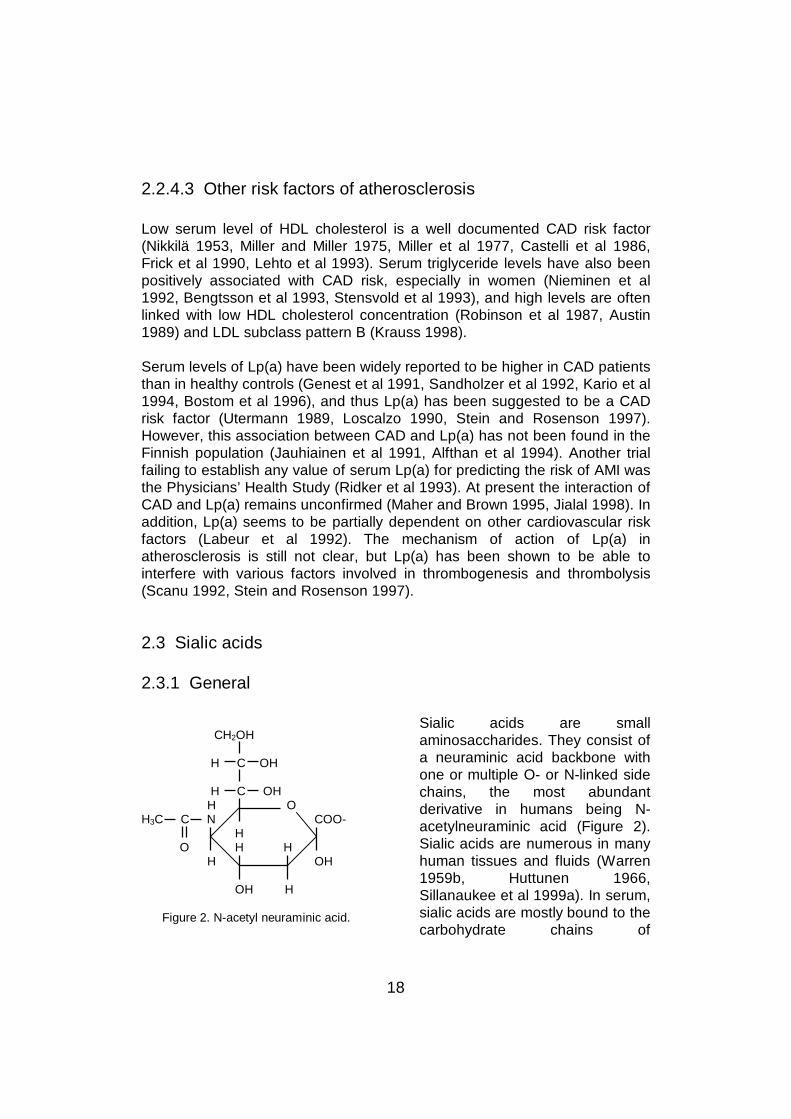
Figure 2. N-acetyl neuraminic acid.
Sialic acids are smallaminosaccharides. They consist ofa neuraminic acid backbone withone or multiple O- or N-linked sidechains, the most abundantderivative in humans being N-acetylneuraminic acid (Figure 2).Sialic acids are numerous in manyhuman tissues and fluids (Warren1959b, Huttunen 1966,Sillanaukee et al 1999a). In serum,sialic acids are mostly bound to thecarbohydrate chains of
19
glycoproteins and glycolipids. Sialic acid residues are located in the terminalends of carbohydrate chains of many glycoproteins, for instanceimmunoglobulins and peptide hormones, and when the terminal sialic acid isremoved by neuraminidase of the vascular endothelium, a galactosemolecule is revealed. Specific receptors on hepatocytes recognize theseasialoglyco-proteins and remove them from the circulation (Stryer 1988).
2.3.2 Serum sialic acid
A high serum total sialic acid (TSA) concentration has been associated with anumber of conditions (Sillanaukee et al 1999a). Many studies havedemonstrated a positive association between high TSA levels and theincidence of CAD (Lindberg et al 1991, Watts et al 1995a, Råstam et al1996). TSA seems to be linked with other CAD risk factors, namely highserum cholesterol and triglyceride concentrations and low HDL cholesterolconcentration (Wakabayashi et al 1992, Wu et al 1999), high serumconcentration of apo B and smoking and physical inactivity (Lindberg et al1993), and high serum Lp(a) levels (Kario et al 1994). TSA concentration hasbeen also shown to be higher in American than Japanese subjects even afteradjustment for other CAD risk factors, this being in accordance with thehigher prevalence of CAD in American population (Lindberg et al 1997).However, other studies have not found any difference in TSA level betweenangiographically separated CAD and non-CAD subjects (Salomone et al1998, Wu et al 1999).
Serum TSA level has been shown to be elevated in DM (Crook et al 1993,Crook et al 1996), and associated with CAD risk in diabetic subjects (Pickupet al 1995). TSA is also an indicator of microalbuminuria in DM both in cross-sectional (Crook et al 1994, Chen et al 1996) and follow-up studies(Yokoyama et al 1996). Besides, TSA concentration is higher in women thanin men with DM, which could explain why DM increases the relative risk ofCAD more in women than in men (Pickup et al 1997). TSA level is alsoelevated in many malignant diseases (Sillanaukee et al 1999a), duringpregnancy (Crook et al 1997a), and in alcohol abuse (Sillanaukee et al1999b). It is also high in inflammation (Sillanaukee et al 1999a) and positivelycorrelated with erythrocyte sedimentation rate (Miettinen and Nikkilä 1960,Crook et al 1997b) and serum concentration of C-reactive protein (Wu et al1999). Accordingly, serum TSA level seems to be an unspecific but sensitivemarker of acute phase reactions, probably because of the pronouncedcontent of sialic acid in many acute phase proteins.
20
2.3.3 Sialic acid in vascular tissue
Freshly infarcted myocardium has been shown to have a greater sialic acidcontent than normal myocardium (Huttunen et al 1972). Both vascularendothelium and red blood cells are rich in sialic acid and thus have a highnegative charge creating a repulsion between endothelium and blood cells,which can inhibit blood clotting (Born and Palinski 1985). Treatment of cellswith neuraminidase that removes a major part of their surface sialic acids hasbeen shown to promote the uptake of LDL into the cells (Görög and Born1982, Görög and Pearson 1984).
2.3.4 Sialic acid in lipoproteins
2.3.4.1 Chylomicrons, VLDL and IDL
The sialic acid content of chylomicrons has not been studied, while a fewresearchers have investigated the sialic acid content of other triglyceride-richlipoproteins. An early study found the highest sialic acid content in VLDL,whereas levels in IDL, LDL and HDL did not differ from each other (Fontaineand Malmendier 1975). Similarly, two recent studies reported that the sialicacid content decreased with increasing lipoprotein density from light VLDL todense IDL, then being similar until dense LDL (Anber et al 1997, Millar et al1999). Another study described decreasing sialic acid content from VLDL toLDL and further to HDL (Harada et al 1998). In these studies, the sialic acidcontents of the protein and lipid parts were not separated.
2.3.4.2 LDL
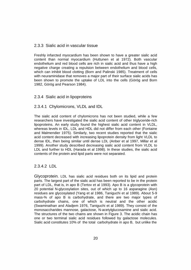
Glycoprotein. LDL has sialic acid residues both on its lipid and proteinparts. The largest part of the sialic acid has been reported to be in the proteinpart of LDL, that is, in apo B (Tertov et al 1993). Apo B is a glycoprotein with20 potential N-glycosylation sites, out of which up to 16 asparagine (Asn)residues are glycosylated (Yang et al 1986, Taniguchi et al 1989). About 5-9mass-% of apo B is carbohydrate, and there are two major types ofcarbohydrate chains, one of which is neutral and the other acidic(Swaminathan and Aladjem 1976, Taniguchi et al 1989). They consist of themonosaccharides mannose, galactose, N-acetylglucosamine and sialic acid.The structures of the two chains are shown in Figure 3. The acidic chain hasone or two terminal sialic acid residues followed by galactose molecules.Sialic acid constitutes 10% of the total carbohydrate in apo B, but unlike the

21
Sialic acid Sialic acid
Man Man Gal Gal
Man Man GlcNAc GlcNAc
Man Man Man Man
Man Man
GlcNAc GlcNAc
GlcNAc GlcNAc
Figure 3. The two major carbohydratechains of LDL apo B. Both are N-linkedsugar chains and bound to 16 Asnresidues distributed along the apo B-100polylpeptide chain. Gal = galactose;GlcNAc = N-acetylglucosamine; Man =mannose.
other monosaccharides, thecontents of which are fairlyconstant, the amount of sialic acidvaries markedly betweenindividuals (Swaminathan andAladjem 1976). The apo Bmolecule in LDL is calculated tocontain usually 12-14 sialic acidresidues per particle (Taniguchi etal 1989). LDL apo B differs frommost serum glycoproteins byhaving more galactose than sialicacid residues (Taniguchi et al1989), and the amount ofmonosialylated oligosaccharides,reflecting the number of freegalactose residues, can becalculated to be on the averagefour per LDL particle. A recentstudy confirmed that all LDL
particles in plasma have free galactose residues at the ends of thecarbohydrate chains, indicating that LDL apo B is always partially desialylated(Bartlett and Stanley 1998). The sialic acid content of LDL has a very strongpositive correlation with LDL apo B concentration (Fontaine and Malmendier1978), attesting that a large part of the sialic acid in LDL is bound to apo B.
Glycolipids. Gangliosides are sialic acid -rich glycosphingolipids transportedin serum by lipoproteins, 66% of total serum gangliosides being transportedby LDL and smaller amounts by other lipoproteins, and thus they alsocontribute to LDL sialic acid content (Dawson et al 1976, Senn et al 1989).Gangliosides probably contain all the sialic acid associated with the lipid partof LDL. In addition to the monosaccharides of LDL apo B, the lipid part ofLDL contains galactosamine and glucose but no mannose (Tertov et al1993). Sialic acid constitutes 6-7% of the total carbohydrate in LDL lipid. Thecontent of galactose is markedly higher than that of sialic acid also in the lipidpart of LDL (Tertov et al 1993), thus contributing to the number of freegalactose residues in the lipoprotein particle.
22
2.3.4.3 Cell culture studies
Many forms of modifications have been observed to increase theatherogenicity of LDL (Steinberg et al 1989). Regarding the role of sialic acidin the metabolism of glycoproteins, its content in LDL could affect theatherogenicity of LDL. This has been studied in cell cultures. Incubation withneuraminidase leading to desialylation of either aortic cells, LDL, or both,caused a rise in aortic uptake of LDL (Day 1976). Furthermore, desialylationof LDL was shown to double its binding and uptake into human fibroblasts(Filipovic and Buddecke 1979), and there was a positive correlation betweenthe degree of desialylation and the enhancement of uptake (Filipovic et al1979). In addition, a recent study described accelerated uptake of cholesterolfrom desialylated LDL to macrophages (Harada et al 1998). Harada et al(1998) also reported that desialylation of LDL increased the transport ofcholesterol esters from HDL to LDL by CETP, and desialylation of HDLdecreased its capacity to remove cholesterol from cells, both effectspromoting atherogenesis. However, two other studies found no difference inthe cellular binding and degradation of native and desialylated LDL (Attie etal 1979, Shireman and Fisher 1979).
LDL with low sialic acid content formed insoluble complexes with arterialproteoglycans avidly, and there was a significant negative correlationbetween the sialic acid content of LDL and the relative avidity to form thesecomplexes (Camejo et al 1985). This finding was confirmed in a recent study,where a fall in the sialic acid content of LDL raised its ability to interact witharterial proteoglycans (Millar et al 1999). Since the arterial proteoglycanshave a strong electronegative charge (Camejo et al 1991) and desialylationof LDL diminishes its negative charge, the increased affinity could partially bedue to reduced electronegative repulsion.
Enzymatic treatment of LDL with the combination of trypsin, cholesterolesterase and neuraminidase turns LDL into non-homogeneous lipid particlessimilar to those found in atherosclerotic lesions (Bhakdi et al 1995). Thismodified LDL does not contain oxidized lipids but has a high negative charge.It accumulates at a high rate into macrophages, this being mediated at leastpartially by the scavenger receptor pathway, and induces immunologicalreactions that promote early development of atherosclerotic lesions, such asactivation of the complement system, and chemotaxis, adhesion andtransendothelial migration of blood monocytes (Bhakdi et al 1995, Klouche etal 1998, Klouche et al 1999). All these effects were stronger for theenzymatically modified LDL than for acetylated or oxidized LDL, but were notdetectable in LDL treated with only one or two of the above-mentionedenzymes.

23
Apart from desialylation, another modification of LDL, acetylation, augmentsits cellular uptake and degradation, and this is mediated not by LDL receptorsbut by scavenger receptors (Goldstein et al 1979, Wiklund et al 1991). Suchreceptors also seem to be involved in the catabolism of desialylatedglycoproteins, including LDL. Desialylated glycopeptides bind to a hepaticlectin, the avidity of this binding being highly dependent on the number offree terminal galactose residues, that is, on the degree of desialylation (Leeet al 1983). Furthermore, a high density of galactose residues increases thebinding of asialo-orosomucoid both to a lectin receptor on hepatocytes, andto an asialoglycoprotein receptor on macrophages (Ozaki et al 1995).Assumably, desialylated LDL can also be taken up by these receptors.Indeed, LDL and chylomicron remnants have been reported to be able tobind to an asialoglycoprotein receptor on hepatocytes (Windler et al 1991),and desialylation of chylomicrons increases their hepatic uptake anddegradation, probably due to interaction with this asialoglycoprotein receptor(Guldur et al 1997). Moreover, LDL is documented to enter cells besidesthrough the LDL receptor pathway, also through a galactose-specific lectinreceptor pathway (Grewal et al 1996).
2.3.4.4 Coronary artery disease
Since the sialic acid content in LDL seems to be associated withatherogenesis, it might have a role in CAD. A Russian group has carried outextensive studies on the relations of LDL sialic acid content withatherosclerosis and CAD. They first discovered that serum or LDL of CADpatients caused a significant rise in cholesterol content of normal aortic cellsduring incubation, while serum or LDL from healthy subjects did not change it(Orekhov et al 1988, Tertov et al 1989a). Lipid and protein compositions ofthese LDLs were similar, but the sialic acid content was markedly lower inLDL of CAD patients (Orekhov et al 1989, Orekhov et al 1991b, Tertov et al1992a, Tertov et al 1992b). Neuraminidase treatment of LDL from healthysubjects made it atherogenic, and there was a strong negative correlationbetween LDL sialic acid content and the amount of cholesterol accumulatedintracellularly (Orekhov et al 1989, Orekhov et al 1992, Tertov et al 1992a,Tertov et al 1992b). In addition, LDL separated from normal aortic intima andfrom fatty streaks had a lower sialic acid content than plasma LDL (Tertov etal 1996a). These findings together support the hypothesis that desialylation isan atherogenic LDL modification taking place in vivo (Sobenin et al 1991,Orekhov et al 1992). During incubation, a decrease in the sialic acid contentwas the first modification observed in LDL, followed by gain of atherogenicityand decrease in the lipid content and size of the particles (Tertov et al 1998).

24
Moreover, it has been proved that sialic acid is implicated in the antigenicsites of LDL (Goldstein and Chapman 1981), and Orekhov et al (1991a)found, compared with healthy controls, in sera of CAD patients a thirty-foldgreater amount of autoantibodies against LDL that had a strong affinity fordesialylated LDL. Circulating immune complexes from blood of CAD patientswere discovered to be atherogenic (Tertov et al 1990, Tertov et al 1996b),and LDL from circulating immune complexes was sialic acid -poor (Tertov etal 1996b). LDL was separated from both CAD patients and healthy subjectsto sialic acid -rich and sialic acid -poor fractions by affinity chromatography,and it turned out that the proportion of desialylated LDL was considerablyhigher in CAD patients than in controls (Tertov et al 1992b). Desialylated LDLfrom both groups raised the intracellular lipid content, but this effect wasmuch stronger for desialylated LDL from CAD patients. Sialic acid -poor LDLand LDL from CAD patients also formed aggregates during incubation whilesialylated LDL or LDL from healthy subjects did not (Tertov et al 1992b,Tertov et al 1992c), and a strong positive correlation between LDLaggregation and intracellular cholesterol accumulation was demonstrated(Tertov et al 1989b, Tertov et al 1992c).
Other groups have come up with controversial results about differences in thesialic acid content of LDL between CAD patients and controls. Ruelland et al(1993) found lower a LDL sialic acid content in CAD patients than inangiographically verified non-CAD subjects, but LDL sialic acid content wasnot correlated with the severity of CAD. Furthermore, other studies withasymptomatic hypercholesterolemic subjects revealed no differences in LDLsialic acid content between subjects who in angiography were seen to haveeither no atherosclerotic plaques or one or more plaques (Chappey et al1995, Chappey et al 1998). These results imply that the sialic acid content ofLDL is not associated with the extent of CAD. In addition, LDL sialic acidcontent was even slightly higher in subjects with both CAD and peripheralatherosclerosis and in patients suffering from AMI than in those with CADonly and in controls (Chappey et al 1998), while another research groupreported decreased LDL sialic acid content immediately after coronary arteryby-pass surgery (Jahangiri et al 1999). However, a recent study found lowersialic acid content in both light and dense LDL of CAD patients comparedwith healthy controls (Millar et al 1999).
LDL from DM patients also turned out to be poor in sialic acid and able tocause intracellular cholesterol deposition, and as in CAD patients andcontrols there was a strong negative correlation between the sialic acidcontent of LDL and the degree of atherogenicity (Tertov et al 1992a, Sobeninet al 1993, Sobenin et al 1994). A more recent study again found no
25
difference in LDL sialic acid content between DM patients and controlsubjects (Ruelland et al 1997).
Little is known about the importance of LDL sialic acid content in themetabolism of LDL, but two studies have reported that a reduction of LDLsialic acid content by neuraminidase treatment leads to faster LDL catabolism(Hendrix et al 1975, Malmendier et al 1980).
Only a couple of studies have investigated LDL sialic acid content inhyperlipidemia. Barbosa et al (1995) stated the sialic acid content of bothlight and dense LDL to be high in combined hyperlipidemia but low inhypercholesterolemia compared with normolipidemia. Another study showedlow LDL sialic acid content associated with hypercholesterolemia in DMpatients (Maruhama et al 1983).
Two studies have shown an inverse connection between LDL sialic acidcontent and serum triglyceride levels (La Belle and Krauss 1990, Chappey etal 1995), and a negative correlation with LDL cholesterol and positive withHDL cholesterol level have also been documented (La Belle and Krauss1990). However, Millar et al (1999) found no correlation between serumcholesterol or triglyceride concentrations and LDL sialic acid content.
The sialic acid -containing gangliosides, transported in serum mainly by LDL,have also been shown to be associated with atherosclerosis (Prokazova andBergelson 1994). Gangliosides stimulate LDL aggregation (Mikhailenko et al1991), and LDL incubated with gangliosides is avidly taken up bymacrophages (Prokazova et al 1991) and fibroblasts (Filipovic et al 1981) butpoorly by hepatocytes (Prokazova et al 1986) thus probably interfering withthe clearance of LDL from the circulation to the liver. Furthermore, serumganglioside concentration is high in hypercholesterolemia proportionately tothe increase in cholesterol concentration (Dawson et al 1976, Senn et al1992), and antibodies against gangliosides have been detected in serum ofCAD patients (Golovanova et al 1998). In addition, high amounts ofgangliosides have been found in atherosclerotic lesions compared tounaffected intima (Breckenridge et al 1975, Mukhin et al 1989).
2.3.4.5 LDL density classes
Within the LDL density range, the sialic acid content falls with growingparticle density and diminishing size (La Belle and Krauss 1990, Millar et al1999). Furthermore, Tertov et al demonstrated that desialylated LDLseparated from total LDL was smaller and denser than sialic acid -rich LDL,
26
and poor in cholesterol and triglycerides (Tertov et al 1992b, Tertov et al1996b). In addition, as the density of the particles rose, their sialic acidcontent decreased and their ability to accumulate cholesterol into cellsincreased (Tertov et al 1992b). In accordance with these results, La Belle andKrauss (1990) also reported that subjects with LDL subclass pattern B had alower sialic acid content in LDL than those with subclass pattern A, and thedifference appeared to be in the glycolipid and not in the protein part of theparticle.
2.3.4.6 Oxidation and electronegativity
Tertov et al (1992b) found no differences in the content of oxidation productsin sialic acid -poor and sialic acid -rich LDL particles, and another study foundthat the desialylation of LDL had no effect on its susceptibility to peroxidation(Sattler et al 1991). However, in two other studies neuraminidase treatmentdecreased the susceptibility of LDL to oxidation (Dousset et al 1994, Myara etal 1995). A recent study reported that desialylation of LDL withneuraminidase caused physical and chemical modifications in the structure ofapo B, which could account for its lowered susceptibility to oxidation (Doussetet al 1998). Barbosa et al (1995) showed that the effect of desialylation onLDL oxidizability was dependent on the density of LDL and on the type ofhyperlipidemia; light LDL was more resistant to oxidation in both type IIa andIIb hyperlipidemia than in controls, but dense LDL of IIb hyperlipidemicsubjects was more susceptible to oxidation than that of normolipidemicsubjects, and its desialylation raised its resistance to oxidation, while itdecreased the resistance to oxidation of dense LDL in type IIahyperlipidemia. On the other hand, a fall in LDL sialic acid content duringperoxidation has also been reported (Tanaka et al 1997).
An electronegative subfraction of LDL was found to be sialic acid -poor andatherogenic, and it was suggested that this electronegative subfraction couldbe identical with the desialylated subfraction (Tertov et al 1995, Tertov et al1996a). However, another group separated an electronegative LDLsubfraction with a high sialic acid content, and its proportion of total LDL waspositively correlated with LDL cholesterol concentration (Demuth et al 1996).
2.3.4.7 Lp(a)
Lp(a), which consists of LDL and apo(a), contains six times as much sialicacid as LDL (Ehnholm et al 1972, Utermann 1989). Since its density rangeoverlaps that of LDL, it is possible that LDL separated by ultracentrifugation
27
contains some Lp(a), this possibly affecting its sialic acid content in analysis.However, the sialic acid content of LDL has been proved not to be correlatedwith serum Lp(a) levels (Chappey et al 1995).
Lp(a) is more resistant to peroxidation than LDL (Sattler et al 1991,Beaudeux et al 1996), but neuraminidase treatment was shown to either notaffect (Beaudeux et al 1996) or reduce (Sattler et al 1991) the resistance, thelatter result leading to the suggestion that the higher resistance of nativeLp(a) to peroxidation could be due to its higher sialic acid content ascompared with LDL. Like LDL, Lp(a) from CAD patients was reported to besialic acid -poor and atherogenic in cell culture compared with Lp(a) fromhealthy controls; unlike native Lp(a) desialylated Lp(a) formed aggregates,and their formation was strongly correlated with the extent of intracellularcholesterol deposition (Tertov and Orekhov 1994).
2.4 Cholesterol metabolism
2.4.1 Cholesterol absorption
Cholesterol is derived to the body from two sources, diet and cellularsynthesis. Cholesterol elimination from the body is mediated by the liver,which can transform cholesterol into bile acids. Both free cholesterol and bileacids are secreted into bile. Dietary and biliary cholesterol is absorbed fromthe small intestine into enterocytes, where it is incorporated into chylomicronsand secreted into the circulation. Unabsorbed cholesterol and bile acids areexcreted from the body in feces as neutral steroids and bile acids. Theabsorption of cholesterol from the intestine depends on the amount ofcholesterol in diet and in bile, and on the absorption efficiency of cholesterol,which varies considerably interindividually, the usual range being 30 to 80 %(Grundy 1983). Cholesterol absorption efficiency has been reported toregulate serum cholesterol levels in Finnish men (Kesäniemi and Miettinen1987), so that cholesterol absorption efficiency was correlated positively withserum and LDL cholesterol levels (Kesäniemi and Miettinen 1987, Miettinenand Kesäniemi 1989, Vanhanen and Miettinen 1995), and negatively withcholesterol synthesis (Kesäniemi and Miettinen 1987, Gylling and Miettinen1989, Miettinen and Kesäniemi 1989). However, another study reported nocorrelation of cholesterol absorption with serum and LDL cholesterol levels(Gylling and Miettinen 1988). Lowered cholesterol absorption efficiency hasbeen observed in many conditions, including obesity (Miettinen andKesäniemi 1989) and DM (Gylling and Miettinen 1997).
28
Small amounts of plant sterols are present in human serum, and the mostcommon of these are campesterol and sitosterol. They are totally of dietaryorigin, and are absorbed in the small intestine similarly to cholesterol, but lesseffectively. In serum, they are transported by lipoproteins and thus theirconcentration is highly dependent on serum cholesterol concentration. Theratios of campesterol and sitosterol to cholesterol are positively correlatedwith cholesterol absorption efficiency (Tilvis and Miettinen 1986, Vuoristo et al1988, Miettinen et al 1990), and the measurement of their concentrationsfrom serum represents a simple method for the estimation of cholesterolabsorption efficiency.
Cholestanol is a metabolite of cholesterol found in small amounts in blood,and transported by lipoproteins. Its ratio to cholesterol is strongly correlatedwith the ratios of campesterol and sitosterol (Miettinen et al 1998), and, likethese plant sterols, it is a reliable indicator of cholesterol absorption efficiency(Miettinen et al 1989).
2.4.2 Cholesterol synthesis
Cholesterol synthesis takes place mainly in the liver, but nearly all humancells are capable of synthesizing cholesterol. The initial step in cholesterolsynthesis is the conversion of 3-hydroxy-3-methylglutaryl coenzyme A (HMG-CoA) into mevalonic acid by HMG-CoA reductase, which is the rate limitingenzyme in cholesterol synthesis. Mevalonic acid is then transformed intosqualene, and after many stages finally into cholesterol. Some of thecholesterol precursor sterols that are part of the chain leading from squaleneto cholesterol are present in detectable amounts in serum, particularly∆8-lathosterol, desmosterol and lathosterol (Miettinen 1968, Miettinen 1969).They are transported in serum by lipoproteins similarly to cholesterol. Theratios of these precursor sterols to cholesterol are correlated positively withcholesterol synthesis rate (Björkhem et al 1987, Miettinen et al 1990), andnegatively with cholesterol absorption efficiency. Thus, the precursor sterolratios can be used as indicators of cholesterol synthesis rate.
Cholesterol synthesis in the body is strictly regulated (Brown and Goldstein1986). Circulating lipoproteins, especially LDL, deliver cholesterol toperipheral and liver cells, and enter the cells principally via the LDL receptor.In cells, cholesterol esters are hydrolyzed to free cholesterol, which can beused as a constituent of cell membranes, in the biosynthesis of steroidhormones, or re-esterified for storage. Cholesterol entering the cell from LDLsuppresses the cell’s own cholesterol synthesis thus keeping up ahomeostasis in the cholesterol content of the cell. In hepatocytes, cholesterol
29
can also be secreted back to the blood in lipoprotein particles or it can beconverted into bile acids or secreted as free cholesterol with bile acids to thebile.
Cholesterol synthesis averages 9-13 mg/kg of body weight in humans perday (Nestel et al 1969). It is high in obesity (Miettinen 1970),hypertriglyceridemia (Briones et al 1986), and DM (Bennion and Grundy1977, Briones et al 1986, Gylling and Miettinen 1997), and low inhypercholesterolemia (Miettinen 1970, Miettinen 1971). In FH, low cholesterolsynthesis rate is associated with high CAD mortality (Miettinen and Gylling1988). Both positive (Gylling and Miettinen 1989) and negative (Miettinen etal 1992, Gylling and Miettinen 1997) correlations have been reportedbetween cholesterol synthesis rate and serum and LDL cholesterolconcentrations, but most studies (Gylling and Miettinen 1988, Miettinen andKesäniemi 1989, Miettinen et al 1989, Gylling et al 1994) found noassociation between these. Thus it seems that serum cholesterol levels aremore dependent on cholesterol absorption than synthesis. However, in type 2DM cholesterol synthesis seems to have an inverse association with LDLcholesterol level (Gylling and Miettinen 1996a, Gylling and Miettinen 1997).
2.4.3 Cholesterol and lipoprotein metabolism
Few studies have investigated the associations between cholesterolsynthesis and absorption on the one hand, and production and catabolism oflipoproteins on the other hand. Miettinen et al (1987) reported no clearcorrelations between the synthesis and elimination of cholesterol and, onanother side, concentration and catabolism of LDL apo B, which suggeststhat the changes in cholesterol metabolism are compensated in the liverwithout affecting the metabolism of LDL apo B. However, bile acidmalabsorption enhanced both cholesterol synthesis and the catabolism ofLDL apo B, while cholesterol malabsorption caused by different mechanismsonly increased cholesterol synthesis but affected LDL apo B kineticsinconsistently (Miettinen et al 1987). Later, other studies found positivecorrelations between cholesterol synthesis rate and the catabolism of LDLapo B, and between fractional cholesterol absorption and production of LDLapo B (Miettinen et al 1992, Gylling et al 1994). Positive correlations foundrecently between the hepatic secretion of VLDL apo B and indicators ofcholesterol synthesis suggest that the production of apo B from the liver isregulated by cholesterol synthesis rate (Watts et al 1995c, Riches et al 1997).
30
2.5 Statin treatment
The first statin, initially called mevinolin, was discovered over twenty yearsago (Endo et al 1976, Alberts et al 1980). Statins are specific inhibitors ofHMG-CoA reductase, the rate limiting enzyme in cholesterol synthesis.Inhibition of cholesterol synthesis accelerates the activity of LDL receptors(Kovanen et al 1981, Traber and Kayden 1984, Berglund et al 1989, Ravehet al 1990, Reihner et al 1990, Angelin 1991). However, the clearance of LDLparticles from the circulation has been reported to either increase (Kovanenet al 1981, Bilheimer et al 1983, Malmendier et al 1989, Vega and Grundy1991, Gylling and Miettinen 1996b), decrease (Vega et al 1988), or remainunchanged (Grundy and Vega 1985, Arad et al 1990, Vega et al 1990,Cuchel et al 1997) during statin treatment. Lowered production of LDL apo Bduring statin treatment has been more consistently reported (Kovanen et al1981, Grundy and Vega 1985, Vega et al 1988, Arad et al 1990, Vega et al1990, Gaw et al 1993, Gylling and Miettinen 1996b, Cuchel et al 1997), and itis considered to result from increased clearance of VLDL and IDL particles byLDL receptors before conversion into LDL. However, in combinedhyperlipidemia a rise in the catabolic rate of LDL apo B without a significantchange in its production during statin treatment has been demonstrated(Vega and Grundy 1991, Parhofer et al 1993, Schonfeld et al 1998). Somestudies have also shown decreased production of VLDL apo B by the liverduring statin treatment (Cortner et al 1993, Watts et al 1995b, Burnett et al1997), possibly due to the reduction in cholesterol synthesis regulating theproduction of apo B in the liver to some extent (Thompson et al 1996, Richeset al 1997, Watts et al 1997). Furthermore, the basal rate of lipoproteinmetabolism and the dose of the statin probably affect the changes caused bystatins (Aguilar-Salinas et al 1998). In addition to increased LDL receptoractivity and decreased production of LDL apo B, statin treatment was recentlyshown to decrease the affinity of LDL particles for the LDL receptor, whichcould explain why the catabolism of LDL is only modestly increased by statins(Berglund et al 1998). Thus, the mechanism by which statins lower levels ofLDL cholesterol seems to be more complicated than just up-regulation of LDLreceptors. The effect of statins on the sialic acid content of lipoproteins hasnot been studied, whereas fibrates seem to diminish it (Anber et al 1997,Millar et al 1999).
Nowadays, statins are widely used in the treatment of hypercholesterolemia.Their efficiency in lowering serum and LDL cholesterol levels and thus themorbidity and mortality related to CAD has been confirmed in many largestudies (Scandinavian Simvastatin Survival Study Group 1994, West ofScotland Coronary Prevention Study Group 1995, Cholesterol and RecurrentEvents Trial Investigators 1996, AFCAPS/TexCAPS Research Group 1998,
31
The Long-Term Intervention with Pravastatin in Ischaemic Disease (LIPID)Study Group 1998). They have also been shown to inhibit progression ofatherosclerosis in coronary arteries (Watanabe et al 1988, Khachadurian et al1991, Kroon et al 1993). Lately, other mechanisms apart from the lowering ofcholesterol levels have been suggested to account partially for the good andrelatively rapid efficiency of statins in diminishing the incidence of CADevents and CAD mortality, e.g. inhibition of the action of macrophages,promotion of endothelial relaxation, and decrease in hypercoagulability byaffecting multiple factors associated with blood clotting (Vaughan et al 1996).
Out of LDL subfractions, statin treatment reduced only the concentration ofthe most dense fraction in one study (Nozaki et al 1990), but evenly theconcentrations of all subfractions in another study (Yuan et al 1991). Theratio of cholesterol to apo B in LDL has been reported to either decrease(Nozaki et al 1990) or remain unchanged (Vega et al 1990, Yuan et al 1991,Gylling and Miettinen 1996b) during statin therapy, whereas the mean size ofLDL particles enlarges (Yuan et al 1991, Zhao et al 1991) or keepsunchanged (Vega et al 1990, Cheung et al 1993, Superko et al 1997).Overall, LDL subclass pattern seems to persist during statin treatment (Vegaet al 1990, Franceschini et al 1994, Homma et al 1995, Superko et al 1997).Statins have been shown to diminish the ratios of cholesterol precursorsterols to cholesterol and elevate the ratios of plant sterols, reflectingdecreased synthesis and increased intestinal absorption of cholesterol(Vanhanen and Miettinen 1995, Gylling and Miettinen 1996b). A high ratio ofcholestanol to cholesterol at baseline, indicating low cholesterol synthesisrate, predicts poor response to statin treatment (Miettinen et al 1998).
While statins efficaciously reduce the concentrations of serum and LDLcholesterol, their effect on serum Lp(a) levels is controversial. Many studieshave found no change in Lp(a) levels during statin treatment (Berg and Leren1989, Fieseler et al 1991, Cheung et al 1993, Hunninghake et al 1993,Haffner et al 1995, Raal et al 1997), some have reported even elevatedlevels (Jürgens et al 1989, Kostner et al 1989, Broijersen et al 1994, Branchiet al 1995), while also lowered levels have been described (Leren et al 1992).The importance of the apparent uneffectiveness of statins on high Lp(a)levels is unclear, but possibly subjects with very high levels would benefitfrom reduction of Lp(a).
32
3 AIMS OF THE STUDY
The deposition of cholesterol from lipoproteins to artery walls leads toatherosclerosis, and this is undoubtedly mediated by multiple mechanisms.According to data from previous studies referred to above, sialic acid asstructural part of LDL particles seems to have a role in the evaluation of theatherogenic potential of LDL. A low sialic acid content in LDL of CAD patientshas been reported in many but not all studies, leaving the subjectcontroversial. Serum total and LDL cholesterol levels are regulated in generalby the metabolism of cholesterol and LDL apo B, but there is no informationon whether the sialic acid content of LDL is related with this. There is someevidence that LDL sialic acid content has associations with serum andlipoprotein lipid concentrations, but former data are not consistent, and thereare no data on the connections of sialic acid with lipid and apo B contents inLDL subfractions. Furthermore, the effect of statins, widely used asefficacious hypocholesterolemic agents, on LDL sialic acid content has notbeen studied.
Thus, the aims of this study were:
• To evaluate the relations of LDL sialic acid content with the concentrationsof lipids and lipoproteins, and with the composition of LDL particles
• To investigate the sialic acid content of LDL in CAD and in conditionsassociated with an increased risk of CAD, namely type 2 DM and type IIaand IIb hyperlipidemia
• To find out possible associations of LDL sialic acid content with themetabolism of LDL, and with synthesis and absorption of cholesterol
• To study the effect of statin treatment on LDL sialic acid content
33
4 PATIENTS AND METHODS
4.1 Patients and study designs
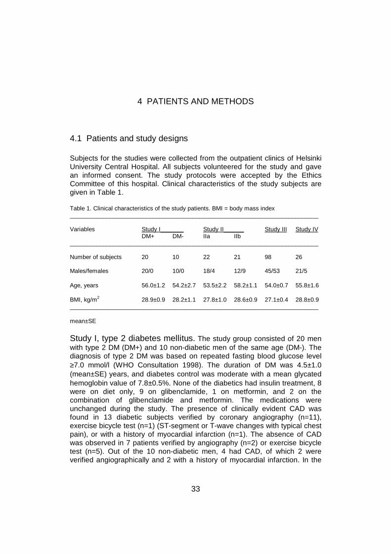
Subjects for the studies were collected from the outpatient clinics of HelsinkiUniversity Central Hospital. All subjects volunteered for the study and gavean informed consent. The study protocols were accepted by the EthicsCommittee of this hospital. Clinical characteristics of the study subjects aregiven in Table 1.
Table 1. Clinical characteristics of the study patients. BMI = body mass index___________________________________________________________________________
Variables Study I_______ Study II______ Study III Study IVDM+ DM- IIa IIb
___________________________________________________________________________
Number of subjects 20 10 22 21 98 26
Males/females 20/0 10/0 18/4 12/9 45/53 21/5
Age, years 56.0±1.2 54.2±2.7 53.5±2.2 58.2±1.1 54.0±0.7 55.8±1.6
BMI, kg/m2 28.9±0.9 28.2±1.1 27.8±1.0 28.6±0.9 27.1±0.4 28.8±0.9___________________________________________________________________________
mean±SE
Study I, type 2 diabetes mellitus. The study group consisted of 20 menwith type 2 DM (DM+) and 10 non-diabetic men of the same age (DM-). Thediagnosis of type 2 DM was based on repeated fasting blood glucose level≥7.0 mmol/l (WHO Consultation 1998). The duration of DM was 4.5±1.0(mean±SE) years, and diabetes control was moderate with a mean glycatedhemoglobin value of 7.8±0.5%. None of the diabetics had insulin treatment, 8were on diet only, 9 on glibenclamide, 1 on metformin, and 2 on thecombination of glibenclamide and metformin. The medications wereunchanged during the study. The presence of clinically evident CAD wasfound in 13 diabetic subjects verified by coronary angiography (n=11),exercise bicycle test (n=1) (ST-segment or T-wave changes with typical chestpain), or with a history of myocardial infarction (n=1). The absence of CADwas observed in 7 patients verified by angiography (n=2) or exercise bicycletest (n=5). Out of the 10 non-diabetic men, 4 had CAD, of which 2 wereverified angiographically and 2 with a history of myocardial infarction. In the
34
remaining 6 subjects, CAD was excluded by exercise bicycle test. Three DMpatients and 1 control subject were smokers.
Study II, type IIa and IIb hyperlipidemia. Forty-three patients, 30 menand 13 women, were divided into two groups according to their lipidphenotype obtained in two fasting serum samples a week apart. Twenty-twosubjects (18 men and 4 women) were categorized into the primary moderatehypercholesterolemia group (type IIa; LDL cholesterol 3.5-5.5 mmol/l andserum triglycerides <2.5 mmol/l), and 21 subjects (12 men and 9 women) intothe combined hyperlipidemia group (type IIb; LDL cholesterol 3.5-5.5 mmol/land serum triglycerides >2.5 mmol/l). Thirty-three subjects had CAD verifiedeither by exercise bicycle test (n=4), or by coronary angiography and/or ahistory of myocardial infarction at least six months prior to the study (n=29),while the remaining 10 subjects had no evidence of CAD in exercise bicycletest. One patient in the IIa group had oral medication for type 2 DM, and 3 inIIa and 4 in IIb group had DM treated only with diet. Eleven volunteer subjectsfrom IIa group and 16 from IIb group participated in the kinetic studies.
Study III, coronary artery disease. Ninety-eight subjects, 53 women and45 men, were recruited for the study. Fifty-six subjects (CAD+), 30 men and26 women, had CAD diagnosed either by a history of myocardial infarction(n=37), by coronary angiography (n=14) or by exercise bicycle test (n=5),while the remaining 42 subjects (CAD-) had no symptoms or manifestationsof CAD in clinical interview and examination, or in ECG. None of the subjectshad DM. They had been councelled a low-fat low-cholesterol diet at least sixmonths earlier, and they kept the diet unchanged. Forty-four subjects wereon beta-blocking agents, 6 subjects were taking thiazide diuretics, and 15 outof the 53 women had hormone replacement therapy. Twenty-four subjectswere current smokers. In order to examine differences in CAD+ and CAD-subjects at different sialic acid levels, both CAD+ and CAD- subjects weredivided into quartiles according to the sialic acid to apo B ratios of total anddense LDL, with equal numbers of subjects in each quartile. The quartiles aredesignated as Q1, Q2, Q3 and Q4 from low to high sialic acid ratios. Fifty-eightvolunteers out of the 98 subjects participated in the kinetic study.
Study IV, statin treatment. The study group consisted of 26 moderatelyhypercholesterolemic patients (LDL cholesterol >3.3 mmol/l), 21 males and 5females. All except 4 patients had CAD verified either by angiography and/ora history of acute myocardial infarction at least six months before the study(n=20), or by exercise bicycle test (n=2). Five patients had DM treated withdiet only. Sixteen patients were on beta-blocker treatment at the time of thestudy, and 2 used thiazide diuretics for hypertension. The medications wereunchanged during the study. None of the women used hormone replacement
35
therapy. Four patients were current smokers. After recruitment to the study,possible lipid lowering medication was discontinued, and the patients starteda low-fat low-cholesterol diet, containing a scheduled amount of 30 energy-%of fat and less than 300 mg/day of cholesterol. After six weeks on the diet,baseline lipid analyses and metabolic studies were performed. Subsequently,the patients continued the diet and started statin treatment, with 10 patientsreceiving on the average 0.2 mg/day (0.1-0.3 mg) of cerivastatin, 9 patientson the average 30 mg/day (20-40 mg) of fluvastatin, and 7 patients 20mg/day of simvastatin. When the patients had been on statins for two tothree months, the lipid analyses and metabolic studies were repeated.Thirteen subjects volunteered to participate in both kinetic studies.
None of the subjects in the studies had gastrointestinal, liver, renal, or thyroiddisease, except 3 subjects in Study III who had thyroxin treatment forhypothyroidism, and had been euthyroid for at least two years before thestudy. No one had tendon or cutaneous xanthomas or xanthelasmas. Thesubjects were not on any lipid lowering medication at the baseline of thestudies.
4.2 Methods
4.2.1 Separation of lipoproteins
Serum samples were drawn after a 12-hour fast. Serum lipoproteins wereseparated with serial ultracentrifugations into VLDL, IDL, LDL and HDL(Havel et al 1955, Lipid Research Clinics Program 1982). After the separationof total LDL (d = 1.019-1.063 g/ml), it was fractionated into three subfractions:light (d = 1.019-1.036 g/ml), dense (d = 1.037-1.055 g/ml) and very dense(d = 1.056-1.063 g/ml), by density gradient ultracentrifugation. The densitygradient was prepared in a 40-ml polyallomer Quick-Seal tube as follows:11.3 ml of NaBr-NaCl salt solution (d = 1.065 g/ml) was carefully overlayeredby 11.3 ml of LDL (d = 1.04 g/ml), which in turn was overlayered by NaBr-NaCl salt solution of d = 1.025 g/ml. The fractionation was completed byultracentrifugation (44 h at 58,000 rpm at +10°C) in a Ti 60 fixed-angle rotor(Beckman Instruments).
4.2.2 Analyses of lipids, apolipoproteins and non-cholesterolsterols
The concentrations of total and free cholesterol, triglycerides, andphospholipids were analyzed from serum and from all lipoproteins with
36
commercial kits (Boehringer Diagnostica, Mannheim, Germany; and WakoChemicals GMBH, Neuss, Germany). Concentrations of apo B, apo A-I andLp(a) were analyzed with commercial kits (Orion Diagnostica, Espoo, Finland;and Mercodia AB, Uppsala, Sweden) from serum, and that of apo B also fromtotal LDL and its subfractions. The coefficients of variation of the analyseswere <2% for lipids and <5% for apolipoproteins. Mean values of threeanalyses from samples taken at one-week intervals are given for serum andlipoprotein lipids and apolipoproteins. Recoveries for serum lipoprotein lipidconcentrations were always >90% and mostly >95%. In LDL density gradientthe recovery percents were occasionally more variable, and therefore the lipidconcentrations of LDL subfractions are given recovery corrected except inStudy I. Apo E phenotypes were analyzed by electrophoresis and isoelectricfocusing from serum (Havekes et al 1987). Total protein contents of thelipoproteins were analyzed in Study II with the Lowry method (Lowry et al1951).
Serum non-cholesterol sterols, including the cholesterol precursor sterols∆8-lathosterol, desmosterol and lathosterol, the plant sterols campesterol andsitosterol, and the cholesterol metabolite cholestanol, were determined fromnon-saponifiable serum extract by gas-liquid chromatography on a 50-m longSE-30 capillary column (Hewlett Packard, Ultra I column) (Miettinen 1988).Being transported in serum by lipoproteins similarly to cholesterol, theconcentrations of the non-cholesterol sterols are highly dependent on serumcholesterol levels. Thus the values are expressed as 102
x mmol/mol ofserum cholesterol, i.e. as ratios to cholesterol, to eliminate the effect ofvariation in cholesterol levels.
4.2.3 Analysis of sialic acids
The concentration of sialic acids in total LDL and its subfractions, and inStudy II, also in VLDL, IDL and HDL, was analyzed according to theresorcinol method by Svennerholm (1957) modified by Miettinen and Takki-Luukkainen (1959), using N-acetylneuraminic acid (Sigma ChemicalCompany, St Louis, MO, USA) as the standard. Shortly, sialic acids were firstdissociated from LDL by acid hydrolysis in 0.1-N H2SO4 at +80°C for onehour. Subsequently, resorcinol-hydrochloric acid was used to cause a colorreaction with sialic acid. Finally, the pigment was extracted with butyl acetateand its intensity measured with a spectrophotometer. In Study I, the sialicacid concentration was determined with the thiobarbituric acid assay (Warren1959a) in addition to the resorcinol method, and the results were consistentwith each other with a correlation coefficient of r=0.960 (n=16). Because ofthe high dependence of LDL sialic acid concentration on LDL apo B
37
concentration (Fontaine and Malmendier 1978), observed also in the presentstudy, the sialic acid content of total LDL and its subfractions is given as theratio of sialic acid to apo B and not as sialic acid concentration as in all earlierstudies, and it is designated as the sialic acid ratio. In Study II, the sialic acidcontent of lipoproteins in the comparison of VLDL, IDL, LDL and HDL is givenas the ratio of sialic acid to total protein.
4.2.4 Lipoprotein kinetic studies
For the kinetic studies, 50 ml of fasting EDTA plasma was drawn, andautologous total LDL and HDL were separated by serial densityultracentrifugations. Apo A-I was isolated from HDL as described in detail byGylling et al (1992). Briefly, HDL was incubated with guanidine hydrochloride,dialyzed, density-adjusted with NaBr, and ultracentrifugated, after which pureapo A-I was collected from the bottom and extensively dialyzed. LDL wasfractionated by serial density ultracentrifugations into three subclasses aspreviously described, and dialyzed extensively. Dense LDL apo B and apoA-I were iodinated with
125I, and total LDL apo B with 131I by a modification of
the iodine-monochloride method (McFarlane 1958, Bilheimer et al 1972).Three days before injection the subjects started to take peroral potassiumiodide to protect the thyroid gland. Approximately 1 mg of a mixture of thelabeled total and dense LDL apo B and apo A-I were mixed with 5% humanserum albumin, filtered, and injected. The total amount of radioactivity did notexceed 60 µCi.
After the injection, serial blood samples of 10 ml were collected for 14 daysand counted. The die-away curves were constructed from plasma for 131I-LDLand after ultracentrifugal separation for 125I-dense LDL and
125I-HDL.Fractional catabolic rates (FCRs) for total and dense LDL apo B and HDL apoA-I were determined using a two-pool model (Matthews 1957). Transport rate(TR) was calculated by multiplying FCR by the pool size. Pool size was theapoprotein plasma concentration multiplied by plasma volume, which wascalculated to be 4.5% of body weight. FCR is expressed in pools/day andreflects the rate at which the lipoprotein is removed from the circulation, whileTR, expressed in mg/kg/day, reflects the rate of production of the lipoprotein.
4.2.5 Statistics
Analyses were performed with Microsoft Excel, Primer of Biostatistics, andBiomedical Data Program (BMDP). Statistical significancies of differencesbetween groups were tested with analysis of variance (ANOVA) and, when
38
appropriate, with Student's two-tailed t-test. X2-test or Fisher exact test wasused for nominal scale variables. In Study IV, paired t-test was used todetermine the change caused by the statin treatment. Correlations wereanalyzed by calculating Pearson’s product-moment correlation coefficient fornormally distributed variables. In case of skewed distributions, eitherlogarithmic transformations or Spearman's rank-order correlation coefficientwas applied. Values are given as mean±SE. A P-value of less than 0.05 wasconsidered statistically significant.
In Study I, the variability in the FCR for LDL apo B was examined with amultiple linear regression analysis with the presence of DM, LDL and HDLcholesterol and serum triglyceride concentrations, and LDL sialic acid ratio asthe independent variables. Similarly, another linear regression model wasmade with FCR for dense LDL apo B as the dependent variable, and thepresence of DM and cholesterol and triglyceride concentrations and the sialicacid ratio of dense LDL as the independent variables. In Study III, a stepwiselogistic regression analysis was carried out with the presence of CAD as thedependent variable, and LDL cholesterol and triglyceride concentrations, LDLsialic acid ratio, and FCR and TR for LDL apo B as the independentvariables. To explain the variability in LDL sialic acid ratio, a stepwiseregression analysis was performed with the presence of CAD, theconcentrations of cholesterol and triglycerides in LDL, and FCR and TR forLDL apo B as the independent variables.
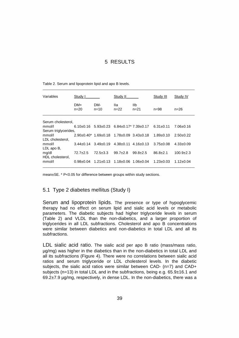
39
5 RESULTS
Table 2. Serum and lipoprotein lipid and apo B levels.___________________________________________________________________________
Variables Study I_______ Study II______ Study III Study IV
DM+ DM- IIa IIbn=20 n=10 n=22 n=21 n=98 n=26
___________________________________________________________________________
Serum cholesterol,mmol/l 6.10±0.16 5.93±0.23 6.84±0.17* 7.39±0.17 6.31±0.11 7.06±0.16Serum triglycerides,mmol/l 2.90±0.40* 1.69±0.18 1.78±0.09 3.43±0.18 1.89±0.10 2.50±0.22LDL cholesterol,mmol/l 3.44±0.14 3.49±0.19 4.38±0.11 4.16±0.13 3.75±0.08 4.33±0.09LDL apo B,mg/dl 72.7±2.5 72.5±3.3 99.7±2.8 99.8±2.5 86.8±2.1 100.9±2.3HDL cholesterol,mmol/l 0.98±0.04 1.21±0.13 1.18±0.06 1.06±0.04 1.23±0.03 1.12±0.04___________________________________________________________________________
mean±SE. * P<0.05 for difference between groups within study sections.
5.1 Type 2 diabetes mellitus (Study I)
Serum and lipoprotein lipids. The presence or type of hypoglycemictherapy had no effect on serum lipid and sialic acid levels or metabolicparameters. The diabetic subjects had higher triglyceride levels in serum(Table 2) and VLDL than the non-diabetics, and a larger proportion oftriglycerides in all LDL subfractions. Cholesterol and apo B concentrationswere similar between diabetics and non-diabetics in total LDL and all itssubfractions.
LDL sialic acid ratio. The sialic acid per apo B ratio (mass/mass ratio,µg/mg) was higher in the diabetics than in the non-diabetics in total LDL andall its subfractions (Figure 4). There were no correlations between sialic acidratios and serum triglyceride or LDL cholesterol levels. In the diabeticsubjects, the sialic acid ratios were similar between CAD- (n=7) and CAD+subjects (n=13) in total LDL and in the subfractions, being e.g. 65.9±16.1 and69.2±7.9 µg/mg, respectively, in dense LDL. In the non-diabetics, there was a
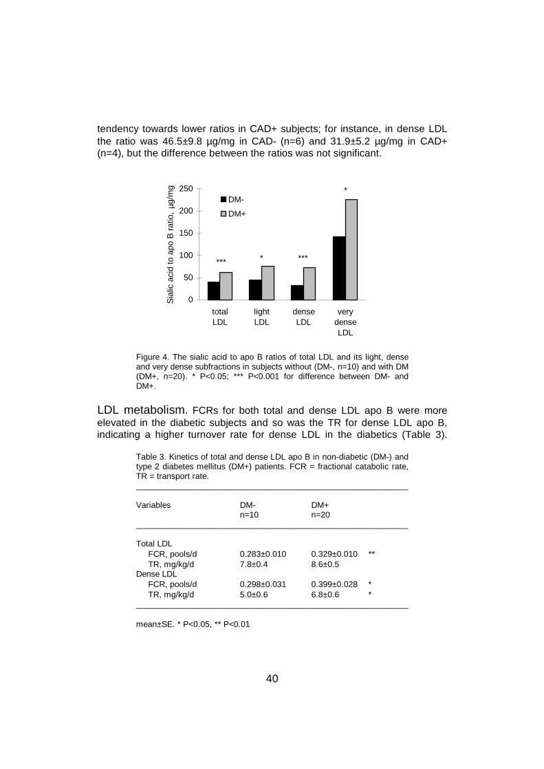
40
tendency towards lower ratios in CAD+ subjects; for instance, in dense LDLthe ratio was 46.5±9.8 µg/mg in CAD- (n=6) and 31.9±5.2 µg/mg in CAD+(n=4), but the difference between the ratios was not significant.
0
50
100
150
200
250
totalLDL
lightLDL
denseLDL
verydenseLDL
Sia
lic a
cid
to a
po B
rat
io,
µg/m
g
DM-
DM+
*** ****
*
Figure 4. The sialic acid to apo B ratios of total LDL and its light, denseand very dense subfractions in subjects without (DM-, n=10) and with DM(DM+, n=20). * P<0.05; *** P<0.001 for difference between DM- andDM+.
LDL metabolism. FCRs for both total and dense LDL apo B were moreelevated in the diabetic subjects and so was the TR for dense LDL apo B,indicating a higher turnover rate for dense LDL in the diabetics (Table 3).
Table 3. Kinetics of total and dense LDL apo B in non-diabetic (DM-) andtype 2 diabetes mellitus (DM+) patients. FCR = fractional catabolic rate,TR = transport rate.___________________________________________________________
Variables DM- DM+n=10 n=20
___________________________________________________________
Total LDL FCR, pools/d 0.283±0.010 0.329±0.010 ** TR, mg/kg/d 7.8±0.4 8.6±0.5Dense LDL FCR, pools/d 0.298±0.031 0.399±0.028 * TR, mg/kg/d 5.0±0.6 6.8±0.6 *___________________________________________________________
mean±SE. * P<0.05, ** P<0.01
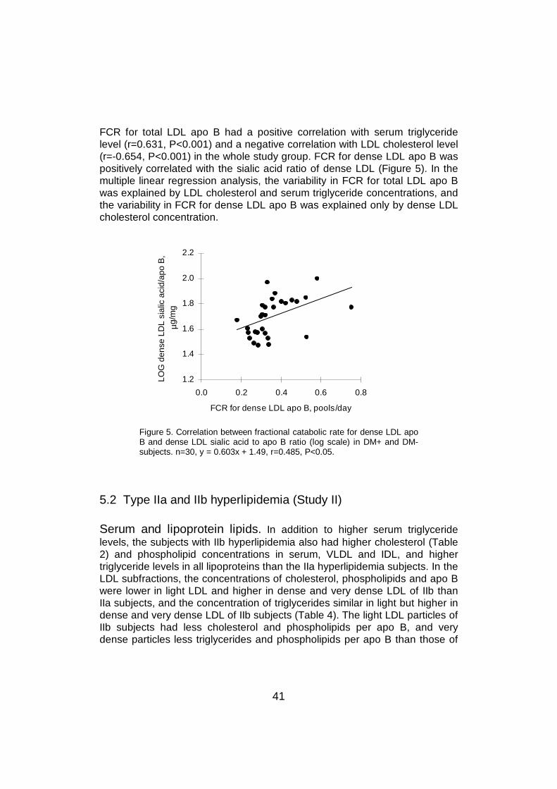
41
FCR for total LDL apo B had a positive correlation with serum triglyceridelevel (r=0.631, P<0.001) and a negative correlation with LDL cholesterol level(r=-0.654, P<0.001) in the whole study group. FCR for dense LDL apo B waspositively correlated with the sialic acid ratio of dense LDL (Figure 5). In themultiple linear regression analysis, the variability in FCR for total LDL apo Bwas explained by LDL cholesterol and serum triglyceride concentrations, andthe variability in FCR for dense LDL apo B was explained only by dense LDLcholesterol concentration.
1.2
1.4
1.6
1.8
2.0
2.2
0.0 0.2 0.4 0.6 0.8
FCR for dense LDL apo B, pools/day
LOG
den
se L
DL
sial
ic a
cid/
apo
B,
µg/m
g
Figure 5. Correlation between fractional catabolic rate for dense LDL apoB and dense LDL sialic acid to apo B ratio (log scale) in DM+ and DM-subjects. n=30, y = 0.603x + 1.49, r=0.485, P<0.05.
5.2 Type IIa and IIb hyperlipidemia (Study II)
Serum and lipoprotein lipids. In addition to higher serum triglyceridelevels, the subjects with IIb hyperlipidemia also had higher cholesterol (Table2) and phospholipid concentrations in serum, VLDL and IDL, and highertriglyceride levels in all lipoproteins than the IIa hyperlipidemia subjects. In theLDL subfractions, the concentrations of cholesterol, phospholipids and apo Bwere lower in light LDL and higher in dense and very dense LDL of IIb thanIIa subjects, and the concentration of triglycerides similar in light but higher indense and very dense LDL of IIb subjects (Table 4). The light LDL particles ofIIb subjects had less cholesterol and phospholipids per apo B, and verydense particles less triglycerides and phospholipids per apo B than those of
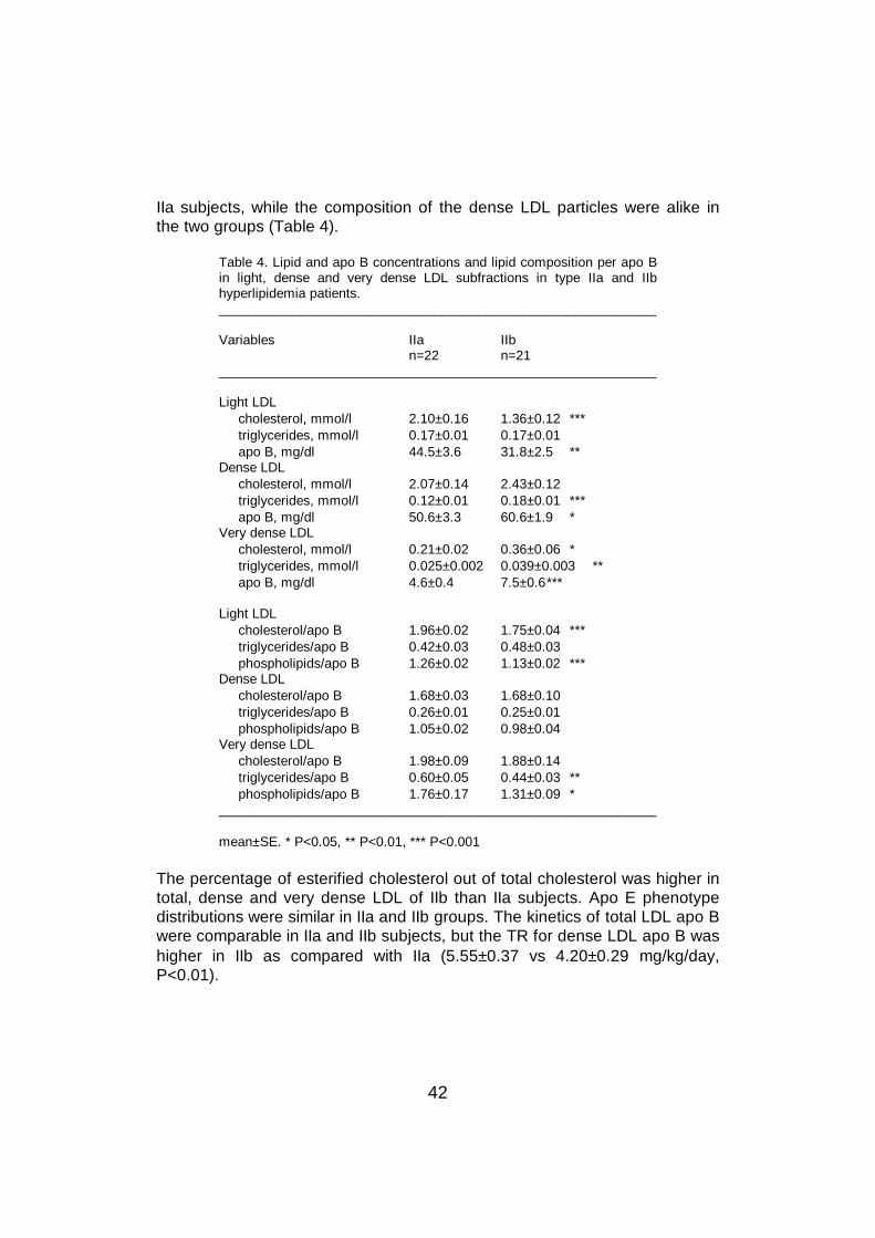
42
IIa subjects, while the composition of the dense LDL particles were alike inthe two groups (Table 4).
Table 4. Lipid and apo B concentrations and lipid composition per apo Bin light, dense and very dense LDL subfractions in type IIa and IIbhyperlipidemia patients.___________________________________________________________
Variables IIa IIbn=22 n=21
___________________________________________________________
Light LDL cholesterol, mmol/l 2.10±0.16 1.36±0.12 *** triglycerides, mmol/l 0.17±0.01 0.17±0.01 apo B, mg/dl 44.5±3.6 31.8±2.5 **Dense LDL cholesterol, mmol/l 2.07±0.14 2.43±0.12 triglycerides, mmol/l 0.12±0.01 0.18±0.01 *** apo B, mg/dl 50.6±3.3 60.6±1.9 *Very dense LDL cholesterol, mmol/l 0.21±0.02 0.36±0.06 * triglycerides, mmol/l 0.025±0.002 0.039±0.003 ** apo B, mg/dl 4.6±0.4 7.5±0.6***
Light LDL cholesterol/apo B 1.96±0.02 1.75±0.04 *** triglycerides/apo B 0.42±0.03 0.48±0.03 phospholipids/apo B 1.26±0.02 1.13±0.02 ***Dense LDL cholesterol/apo B 1.68±0.03 1.68±0.10 triglycerides/apo B 0.26±0.01 0.25±0.01 phospholipids/apo B 1.05±0.02 0.98±0.04Very dense LDL cholesterol/apo B 1.98±0.09 1.88±0.14 triglycerides/apo B 0.60±0.05 0.44±0.03 ** phospholipids/apo B 1.76±0.17 1.31±0.09 *___________________________________________________________
mean±SE. * P<0.05, ** P<0.01, *** P<0.001
The percentage of esterified cholesterol out of total cholesterol was higher intotal, dense and very dense LDL of IIb than IIa subjects. Apo E phenotypedistributions were similar in IIa and IIb groups. The kinetics of total LDL apo Bwere comparable in IIa and IIb subjects, but the TR for dense LDL apo B washigher in IIb as compared with IIa (5.55±0.37 vs 4.20±0.29 mg/kg/day,P<0.01).
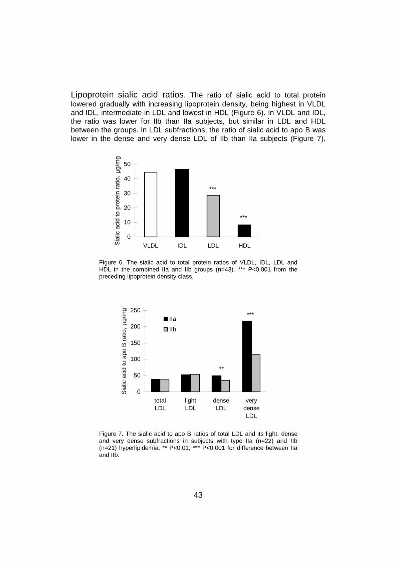
43
Lipoprotein sialic acid ratios. The ratio of sialic acid to total proteinlowered gradually with increasing lipoprotein density, being highest in VLDLand IDL, intermediate in LDL and lowest in HDL (Figure 6). In VLDL and IDL,the ratio was lower for IIb than IIa subjects, but similar in LDL and HDLbetween the groups. In LDL subfractions, the ratio of sialic acid to apo B waslower in the dense and very dense LDL of IIb than IIa subjects (Figure 7).
0
10
20
30
40
50
VLDL IDL LDL HDLSia
lic a
cid
to p
rote
in r
atio
, µg
/mg
***
***
Figure 6. The sialic acid to total protein ratios of VLDL, IDL, LDL andHDL in the combined IIa and IIb groups (n=43). *** P<0.001 from thepreceding lipoprotein density class.
0
50
100
150
200
250
totalLDL
lightLDL
denseLDL
verydenseLDL
Sia
lic a
cid
to a
po B
rat
io,
µg/m
g
IIa
IIb
***
**
Figure 7. The sialic acid to apo B ratios of total LDL and its light, denseand very dense subfractions in subjects with type IIa (n=22) and IIb(n=21) hyperlipidemia. ** P<0.01; *** P<0.001 for difference between IIaand IIb.
44
CAD+ and -CAD subjects had similar sialic acid ratios in IIa and IIb groupsseparately, and when the groups were combined, the ratio did not differbetween CAD+ and CAD- subjects in total, light or dense LDL, but was higherin the very dense LDL of CAD (n=33) than non-CAD (n=10) subjects(178.9±20.1 and 129.7±12.6 µg/mg, respectively, P=0.04). This ispresumably associated with the presence of Lp(a) in this fraction, and serumLp(a) levels were unsignificantly higher in CAD+ than CAD- subjects (345±59vs 199±55 U/l, respectively, P=0.08). The 8 subjects with DM had similar LDLsialic acid ratios than the rest of the subjects.
Correlations. In IIa and IIb hyperlipidemia groups combined, the sialic acidratios of dense and very dense LDL were negatively related with theircholesterol (r=-0.526 and r=-0.597, respectively, P<0.001 for both) andtriglyceride concentrations (r=-0.486 and r=-0.463, respectively, P<0.01 forboth), and with the TR for dense LDL apo B (r=-0.575 and r=-0.624, P<0.01for both). TR for dense LDL apo B was positively correlated with thecholesterol concentration of dense LDL, serum triglyceride level, and theesterification percentage of total LDL cholesterol (r=0.675, P<0.001).
5.3 Coronary artery disease (Study III)
Clinical characteristics and LDL sialic acid ratios. Sialic acid ratios oftotal LDL and its subfractions did not differ between males and females, orsmokers and non-smokers. The use of thiazide diuretics or, in females,hormone replacement therapy, had no effect on LDL sialic acid ratios. In thewhole study group and in the CAD+ group, LDL sialic acid ratios were notaffected by beta-blocker treatment, but in hypertensive CAD- subjects onbeta-blockers (n=6) the sialic acid ratio was lower in total LDL than in CAD-subjects not on beta-blockers (n=34) (33.5±3.1 vs 44.9±2.3 µg/mg,respectively, P<0.01). CAD+ and CAD- subjects were of similar age, bodymass index (BMI), and sex and apo E phenotype distributions.
LDL sialic acid ratio and CAD. LDL cholesterol concentration was higherin CAD+ than CAD- subjects, while levels of HDL cholesterol, serumtriglycerides and Lp(a) were similar between the groups. The mean sialic acidratios of total LDL and its dense and very dense subfractions were lower inCAD+ than CAD- subjects (Figure 8). When the CAD+ and CAD- subjectswere divided into quartiles by LDL sialic acid ratio, the ratios of both total anddense LDL were markedly lower in CAD+ than CAD- subjects of the threehighest quartiles but not in those of the lowest quartile. However, in thestepwise regression analysis with the presence of CAD as the dependent
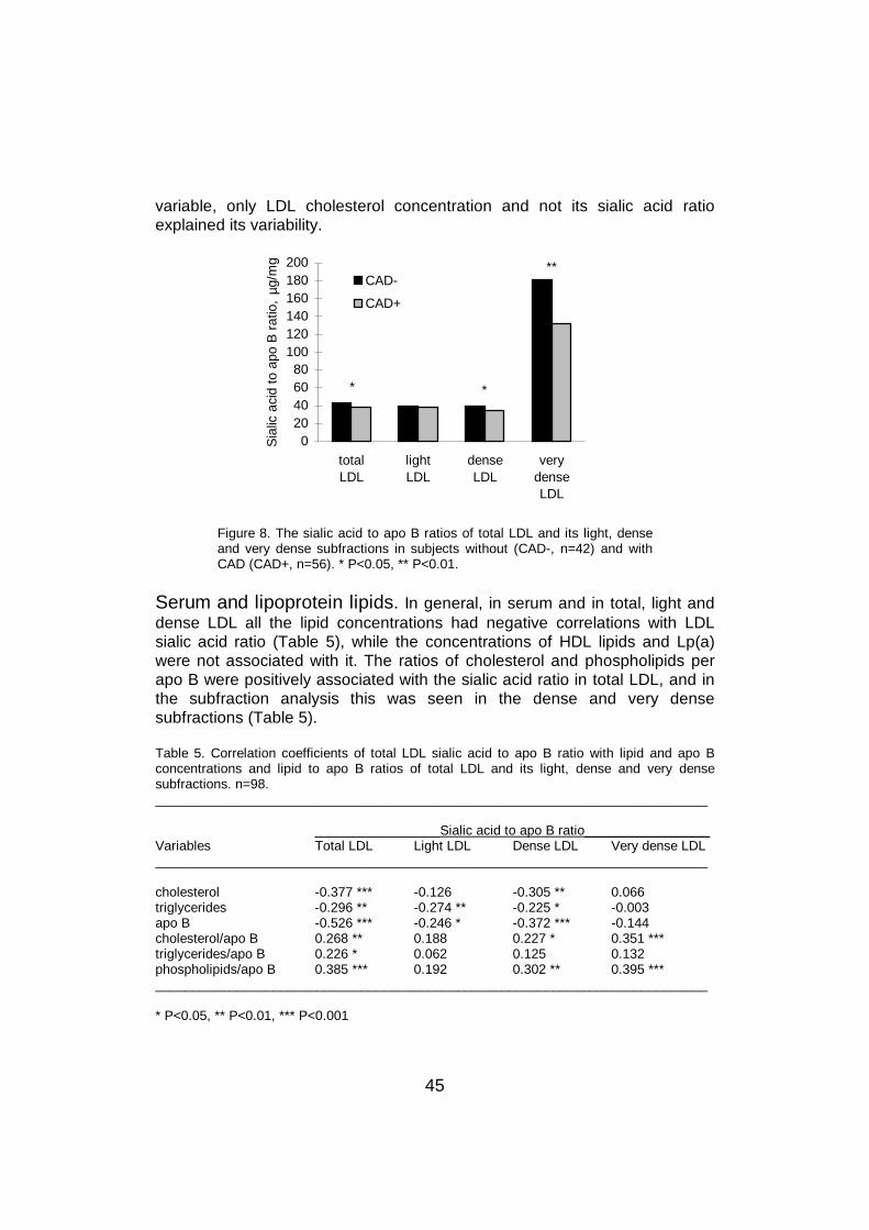
45
variable, only LDL cholesterol concentration and not its sialic acid ratioexplained its variability.
020406080
100120140160180200
totalLDL
lightLDL
denseLDL
verydenseLDL
Sia
lic a
cid
to a
po B
rat
io,
µg/m
gCAD-
CAD+
**
* *
Figure 8. The sialic acid to apo B ratios of total LDL and its light, denseand very dense subfractions in subjects without (CAD-, n=42) and withCAD (CAD+, n=56). * P<0.05, ** P<0.01.
Serum and lipoprotein lipids. In general, in serum and in total, light anddense LDL all the lipid concentrations had negative correlations with LDLsialic acid ratio (Table 5), while the concentrations of HDL lipids and Lp(a)were not associated with it. The ratios of cholesterol and phospholipids perapo B were positively associated with the sialic acid ratio in total LDL, and inthe subfraction analysis this was seen in the dense and very densesubfractions (Table 5).
Table 5. Correlation coefficients of total LDL sialic acid to apo B ratio with lipid and apo Bconcentrations and lipid to apo B ratios of total LDL and its light, dense and very densesubfractions. n=98.___________________________________________________________________________
_________________Sialic acid to apo B ratio_________________Variables Total LDL Light LDL Dense LDL Very dense LDL___________________________________________________________________________
cholesterol -0.377 *** -0.126 -0.305 ** 0.066triglycerides -0.296 ** -0.274 ** -0.225 * -0.003apo B -0.526 *** -0.246 * -0.372 *** -0.144cholesterol/apo B 0.268 ** 0.188 0.227 * 0.351 ***triglycerides/apo B 0.226 * 0.062 0.125 0.132phospholipids/apo B 0.385 *** 0.192 0.302 ** 0.395 ***___________________________________________________________________________
* P<0.05, ** P<0.01, *** P<0.001
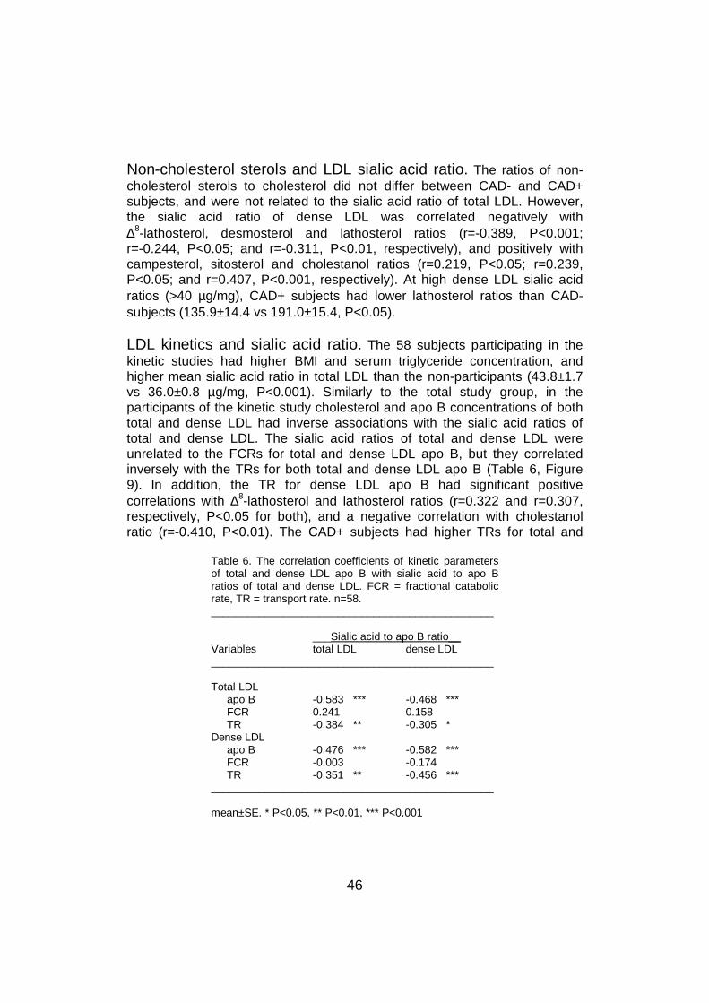
46
Non-cholesterol sterols and LDL sialic acid ratio. The ratios of non-cholesterol sterols to cholesterol did not differ between CAD- and CAD+subjects, and were not related to the sialic acid ratio of total LDL. However,the sialic acid ratio of dense LDL was correlated negatively with∆8-lathosterol, desmosterol and lathosterol ratios (r=-0.389, P<0.001;r=-0.244, P<0.05; and r=-0.311, P<0.01, respectively), and positively withcampesterol, sitosterol and cholestanol ratios (r=0.219, P<0.05; r=0.239,P<0.05; and r=0.407, P<0.001, respectively). At high dense LDL sialic acidratios (>40 µg/mg), CAD+ subjects had lower lathosterol ratios than CAD-subjects (135.9±14.4 vs 191.0±15.4, P<0.05).
LDL kinetics and sialic acid ratio. The 58 subjects participating in thekinetic studies had higher BMI and serum triglyceride concentration, andhigher mean sialic acid ratio in total LDL than the non-participants (43.8±1.7vs 36.0±0.8 µg/mg, P<0.001). Similarly to the total study group, in theparticipants of the kinetic study cholesterol and apo B concentrations of bothtotal and dense LDL had inverse associations with the sialic acid ratios oftotal and dense LDL. The sialic acid ratios of total and dense LDL wereunrelated to the FCRs for total and dense LDL apo B, but they correlatedinversely with the TRs for both total and dense LDL apo B (Table 6, Figure9). In addition, the TR for dense LDL apo B had significant positivecorrelations with ∆8-lathosterol and lathosterol ratios (r=0.322 and r=0.307,respectively, P<0.05 for both), and a negative correlation with cholestanolratio (r=-0.410, P<0.01). The CAD+ subjects had higher TRs for total and
Table 6. The correlation coefficients of kinetic parametersof total and dense LDL apo B with sialic acid to apo Bratios of total and dense LDL. FCR = fractional catabolicrate, TR = transport rate. n=58._______________________________________________
___Sialic acid to apo B ratio__Variables total LDL dense LDL_______________________________________________
Total LDL apo B -0.583 *** -0.468 *** FCR 0.241 0.158 TR -0.384 ** -0.305 *Dense LDL apo B -0.476 *** -0.582 *** FCR -0.003 -0.174 TR -0.351 ** -0.456 ***_______________________________________________
mean±SE. * P<0.05, ** P<0.01, *** P<0.001
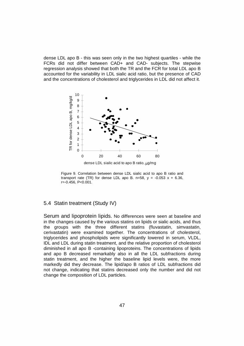
47
dense LDL apo B - this was seen only in the two highest quartiles - while theFCRs did not differ between CAD+ and CAD- subjects. The stepwiseregression analysis showed that both the TR and the FCR for total LDL apo Baccounted for the variability in LDL sialic acid ratio, but the presence of CADand the concentrations of cholesterol and triglycerides in LDL did not affect it.
0
1
2
3
4
5
6
7
8
9
10
0 20 40 60 80
dense LDL sialic acid to apo B ratio, µg/mg
TR
for
dens
e LD
L ap
o B
, mg/
kg/d
Figure 9. Correlation between dense LDL sialic acid to apo B ratio andtransport rate (TR) for dense LDL apo B. n=58, y = -0.053 x + 6.36,r=-0.456, P<0.001.
5.4 Statin treatment (Study IV)
Serum and lipoprotein lipids. No differences were seen at baseline andin the changes caused by the various statins on lipids or sialic acids, and thusthe groups with the three different statins (fluvastatin, simvastatin,cerivastatin) were examined together. The concentrations of cholesterol,triglycerides and phospholipids were significantly lowered in serum, VLDL,IDL and LDL during statin treatment, and the relative proportion of cholesteroldiminished in all apo B -containing lipoproteins. The concentrations of lipidsand apo B decreased remarkably also in all the LDL subfractions duringstatin treatment, and the higher the baseline lipid levels were, the moremarkedly did they decrease. The lipid/apo B ratios of LDL subfractions didnot change, indicating that statins decreased only the number and did notchange the composition of LDL particles.
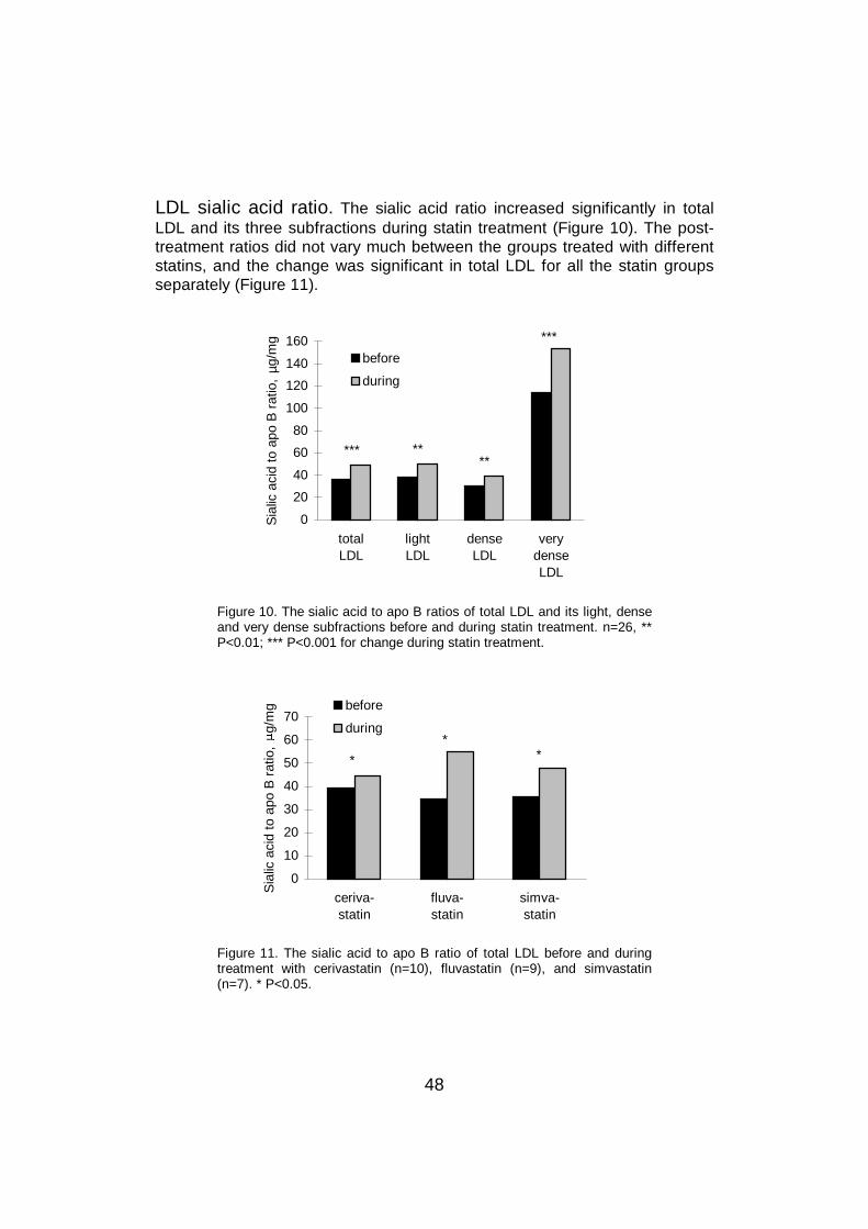
48
LDL sialic acid ratio. The sialic acid ratio increased significantly in totalLDL and its three subfractions during statin treatment (Figure 10). The post-treatment ratios did not vary much between the groups treated with differentstatins, and the change was significant in total LDL for all the statin groupsseparately (Figure 11).
0
20
40
60
80
100
120
140
160
totalLDL
lightLDL
denseLDL
verydenseLDL
Sia
lic a
cid
to a
po B
rat
io,
µg/m
g
before
during
***
***
****
Figure 10. The sialic acid to apo B ratios of total LDL and its light, denseand very dense subfractions before and during statin treatment. n=26, **P<0.01; *** P<0.001 for change during statin treatment.
0
10
20
30
40
50
60
70
ceriva-statin
fluva-statin
simva-statin
Sia
lic a
cid
to a
po B
rat
io,
g/m
g before
during
*
**
Figure 11. The sialic acid to apo B ratio of total LDL before and duringtreatment with cerivastatin (n=10), fluvastatin (n=9), and simvastatin(n=7). * P<0.05.
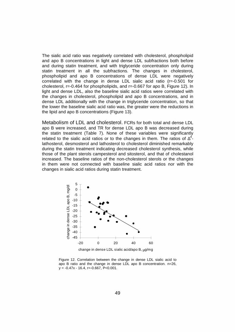
49
The sialic acid ratio was negatively correlated with cholesterol, phospholipidand apo B concentrations in light and dense LDL subfractions both beforeand during statin treatment, and with triglyceride concentration only duringstatin treatment in all the subfractions. The changes in cholesterol,phospholipid and apo B concentrations of dense LDL were negativelycorrelated with the change in dense LDL sialic acid ratio (r=-0.501 forcholesterol, r=-0.464 for phospholipids, and r=-0.667 for apo B, Figure 12). Inlight and dense LDL, also the baseline sialic acid ratios were correlated withthe changes in cholesterol, phospholipid and apo B concentrations, and indense LDL additionally with the change in triglyceride concentration, so thatthe lower the baseline sialic acid ratio was, the greater were the reductions inthe lipid and apo B concentrations (Figure 13).
Metabolism of LDL and cholesterol. FCRs for both total and dense LDLapo B were increased, and TR for dense LDL apo B was decreased duringthe statin treatment (Table 7). None of these variables were significantlyrelated to the sialic acid ratios or to the changes in them. The ratios of ∆8-lathosterol, desmosterol and lathosterol to cholesterol diminished remarkablyduring the statin treatment indicating decreased cholesterol synthesis, whilethose of the plant sterols campesterol and sitosterol, and that of cholestanolincreased. The baseline ratios of the non-cholesterol sterols or the changesin them were not connected with baseline sialic acid ratios nor with thechanges in sialic acid ratios during statin treatment.
-45
-40
-35
-30
-25
-20
-15
-10
-5
0
5
-20 0 20 40 60
change in dense LDL sialic acid/apo B, µg/mg
chan
ge in
den
se L
DL
apo
B, m
g/dl
Figure 12. Correlation between the change in dense LDL sialic acid toapo B ratio and the change in dense LDL apo B concentration. n=26,y = -0.47x - 16.4, r=-0.667, P<0.001.
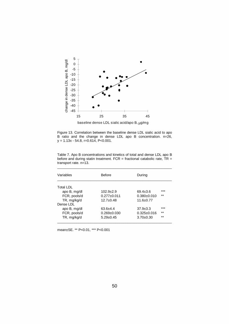
50
-45
-40
-35
-30
-25
-20
-15
-10
-5
0
5
15 25 35 45
baseline dense LDL sialic acid/apo B, µg/mg
chan
ge in
den
se L
DL
apo
B, m
g/dl
Figure 13. Correlation between the baseline dense LDL sialic acid to apoB ratio and the change in dense LDL apo B concentration. n=26,y = 1.13x - 54.8, r=0.614, P<0.001.
Table 7. Apo B concentrations and kinetics of total and dense LDL apo Bbefore and during statin treatment. FCR = fractional catabolic rate, TR =transport rate. n=13.___________________________________________________________
Variables Before During___________________________________________________________
Total LDL apo B, mg/dl 102.9±2.9 69.4±3.6 *** FCR, pools/d 0.277±0.011 0.380±0.010 ** TR, mg/kg/d 12.7±0.48 11.6±0.77Dense LDL apo B, mg/dl 63.6±4.4 37.9±3.3 *** FCR, pools/d 0.269±0.030 0.325±0.016 ** TR, mg/kg/d 5.29±0.45 3.70±0.30 **___________________________________________________________
mean±SE. ** P<0.01, *** P<0.001
51
6 DISCUSSION
6.1 General
The function of sialic acids in the metabolism of some glycoproteins (Stryer1988) and their presence in lipoproteins, especially LDL (Swaminathan andAladjem 1976), make their role in atherosclerosis an intriguing subject. In thelate 1970’s a few studies reported that desialylation of LDL affects itcatabolism both in vitro (Filipovic and Buddecke 1979, Filipovic et al 1979)and in vivo (Hendrix et al 1975, Malmendier et al 1980). Ten years later thesialic acid content of LDL and its association with atherosclerosis came tofocus again, first in the numerous studies by Orekhov et al (1989, 1991b,1992) and Tertov et al (1992a, 1992b), and later in the mid- to late 1990’salso in studies by other groups (Ruelland et al 1993, Chappey et al 1995,Grewal et al 1996, Anber et al 1997, Bartlett et al 1998, Harada et al 1998,Millar et al 1999).
6.2 Clinical characteristics
In this study, the sialic acid ratios of total LDL and its subfractions did notdiffer consistently between males and females. The mean age of the subjectswas around 55 years, and the females were practically all postmenopausal.In Study III, 15 females out of 53 were on hormone replacement therapy, andtheir LDL sialic acid ratios were similar to those of the women not takinghormones and to those of the men. The age of the subjects was notconnected with LDL sialic acid ratio, nor were there consistent associationsbetween BMI and LDL sialic acid ratio. Current smokers had LDL sialic acidratios approximately corresponding to those of non-smokers. Apo Ephenotypes had no relation with LDL sialic acid content. Consequently, LDLsialic acid ratio seems to be independent of demographic factors associatedwith an increased risk of CAD, that is, male sex, old age, overweight, andsmoking, and also of apo E phenotypes 4/3 and 4/4 known to associate withhigh CAD risk (Davignon et al 1988).
Beta-blocking agents are known to change plasma lipid profiles to a moreatherogenic direction (Lehtonen and Viikari 1979, Rohlfing and Brunzell1986, Cruickshank 1990, Superko et al 1993), but they reduce CAD mortality(Cruickshank 1990, Wikstrand et al 1991, Goldstein 1996). Overall, nosignificant lipid variations were found between beta-blocker users and non-
52
users in any of the present studies. However, in Study III in the CAD-subgroup, beta-blocker users had lower total LDL sialic acid ratio than non-users, while in the CAD+ subjects, who included most of the beta-blockerusers, there was no difference whatsoever in the sialic acid ratios betweenusers and non-users (39.0±1.6 vs 37.1±2.1 µg/mg for total LDL, respectively).The small number of beta-blocker users in the CAD- subgroup (6 out of 42subjects) does not allow to draw firm conclusions from the difference in thisgroup and, according to all available data, it is inferred that beta-blockersseem not to have a significant effect on LDL sialic acid levels. Furthermore,the use of thiazide diuretics, or the type of hypoglycemic therapy in DMsubjects did not affect the LDL sialic acid ratio.
6.3 Measurement of sialic acid ratio
The sialic acid content of lipoproteins was measured mainly with theresorcinol method (Svennerholm 1957, Miettinen and Takki-Luukkainen1959), and in Study I, also with the thiobarbituric acid assay (Warren 1959a).Both methods are very practicable and reliable. Results from repeatedanalyses with the resorcinol method were constant, with an intra-assay andinterassay variability of less than 5% in the present study. The resorcinolmethod was preferably used because no time-consuming dialysis of thesamples is necessary, and the reagents are more stable than in thethiobarbituric acid assay. Most other investigators have employed thethiobarbituric acid assay, while the resorcinol method has been used by aminority of groups (Fontaine and Malmendier 1975, Fontaine and Malmendier1978, La Belle and Krauss 1990, Sobenin et al 1993, Tertov et al 1996b,Millar et al 1999). The two methods, utilized in parallel in Study I, gave similarresults (r=0.960) in conformity with other reports (Orekhov et al 1991b,Orekhov et al 1992).
The overall sialic acid to apo B ratios of LDL were on similar level in all thesubject groups, the mean values of the groups ranging from 36.7 to 43.7µg/mg apo B for total LDL, excluding the diabetic subjects in Study I who hada clearly higher ratio (mean of 62.3 µg/mg). These levels were close to thosereported in some studies (Orekhov et al 1989, La Belle and Krauss 1990,Orekhov et al 1991b, Grewal et al 1996), but markedly higher than in mostother papers (Maruhama et al 1983, Orekhov et al 1992, Tertov et al 1992a,Ruelland et al 1993, Sobenin et al 1993, Tertov et al 1993, Chappey et al1995, Myara et al 1995, Ruelland et al 1997, Chappey et al 1998, Harada etal 1998), in which the sialic acid levels were between 6 and 13 µg/mg protein.Thus, there are large variations in the sialic acid ratios in studies by differentgroups, and the results of this study are not the only ones that deviate from

53
the majority of reports. The variability is not a question of methodology,because the two methods employed in the earlier studies and the presentone give similar and reliable results as already discussed. Therefore, thelarge differences in the sialic acid levels can not be explained, but with asimilar range in all the sections of the present study the measurements usedin them can be considered reliable.
The sialic acid content of LDL is expressed as ratio to the concentration ofapo B in LDL because of the high dependence of its concentration on that ofapo B, as already pointed out in the Methods chapter, and this having beenthe praxis in all previous studies. In the present study, the concentration ofapo B was measured with an immunoturbidometric method (Riepponen et al1987), while in most other studies the total protein of LDL has beenmeasured with the Lowry method (Lowry et al 1951). Apo B is the onlyprotein in LDL, and its concentration is very close to the total proteinconcentration in LDL. The reproducibility of results with the apo B analysismethod is good, and the levels are closely similar to those obtained with theLowry method (apo B 35.1±1.6 mg/dl and total protein 38.7±1.7 mg/dl indense LDL, n=90, r=0.967). Thus, variability in protein measurements cannotexplain the differences in the sialic acid to apo B ratios observed in differentstudies.
6.4 Lipoproteins and sialic acids
Negative associations between serum and lipoprotein lipid levels and LDLsialic acid ratio have been described also earlier (La Belle and Krauss 1990,Chappey et al 1995). In the present study, serum total and LDL cholesterollevels had negative correlations with the sialic acid ratio of total LDL in thelarge population of Study III. This indicates that high LDL cholesterol level isassociated with low LDL sialic acid ratio, both presumably acceleratingatherogenesis. However, in the smaller patient groups of Studies I, II and IV,the negative associations between serum or LDL cholesterol concentrationsand LDL sialic acid ratios were not significant, similarly to two other studies(Chappey et al 1995, Millar et al 1999), while La Belle and Krauss (1990) alsofound a weak but significant negative association between the cholesterolconcentration and the sialic acid ratio of LDL. This can simply be due to theconnection not being strong enough to be seen in all populations.
Serum triglyceride levels have been inversely associated with LDL sialic acidratio (La Belle and Krauss 1990, Chappey et al 1995). In the present study,the sialic acid ratio of total LDL had no association with serum triglyceridelevels, but in Study III it was negatively related with the triglyceride
54
concentration of LDL. Furthermore, a clear negative association was seen fordense and very dense LDL sialic acid ratios and serum triglyceride levels inStudies II and III, and only for very dense LDL sialic acid ratio in Study IV. InStudy II, the subjects were divided into groups according to their serumtriglyceride level, and in the IIb hyperlipidemics high triglyceride levels wereassociated with low sialic acid ratios in dense and very dense LDL, but not intotal LDL. Consequently, an inverse association seems to exist betweenserum triglyceride levels and the sialic acid ratios of the denser LDLsubfractions.
High concentrations of apo B in serum and LDL are considered a remarkablerisk factor for CAD (Sniderman et al 1980). The sialic acid ratios of total anddense LDL had strong negative associations with serum and LDL apo Blevels in Study III. However, in the smaller populations of Studies II and IV,the relation with serum apo B level was significant only for very dense, and inStudy II, also for dense LDL sialic acid ratio. Again, the significant findings inthe large population suggest that there is a correlation that is not seen insmaller populations. Overall, these associations of LDL sialic acid ratios withserum and LDL lipid and apo B levels indicate that atherogenic lipid profiles,specifically high cholesterol, triglyceride and apo B concentrations in LDL, areconnected with low LDL sialic acid ratios, especially in the dense and verydense subfractions. On the contrary, no association between LDL sialic acidratios and HDL cholesterol level was seen in any of the studies.
The sialic acid ratios of the light, dense and very dense LDL subfractionswere in general negatively associated with the respective cholesterol,triglyceride, phospholipid and apo B concentrations, the inverse associationbeing strongest for apo B concentrations. The correlations were significant indense LDL for all the lipid and apo B levels in every study section, and forlight and very dense LDL in almost all of them. As each LDL particle containsone apo B molecule, the concentration of apo B reflects the number of LDLparticles. Thus, the inverse correlation between apo B concentration andsialic acid ratio suggests that when the number of LDL particles is high, theirsialic acid content is low. In addition, the sialic acid ratios were positivelyassociated with the lipid to apo B ratios of total LDL and its subfractions,which reflect the size of LDL particles. These observations together suggestthat low LDL sialic acid content is associated with a large number of relativelysmall LDL particles, generally known to be atherogenic, and are also inconcordance with an earlier finding that subjects with small dense LDL havea lower LDL sialic acid content (La Belle and Krauss 1990).
The ratio of sialic acid to protein was negatively associated with the density oflipoproteins so that the ratio lowered from VLDL and IDL to LDL. Similardecreases were described in three recent studies (Anber et al 1997, Harada
55
et al 1998, Millar et al 1999). There is a number of possible explanations forthis loss of sialic acid from lipoproteins when density increases. First,apolipoproteins other than apo B are lost during the conversion of VLDL intoIDL and further into LDL; second, the removal of sialic acid -rich apo B-containg lipoproteins could be enhanced, and third, apo B could bedesialylated by a neuraminidase during this conversion into denser particlesowing to longer residence time in the circulation. The recently reported findingthat in plasma, LDL apo B is always partially desialylated (Bartlett andStanley 1998) suggests that desialylation of apo B does take place in plasmaand can contribute to the observed decrease in lipoprotein sialic acid ratiowith increasing density. On the other hand, LDL is richer in gangliosides thanVLDL (Senn et al 1989), and as the amount of sialic acid in the glycolipid partof lipoproteins is thus higher in LDL than VLDL, the decrease in the sialic acidcontent of the lipoproteins with delipidation indeed seems to ensue from aloss of sialic acid from the glycoprotein, that is, apo B.
HDL was found to have a lower sialic acid ratio than any of the apo B-containg lipoproteins, in concordance with a previous study (Harada et al1998). This is undoubtedly due to the major apolipoproteins in HDL, apo A-Iand A-II, not being glycoproteins, the small amount of sialic acid thus residingin the glycolipids of HDL and in some tightly bound glycoproteins like LCAT,PLTP and CETP.
The sialic acid ratios have been reported to diminish also by increasingdensity of LDL particles (La Belle and Krauss 1990, Millar et al 1999), incontinuation of the diminution in sialic acid ratio from triglyceride-rich apo B-containing lipoproteins to LDL. Results from the present study partly supportprevious findings, but are not totally concordant. In Study II, the sialic acidratio fell significantly from light to dense LDL only in the IIb but not in the IIagroup; however, the decrease was significant when both groups werecombined (53.2±2.2 to 42.1±2.2 µg/mg, P=0.002). In Study IV, the ratiodecreased clearly from light to dense LDL both at baseline and during thestatin treatment (P<0.01 for both). On the contrary, in Study I the sialic acidratios of the diabetic patients were similar in light and dense LDL, and in thenon-diabetic group, there was a non-significant decrease from light to densesubfraction (P=0.09). In Study III, no significant decrease was seen either forall the patients together or for CAD+ and CAD- subjects separately. However,the positive associations of LDL sialic acid ratios with lipid to apo B ratiossuggest that when the sialic acid ratios are low, the particles are lipid-depleted and thus denser and smaller in size. In summary, at least atendency towards decreasing sialic acid ratios with increasing particle densitywas seen in most of the present study populations, and desialylation of LDLapo B possibly contributes to this loss of sialic acid, supported by the multiplefindings of desialylated LDL in human plasma (Orekhov et al 1992, Tertov et
56
al 1992a, Tertov et al 1992b, Sobenin et al 1993, Tertov et al 1993, Bartlettand Stanley 1998).
In all the study sections and patient groups, the sialic acid ratio was clearlyhighest in very dense LDL. This is most presumably due to the presence ofLp(a), which is highly sialylated (Ehnholm et al 1972) and overlaps with thedensity range of LDL. Separation of dense and very dense LDL wasperformed to minimize the amount of Lp(a) in the dense subfraction andapparently this succeeded, because the dense LDL subfraction, having aremarkably lower sialic acid ratio, probably did not contain significantamounts of Lp(a). Furthermore, separate measurements showed that lessthan 30% of the subjects, all of whom had relatively high Lp(a) concentrationsin serum, had detectable amounts of Lp(a) in dense LDL, and in thesesubjects its mean concentration in dense LDL was 16.6±2.9 U/l or 4.7% ofserum total Lp(a). Similarly, very dense LDL contained 8.3% of serum Lp(a).
6.5 LDL sialic acid content in different conditions
6.5.1 Type 2 diabetes mellitus
The sialic acid ratio of total LDL and its three subfractions was higher in DMsubjects than in the controls in Study I. The few DM patients in Studies II andIV (8 and 5, respectively) did not differ significantly from the non-DM subjects,but in both studies there was a non-significant tendency towards higher sialicacid ratios in the DM subjects. These results are contradictory to earlierobservations by Tertov et al (1992a) and Sobenin et al (1993), who foundlower content of sialic acid in the LDL of DM patients compared withnormoglycemic controls. In addition, other studies have reported nodifferences in LDL sialic acid ratio in normolipidemic subjects with or withoutDM (Maruhama et al 1983, Ruelland et al 1997). The reason for the very highsialic acid levels in the diabetic subjects of the present study might be in thecatabolism of LDL, which also differed from that of the non-DM subjects. Bothin Studies I and II the FCR for LDL apo B, and especially the FCR for denseLDL apo B, was significantly higher in the DM subjects compared withnormoglycemic controls. This is opposite to what was expected, because theFCR for LDL apo B has been reported to be decreased in DM (Howard et al1987). Thus, the higher sialic acid ratio in LDL was associated with anaccelerated removal of LDL from the circulation in DM. On the other hand,hypertriglyceridemia is often related with enhanced clearance of LDL (Vegaand Grundy 1989). The diabetic subjects in Study I had higher serumtriglyceride levels than the non-DM controls, which could explain their fasterLDL catabolism. The FCR for LDL apo B also had a positive correlation with
57
serum triglyceride levels, so that both high LDL sialic acid ratio and highserum triglyceride levels were connected with increased catabolism of LDLapo B in DM.
6.5.2 Type IIa and IIb hyperlipidemia
Even though the study population was selected according to their lipid profileand matched for age, sex and BMI, the composition of lipoproteins, especiallyof LDL, was similar in type IIb to that presented for familial combinedhyperlipidemia (Kane and Havel 1995). As compared with IIa, in IIbhyperlipidemia light LDL particles were lipid-depleted and fewer. Thecomposition of dense particles was similar in the two groups while in IIb thenumbers of both dense and very dense LDL particles were higher, and verydense particles were poor in triglycerides and phospholipids. Thus, in IIbsubjects both the number and the production rate of dense LDL particleswere elevated.
The sialic acid ratios of dense and very dense LDL subfractions were lower inIIb than IIa hyperlipidemia, in accordance with the negative correlation of thesialic acid ratios of dense and very dense LDL with serum triglyceride levels.In addition, LDL sialic acid ratio was low both in IIa and IIb hyperlipidemiacompared with a healthy female population. Both type IIa and IIbhyperlipidemic subjects have an increased risk of CAD. In IIb hyperlipidemia,elevated triglyceride levels are often associated with high concentrations ofdense LDL and low HDL cholesterol levels, this lipid profile being especiallyatherogenic (Grundy 1995a). This was the case also in the IIb hyperlipidemicsubjects of the present study with high concentrations of dense LDL and non-significantly lower HDL cholesterol levels compared with IIa subjects. It canbe assumed that the risk of developing CAD is even higher for IIb than IIasubjects, and the lower sialic acid content in the atherogenic dense LDLsubfraction of IIb subjects could contribute to their increased risk.
6.5.3 Coronary artery disease
Low sialic acid content has been found in LDL of CAD patients in many(Orekhov et al 1989, Orekhov et al 1991b, Tertov et al 1992a, Tertov et al1992b, Ruelland et al 1993, Millar et al 1999) but not in all studies (Chappeyet al 1998). It has been suggested that desialylation is an atherogenicmodification of LDL taking place in the circulation, since sialic acid -poor LDLhas been found in blood, especially in that of CAD patients (Tertov et al1992b). LDL with a low sialic acid content causes lipid deposition into cells
58
(Filipovic et al 1979, Orekhov et al 1992) and binds to arterial proteoglycans(Camejo et al 1985), which both phenomena are characteristic toatheromatous lesions. Desialylized LDL can be considered as a minimally-modified LDL that avidly internalizes in macrophage-foam cells. Thus, sialicacid -poor LDL could be one relevant factor leading to the development ofCAD.
In the large patient groups of Study III, CAD+ patients had significantly lowersialic acid ratios in total, dense and very dense LDL than CAD- subjects. Thisgives further support to the theory that the sialic acid content of LDL mightaffect its atherogenicity and thus predispose to the development of CAD.However, at low levels of total and dense LDL sialic acid ratio (in Q1), CAD+and CAD- subjects had similar mean sialic acid ratios, while at higher values(Q2-Q4) CAD+ patients had lower mean sialic acid ratios than CAD- subjects.This implies that the generally lower LDL sialic acid ratios in CAD+ than CAD-subjects may not be seen in populations with low LDL sialic acid ratios. Thiscould partially explain the inconsistencies of previous studies in findingdifferences in LDL sialic acid ratio between CAD+ and CAD- subjects.Furthermore, LDL cholesterol level was inversely associated with LDL sialicacid ratio and was higher in CAD+ than CAD- subjects. Thus, in stepwiselogistic regression analysis only the concentration of cholesterol in LDL, andnot its sialic acid ratio, was associated with the presence of CAD, whichindicates that LDL cholesterol concentration is more strongly associated withCAD than is its sialic acid ratio.
However, in the other sections of the present study no clear associationbetween low sialic acid ratio and CAD was observed, but this can beexplained for instance by the following factors. The diabetic subjects, knownto have a very high CAD risk (Kannel and McGee 1979), had high LDL sialicacid levels in Study I, and since the difference in LDL sialic acid ratio seemsto be strongest at high levels of sialic acid ratios, the diabetics should exhibitmarked differences between CAD+ and CAD- subjects. However, diabeticCAD+ and CAD- subjects had equally high sialic acid ratios, and thus theyseem not to be comparable with non-diabetics. In the 10 normoglycemicsubjects in Study I, the sialic acid ratio did not differ significantly betweenCAD+ and CAD- subjects in total LDL or its subfractions, but there was atendency towards lower sialic acid ratios in dense LDL of CAD+ subjects. InStudy II, the sialic acid ratios of total and dense LDL were very similarbetween CAD+ and CAD- subjects, but this could be due to 8 out of the 10CAD- subjects belonging to the IIb hyperlipidemia group and thus having ahigh serum triglyceride level which was associated with a low LDL sialic acidratio. In Study IV, only 4 subjects did not have CAD, and their small numberdoes not allow to draw conclusions about the lack of differences in LDL sialicacid ratio between CAD- and CAD+ subjects.
59
Considering these confounding factors in the other sections of the presentstudy on this aspect, and the association being evident in Study III with thelargest number of patients, it can be deduced that non-diabetic CAD patientshave in general lower LDL sialic acid ratios than comparable subjects withoutCAD. On the other hand, since to this day there are no follow-up studies onthe matter, it can not be excluded that subjects with a high LDL sialic acidratio could have an elevated rate of fatal AMIs, those with low values havingnon-fatal AMIs; this would lead to surviving CAD patients having low LDLsialic acid content compared with the general population. In the future, follow-up studies will be crucial in the evaluation of this aspect.
In the combined IIa and IIb group in Study II, the sialic acid ratio was higher invery dense LDL of CAD+ than CAD- subjects, while no differences weredetected in total LDL or in the other subfractions. This is probably related to ahigher level of Lp(a), known to be associated with CAD in many populations(Genest et al 1991, Sandholzer et al 1992, Kario et al 1994, Bostom et al1996). If total LDL or its light and dense subfractions were contaminated withsignificant amounts of Lp(a), it probably would raise their sialic acid content.Accordingly, it could be assumed that CAD+ patients would have a higherLDL sialic acid ratio. However, even though CAD+ patients tended to have ahigher mean Lp(a) level in Studies II and III, they had generally lower LDLsialic acid ratios than CAD- subjects. In addition, there was no correlationbetween LDL sialic acid ratio and serum concentration of Lp(a) in the presentstudy, consistently with previous results (Chappey et al 1995). Moreover,subjects with serum Lp(a) levels in the low or high end of the range had sialicacid ratios in LDL and all its subfractions similar to those of all the subjectstogether. These findings strongly suggest that the possible contamination ofLDL with Lp(a), except in the case of the very dense subfraction, was sosmall that it can be considered insignificant. Furthermore, it can be presumedthat the fractions were equally contaminated with Lp(a) in CAD+ and CAD-subjects, and in the case of a real contamination, the difference in the sialicacid ratio between CAD+ and CAD- subjects would be even more marked inpure LDL.
6.6 LDL sialic acid content and lipoprotein metabolism
6.6.1 LDL catabolism
Multiple types of scavenger receptors with the ability to mediate uptake anddegradation of modified lipoproteins have been found in atheroscleroticlesions (Ylä-Herttuala 1996, Greaves et al 1998), while cells of these lesionsseem not to express LDL receptors (Ylä-Herttuala et al 1991, Luoma et al
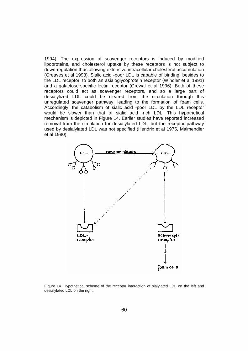
60
1994). The expression of scavenger receptors is induced by modifiedlipoproteins, and cholesterol uptake by these receptors is not subject todown-regulation thus allowing extensive intracellular cholesterol accumulation(Greaves et al 1998). Sialic acid -poor LDL is capable of binding, besides tothe LDL receptor, to both an asialoglycoprotein receptor (Windler et al 1991)and a galactose-specific lectin receptor (Grewal et al 1996). Both of thesereceptors could act as scavenger receptors, and so a large part ofdesialylized LDL could be cleared from the circulation through thisunregulated scavenger pathway, leading to the formation of foam cells.Accordingly, the catabolism of sialic acid -poor LDL by the LDL receptorwould be slower than that of sialic acid -rich LDL. This hypotheticalmechanism is depicted in Figure 14. Earlier studies have reported increasedremoval from the circulation for desialylated LDL, but the receptor pathwayused by desialylated LDL was not specified (Hendrix et al 1975, Malmendieret al 1980).
Figure 14. Hypothetical scheme of the receptor interaction of sialylated LDL on the left anddesialylated LDL on the right.
61
In Study I, a positive correlation between the sialic acid ratio of dense LDLand the FCR for dense LDL apo B was seen, while the correlation betweenthese parameters in total LDL did not quite reach significance. The subjectsin this study were mainly diabetics with high LDL sialic acid ratios and highFCRs for total and dense LDL. However, in the mostly non-diabetic subjectsof the other study sections no clear association came out. In Study II with 8out of 43 subjects being diabetic, a significant positive correlation wasestablished between the FCR for total LDL apo B and the sialic acid ratio oflight but not that of total or dense LDL. Due to the small number of subjects inthe kinetic analyses of Study IV no conclusion can be drawn from the lack ofcorrelations between LDL sialic acid and metabolic parameters. In thenormoglycemic population in Study III, the FCRs for total and dense LDL apoB were not significantly associated with their sialic acid ratios, although therewas a tendency towards a positive correlation between total LDL sialic acidratio and the FCR for LDL apo B (r=0.241, P=0.08). Interestingly, however, instepwise regression analysis the FCR for LDL apo B entered the modelexplaining the variability of LDL sialic acid ratio along with the TR for LDL apoB when the effect of the latter was removed. This suggests that both theproduction and the catabolism of LDL are connected to some extent with itssialic acid content. Globally, these data indicate that the sialic acid content ofLDL may have a weak positive correlation with its catabolism, the associationbeing stronger in diabetic subjects.
6.6.2 LDL production
The TR for dense LDL apo B was higher in IIb than IIa hyperlipidemia, andhad a strong positive correlation with serum triglyceride levels, implying thatin the presence of high triglyceride levels, the production of dense LDLparticles increases. This is supported by the fact that in FCHL (Kissebah et al1984, Teng et al 1986, Venkatesan et al 1993) and in hypertriglyceridemia(Vega et al 1985, Vega and Grundy 1989, Vega and Grundy 1998), there isan enhanced production of apo B from the liver, and production rates forVLDL and LDL are high. Thus, also more dense LDL particles are likely to beformed. The TR for dense LDL apo B was also strongly correlated with theesterification percentage of total LDL cholesterol in the IIa and IIbhyperlipidemia subjects, which indicates that the more LDL and specificallydense LDL is produced, the greater part of its cholesterol content isesterified. Cholesterol esterification and ester transfer rates have beenreported to be higher in subjects with hypertriglyceridemia or combinedhyperlipidemia than in hypercholesterolemic or normolipidemic subjects(Kudchodkar and Sodhi 1976, Murakami et al 1995), and to be positivelycorrelated with serum triglyceride level (Murakami et al 1995). This is in
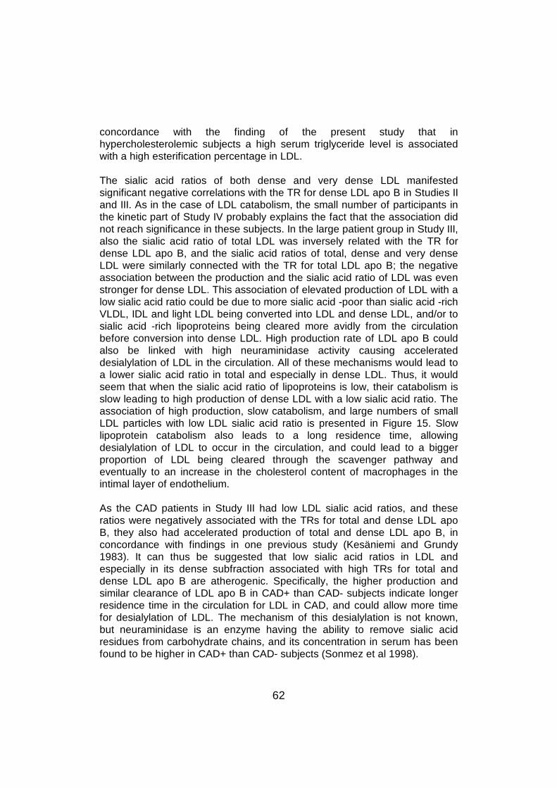
62
concordance with the finding of the present study that inhypercholesterolemic subjects a high serum triglyceride level is associatedwith a high esterification percentage in LDL.
The sialic acid ratios of both dense and very dense LDL manifestedsignificant negative correlations with the TR for dense LDL apo B in Studies IIand III. As in the case of LDL catabolism, the small number of participants inthe kinetic part of Study IV probably explains the fact that the association didnot reach significance in these subjects. In the large patient group in Study III,also the sialic acid ratio of total LDL was inversely related with the TR fordense LDL apo B, and the sialic acid ratios of total, dense and very denseLDL were similarly connected with the TR for total LDL apo B; the negativeassociation between the production and the sialic acid ratio of LDL was evenstronger for dense LDL. This association of elevated production of LDL with alow sialic acid ratio could be due to more sialic acid -poor than sialic acid -richVLDL, IDL and light LDL being converted into LDL and dense LDL, and/or tosialic acid -rich lipoproteins being cleared more avidly from the circulationbefore conversion into dense LDL. High production rate of LDL apo B couldalso be linked with high neuraminidase activity causing accelerateddesialylation of LDL in the circulation. All of these mechanisms would lead toa lower sialic acid ratio in total and especially in dense LDL. Thus, it wouldseem that when the sialic acid ratio of lipoproteins is low, their catabolism isslow leading to high production of dense LDL with a low sialic acid ratio. Theassociation of high production, slow catabolism, and large numbers of smallLDL particles with low LDL sialic acid ratio is presented in Figure 15. Slowlipoprotein catabolism also leads to a long residence time, allowingdesialylation of LDL to occur in the circulation, and could lead to a biggerproportion of LDL being cleared through the scavenger pathway andeventually to an increase in the cholesterol content of macrophages in theintimal layer of endothelium.
As the CAD patients in Study III had low LDL sialic acid ratios, and theseratios were negatively associated with the TRs for total and dense LDL apoB, they also had accelerated production of total and dense LDL apo B, inconcordance with findings in one previous study (Kesäniemi and Grundy1983). It can thus be suggested that low sialic acid ratios in LDL andespecially in its dense subfraction associated with high TRs for total anddense LDL apo B are atherogenic. Specifically, the higher production andsimilar clearance of LDL apo B in CAD+ than CAD- subjects indicate longerresidence time in the circulation for LDL in CAD, and could allow more timefor desialylation of LDL. The mechanism of this desialylation is not known,but neuraminidase is an enzyme having the ability to remove sialic acidresidues from carbohydrate chains, and its concentration in serum has beenfound to be higher in CAD+ than CAD- subjects (Sonmez et al 1998).
63
Figure 15. Schematic presentation of the metabolism of LDL and the size and concentration ofLDL particles in the case of high and low sialic acid to apo B ratio.
6.7 LDL sialic acid content and cholesterol metabolism
Serum ratios of the cholesterol precursor sterols ∆8-lathosterol, desmosteroland lathosterol to cholesterol reflect cholesterol synthesis, while the ratios ofthe plant sterols campesterol and sitosterol and the ratio of cholestanol reflectcholesterol absorption (Miettinen et al 1990). The correlations of dense LDLsialic acid ratio with serum non-cholesterol sterol ratios in the present studyindicate that a low sialic acid ratio in dense LDL was associated with highsynthesis and low absorption of cholesterol, and these were all connectedwith a high TR for dense LDL apo B. A positive correlation betweencholesterol synthesis and TR for LDL apo B has also been observed earlier inelderly men (Gylling et al 1994), but in middle-aged men no such correlationwas found, and contrary to the results now obtained, a positive correlationbetween cholesterol absorption and TR for LDL apo B has been reported(Miettinen et al 1992, Gylling et al 1994). However, recent studies with stableisotopes have demonstrated a positive correlation between cholesterolsynthesis and hepatic production of VLDL apo B (Watts et al 1995c, Richeset al 1997), and since in hypercholesterolemia both TRs for VLDL apo B andLDL apo B are elevated (Teng et al 1986), also more dense LDL apo B ispresumably formed. This is in accordance with the present results whichsuggest that the more cholesterol is synthesized, the more dense LDL isproduced, and the lower is its sialic acid ratio.
64
No consistent differences in cholesterol metabolism have been reportedbetween CAD+ and CAD- subjects previously, but low cholesterol synthesisrate was found to predispose to CAD in a study with FH patients (Miettinenand Gylling 1988). This is in concordance with the finding of this study that athigh dense LDL sialic acid ratios, CAD+ subjects had lower lathosterol ratiosthan CAD- subjects. The reason for this not being seen at lower sialic acidratios may be due to all the subjects with low sialic acid ratios having high TRfor dense LDL apo B, which in turn was associated with high lathosterolratios, and thus subjects with low LDL sialic acid ratios had high lathosterolratios independently of CAD status.
6.8 The effect of statin treatment on LDL sialic acid content
The change in the sialic acid ratios of total LDL and all its subfractions wasapproximately similar in the groups treated with different statins, i.e.cerivastatin (n=10), fluvastatin (n=9) and simvastatin (n=7). The cholesterolconcentrations of light and dense LDL subfractions at baseline werenegatively correlated with the respective baseline sialic acid ratios so thatwhen the cholesterol contents were decreased by statins, the sialic acidratios increased. Further, the more marked the decrease in cholesterol or apoB concentration of dense LDL was, the greater was the increase in its sialicacid ratio. Thus, since the strongest lipid lowering of LDL subfractions wasfound in the most hypercholesterolemic subjects, it could be assumed thatthese patients also had the biggest rise in sialic acid ratios. However, basallipid levels were not correlated with the change observed in the sialic acidratios.
Considering that desialylated LDL is supposed to be atherogenic, the newfinding that LDL sialic acid ratio rises during statin treatment could contributeto the well-known observation that statin treatment is associated with aregression of atherosclerosis. The decrease in dense LDL lipid and apo Bconcentrations was related to the basal sialic acid ratio of dense LDL so thata low sialic acid ratio at baseline predicted effective lowering of lipid and apoB concentrations.
In a recent study by Orekhov et al (1997), the sialic acid content of LDLincreased in patients treated with amlodipine alone or with the combination ofamlodipine and lovastatin. The augmentation was seen more rapidly with thecombination treatment than with amlodipine alone, but after a treatment offour weeks, the final level of sialic acid was the same whether or notlovastatin was used. In contrast to these findings, results of the present studyimply that statin treatment alone causes a rise in LDL sialic acid ratio. Millar et
65
al (1999) reported that ciprofibrate treatment reduced the sialic acid ratio oflipoproteins, and although the sialic acid content was inversely associatedwith the formation of atherogenic lipoprotein-proteoglycan complexes, theformation of these complexes diminished during ciprofibrate treatment.
LDL sialic acid ratio rose in most but not in all subjects during the statintreatment. The 5 subjects with a decreasing sialic acid ratio had a higher LDLsialic acid ratio at baseline than the other subjects, but otherwise they hadidentical clinical characteristics, baseline lipid and apo B concentrations andLDL kinetic parameters, and the changes in these parameters during statintreatment were comparable to those in patients with increasing LDL sialicacid content. Because of the good reproducibility of sialic acid measurementsdiscussed earlier, the decreasing sialic acid ratios in some subjects can noteither be explained by error in the measurement. Consequently, it is difficultto determine why their LDL sialic acid content responded differently.
As the lipid contents of lipoproteins fell during the statin treatment, theirmetabolism was also affected. The FCRs for total and dense LDL apo Bincreased, implying an accelerated catabolism. Furthermore, there was adecrease in the TR for dense LDL apo B, reflecting a diminished productionof this subfraction, while the TR for total LDL apo B was not affected. Thissuggests that statin treatment reduced the conversion of light LDL into themore atherogenic dense subfraction, which has also been associated with alower sialic acid content (La Belle and Krauss 1990). However, there was noconnection between baseline sialic acid ratios and the changes in LDLmetabolic variables, but it should be noted that the number of subjects in thekinetic study was limited.
The observed increases in LDL sialic acid ratio and LDL catabolic rate are inconcordance with the finding in Study I that high LDL sialic acid ratio isassociated with high FCR for LDL apo B, especially in diabetics. It is notknown which of these changes causes the other. Statin treatment augmentsLDL catabolism by upregulating the activity of LDL receptors (Kovanen et al1981, Reihner et al 1990), and TR for dense LDL apo B is apparentlydecreased because of effective removal of lipoproteins of lower density. So, itcould be assumed that when the sialic acid content of LDL is high, itscatabolism by the LDL receptor accelerates, which also leads to lowerproduction of dense LDL. On the other hand, high LDL catabolic rate couldlead to a higher sialic acid content by leaving less time for desialylation ofLDL in the circulation. Both of these mechanisms can link high LDL sialic acidratio to enhanced catabolism of LDL and to low production of dense LDL.Further confirmation to this would be given by investigating FH subjects, whohave a slow LDL catabolism and therefore presumably a low LDL sialic acidratio.
66
7 SUMMARY AND CONCLUSIONS
The sialic acid content of lipoproteins has been suggested to affectlipoprotein metabolism and the development of atherosclerosis. Therefore,the sialic acid content of lipoproteins, especially that of LDL and its threesubfractions, and its relations to different lipid variables and lipoprotein andcholesterol metabolism, as well as to DM, type IIa and IIb hyperlipidemias,and CAD were investigated. Furthermore, the effect of statin treatment onLDL sialic acid content was studied.
To study these aspects, subjects were examined as follows: 20 type 2 DMsubjects and 10 non-diabetic controls (Study I), 22 type IIa and 21 type IIbhyperlipidemic subjects (Study II), 56 CAD and 42 non-CAD subjects (StudyIII), and 26 hypercholesterolemic subjects before and during statin treatment(Study IV). Lipoproteins were separated by ultracentrifugation, and sialic acidcontents were measured with the resorcinol method. The metabolism oflipoproteins was investigated with radioactive iodine labeling, and that ofcholesterol by measuring non-cholesterol sterols from serum.
The sialic acid content of apo B -containing lipoproteins decreased withincreasing particle density from VLDL to LDL, and less consistently from lightto dense LDL. The sialic acid content of LDL was negatively correlated withthe concentrations of cholesterol, triglycerides, phospholipids, and apo Bespecially in the dense LDL subfraction, and low sialic acid content wasassociated with large amounts of relatively small LDL particles.
LDL sialic acid content had a weak positive correlation with the catabolism ofLDL apo B, the association being stronger in type 2 diabetes mellitus. Theproduction rate of LDL apo B was inversely related with LDL sialic acidcontent, especially in dense LDL. Low sialic acid content in dense LDL wasalso associated with high synthesis and low absorption of cholesterol, whichin turn were also related with high production rate of dense LDL apo B.
CAD patients had a lower sialic acid content in LDL, especially at high sialicacid ratios, than non-CAD subjects. Type IIb hyperlipidemia was associatedwith a low sialic acid content in dense and very dense LDL compared withtype IIa hyperlipidemia. Type 2 diabetic patients had higher sialic acidcontents in total LDL and its subfractions than non-diabetics, connected withhigher catabolism of LDL apo B. Age, sex, BMI, smoking and apo E
67
phenotype had no effect on LDL sialic acid ratio, neither had the use of beta-blockers, thiazide diuretics or hormone replacement therapy.
Statin treatment increased the sialic acid ratio of LDL and its subfractionsproportionately to the decrease in cholesterol levels, and in dense LDL lowsialic acid content at baseline predicted effective lowering of cholesterol levelin the subfraction. Production of dense but not that of total LDL apo Bdecreased during statin treatment, while catabolism of both total and denseLDL apo B increased. The number of subjects in the kinetic study before andduring statin treatment was, however, so small that no associations werefound between the changes in kinetics and the sialic acid ratios of LDL.
In summary, a low sialic acid content in LDL and especially in its densesubfraction was associated with CAD and with multiple variables related withan increased risk of CAD, namely high serum and lipoprotein lipid levels, highnumbers of small LDL particles, and high production rate of dense LDL apoB. In addition, statins increased the sialic acid content of LDL. Thus, low LDLsialic acid content seems to be a risk factor of atherosclerosis and connectswith high lipoprotein lipid levels, and LDL sialic acid level is associated withthe metabolism of LDL apo B and cholesterol and is increased by statintreatment proportionately to the decrease in lipid levels.
68
ACKNOWLEDGEMENTS
This study was started in 1993 with most of the work having been carried outduring the years 1997-2000 in the laboratory of professor Tatu Miettinen anddocent Helena Gylling in the Department of Medicine, University of Helsinki. Iwish to thank professor Reijo Tilvis, head of the Department, and morerecently, professor Pertti Pentikäinen, for providing me excellent researchfacilities.
I want to express my deepest gratitude to my supervisors professor TatuMiettinen and docent Helena Gylling. Tatu’s unique knowledge and extensiveexperience in the world of cholesterol metabolism have been ofindispensable value in this work. Helena has always been there for me, andhas resolved the most complicated problems as well as patiently answeredmy most trivial questions. I could not imagine a more encouraging, positiveand warm person to guide me throughout this work.
Professor Jorma Viikari and docent Matti Jauhiainen carefully reviewed thismanuscript and gave constructive remarks. I greatly thank them for helping toclarify things out and for their encouraging attitude.
Working in the Scientific Laboratory has been a real pleasure. Special thanksgo to Leena Saikko for expertly teaching me the techniques of the mostimportant laboratory analyses of this work in the beginning of my scientificcareer. Leena Kaipiainen has been of incomparable help in any technical andpractical problems, and her patience and warm personality have created apleasant atmosphere in the lab. Pia Hoffström has been of great help in allcomputer-related questions, and Orvokki Ahlroos, Anne Honkonen, TainaNieminen and Ritva Nissilä have provided skillful technical assistance andnice company. Kaiju Sysikumpu, Raija Selivuo, Soile Huumonen and MarjaAarnio are thanked for their secretarial and practical help.
I have been lucky to have collegues who have been of joyful company andwho have become good friends in the course of the years. Working in theotherwise distressing underground workroom was even fun at times thanks tolong and intriguing conversations on both scientific and whatever matters withTatu E Miettinen (Ekku), and more recently with Heikki Relas. RadhakrishnanRajaratnam has also always brought my spirits up with his positiveness. SamiMustajoki has been a warm friend to talk with about work and life. In additionto Ekku, Heikki and Sami, Satu Vehkavaara and Jaakko Kaukonen haveregularly participated in the Wednesday jogging group, which was importantboth for my physical and mental health. I am grateful to them for introducing

69
me to this new hobby, even though it took a while before I believed that Iwould be able to keep up with their speed.
I want to thank all my friends for being my friends and for sharingunforgettable moments with me during these years and, hopefully, for manyyears to come. Kaisa Pekari has been my close friend since we were eleven,and her warm friendship and optimistic and spontaneous personality havebrought happiness into my life.
My aunt and godmother Riitta Melajärvi has been a very close person to me,and I thank her for giving me so much throughout my life. I also owe her veryspecial thanks for carefully revising the language of this manuscript. Mymother-in-law Anneli Lindbohm, and my brothers- and sisters-in-law with theirwonderful children have also been important in my life.
My warmest thanks belong to my dear parents Tarja and Antti Melajärvi.Without their endless and overwhelming love and extremely supportiveattitude since the day I was born I would never even have started let aloneaccomplished this work. I am honoured to have such intelligent, warm andunderstanding persons as my parents, and I hope that as a future parentmyself I could be even half as good as they have been. I also want to thankmy lovely sister Anna Melajärvi for always being there for me, and especiallyfor using her great artistic skill in drawing the pictures to this work. IlkkaPakkala is also thanked for his positive personality, and especially for makingAnna so happy.
Finally, I owe my most special thanks to my dear husband Jaakko. He hasgiven me so much love I could never have imagined and has really made mylife worth living. He has also excellently supported me throughout this work,and has kept our home up when I have not ”had the time”. We have had awonderful life together so far, and no doubt the rest of it will be wonderful,too. In addition to me finishing this work, another even bigger and magnificentchange is going to take place this spring as we will become parents. I alsowant to thank the little one inside of me for tolerating the long hours ofwriting, and for already making me the happiest person on earth.
This study was financially supported by Helsinki University Central Hospitaland The Finnish Medical Foundation.
Helsinki, February 2000
Nina Lindbohm
70
REFERENCES
AFCAPS/TexCAPS Research Group, Downs JR, Clearfield M, Weis S, Whitney E, ShapiroDR, Beere PA, Langendorfer A, Stein EA, Kruyer W, Gotto AM Jr. Primary prevention of acutecoronary events with lovastatin in men and women with average cholesterol levels. Results ofAFCAPS/TexCAPS. JAMA 1998; 279: 1615-1622
Aguilar-Salinas CA, Barrett PHR, Pulai J, Zhu XL, Schonfeld G. A familial combinedhyperlipidemic kindred with impaired apolipoprotein B catabolism. Kinetics of apolipoprotein Bduring placebo and pravastatin therapy. Arterioscler Thromb Vasc Biol 1997; 17: 72-82
Aguilar-Salinas CA, Barrett H, Schonfeld G. Metabolic modes of action of the statins in thehyperlipoproteinemias. Atherosclerosis 1998; 141: 203-207
Alberts AW, Chen J, Kuron G, Hunt V, Huff J, Hoffman C, Rothrock J, Lopez M, Joshua H,Harris E, Patchett A, Monaghan R, Currie S, Stapley E, Albers-Schonberg G, Hensens O,Hirshfield J, Hoogsteen K, Liesch J, Springer J. Mevinolin: a highly potent competitive inhibitorof hydroxymethylglutaryl-coenzyme A reductase and a cholesterol-lowering agent. Proc NatlAcad Sci USA 1980; 77: 3957-3961
Alfthan G, Pekkanen J, Jauhiainen M, Pitkäniemi J, Karvonen M, Tuomilehto J, Salonen JT,Ehnholm C. Relation of serum homocysteine and lipoprotein(a) concentrations toatherosclerotic disease in a prospective Finnish population based study. Atherosclerosis 1994;106: 9-19
Anber V, Griffin BA, McConnell M, Packard CJ, Shepherd J. Influence of plasma lipid andLDL-subfraction profile on the interaction between low density lipoprotein with human arterialwall proteoglycans. Atherosclerosis 1996; 124: 261-271
Anber V, Millar JS, McConnell M, Shepherd J, Packard CJ. Interaction of very-low-density,intermediate-density, and low-density lipoproteins with human arterial wall proteoglycans.Arterioscler Thromb Vasc Biol 1997; 17: 2507-2514
Angelin B. Regulation of hepatic cholesterol metabolism in man. Ann Med 1991; 23: 177-180
Arad Y, Ramakrishnan R, Ginsberg HN. Lovastatin therapy reduces low density lipoproteinapoB levels in subjects with combined hyperlipidemia by reducing the production of apoB-containing lipoproteins: implications for the pathophysiology of apoB production. J Lipid Res1990; 31: 567-582
Attie AD, Weinstein DB, Freeze HH, Pittman RC, Steinberg D. Unaltered catabolism ofdesialylated low-density lipoprotein in the pig and in cultured rat hepatocytes. Biochem J 1979;180: 647-654
Austin MA, Krauss RM. Genetic control of low-density-lipoprotein subclasses. Lancet 1986; 2:592-595
Austin MA, Breslow JL, Hennekens CH, Buring JE, Willett WC, Krauss RM. Low-densitylipoprotein subclass patterns and risk of myocardial infarction. JAMA 1988; 260: 1917-1921
71
Austin MA. Plasma triglyceride as a risk factor for coronary heart disease. The epidemiologicevidence and beyond. Am J Epidemiol 1989; 129: 249-259
Austin MA, Brunzell JD, Fitch WL, Krauss RM. Inheritance of low density lipoprotein subclasspatterns in familial combined hyperlipidemia. Arteriosclerosis 1990a; 10: 520-530
Austin MA, King MC, Vranizan KM, Krauss RM. Atherogenic lipoprotein phenotype. Aproposed genetic marker for coronary heart disease risk. Circulation 1990b; 82: 495-506
Austin MA. Genetics of low-density lipoprotein subclasses. Curr Opin Lipidol 1993; 4: 125-132
Austin MA, Mykkänen L, Kuusisto J, Edwards KL, Nelson C, Haffner SM, Pyörälä K, LaaksoM. Prospective study of small LDLs as a risk factor for non-insulin dependent diabetes mellitusin elderly men and women. Circulation 1995; 92: 1770-1778
Barbosa DS, Maranhão RC, Araújo FB, Chang YH, Hirata MH, Abdalla DSP. Sialic acid andoxidizability of low density lipoprotein subfractions of hyperlipidemic patients. Clin Biochem1995; 28: 435-441
Bartlett AL, Stanley KK. All low density lipoprotein particles are partially desialylated inplasma. Atherosclerosis 1998; 138: 237-245
Beaudeux JL, Gardes-Albert M, Delattre J, Legrand A, Rousselet F, Peynet J. Resistance oflipoprotein(a) to lipid peroxidation induced by oxygenated free radicals produced by γradiolysis: a comparison with low-density lipoprotein. Biochem J 1996; 314: 277-284
Bengtsson C, Björkelund C, Lapidus L, Lissner L. Associations of serum lipid concentrationsand obesity with mortality in women: 20 year follow up of participants in prospectivepopulation study in Gothenburg, Sweden. BMJ 1993; 307: 1385-1388
Bennion LJ, Grundy SM. Effects of diabetes mellitus on cholesterol metabolism in man. NEngl J Med 1977; 296: 1365-1371
Berg K, Leren TP. Unchanged serum lipoprotein (a) concentrations with lovastatin. Lancet1989; 2: 812
Berglund L, Sharkey MF, Elam RL, Witztum JL. Effects of lovastatin therapy on guinea pig lowdensity lipoprotein composition and metabolism. J Lipid Res 1989; 30: 1591-1600
Berglund L, Witztum JL, Galeano NF, Khouw AS, Ginsberg HN, Ramakrishnan R. Three-foldeffect of lovastatin treatment on low density lipoprotein metabolism in subjects withhyperlipidemia: increase in receptor activity, decrease in apoB production, and decrease inparticle affinity for the receptor. Results from a novel triple-tracer approach. J Lipid Res 1998;39: 913-924
Bhakdi S, Dorweiler B, Kirchmann R, Torzewski J, Weise E, Tranum-Jensen J, Walev I,Wieland E. On the pathogenesis of atherosclerosis: enzymatic transformation of human lowdensity lipoprotein to an atherogenic moiety. J Exp Med 1995; 182: 1959-1971
Bilheimer DW, Eisenberg S, Levy RI. The metabolism of very low density lipoprotein proteins.I. Preliminary in vitro and in vivo observations. Biochim Biophys Acta 1972; 260: 212-221
72
Bilheimer DW, Grundy SM, Brown MS, Goldstein JL. Mevinolin and colestipol stimulatereceptor-mediated clearance of low density lipoprotein from plasma in familialhypercholesterolemia heterozygotes. Proc Natl Acad Sci USA 1983; 80: 4124-4128
Björkhem I, Miettinen T, Reihnér E, Ewerth S, Angelin B, Einarsson K. Correlation betweenserum levels of some cholesterol precursors and activity of HMG-CoA reductase in humanliver. J Lipid Res 1987; 28: 1137-1143
Bondjers G, Wiklund O, Fager G, Camejo EH, Camejo G. Transfer of lipoproteins fromplasma to the cell populations of the normal and atherosclerotic arterial tissue. Eur Heart J1990; 11 Suppl E: 158-163
Born GVR, Palinski W. Unusually high concentrations of sialic acids on the surface of vascularendothelia. Br J Exp Pathol 1985; 66: 543-549
Bostom AG, Cupples LA, Jenner JL, Ordovas JM, Seman LJ, Wilson PWF, Schaefer EJ,Castelli WP. Elevated plasma lipoprotein(a) and coronary heart disease in men aged 55 yearsand younger. A prospective study. JAMA 1996; 276: 544-548
Branchi A, Rovellini A, Fiorenza AM, Sommariva D. Effects of bezafibrate and of 2 HMG-CoAreductase inhibitors on lipoprotein (a) level in hypercholesterolemic patients. Int J ClinPharmacol Ther 1995; 33: 345-350
Breckenridge WC, Halloran JL, Kovacs K, Silver MD. Increase of gangliosides inatherosclerotic human aortas. Lipids 1975; 10: 256-259
Briones ER, Steiger DL, Palumbo PJ, O’Fallon WM, Langworthy AL, Zimmerman BR, KottkeBA. Sterol excretion and cholesterol absorption in diabetics and nondiabetics with and withouthyperlipidemia. Am J Clin Nutr 1986; 44: 353-361
Bröijersén A, Eriksson M, Larsson PT, Beck O, Berglund L, Angelin B, Hjemdahl P. Effects ofselective LDL-apheresis and pravastatin therapy on platelet function in familialhypercholesterolaemia. Eur J Clin Invest 1994; 24: 488-498
Brown MS, Goldstein JL. Receptor-mediated control of cholesterol metabolism. Study ofhuman mutants has disclosed how cells regulate a substance that is both vital and lethal.Science 1976; 191: 150-154
Brown MS, Kovanen PT, Goldstein JL. Regulation of plasma cholesterol by lipoproteinreceptors. Science 1981; 212: 628-635
Brown MS, Goldstein JL. Lipoprotein metabolism in the macrophage: implications forcholesterol deposition in atherosclerosis. Annu Rev Biochem 1983; 52: 223-261
Brown MS, Goldstein JL. A receptor-mediated pathway for cholesterol homeostasis. Science1986; 232: 34-47
Brunner D, Weisbort J, Meshulam N, Schwartz S, Gross J, Saltz-Rennert H, Altman S, LoeblK. Relation of serum total cholesterol and high-density lipoprotein cholesterol percentage tothe incidence of definite coronary events: twenty-year follow-up of the Donolo-Tel Avivprospective coronary artery disease study. Am J Cardiol 1987; 59: 1271-1276

73
Brunzell JD, Chait A, Albers JJ, Foster DM, Failor RA, Bierman EL. Metabolic consequencesof genetic heterogeneity of lipoprotein composition (lipoprotein heterogeneity). Am Heart J1987; 113: 583-588
Burnett JR, Wilcox LJ, Telford DE, Kleinstiver SJ, Barrett PHR, Newton RS, Huff MW.Inhibition of HMG-CoA reductase by atorvastatin decreases both VLDL and LDL apolipoproteinB production in miniature pigs. Arterioscler Thromb Vasc Biol 1997; 17: 2589-2600
Camejo G, Lopez A, Vegas H, Paoli H. The participation of aortic proteins in the formation ofcomplexes between low density lipoproteins and intima-media extracts. Atherosclerosis 1975;21: 77-91
Camejo G, Waich S, Quintero G, Berrizbeitia ML, Lalaguna F. The affinity of low densitylipoproteins for an arterial macromolecular complex. A study in ischemic heart disease andcontrols. Atherosclerosis 1976; 24: 341-354
Camejo G, Lalaguna F, López F, Starosta R. Characterization and properties of a lipoprotein-complexing proteoglycan from human aorta. Atherosclerosis 1980; 35: 307-320
Camejo G, López A, López F, Quiñones J. Interaction of low density lipoproteins with arterialproteoglycans. The role of charge and sialic acid content. Atherosclerosis 1985; 55: 93-105
Camejo G, Linden T, Olsson U, Wiklund O, Lopez F, Bondjers G. Binding parameters andconcentration modulate formation of complexes between LDL and arterial proteoglycans inserum. Atherosclerosis 1989; 79: 121-128
Camejo G, Hurt E, Thubrikar M, Bondjers G. Modification of low density lipoproteinassociation with the arterial intima. A possible environment for the antiatherogenic action of β-blockers. Circulation 1991; 84: VI17-VI22
Camejo G, Hurt-Camejo E, Wiklund O, Bondjers G. Association of apo B lipoproteins witharterial proteoglycans: pathological significance and molecular basis. Atherosclerosis 1998;139: 205-222
Campos H, McNamara JR, Wilson PWF, Ordovas JM, Schaefer EJ. Differences in low densitylipoprotein subfractions and apolipoproteins in premenopausal and postmenopausal women. JClin Endocrinol Metab 1988; 67: 30-35
Campos H, Blijlevens E, McNamara JR, Ordovas JM, Posner BM, Wilson PWF, Castelli WP,Schaefer EJ. LDL particle size distribution. Results from the Framingham Offspring Study.Arterioscler Thromb 1992a; 12: 1410-1419
Campos H, Genest JJ Jr, Blijlevens E, McNamara JR, Jenner JL, Ordovas JM, Wilson PWF,Schaefer EJ. Low density lipoprotein particle size and coronary artery disease. ArteriosclerThromb 1992b; 12: 187-195
Campos H, Roederer GO, Lussier-Cacan S, Davignon J, Krauss RM. Predominance of largeLDL and reduced HDL2 cholesterol in normolipidemic men with coronary artery disease.Arterioscler Thromb Vasc Biol 1995; 15: 1043-1048
Castelli WP, Garrison RJ, Wilson PWF, Abbott RD, Kalousdian S, Kannel WB. Incidence ofcoronary heart disease and lipoprotein cholesterol levels. The Framingham Study. JAMA1986; 256: 2835-2838
74
Chait A, Brazg RL, Tribble DL, Krauss RM. Susceptibility of small, dense, low-densitylipoproteins to oxidative modification in subjects with the atherogenic lipoprotein phenotype,pattern B. Am J Med 1993; 94: 350-356
Chappey B, Myara I, Giral P, Kerharo G, Plainfosse MC, Levenson J, Simon A, Moatti N, andthe PCVMETRA Group. Evaluation of the sialic acid content of LDL as a marker of coronarycalcification and extracoronary atherosclerosis in asymptomatic hypercholesterolemicsubjects. Arterioscler Thromb Vasc Biol 1995; 15: 334-339
Chappey B, Beyssen B, Foos E, Ledru F, Guermonprez JL, Gaux JC, Myara I. Sialic acidcontent of LDL in coronary artery disease: no evidence of desialylation in subjects withcoronary stenosis and increased levels in subjects with extensive atherosclerosis and acutemyocardial infarction. Relation between desialylation and in vitro peroxidation. ArteriosclerThromb Vasc Biol 1998; 18: 876-883
Chen J, Gall MA, Yokoyama H, Jensen JS, Deckert M, Parving HH. Raised serum sialic acidconcentration in NIDDM patients with and without diabetic nephropathy. Diabetes Care 1996;19: 130-134
Cheung MC, Austin MA, Moulin P, Wolf AC, Cryer D, Knopp RH. Effects of pravastatin onapolipoprotein-specific high density lipoprotein subpopulations and low density lipoproteinsubclass phenotypes in patients with primary hypercholesterolemia. Atherosclerosis 1993;102: 107-119
Cholesterol and Recurrent Events Trial Investigators, Sacks FM, Pfeffer MA, Moye LA,Rouleau JL, Rutherford JD, Cole TG, Brown L, Warnica JW, Arnold JMO, Wun CC, Davis BR,Braunwald E. The effect of pravastatin on coronary events after myocardial infarction inpatients with average cholesterol levels. N Engl J Med 1996; 335: 1001-1009
Cohn JS, Wagner DA, Cohn SD, Millar JS, Schaefer EJ. Measurement of very low density andlow density lipoprotein apolipoprotein (apo) B-100 and high density lipoprotein apo A-Iproduction in human subjects using deuterated leucine. Effect of fasting and feeding. J ClinInvest 1990; 85: 804-811
Coresh J, Kwiterovich PO Jr, Smith HH, Bachorik PS. Association of plasma triglycerideconcentration and LDL particle diameter, density, and chemical composition with prematurecoronary artery disease in men and women. J Lipid Res 1993; 34: 1687-1697
Cortner JA, Bennett MJ, Le NA, Coates PM. The effect of lovastatin on very low-densitylipoprotein apolipoprotein B production by the liver in familial combined hyperlipidaemia. JInherit Metab Dis 1993; 16: 127-134
Crook M, Constable S, Lumb P, Rymer J. Elevated serum sialic acid in pregnancy. J ClinPathol 1997a; 50: 494-495
Crook MA, Tutt P, Pickup JC. Elevated serum sialic acid concentration in NIDDM and itsrelationship to blood pressure and retinopathy. Diabetes Care 1993; 16: 57-60
Crook MA, Earle K, Morocutti A, Yip J, Viberti G, Pickup JC. Serum sialic acid, a risk factor forcardiovascular disease, is increased in IDDM patients with microalbuminuria and clinicalproteinuria. Diabetes Care 1994; 17: 305-310
Crook MA, Couchman S, Tutt P. Plasma fibrinogen and its relationship to plasma sialic acid innon-insulin-dependent diabetes mellitus. Blood Coagul Fibrinolysis 1996; 7: 586-589
75
Crook MA, Haq M, Tutt P. Serum sialic acid and its relationship to various haematologicalparameters, including erythrocyte sedimentation rate. Br J Biomed Sci 1997b; 54: 100-103
Crouse JR, Parks JS, Schey HM, Kahl FR. Studies of low density lipoprotein molecular weightin human beings with coronary artery disease. J Lipid Res 1985; 26: 566-574
Cruickshank JM. β-blockers, plasma lipids, and coronary heart disease. Circulation 1990; 82:II60-II65
Cuchel M, Schaefer EJ, Millar JS, Jones PJH, Dolnikowski GG, Vergani C, Lichtenstein AH.Lovastatin decreases de novo cholesterol synthesis and LDL apo B-100 production rates incombined-hyperlipidemic males. Arterioscler Thromb Vasc Biol 1997; 17: 1910-1917
Davignon J, Gregg RE, Sing CF. Apolipoprotein E polymorphism and atherosclerosis.Arteriosclerosis 1988; 8: 1-21
Dawson G, Kruski AW, Scanu AM. Distribution of glycosphingolipids in the serum lipoproteinsof normal human subjects and patients with hypo- and hyperlipidemias. J Lipid Res 1976; 17:125-131
Day CE. Control of the interaction of cholesterol ester-rich lipoproteins with arterial receptors.Atherosclerosis 1976; 25: 199-204
Deckelbaum RJ. Structure and composition of human plasma lipoproteins. In: Atherosclerosis.Biology and Clinical Science. Olsson AG, editor. Churchill Livingstone, New York 1987; pp251-255
Dejager S, Bruckert E, Chapman MJ. Dense low density lipoprotein subspecies withdiminished oxidative resistance predominate in combined hyperlipidemia. J Lipid Res 1993;34: 295-308
Demant T, Packard CJ, Demmelmair H, Stewart P, Bedynek A, Bedford D, Seidel D, ShepherdJ. Sensitive methods to study human apolipoprotein B metabolism using stable isotope-labeled amino acids. Am J Physiol 1996; 270: E1022-E1036
Demuth K, Myara I, Chappey B, Vedie B, Pech-Amsellem MA, Haberland ME, Moatti N. Acytotoxic electronegative LDL subfraction is present in human plasma. Arterioscler ThrombVasc Biol 1996; 16: 773-783
Dousset N, Dousset JC, Taus M, Ferretti G, Curatola G, Soléra ML, Valdiguié P. Effect ofdesialylation on low density lipoproteins: comparative study before and after oxidative stress.Biochem Mol Biol Int 1994; 32: 555-563
Dousset N, Ferretti G, Taus M, Solera ML, Dousset JC, Valdiguie P, Curatola G. Physico-chemical modifications in desialylated low density lipoproteins. Nutr Metab Cardiovasc Dis1998; 8: 366-370
Ehnholm C, Garoff H, Renkonen O, Simons K. Protein and carbohydrate composition of Lp(a)lipoprotein from human plasma. Biochemistry 1972; 11: 3229-3232
Endo A, Kuroda M, Tanzawa K. Competitive inhibition of 3-hydroxy-3-methylglutaryl coenzymeA reductase by ML-236A and ML-236B fungal metabolites, having hypocholesterolemicactivity. FEBS Lett 1976; 72: 323-326
76
Ericsson S, Eriksson M, Berglund L, Angelin B. Metabolism of plasma low density lipoproteinsin familial combined hyperlipidaemia: effect of acipimox therapy. J Intern Med 1992; 232: 313-320
Feingold KR, Grunfeld C, Pang M, Doerrler W, Krauss RM. LDL subclass phenotypes andtriglyceride metabolism in non-insulin-dependent diabetes. Arterioscler Thromb 1992; 12:1496-1502
Fieseler HG, Armstrong VW, Wieland E, Thiery J, Schütz E, Walli AK, Seidel D. Serum Lp(a)concentrations are unaffected by treatment with the HMG-CoA reductase inhibitor Pravastatin:results of a 2-year investigation. Clin Chim Acta 1991; 204: 291-300
Filipovic I, Buddecke E. Desialized low-density lipoprotein regulates cholesterol metabolism inreceptor-deficient fibroblasts. Eur J Biochem 1979; 101: 119-122
Filipovic I, Schwarzmann G, Mraz W, Wiegandt H, Buddecke E. Sialic-acid content of low-density lipoproteins controls their binding and uptake by cultured cells. Eur J Biochem 1979;93: 51-55
Filipovic I, Schwarzmann G, Buddecke E. Sphingolipid-induced enhancement of receptor-mediated uptake of low density lipoproteins in normal and receptor-deficient human skinfibroblasts. Biochim Biophys Acta 1981; 647: 112-118
Fisher WR, Zech LA, Bardalaye P, Warmke G, Berman M. The metabolism of apolipoproteinB in subjects with hypertriglyceridemia and polydisperse LDL. J Lipid Res 1980; 21: 760-774
Fisher WR, Zech LA, Stacpoole PW. ApoB metabolism in familial hypercholesterolemia.Inconsistencies with the LDL receptor paradigm. Arterioscler Thromb 1994; 14: 501-510
Fontaine M, Malmendier CL. Carbohydrate content of human VLDL, IDL, LDL and HDLplasma apoproteins from fasting normal and hyperlipemic patients. Clin Chim Acta 1975; 64:91-93
Fontaine M, Malmendier CL. Relationship between the carbohydrate content of apoproteins ofVLDL, IDL and LDL and the plasma level of these lipoproteins in man. Atherosclerosis 1978;30: 285-292
Franceschini G, Cassinotti M, Vecchio G, Gianfranceschi G, Pazzucconi F, Murakami T,Sirtori M, D’Acquarica AL, Sirtori CR. Pravastatin effectively lowers LDL cholesterol in familialcombined hyperlipidemia without changing LDL subclass pattern. Arterioscler Thromb 1994;14: 1569-1575
Frick MH, Elo O, Haapa K, Heinonen OP, Heinsalmi P, Helo P, Huttunen JK, Kaitaniemi P,Koskinen P, Manninen V, Mäenpää H, Mälkönen M, Mänttäri M, Norola S, Pasternack A,Pikkarainen J, Romo M, Sjöblom T, Nikkilä EA. Helsinki Heart Study: primary-prevention trialwith gemfibrozil in middle-aged men with dyslipidemia. Safety of treatment, changes in riskfactors, and incidence of coronary heart disease. N Engl J Med 1987; 317: 1237-1245
Frick MH, Manninen V, Huttunen JK, Heinonen OP, Tenkanen L, Mänttäri M. HDL-cholesterolas a risk factor in coronary heart disease. An update of the Helsinki Heart Study. Drugs 1990;40 Suppl 1: 7-12
77
Galeano NF, Milne R, Marcel YL, Walsh MT, Levy E, Ngu'yen TD, Gleeson A, Arad Y, Witte L,Al-Haideri M, Rumsey SC, Deckelbaum RJ. Apoprotein B structure and receptor recognition oftriglyceride-rich low density lipoprotein (LDL) is modified in small LDL but not in triglyceride-rich LDL of normal size. J Biol Chem 1994; 269: 511-519
Galeano NF, Al-Haideri M, Keyserman F, Rumsey SC, Deckelbaum RJ. Small dense lowdensity lipoprotein has increased affinity for LDL receptor-independent cell surface bindingsites: a potential mechanism for increased atherogenicity. J Lipid Res 1998; 39: 1263-1273
Gardner CD, Fortmann SP, Krauss RM. Association of small low-density lipoprotein particleswith the incidence of coronary artery disease in men and women. JAMA 1996; 276: 875-881
Gaw A, Packard CJ, Murray EF, Lindsay GM, Griffin BA, Caslake MJ, Vallance BD, LorimerAR, Shepherd J. Effects of simvastatin on apoB metabolism and LDL subfraction distribution.Arterioscler Thromb 1993; 13: 170-189
Gaw A, Packard CJ, Lindsay GM, Griffin BA, Caslake MJ, Lorimer AR, Shepherd J.Overproduction of small very low density lipoproteins (Sf 20-60) in moderatehypercholesterolemia: relationships between apolipoprotein B kinetics and plasmalipoproteins. J Lipid Res 1995; 36: 158-171
Genest J Jr, Jenner JL, McNamara JR, Ordovas JM, Silberman SR, Wilson PW, Schaefer EJ.Prevalence of lipoprotein (a) [Lp(a)] excess in coronary artery disease. Am J Cardiol 1991; 67:1039-1045
Goldstein JL, Ho YK, Basu SK, Brown MS. Binding site on macrophages that mediates uptakeand degradation of acetylated low density lipoprotein, producing massive cholesteroldeposition. Proc Natl Acad Sci USA 1979; 76: 333-337
Goldstein JL, Hobbs HH, Brown MS. Familial hypercholesterolemia. In: The Metabolic andMolecular Bases of Inherited Disease. Scriver CR, Beaudet AL, Sly WS, Valle D, editors.McGraw-Hill, New York 1995; 1981-2030
Goldstein S, Chapman MJ. Role of the carbohydrate moiety in the antigenic site(s) of humanserum low-density lipoprotein. Biochemistry 1981; 20: 1025-1032
Goldstein S. β-blockers in hypertensive and coronary heart disease. Arch Intern Med 1996;156: 1267-1276
Golovanova NK, Gracheva EV, Basharova LA, Kozlov SG, Lyakishev AA, Prokazova NV,Bergelson LD. Autoantibodies to gangliosides in sera of atherosclerotic patients. Clin ChimActa 1998; 272: 197-207
de Graaf J, Hak-Lemmers HLM, Hectors MPC, Demacker PNM, Hendriks JCM, StalenhoefAFH. Enhanced susceptibility to in vitro oxidation of the dense low density lipoproteinsubfraction in healthy subjects. Arterioscler Thromb 1991; 11: 298-306
de Graaf J, Swinkels DW, de Haan AFJ, Demacker PNM, Stalenhoef AFH. Both inheritedsusceptibility and environmental exposure determine the low-density lipoprotein-subfractionpattern distribution in healthy Dutch families. Am J Hum Genet 1992; 51: 1295-1310
de Graaf J, Hendriks JCM, Demacker PNM, Stalenhoef AFH. Identification of multiple denseLDL subfractions with enhanced susceptibility to in vitro oxidation among hypertriglyceridemicsubjects. Normalization after clofibrate treatment. Arterioscler Thromb 1993; 13: 712-719
78
de Graaf J, Stalenhoef AFH. Defects of lipoprotein metabolism in familial combinedhyperlipidaemia. Curr Opin Lipidol 1998; 9: 189-196
Greaves DR, Gough PJ, Gordon S. Recent progress in defining the role of scavengerreceptors in lipid transport, atherosclerosis and host defence. Curr Opin Lipidol 1998; 9: 425-432
Grewal T, Bartlett A, Burgess JW, Packer NH, Stanley KK. Desialylated LDL uptake in humanand mouse macrophages can be mediated by a lectin receptor. Atherosclerosis 1996; 121:151-163
Griffin BA, Freeman DJ, Tait GW, Thomson J, Caslake MJ, Packard CJ, Shepherd J. Role ofplasma triglyceride in the regulation of plasma low density lipoprotein (LDL) subfractions:relative contribution of small, dense LDL to coronary heart disease risk. Atherosclerosis 1994;106: 241-253
Grundy SM. Absorption and metabolism of dietary cholesterol. Annu Rev Nutr 1983; 3: 71-96
Grundy SM, Vega GL. Influence of mevinolin on metabolism of low density lipoproteins inprimary moderate hypercholesterolemia. J Lipid Res 1985; 26: 1464-1475
Grundy SM, Vega GL, Kesäniemi YA. Abnormalities in metabolism of low density lipoproteinsassociated with coronary heart disease. Acta Med Scand Suppl 1985; 701: 23-37
Grundy SM, Chait A, Brunzell JD. Meeting summary. Familial combined hyperlipidemiaworkshop. Arteriosclerosis 1987; 7: 203-207
Grundy SM. Atherogenic dyslipidemia: lipoprotein abnormalities and implications for therapy.Am J Cardiol 1995a; 75: 45B-52B
Grundy SM. Role of low-density lipoproteins in atherogenesis and development of coronaryheart disease. Clin Chem 1995b; 41: 139-146
Guldur T, Bravo E, Botham KM, Cantafora A, Mayes PA. Enhanced hepatic uptake andmetabolism of desialylated chylomicrons. Nutr Metab Cardiovasc Dis 1997; 7: 415-424
Gylling H, Miettinen TA. Serum noncholesterol sterols related to cholesterol metabolism infamilial hypercholesterolemia. Clin Chim Acta 1988; 178: 41-50
Gylling H, Miettinen TA. Absorption and metabolism of cholesterol in familialhypercholesterolaemia. Clin Sci 1989; 76: 297-301
Gylling H, Vega GL, Grundy SM. Physiologic mechanisms for reduced apolipoprotein A-Iconcentrations associated with low levels of high density lipoprotein cholesterol in patients withnormal plasma lipids. J Lipid Res 1992; 33: 1527-1539
Gylling H, Strandberg T, Tilvis R, Miettinen TA. Regulation of serum cholesterol level inmiddle-aged and elderly men. Relation of cholesterol absorption and synthesis to lipoproteinmetabolism. Arterioscler Thromb 1994; 14: 694-700
Gylling H, Miettinen TA. Cholesterol absorption and lipoprotein metabolism in type II diabetesmellitus with and without coronary artery disease. Atherosclerosis 1996a; 126: 325-332
79
Gylling H, Miettinen TA. Effects of inhibiting cholesterol absorption and synthesis oncholesterol and lipoprotein metabolism in hypercholesterolemic non-insulin-dependent diabeticmen. J Lipid Res 1996b; 37: 1776-1785
Gylling H, Miettinen TA. Cholesterol absorption, synthesis, and LDL metabolism in NIDDM.Diabetes Care 1997; 20: 90-95
Görög P, Born GVR. Increased uptake of circulating low-density lipoproteins and fibrinogen byarterial walls after removal of sialic acids from their endothelial surface. Br J Exp Pathol 1982;63: 447-451
Görög P, Pearson JD. Surface determinants of low density lipoprotein uptake by endothelialcells. Atherosclerosis 1984; 53: 21-29
Haffner S, Orchard T, Stein E, Schmidt D, LaBelle P. Effect of simvastatin on Lp(a)concentrations. Clin Cardiol 1995; 18: 261-267
Harada LM, Carvalho MDT, Passarelli M, Quintão ECR. Lipoprotein desialylationsimultaneously enhances the cell cholesterol uptake and impairs the reverse cholesteroltransport system: in vitro evidences utilizing neuraminidase-treated lipoproteins and mouseperitoneal macrophages. Atherosclerosis 1998; 139: 65-75
Havekes LM, de Knijff P, Beisiegel U, Havinga J, Smit M, Klasen E. A rapid micromethod forapolipoprotein E phenotyping directly in serum. J Lipid Res 1987; 28: 455-463
Havel RJ, Eder HA, Bragdon JH. The distribution and chemical composition ofultracentrifugally separated lipoproteins in human serum. J Clin Invest 1955; 34: 1345-1353
Havel RJ, Kane JP. Introduction: Structure and metabolism of plasma lipoproteins. In: Themetabolic and molecular bases of inherited disease. Scriver CR, Beaudet AL, Sly WS, Valle D,editors. McGraw-Hill, New York 1995; 1841-1851
Hendrix SG, Swift LL, LeQuire VS. Catabolism of human lipoprotein apoproteins in the rat.Clin Res 1975; 23: 12A
Hoff HF, Bradley WA, Heideman CL, Gaubatz JW, Karagas MD, Gotto AM Jr.Characterization of low density lipoprotein-like particle in the human aorta from grossly normaland atherosclerotic regions. Biochim Biophys Acta 1979; 573: 361-374
Hokanson JE, Krauss RM, Albers JJ, Austin MA, Brunzell JD. LDL physical and chemicalproperties in familial combined hyperlipidemia. Arterioscler Thromb Vasc Biol 1995; 15: 452-459
Homma Y, Ozawa H, Kobayashi T, Yamaguchi H, Sakane H, Nakamura H. Effects ofsimvastatin on plasma lipoprotein subfractions, cholesterol esterification rate, and cholesterylester transfer protein in type II hyperlipoproteinemia. Atherosclerosis 1995; 114: 223-234
Howard BV. Lipoprotein metabolism in diabetes mellitus. J Lipid Res 1987; 28: 613-628
Howard BV, Abbott WGH, Beltz WF, Harper IT, Fields RM, Grundy SM, Taskinen MR.Integrated study of low density lipoprotein metabolism and very low density lipoproteinmetabolism in non-insulin-dependent diabetes. Metabolism 1987; 36: 870-877
80
Hunninghake DB, Stein EA, Mellies MJ. Effects of one year of treatment with pravastatin, anHMG-CoA reductase inhibitor, on lipoprotein a. J Clin Pharmacol 1993; 33: 574-580
Hurt E, Camejo G. Effect of arterial proteoglycans on the interaction of LDL with humanmonocyte-derived macrophages. Atherosclerosis 1987; 67: 115-126
Hurt E, Bondjers G, Camejo G. Interaction of LDL with human arterial proteoglycansstimulates its uptake by human monocyte-derived macrophages. J Lipid Res 1990; 31: 443-454
Hurt-Camejo E, Camejo G, Rosengren B, López F, Ahlström C, Fager G, Bondjers G. Effect ofarterial proteoglycans and glycosaminoglycans on low density lipoprotein oxidation and itsuptake by human macrophages and arterial smooth muscle cells. Arterioscler Thromb 1992;12: 569-583
Hurt-Camejo E, Olsson U, Wiklund O, Bondjers G, Camejo G. Cellular consequences of theassociation of apoB lipoproteins with proteoglycans. Potential contribution to atherogenesis.Arterioscler Thromb Vasc Biol 1997; 17: 1011-1017
Huttunen JK. Neuraminic acid-containing oligosaccharides of human urine. Isolation andidentification of di-N-acetylneuraminyl-3-galactosyl-N-acetylgalactosamine, 62-N-acetyl-neuraminyl-lactose, 62-N-acetylneuraminyl-N-acetyllactosamine and 32-N-acetylneuraminyl-lactose. Ann Med Exp Biol Fenn 1966; 44 Suppl 12: 1-60
Huttunen JK, Maury P, Miettinen TA. Increased urinary excretion of neuraminic acid-containingoligosaccharides after myocardial infarction. J Mol Cell Cardiol 1972; 4: 59-70
Huttunen JK, Frick MH, Heinonen OP, Heinsalmi P, Manninen V, Mänttäri M, Romo M, for theHelsinki Heart Study Group. Helsinki Heart Study. New perspectives in the prevention ofcoronary heart disease. Drugs 1988; 36 Suppl 3: 32-36
International Collaborative Study Group. Metabolic epidemiology of plasma cholesterol.Mechanisms of variation of plasma cholesterol within populations and between populations.Lancet 1986; 2: 991-995
Ismail NAE, Alavi MZ, Moore S. Lipoprotein-proteoglycan complexes from injured rabbit aortasaccelerate lipoprotein uptake by arterial smooth muscle cells. Atherosclerosis 1994; 105: 79-87
Jaakkola O, Solakivi T, Tertov VV, Orekhov AN, Miettinen TA, Nikkari T. Characteristics oflow-density lipoprotein subfractions from patients with coronary artery disease. Coron ArteryDis 1993; 4: 379-385
Jahangiri M, Kovacs IB, Ridler CD, Rees GM, Gorog P. Coronary artery surgery is associatedwith different forms of atherogenic lipoprotein modifications. Ann Thorac Surg 1999; 67: 652-656
Janus ED, Nicoll A, Wootton R, Turner PR, Magill PJ, Lewis B. Quantitative studies of verylow density lipoprotein: conversion to low density lipoprotein in normal controls and primaryhyperlipidaemic states and the role of direct secretion of low density lipoprotein inheterozygous familial hypercholesterolaemia. Eur J Clin Invest 1980; 10: 149-159
81
Jauhiainen M, Koskinen P, Ehnholm C, Frick MH, Mänttäri M, Manninen V, Huttunen JK.Lipoprotein (a) and coronary heart disease risk: a nested case-control study of the HelsinkiHeart Study participants. Atherosclerosis 1991; 89: 59-67
Jialal I. Evolving lipoprotein risk factors: lipoprotein(a) and oxidized low-density lipoprotein.Clin Chem 1998; 44: 1827-1832
Jiang XC, Bruce C, Mar J, Lin M, Ji Y, Francone OL, Tall AR. Targeted mutation of plasmaphospholipid transfer protein gene markedly reduces high-density lipoprotein levels. J ClinInvest 1999; 103: 907-914
Jürgens G, Ashy A, Zenker G. Raised serum lipoprotein during treatment with lovastatin.Lancet 1989; 1: 911-912
Kane JP, Havel RJ. Disorders of the biogenesis and secretion of lipoproteins containing the Bapolipoproteins. In: The Metabolic and Molecular Bases of Inherited Disease. Scriver CR,Beaudet AL, Sly WS, Valle D, editors. McGraw-Hill, New York 1995; 1853-1885
Kannel WB, Castelli WP, Gordon T. Cholesterol in the prediction of atherosclerotic disease.New perspectives based on the Framingham Study. Ann Intern Med 1979; 90: 85-91
Kannel WB, McGee DL. Diabetes and cardiovascular disease. The Framingham study. JAMA1979; 241: 2035-2038
Kario K, Matsuo T, Imiya M, Kayaba K, Kuroda T, Nago N, Matsuo H, Shimada K. Closerelation between lipoprotein (a) levels and atherothrombotic disease in Japanese subjects >75years of age. Am J Cardiol 1994; 73: 1187-1190
Kesäniemi YA, Grundy SM. Significance of low density lipoprotein production in the regulationof plasma cholesterol level in man. J Clin Invest 1982; 70: 13-22
Kesäniemi YA, Grundy SM. Overproduction of low density lipoproteins associated withcoronary heart disease. Arteriosclerosis 1983; 3: 40-46
Kesäniemi YA, Färkkilä M, Kervinen K, Koivisto P, Vuoristo M, Miettinen TA. Regulation oflow-density lipoprotein apolipoprotein B levels. Am Heart J 1987; 113: 508-513
Kesäniemi YA, Miettinen TA. Cholesterol absorption efficiency regulates plasma cholesterollevel in the Finnish population. Eur J Clin Invest 1987; 17: 391-395
Keys A. Coronary heart disease in seven countries. Circulation 1970; 41: I1-I211
Khachadurian AK, Shimamura T, Rozovski SJ, Ananthakrishnan R, Armenian B, Coly E,alHinai A, Martucci C, Schneider SH, Amorosa LF. Pravastatin decreases serum lipids andvascular cholesterol deposition in Watanabe heritable hyperlipidemic (WHHL) rabbits. JpnHeart J 1991; 32: 675-685
Kissebah AH, Alfarsi S, Evans DJ. Low density lipoprotein metabolism in familial combinedhyperlipidemia. Mechanism of the multiple lipoprotein phenotypic expression. Arteriosclerosis1984; 4: 614-624
Klouche M, Gottschling S, Gerl V, Hell W, Husmann M, Dorweiler B, Messner M, Bhakdi S.Atherogenic properties of enzymatically degraded LDL. Selective induction of MCP-1 andcytotoxic effects on human macrophages. Arterioscler Thromb Vasc Biol 1998; 18: 1376-1385
82
Klouche M, May AE, Hemmes M, Meßner M, Kanse SM, Preissner KT, Bhakdi S.Enzymatically modified, nonoxidized LDL induces selective adhesion and transmigration ofmonocytes and T-lymphocytes through human endothelial cell monolayers. ArteriosclerThromb Vasc Biol 1999; 19: 784-793
Knight BL, Thompson GR, Soutar AK. Binding and degradation of heavy and light subfractionsof low density lipoprotein by cultured fibroblasts and macrophages. Atherosclerosis 1986; 59:301-306
Koskinen P, Mänttäri M, Manninen V, Huttunen JK, Heinonen OP, Frick MH. Coronary heartdisease incidence in NIDDM patients in the Helsinki Heart Study. Diabetes Care 1992; 15:820-825
Kostner GM, Gavish D, Leopold B, Bolzano K, Weintraub MS, Breslow JL. HMG CoAreductase inhibitors lower LDL cholesterol without reducing Lp(a) levels. Circulation 1989; 80:1313-1319
Kovanen PT, Bilheimer DW, Goldstein JL, Jaramillo JJ, Brown MS. Regulatory role for hepaticlow density lipoprotein receptors in vivo in the dog. Proc Natl Acad Sci USA 1981; 78: 1194-1198
Krauss RM, Burke DJ. Identification of multiple subclasses of plasma low density lipoproteinsin normal humans. J Lipid Res 1982; 23: 97-104
Krauss RM. Atherogenicity of triglyceride-rich lipoproteins. Am J Cardiol 1998; 81: 13B-17B
Kroon AA, Stalenhoef AFH, Buikema H, Demacker PNM, de Wilde PCM, Lejten PA, van GilstWH. The effect of cholesterol reduction on the endothelial function and progression ofatherosclerosis in WHHL rabbits. Atherosclerosis 1993; 103: 221-230
Kudchodkar BJ, Sodhi HS. Turnover of plasma cholesteryl esters and its relationship to otherparameters of lipid metabolism in man. Eur J Clin Invest 1976; 6: 285-298
Kwiterovich PO Jr. HyperapoB: a pleiotropic phenotype characterized by dense low-densitylipoproteins and associated with coronary artery disease. Clin Chem 1988; 34: B71-B77
La Belle M, Krauss RM. Differences in carbohydrate content of low density lipoproteinsassociated with low density lipoprotein subclass patterns. J Lipid Res 1990; 31: 1577-1588
Labeur C, De Bacquer D, De Backer G, Vincke J, Muyldermans L, Vandekerckhove Y, Van derStichele E, Rosseneu M. Plasma lipoprotein(a) values and severity of coronary artery diseasein a large population of patients undergoing coronary angiography. Clin Chem 1992; 38: 2261-2266
Lahdenperä S, Syvänne M, Kahri J, Taskinen MR. Regulation of low-density lipoproteinparticle size distribution in NIDDM and coronary disease: importance of serum triglycerides.Diabetologia 1996; 39: 453-461
Lee YC, Townsend RR, Hardy MR, Lönngren J, Arnarp J, Haraldsson M, Lönn H. Binding ofsynthetic oligosaccharides to the hepatic Gal/GalNac lectin. Dependence on fine structuralfeatures. J Biol Chem 1983; 258: 199-202
83
Lehto S, Palomäki P, Miettinen H, Penttilä I, Salomaa V, Tuomilehto J, Jauhiainen M, PyöräläK. Serum cholesterol and high density lipoprotein cholesterol distributions in patients withacute myocardial infarction and in the general population of Kuopio province, eastern Finland.J Intern Med 1993; 233: 179-185
Lehtonen A, Viikari J. Long-term effect of sotalol on plasma lipids. Clin Sci 1979; 57: 405s-407s
Leren TP, Hjermann I, Foss OP, Leren P, Berg K. Long-term effect of lovastatin alone and incombination with cholestyramine on lipoprotein (a) level in familial hypercholesterolemicsubjects. Clin Investig 1992; 70: 711-718
Lindberg G, Eklund GA, Gullberg B, Råstam L. Serum sialic acid concentration andcardiovascular mortality. BMJ 1991; 302: 143-146
Lindberg G, Råstam L, Gullberg B, Lundblad A, Nilsson-Ehle P, Hanson BS. Serumconcentrations of total sialic acid and sialoglycoproteins in relation to coronary heart diseaserisk markers. Atherosclerosis 1993; 103: 123-129
Lindberg G, Iso H, Råstam L, Lundblad A, Folsom AR. Serum sialic acid and its correlates incommunity samples from Akita, Japan and Minneapolis, USA. Int J Epidemiol 1997; 26: 58-63
Lindén T, Bondjers G, Camejo G, Bergstrand R, Wilhelmsen L, Wiklund O. Affinity of LDL to ahuman arterial proteoglycan among male survivors of myocardial infarction. Eur J Clin Invest1989; 19: 38-44
Lindgren FT, Jensen LC, Wills RD, Freeman NK. Flotation rates, molecular weights andhydrated densities of the low-density lipoproteins. Lipids 1969; 4: 337-344
Lipid Research Clinics Program. Manual of Laboratory Operations. Lipid and LipoproteinAnalysis. 2nd
edition. National Institutes of Health, Bethesda, MD 1982; pp 63-77
Lipid Research Clinics Program. The Lipid Research Clinics Coronary Primary Prevention Trialresults. I. Reduction in incidence of coronary heart disease. JAMA 1984; 251: 351-364
The Long-Term Intervention with Pravastatin in Ischaemic Disease (LIPID) Study Group.Prevention of cardiovascular events and death with pravastatin in patients with coronary heartdisease and a broad range of initial cholesterol levels. N Engl J Med 1998; 339: 1349-1357
Loscalzo J. Lipoprotein(a). A unique risk factor for atherothrombotic disease. Arteriosclerosis1990; 10: 672-679
Lowry OH, Rosebrough NJ, Farr AL, Randall RJ. Protein measurement with the Folin phenolreagent. J Biol Chem 1951; 193: 265-275
Luoma J, Hiltunen T, Särkioja T, Moestrup SK, Gliemann J, Kodama T, Nikkari T, Ylä-Herttuala S. Expression of α2-macroglobulin receptor/low density lipoprotein receptor-relatedprotein and scavenger receptor in human atherosclerotic lesions. J Clin Invest 1994; 93: 2014-2021
Maher VMG, Brown BG. Lipoprotein (a) and coronary heart disease. Curr Opin Lipidol 1995; 6:229-235
84
Malmendier CL, Delcroix C, Fontaine M. Effect of sialic acid removal on human low densitylipoprotein catabolism in vivo. Atherosclerosis 1980; 37: 277-284
Malmendier CL, Lontie JF, Delcroix C, Magot T. Effect of simvastatin on receptor-dependentlow density lipoprotein catabolism in normocholesterolemic human volunteers. Atherosclerosis1989; 80: 101-109
Martin MJ, Hulley SB, Browner WS, Kuller LH, Wentworth D. Serum cholesterol, bloodpressure, and mortality: implications from a cohort of 361 662 men. Lancet 1986; 2: 933-936
Maruhama Y, Hikichi I, Saito F, Hashimoto T, Kaneko H, Takahashi K, Kaito I. Low densitylipoprotein-sialic acids in patients with non-insulin dependent diabetes mellitus. Tohoku J ExpMed 1983; 141: 199-205
Matthews CME. The theory of tracer experiments with 131I-labelled plasma proteins. Phys Med
Biol 1957; 2: 36-53
McFarlane AS. Efficient trace-labelling of proteins with iodine. Nature 1958; 182: 53.
McNamara JR, Campos H, Ordovas JM, Peterson J, Wilson PWF, Schaefer EJ. Effect ofgender, age, and lipid status on low density lipoprotein subfraction distribution. Results fromthe Framingham Offspring Study. Arteriosclerosis 1987; 7: 483-490
Miettinen T, Takki-Luukkainen IT. Use of butyl acetate in determination of sialic acid. ActaChem Scand 1959; 13: 856-858
Miettinen TA, Nikkilä EA. Serum sialic acid and fucose in myocardial infarction. Ann MedIntern Fenn 1960; 49: 159-163
Miettinen TA. Lanosterol and other methyl sterols in serum of fed and fasted human subjects.Ann Med Exp Biol Fenn 1968; 46: 172-176
Miettinen TA. Serum squalene and methyl sterols as indicators of cholesterol synthesis in vivo.Life Sci 1969; 8: 713-721
Miettinen TA. Detection of changes in human cholesterol metabolism. Ann Clin Res 1970; 2:300-320
Miettinen TA. Cholesterol metabolism in patients with coronary heart disease. Ann Clin Res1971; 3: 313-322
Miettinen TA, Kesäniemi YA, Färkkilä M, Koivisto P, Vuoristo M. Enhanced cholesterolelimination and synthesis related to low-density lipoprotein metabolism. Am Heart J 1987;113: 487-493
Miettinen TA. Cholesterol metabolism during ketoconazole treatment in man. J Lipid Res1988; 29: 43-51
Miettinen TA, Gylling H. Mortality and cholesterol metabolism in familial hypercholesterolemia.Long-term follow-up of 96 patients. Arteriosclerosis 1988; 8: 163-167
Miettinen TA, Kesäniemi YA. Cholesterol absorption: regulation of cholesterol synthesis andelimination and within-population variations of serum cholesterol levels. Am J Clin Nutr 1989;49: 629-635
85
Miettinen TA, Tilvis RS, Kesäniemi YA. Serum cholestanol and plant sterol levels in relation tocholesterol metabolism in middle-aged men. Metabolism 1989; 38: 136-140
Miettinen TA, Tilvis RS, Kesäniemi YA. Serum plant sterols and cholesterol precursors reflectcholesterol absorption and synthesis in volunteers of a randomly selected male population.Am J Epidemiol 1990; 131: 20-31
Miettinen TA, Gylling H, Vanhanen H, Ollus A. Cholesterol absorption, elimination, andsynthesis related to LDL kinetics during varying fat intake in men with different apoprotein Ephenotypes. Arterioscler Thromb 1992; 12: 1044-1052
Miettinen TA, Gylling H, Strandberg T, Sarna S, for the Finnish 4S Investigators. Baselineserum cholestanol as predictor of recurrent coronary events in subgroup of Scandinaviansimvastatin survival study. BMJ 1998; 316: 1127-1130
Mikhailenko IA, Dubrovskaya SA, Korepanova OB, Timofeeva NG, Morozkin AD, ProkazovaNV, Bergelson LD. Interaction of low-density lipoproteins with gangliosides. Biochim BiophysActa 1991; 1085: 299-305
Millar JS, Anber V, Shepherd J, Packard CJ. Sialic acid-containing components of lipoproteinsinfluence lipoprotein-proteoglycan interactions. Atherosclerosis 1999; 145: 253-260
Miller GJ, Miller NE. Plasma-high-density-lipoprotein concentration and development ofischaemic heart-disease. Lancet 1975; 1: 16-19
Miller NE, Thelle DS, Førde OH, Mjøs OD. The Tromsø heart-study. High-density lipoproteinand coronary heart-disease: a prospective case-control study. Lancet 1977; 1: 965-968
Mukhin DN, Prokazova NV, Bergelson LD, Orekhov AN. Ganglioside content and compositionof cells from normal and atherosclerotic human aorta. Atherosclerosis 1989; 78: 39-45
Murakami T, Michelagnoli S, Longhi R, Gianfranceschi G, Pazzucconi F, Calabresi L, SirtoriCR, Franceschini G. Triglycerides are major determinants of cholesterol esterification/transferand HDL remodeling in human plasma. Arterioscler Thromb Vasc Biol 1995; 15: 1819-1828
Myara I, Haberland ME, Demuth K, Chappey B, Moatti N. Susceptibility to copper oxidation ofneuraminidase-treated LDL. Clin Chim Acta 1995; 240: 221-223
Nelson CA, Morris MD. Human low density lipoprotein structure: correlations with serumlipoprotein concentrations. Lipids 1983; 18: 553-557
Nestel PJ, Whyte HM, Goodman DS. Distribution and turnover of cholesterol in humans. J ClinInvest 1969; 48: 982-991
Nieminen MS, Mattila KJ, Aalto-Setälä K, Kuusi T, Kontula K, Kauppinen-Mäkelin R, EhnholmC, Jauhiainen M, Valle M, Taskinen MR. Lipoproteins and their genetic variation in subjectswith and without angiographically verified coronary artery disease. Arterioscler Thromb 1992;12: 58-69
Nikkilä E. Studies on the lipid-protein relationships in normal and pathological sera and theeffect of heparin on serum lipoproteins. Scand J Clin Lab Invest 1953; 5 Suppl 8: 1-101
86
Nikkilä M, Pitkäjärvi T, Koivula T, Solakivi T, Lehtimäki T, Laippala P, Jokela H, Lehtomäki E,Seppä K, Sillanaukee P. Women have a larger and less atherogenic low density lipoproteinparticle size than men. Atherosclerosis 1996; 119: 181-190
Nozaki S, Vega GL, Haddox RJ, Dolan ET, Grundy SM. Influence of lovastatin onconcentrations and composition of lipoprotein subfractions. Atherosclerosis 1990; 84: 101-110
Orekhov AN, Tertov VV, Pokrovsky SN, Adamova IY, Martsenyuk ON, Lyakishev AA, SmirnovVN. Blood serum atherogenicity associated with coronary atherosclerosis. Evidence fornonlipid factor providing atherogenicity of low-density lipoproteins and an approach to itselimination. Circ Res 1988; 62: 421-429
Orekhov AN, Tertov VV, Mukhin DN, Mikhailenko IA. Modification of low density lipoprotein bydesialylation causes lipid accumulation in cultured cells: discovery of desialylated lipoproteinwith altered cellular metabolism in the blood of atherosclerotic patients. Biochem Biophys ResCommun 1989; 162: 206-211
Orekhov AN, Tertov VV, Kabakov AE, Adamova IY, Pokrovsky SN, Smirnov VN.Autoantibodies against modified low density lipoprotein. Nonlipid factor of blood plasma thatstimulates foam cell formation. Arterioscler Thromb 1991a; 11: 316-326
Orekhov AN, Tertov VV, Mukhin DN. Desialylated low density lipoprotein - naturally occurringmodified lipoprotein with atherogenic potency. Atherosclerosis 1991b; 86: 153-161
Orekhov AN, Tertov VV, Sobenin IA, Smirnov VN, Via DP, Guevara J Jr, Gotto AM Jr,Morrisett JD. Sialic acid content of human low density lipoproteins affects their interaction withcell receptors and intracellular lipid accumulation. J Lipid Res 1992; 33: 805-817
Orekhov AN, Tertov VV, Sobenin IA, Akhmedzhanov NM, Pivovarova EM. Antiatheroscleroticand antiatherogenic effects of a calcium antagonist plus statin combination: amlodipine andlovastatin. Int J Cardiol 1997; 62: S67-S77
Ozaki K, Lee RT, Lee YC, Kawasaki T. The differences in structural specificity for recognitionand binding between asialoglycoprotein receptors of liver and macrophages. Glycoconj J1995; 12: 268-274
Packard CJ, Munro A, Lorimer AR, Gotto AM, Shepherd J. Metabolism of apolipoprotein B inlarge triglyceride-rich very low density lipoproteins of normal and hypertriglyceridemicsubjects. J Clin Invest 1984; 74: 2178-2192
Pajukanta P, Nuotio I, Terwilliger JD, Porkka KVK, Ylitalo K, Pihlajamäki J, Suomalainen AJ,Syvänen AC, Lehtimäki T, Viikari JSA, Laakso M, Taskinen MR, Ehnholm C, Peltonen L.Linkage of familial combined hyperlipidaemia to chromosome 1q21-q23. Nat Genet 1998; 18:369-373
Pajukanta P, Terwilliger JD, Perola M, Hiekkalinna T, Nuotio I, Ellonen P, Parkkonen M,Hartiala J, Ylitalo K, Pihlajamäki J, Porkka K, Laakso M, Viikari J, Ehnholm C, Taskinen MR,Peltonen L. Genomewide scan for familial combined hyperlipidemia genes in Finnish families,suggesting multiple susceptibility loci influencing triglyceride, cholesterol, and apolipoprotein Blevels. Am J Hum Genet 1999; 64: 1453-1463
Parhofer KG, Barrett PH, Dunn J, Schonfeld G. Effect of pravastatin on metabolic parametersof apolipoprotein B in patients with mixed hyperlipoproteinemia. Clin Investig 1993; 71: 939-946
87
Pentikäinen MO, Lehtonen EM, Öörni K, Lusa S, Somerharju P, Jauhiainen M, Kovanen PT.Human arterial proteoglycans increase the rate of proteolytic fusion of low density lipoproteinparticles. J Biol Chem 1997; 272: 25283-25288
Pickup JC, Mattock MB, Crook MA, Chusney GD, Burt D, Fitzgerald AP. Serum sialic acidconcentration and coronary heart disease in NIDDM. Diabetes Care 1995; 18: 1100-1103
Pickup JC, Roberts GA, Kehely AM, Pasapula C, Chusney GD, Mather HM. Higher serumsialic acid in women than in men with NIDDM: possible relevance to increased cardiovascularrisk in NIDDM women. Diabetes Care 1997; 20: 1496
Porkka KVK, Nuotio I, Pajukanta P, Ehnholm C, Suurinkeroinen L, Syvänne M, Lehtimäki T,Lahdenkari AT, Lahdenperä S, Ylitalo K, Antikainen M, Perola M, Raitakari OT, Kovanen P,Viikari JSA, Peltonen L, Taskinen MR. Phenotype expression in familial combinedhyperlipidemia. Atherosclerosis 1997; 133: 245-253
Prokazova NV, Mikhailenko IA, Preobrazhensky SN, Ivanov VO, Pokrovsky SN, TimofeevaNG, Martinova MA, Repin VS, Bergelson LD. Interaction of gangliosides with plasma lowdensity lipoproteins. Glycoconj J 1986; 3: 273-286
Prokazova NV, Mikhailenko IA, Bergelson LD. Ganglioside GM3 stimulates the uptake andprocessing of low density lipoproteins by macrophages. Biochem Biophys Res Commun 1991;177: 582-587
Prokazova NV, Bergelson LD. Gangliosides and atherosclerosis. Lipids 1994; 29: 1-5
Raal FJ, Pilcher G, Rubinsztein DC, Lingenhel A, Utermann G. Statin therapy in a kindred withboth apolipoprotein B and low density lipoprotein receptor gene defects. Atherosclerosis 1997;129: 97-102
Raveh D, Israeli A, Arnon R, Eisenberg S. Effects of lovastatin therapy on LDL receptoractivity in circulating monocytes and on structure and composition of plasma lipoproteins.Atherosclerosis 1990; 82: 19-26
Reardon MF, Fidge NH, Nestel PJ. Catabolism of very low density lipoprotein B apoprotein inman. J Clin Invest 1978; 61: 850-860
Reihnér E, Rudling M, Ståhlberg D, Berglund L, Ewerth S, Björkhem I, Einarsson K, Angelin B.Influence of pravastatin, a specific inhibitor of HMG-CoA reductase, on hepatic metabolism ofcholesterol. N Engl J Med 1990; 323: 224-228
Riches FM, Watts GF, Naoumova RP, Kelly JM, Croft KD, Thompson GR. Direct associationbetween the hepatic secretion of very-low-density lipoprotein apolipoprotein B-100 and plasmamevalonic acid and lathosterol concentrations in man. Atherosclerosis 1997; 135: 83-91
Ridker PM, Hennekens CH, Stampfer MJ. A prospective study of lipoprotein(a) and the risk ofmyocardial infarction. JAMA 1993; 270: 2195-2199
Riepponen P, Marniemi J, Rautaoja T. Immunoturbidimetric determination of apolipoproteinsA-1 and B in serum. Scand J Clin Lab Invest 1987; 47: 739-744
88
Robinson D, Ferns GAA, Bevan EA, Stocks J, Williams PT, Galton DJ. High densitylipoprotein subfractions and coronary risk factors in normal men. Arteriosclerosis 1987; 7:341-346
Rohlfing JJ, Brunzell JD. The effects of diuretics and adrenergic-blocking agents on plasmalipids. West J Med 1986; 145: 210-218
Ruelland A, Gallou G, Legras B, Paillard F, Cloarec L. LDL sialic acid content in patients withcoronary artery disease. Clin Chim Acta 1993; 221: 127-133
Ruelland A, Durou MR, Letellier C, Guehenneux E, Legras B, Allannic H, Cloarec L. LDL sialicacid content in type 2 diabetic patients. Clin Chim Acta 1997; 259: 191-193
Råstam L, Lindberg G, Folsom AR, Burke GL, Nilsson-Ehle P, Lundblad A, for the ARICinvestigators. Association between serum sialic acid concentration and carotid atherosclerosismeasured by B-mode ultrasound. Int J Epidemiol 1996; 25: 953-958
Salomone OA, Crook JR, Hossein-Nia M, Holt D, Kaski JC. Serum sialic acid concentration isnot associated with the extent or severity of coronary artery disease in patients with stableangina pectoris. Am Heart J 1998; 136: 620-623
Sandholzer C, Boerwinkle E, Saha N, Tong MC, Utermann G. Apolipoprotein(a) phenotypes,Lp(a) concentration and plasma lipid levels in relation to coronary heart disease in a Chinesepopulation: evidence for the role of the apo(a) gene in coronary heart disease. J Clin Invest1992; 89: 1040-1046
Sattler W, Kostner GM, Waeg G, Esterbauer H. Oxidation of lipoprotein Lp(a). A comparisonwith low-density lipoproteins. Biochim Biophys Acta 1991; 1081: 65-74
Scandinavian Simvastatin Survival Study Group. Randomized trial of cholesterol lowering in4444 patients with coronary heart disease: the Scandinavian Simvastatin Survival Study (4S).Lancet 1994; 344: 1383-1389
Scanu AM. Lipoprotein(a). A genetic risk factor for premature coronary heart disease. JAMA1992; 267: 3326-3329
Schonfeld G, Aguilar-Salina C, Elias N. Role of 3-hydroxy-3-methylglutaryl coenzyme Areductase inhibitors (”statins”) in familial combined hyperlipidemia. Am J Cardiol 1998; 81:43B-46B
Senn HJ, Orth M, Fitzke E, Wieland H, Gerok W. Gangliosides in normal human serum.Concentration, pattern and transport by lipoproteins. Eur J Biochem 1989; 181: 657-662
Senn HJ, Orth M, Fitzke E, Köster W, Wieland H, Gerok W. Human serum gangliosides inhypercholesterolemia, before and after extracorporeal elimination of LDL. Atherosclerosis1992; 94: 109-117
Shen MMS, Krauss RM, Lindgren FT, Forte TM. Heterogeneity of serum low densitylipoproteins in normal human subjects. J Lipid Res 1981; 22: 236-244
Shepherd J, Packard CJ. Lipoprotein metabolism in familial hypercholesterolemia.Arteriosclerosis 1989; 9: I-39-I-42
89
Sherrard B, Simpson H, Cameron J, Wahi S, Jennings G, Dart A. LDL particle size in subjectswith previously unsuspected coronary heart disease: relationship with other cardiovascular riskmarkers. Atherosclerosis 1996; 126: 277-287
Shireman R, Kilgore LL, Fisher WR. Solubilization of apolipoprotein B and its specific bindingby the cellular receptor for low density lipoprotein. Proc Natl Acad Sci USA 1977; 74: 5150-5154
Shireman RB, Fisher WR. The absence of a role for the carbohydrate moiety in the binding ofapolipoprotein B to the low density lipoprotein receptor. Biochim Biophys Acta 1979; 572: 537-540
Sigurdsson G, Nicoll A, Lewis B. Conversion of very low density lipoprotein to low densitylipoprotein. A metabolic study of apolipoprotein B kinetics in human subjects. J Clin Invest1975; 56: 1481-1490
Sillanaukee P, Pönniö M, Jääskeläinen IP. Occurrence of sialic acids in healthy humans anddifferent disorders. Eur J Clin Invest 1999a; 29: 413-425
Sillanaukee P, Pönniö M, Seppä K. Sialic acid: new potential marker of alcohol abuse. AlcoholClin Exp Res 1999b; 23: 1039-1043
Sniderman A, Shapiro S, Marpole D, Skinner B, Teng B, Kwiterovich PO Jr. Association ofcoronary atherosclerosis with hyperapobetalipoproteinemia [increased protein but normalcholesterol levels in human plasma low density (β) lipoproteins]. Proc Natl Acad Sci USA1980; 77: 604-608
Sniderman A, Brown BG, Stewart BF, Cianflone K. From familial combined hyperlipidemia tohyperapoB: unravelling the overproduction of hepatic apolipoprotein B. Curr Opin Lipidol 1992;3: 137-142
Sobenin IA, Tertov VV, Orekhov AN, Smirnov VN. Synergetic effect of desialylated andglycated low density lipoproteins on cholesterol accumulation in cultured smooth muscleintimal cells. Atherosclerosis 1991; 89: 151-154
Sobenin IA, Tertov VV, Koschinsky T, Bünting CE, Slavina ES, Dedov II, Orekhov AN.Modified low density lipoprotein from diabetic patients causes cholesterol accumulation inhuman intimal aortic cells. Atherosclerosis 1993; 100: 41-54
Sobenin IA, Tertov VV, Orekhov AN. Characterization of chemical composition of native andmodified low density lipoprotein occurring in the blood of diabetic patients. Int Angiol 1994; 13:78-83
Sonmez H, Suer S, Ulutin T, Kokoglu E, Ucisik N. The relationship of various factors in thepathogenesis of atherosclerosis. Clin Appl Thromb Hemostasis 1998; 4: 105-110
Stampfer MJ, Krauss RM, Ma J, Blanche PJ, Holl LG, Sacks FM, Hennekens CH. Aprospective study of triglyceride level, low-density lipoprotein particle diameter, and risk ofmyocardial infarction. JAMA 1996; 276: 882-888
Stein JH, Rosenson RS. Lipoprotein Lp(a) excess and coronary heart disease. Arch Intern Med1997; 157: 1170-1176
90
Steinberg D, Parthasarathy S, Carew TE, Khoo JC, Witztum JL. Beyond cholesterol.Modifications of low-density lipoprotein that increase its atherogenicity. N Eng J Med 1989;320: 915-924
Stensvold I, Tverdal A, Urdal P, Graff-Iversen S. Non-fasting serum triglyceride concentrationand mortality from coronary heart disease and any cause in middle aged Norwegian women.BMJ 1993; 307: 1318-1322
Stewart MW, Laker MF, Dyer RG, Game F, Mitcheson J, Winocour PH, Alberti KGMM.Lipoprotein compositional abnormalities and insulin resistance in type II diabetic patients withmild hyperlipidemia. Arterioscler Thromb 1993; 13: 1046-1052
Stryer L. Biochemistry. 3rd edition. W. H. Freeman and Company, New York 1988; pp 344-345
Suehiro T, Ohguro T, Sumiyoshi R, Yasuoka N, Nakauchi Y, Kumon Y, Hashimoto K.Relationship of low-density lipoprotein particle size to plasma lipoproteins, obesity, and insulinresistance in Japanese men. Diabetes Care 1995; 18: 333-338
Superko HR, Haskell WL, Krauss RM. Association of lipoprotein subclass distribution with useof selective and non-selective beta-blocker medications in patients with coronary heartdisease. Atherosclerosis 1993; 101: 1-8
Superko HR, Krauss RM, DiRocco C. Effect of fluvastatin on low-density lipoprotein peakparticle diameter. Am J Cardiol 1997; 80: 78-81
Svennerholm L. Quantitative estimation of sialic acids. II. A colorimetric resorcinol-hydrochloric acid method. Biochim Biophys Acta 1957; 24: 604-611
Swaminathan N, Aladjem F. The monosaccharide composition and sequence of thecarbohydrate moiety of human serum low density lipoproteins. Biochemistry 1976; 15: 1516-1522
Swinkels DW, Demacker PNM, Hendriks JCM, Brenninkmeijer BJ, Stuyt PMJ. The relevanceof a protein-enriched low density lipoprotein as a risk for coronary heart disease in relation toother known risk factors. Atherosclerosis 1989a; 77: 59-67
Swinkels DW, Demacker PNM, Hendriks JCM, van 't Laar A. Low density lipoproteinsubfractions and relationship to other risk factors for coronary artery disease in healthyindividuals. Arteriosclerosis 1989b; 9: 604-613
Tall AR. Plasma lipid transfer proteins. J Lipid Res 1986; 27: 361-367
Tanaka K, Tokomaru S, Kojo S. Possible involvement of radical reactions in desialylation ofLDL. FEBS Lett 1997; 413: 202-204
Taniguchi T, Ishikawa Y, Tsunemitsu M, Fukuzaki H. The structures of the asparagine-linkedsugar chains of human apolipoprotein B-100. Arch Biochem Biophys 1989; 273: 197-205
Teng B, Thompson GR, Sniderman AD, Forte TM, Krauss RM, Kwiterovich PO Jr.Composition and distribution of low density lipoprotein fractions inhyperapobetalipoproteinemia, normolipidemia, and familial hypercholesterolemia. Proc NatlAcad Sci USA 1983; 80: 6662-6666
91
Teng B, Sniderman A, Krauss RM, Kwiterovich PO Jr, Milne RW, Marcel YL. Modulation ofapolipoprotein B antigenic determinants in human low density lipoprotein subclasses. J BiolChem 1985; 260: 5067-5072
Teng B, Sniderman AD, Soutar AK, Thompson GR. Metabolic basis ofhyperapobetalipoproteinemia. Turnover of apolipoprotein B in low density lipoprotein and itsprecursors and subfractions compared with normal and familial hypercholesterolemia. J ClinInvest 1986; 77: 663-672
Tertov VV, Orekhov AN, Martsenyuk ON, Perova NV, Smirnov VN. Low-density lipoproteinsisolated from the blood of patients with coronary heart disease induce the accumulation oflipids in human aortic cells. Exp Mol Pathol 1989a; 50: 337-347
Tertov VV, Sobenin IA, Gabbasov ZA, Popov EG, Orekhov AN. Lipoprotein aggregation as anessential condition of intracellular lipid accumulation caused by modified low densitylipoproteins. Biochem Biophys Res Commun 1989b; 163: 489-494
Tertov VV, Orekhov AN, Saydyan KS, Serebrennikov SG, Kacharava AG, Lyakishev AA,Smirnov VN. Correlation between cholesterol content in circulating immune complexes andatherogenic properties of CHD patients’ serum manifested in cell culture. Atherosclerosis1990; 81: 183-189
Tertov VV, Orekhov AN, Sobenin IA, Gabbasov ZA, Popov EG, Yaroslavov AA, Smirnov VN.Three types of naturally occurring modified lipoproteins induce intracellular lipid accumulationdue to lipoprotein aggregation. Circ Res 1992a; 71: 218-228
Tertov VV, Sobenin IA, Gabbasov ZA, Popov EG, Jaakkola O, Solakivi T, Nikkari T, SmirnovVN, Orekhov AN. Multiple-modified desialylated low density lipoproteins that causeintracellular lipid accumulation. Isolation, fractionation and characterization. Lab Invest 1992b;67: 665-675
Tertov VV, Sobenin IA, Gabbasov ZA, Popov EG, Yaroslavov AA, Jauhiainen M, Ehnholm C,Smirnov VN, Orekhov AN. Three types of naturally occurring modified lipoproteins induceintracellular lipid accumulation in human aortic intimal cells - the role of lipoproteinaggregation. Eur J Clin Chem Clin Biochem 1992c; 30: 171-178
Tertov VV, Orekhov AN, Sobenin IA, Morrisett JD, Gotto AM Jr, Guevara JG Jr. Carbohydratecomposition of protein and lipid components in sialic acid-rich and -poor low densitylipoproteins from subjects with and without coronary artery disease. J Lipid Res 1993; 34: 365-375
Tertov VV, Orekhov AN. Effect of lipoprotein(a) on lipid metabolism of cultured human intimalaortic cells. Chem Phys Lipids 1994; 67/68: 161-166
Tertov VV, Bittolo-Bon G, Sobenin IA, Cazzolato G, Orekhov AN, Avogaro P. Naturallyoccurring modified low density lipoproteins are similar if not identical: more electronegativeand desialylated lipoprotein subfractions. Exp Mol Pathol 1995; 62: 166-172
Tertov VV, Sobenin IA, Orekhov AN. Similarity between naturally occurring modifieddesialylated, electronegative and aortic low density lipoprotein. Free Radic Res 1996a; 25:313-319
92
Tertov VV, Sobenin IA, Orekhov AN, Jaakkola O, Solakivi T, Nikkari T. Characteristics of lowdensity lipoprotein isolated from circulating immune complexes. Atherosclerosis 1996b; 122:191-199
Tertov VV, Kaplun VV, Sobenin IA, Orekhov AN. Low-density lipoprotein modificationoccurring in human plasma. Possible mechanism of in vivo lipoprotein desialylation as aprimary step of atherogenic modification. Atherosclerosis 1998; 138: 183-195
Thompson GR, Teng B, Sniderman AD. Kinetics of LDL subfractions. Am Heart J 1987; 113:514-517
Thompson GR, Naoumova RP, Watts GF. Role of cholesterol in regulating apolipoprotein Bsecretion by the liver. J Lipid Res 1996; 37: 439-447
Tilvis RS, Miettinen TA: Serum plant sterols and their relation to cholesterol absorption. Am JClin Nutr 1986; 43: 92-97
Tornvall P, Karpe F, Carlson LA, Hamsten A. Relationships of low density lipoproteinsubfractions to angiographically defined coronary artery disease in young survivors ofmyocardial infarction. Atherosclerosis 1991; 90: 67-80
Tornvall P, Båvenholm P, Landou C, de Faire U, Hamsten A. Relation of plasma levels andcomposition of apolipoprotein B-containing lipoproteins to angiographically defined coronaryartery disease in young patients with myocardial infarction. Circulation 1993; 88: 2180-2189
Traber MG, Kayden HJ. Inhibition of cholesterol synthesis by mevinolin stimulates low densitylipoprotein receptor activity in human monocyte-derived macrophages. Atherosclerosis 1984;52: 1-11
Tribble DL, Holl LG, Wood PD, Krauss RM. Variations in oxidative susceptibility among six lowdensity lipoprotein subfractions of differing density and particle size. Atherosclerosis 1992; 93:189-199
Utermann G. The mysteries of lipoprotein(a). Science 1989; 246: 904-910
Utermann G. Lipoprotein(a). In: The Metabolic and Molecular Bases of Inherited Disease.Scriver CR, Beaudet AL, Sly WS, Valle D, editors. McGraw-Hill, New York 1995; 1887-1912
Uusitupa MIJ, Niskanen LK, Siitonen O, Voutilainen E, Pyörälä K. 5-year incidence ofatherosclerotic vascular disease in relation to general risk factors, insulin level, andabnormalities in lipoprotein composition in non-insulin-dependent diabetic and nondiabeticsubjects. Circulation 1990; 82: 27-36
Vanhanen HT, Miettinen TA. Cholesterol absorption and synthesis during pravastatin,gemfibrozil and their combination. Atherosclerosis 1995; 115: 135-146
Vaughan CJ, Murphy MB, Buckley BM. Statins do more than just lower cholesterol. Lancet1996; 348: 1079-1082
Vega GL, Beltz WF, Grundy SM. Low density lipoprotein metabolism in hypertriglyceridemicand normolipidemic patients with coronary heart disease. J Lipid Res 1985; 26: 115-126
Vega GL, East C, Grundy SM. Lovastatin therapy in familial dysbetalipoproteinemia: effects onkinetics of apolipoprotein B. Atherosclerosis 1988; 70: 131-143
93
Vega GL, Grundy SM. Studies on mechanisms for enhanced clearance of low-densitylipoproteins in patients with primary hypertriglyceridaemia. J Intern Med 1989; 226: 5-15
Vega GL, Krauss RM, Grundy SM. Pravastatin therapy in primary moderatehypercholesterolaemia: changes in metabolism of apolipoprotein B-containing lipoproteins. JIntern Med 1990; 227: 81-94
Vega GL, Grundy SM. Influence of lovastatin therapy on metabolism of low densitylipoproteins in mixed hyperlipidaemia. J Intern Med 1991; 230: 341-350
Vega GL, Grundy SM. Effect of statins on metabolism of apo-B-containing lipoproteins inhypertriglyceridemic men. Am J Cardiol 1998; 81: 36B-42B
Venkatesan S, Cullen P, Pacy P, Halliday D, Scott J. Stable isotopes show a direct relationbetween VLDL apoB overproduction and serum triglyceride levels and indicate a metabolicallyand biochemically coherent basis for familial combined hyperlipidemia. Arterioscler Thromb1993; 13: 1110-1118
Vijayagopal P, Srinivasan SR, Radhakrishnamurthy B, Berenson GS. Human monocyte-derived macrophages bind low-density-lipoprotein-proteoglycan complexes by a receptordifferent from the low-density-lipoprotein receptor. Biochem J 1993; 289: 837-844
Vuoristo M, Tilvis R, Miettinen TA. Serum plant sterols and lathosterol related to cholesterolabsorption in coeliac disease. Clin Chim Acta 1988; 174: 213-224
Wakabayashi I, Sakamoto K, Yoshimoto S, Masui H. Relation of serum sialic acid to lipidconcentrations. BMJ 1992; 305: 562-563
Warren L. The thiobarbituric acid assay of sialic acids. J Biol Chem 1959a; 234: 1971-1975
Warren L. Sialic acid in human semen and in the male genital tract. J Clin Invest 1959b; 38:755-761
Watanabe Y, Ito T, Shiomi M, Tsujita Y, Kuroda M, Arai M, Fukami M, Tamura A. Preventiveeffect of pravastatin sodium, a potent inhibitor of 3-hydroxy-3-methylglutaryl coenzyme Areductase, on coronary atherosclerosis and xanthoma in WHHL rabbits. Biochim Biophys Acta1988; 960: 294-302
Watts GF, Crook MA, Haq S, Mandalia S. Serum sialic acid as an indicator of change incoronary artery disease. Metabolism 1995a; 44: 147-148
Watts GF, Cummings MH, Umpleby M, Quiney JR, Naoumova R, Thompson GR, SönksenPH. Simvastatin decreases the hepatic secretion of very-low-density lipoprotein apolipoproteinB-100 in heterozygous familial hypercholesterolaemia: pathophysiological and therapeuticimplications. Eur J Clin Invest 1995b; 25: 559-567
Watts GF, Naoumova R, Cummings MH, Umpleby AM, Slavin BM, Sönksen PH, ThompsonGR. Direct correlation between cholesterol synthesis and hepatic secretion of apolipoprotein B-100 in normolipidemic subjects. Metabolism 1995c; 44: 1052-1057
Watts GF, Naoumova RP, Kelly JM, Riches FM, Croft KD, Thompson GR. Inhibition ofcholesterogenesis decreases hepatic secretion of apoB-100 in normolipidemic subjects. Am JPhysiol 1997; 273: E462-E470
94
West of Scotland Coronary Prevention Study Group, Shepherd J, Cobbe SM, Ford I, Isles CG,Lorimer AR, Macfarlane PW, McKillop JH, Packard CJ. Prevention of coronary heart diseasewith pravastatin in men with hypercholesterolemia. N Engl J Med 1995; 333: 1301-1307
WHO Consultation, Alberti KGMM, Zimmet PZ. Definition, diagnosis and classification ofdiabetes mellitus and its complications. Part I: diagnosis and classification of diabetesmellitus. Provisional report of a WHO consultation. Diabet Med 1998; 15: 539-553
Wiklund O, Mattsson L, Björnheden T, Camejo G, Bondjers G. Uptake and degradation of lowdensity lipoproteins in atherosclerotic rabbit aorta: role of local LDL modification. J Lipid Res1991; 32: 55-62
Wiklund O, Bondjers G, Wright I, Camejo G. Insoluble complex formation between LDL andarterial proteoglycans in relation to serum lipid levels and effects of lipid lowering drugs.Atherosclerosis 1996; 119: 57-67
Wikstrand J, Berglund G, Tuomilehto J. β-blockade in the primary prevention of coronary heartdisease in hypertensive patients. Review of present evidence. Circulation 1991; 84: VI93-VI100
Windler E, Greeve J, Levkau B, Kolb-Bachofen V, Daerr W, Greten H. The humanasialoglycoprotein receptor is a possible binding site for low-density lipoproteins andchylomicron remnants. Biochem J 1991; 276: 79-87
Wu EB, Lumb P, Chambers JB, Crook MA. Plasma sialic acid and coronary arteryatheromatous load in patients with stable chest pain. Atherosclerosis 1999; 145: 261-266
Yang CY, Chen SH, Gianturco SH, Bradley WA, Sparrow JT, Tanimura M, Li WH, SparrowDA, DeLoof H, Rosseneu M, Lee FS, Gu ZW, Gotto AM Jr, Chan L. Sequence, structure,receptor-binding domains and internal repeats of human apolipoprotein B-100. Nature 1986;323: 738-742
Ylä-Herttuala S, Rosenfeld ME, Parthasarathy S, Sigal E, Särkioja T, Witztum JL, Steinberg D.Gene expression in macrophage-rich human atherosclerotic lesions. 15-lipoxygenase andacetyl low density lipoprotein receptor messenger RNA colocalize with oxidation specific lipid-protein adducts. J Clin Invest 1991; 87: 1146-1152
Ylä-Herttuala S. Expression of lipoprotein receptors and related molecules in atheroscleroticlesions. Curr Opin Lipidol 1996; 7: 292-297
Yokoyama H, Jensen JS, Myrup B, Mathiesen ER, Ronn B, Deckert T. Raised serum sialicacid concentration precedes onset of microalbuminuria in IDDM. A 10-year follow-up study.Diabetes Care 1996; 19: 435-440
Yuan J, Tsai MY, Hegland J, Hunninghake DB. Effects of fluvastatin (XU 62-320), an HMG-CoA reductase inhibitor, on the distribution and composition of low density lipoproteinsubspecies in humans. Atherosclerosis 1991; 87: 147-157
Zhao SP, Hollaar L, van 't Hooft FM, Smelt AHM, Gevers Leuven JA, van der Laarse A. Effectof simvastatin on the apparent size of LDL particles in patients with type IIBhyperlipoproteinemia. Clin Chim Acta 1991; 203: 109-118
Top Related