


Languages
Pages
Legal

Masthead LogoUniversity of Pennsylvania
ScholarlyCommons
Master of Chemical Sciences Capstone Projects Department of Chemistry
5-2019
Reverse-Polarity Activity-Based Protein ProfilingSuzanne [email protected]
Follow this and additional works at: https://repository.upenn.edu/mcs_capstones
This paper is posted at ScholarlyCommons. https://repository.upenn.edu/mcs_capstones/19For more information, please contact [email protected].
Dettling, Suzanne, "Reverse-Polarity Activity-Based Protein Profiling" (2019). Master of Chemical Sciences Capstone Projects. 19.https://repository.upenn.edu/mcs_capstones/19

Reverse-Polarity Activity-Based Protein Profiling
AbstractReverse-polarity activity-based protein profiling (RP-ABPP) is a chemical proteomics approach that usesclickable, nucleophilic hydrazine probes to capture and identify protein-bound electrophiles in cells. The RP-ABPP approach is used to characterize the structure and function of reactive electrophilic PTMs and theproteins that harbor them, which may uncover unknown or novel functions of proteins in an endogenoussetting. RP-ABPP has demonstrated utility as a versatile method to monitor metabolic regulation ofelectrophilic cofactors, as was done with the pyruvoyl cofactor in S-adenosyl-L- methionine decarboxylase(AMD1) and discover novel types of electrophilic modifications on proteins in human cells, as was done withthe glyoxylyl modification on secernin-3 (SCRN3). These cofactors cannot be predicted by sequence and assuch this area is relatively undeveloped. RP-ABPP is the only global unbiased approach to discover theseelectrophiles. Here, the utility of these experiments is described and a detailed protocol is provided for denovo discovery, quantitation, and global profiling of electrophilic functionality of proteins through the use ofnitrogenous nucleophilic probes deployed directly to living cells in culture.
Keywordsreverse-polarity activity-based protein profiling, chemical proteomics, enzyme activity, enzyme cofactors,post-translational modifications, de novo PTM discovery
Creative Commons LicenseCreativeCommonsLicenseThis work is licensed under a Creative Commons Attribution-Noncommercial-Share Alike 4.0 License.
This capstone report is available at ScholarlyCommons: https://repository.upenn.edu/mcs_capstones/19

AN ABSTRACT OF THE CAPSTONE REPORT OF
Suzanne E. Dettling for the degree of Master of Chemical Sciences
Title: Reverse-Polarity Activity-Based Protein Profiling
Project conducted at: University of Pennsylvania Chemistry Department
231 S. 34 Street, Philadelphia, PA 19104-6323
Supervisor: Megan L. Matthews, Assistant Professor of Chemistry
Dates of Project: 5/21/18-4/24/19
Abstract approved:
Prof. Megan L. Matthews, Academic Advisor
Reverse-polarity activity-based protein profiling (RP-ABPP) is a chemical
proteomics approach that uses clickable, nucleophilic hydrazine probes to capture and
identify protein-bound electrophiles in cells. The RP-ABPP approach is used to
characterize the structure and function of reactive electrophilic PTMs and the proteins
that harbor them, which may uncover unknown or novel functions of proteins in an
endogenous setting. RP-ABPP has demonstrated utility as a versatile method to monitor
metabolic regulation of electrophilic cofactors, as was done with the pyruvoyl cofactor in
S-adenosyl-L- methionine decarboxylase (AMD1) and discover novel types of
electrophilic modifications on proteins in human cells, as was done with the glyoxylyl
modification on secernin-3 (SCRN3). These cofactors cannot be predicted by sequence
and as such this area is relatively undeveloped. RP-ABPP is the only global unbiased
approach to discover these electrophiles. Here, the utility of these experiments is
described and a detailed protocol is provided for de novo discovery, quantitation, and
global profiling of electrophilic functionality of proteins through the use of nitrogenous
nucleophilic probes deployed directly to living cells in culture.

ii
Reverse-Polarity Activity-Based Protein Profiling
by
Suzanne E. Dettling
A CAPSTONE REPORT
submitted to the
University of Pennsylvania
in partial fulfillment of
the requirements for
the degree of
Master of Chemical Sciences
Presented April 24, 2019
Commencement May 20, 2019
iii
Master of Chemical Sciences Capstone Report of Suzanne E. Dettling
presented on April 24, 2019.
APPROVED:
_____________________________________________________________
Megan L. Matthews, representing Organic Chemistry
I understand that my Capstone Report will become part of the permanent
collection of the University of Pennsylvania Master of Chemical Sciences
Program. My signature below authorizes release of my final report to any reader
upon request.
_____________________________________________________________
Suzanne E. Dettling, Author

iv
Acknowledgements
I would first and foremost like to thank my advisor, Dr. Megan Matthews, for the
opportunities, support, and training she has given me. I would also like to thank my other
committee members, Dr. Benjamin Garcia and Dr. Ana-Rita Mayol for their assistance
and guidance throughout this process. Finally, I would like to thank my fellow lab
members, Dr. Zongtao (Tom) Lin, Dr. Xie Wang, Katelyn Bustin, and Sara Martin for
their continuous help, instruction, and support both inside and outside the laboratory. I
have learned much while pursuing my master’s degree and am truly grateful for the
opportunities and education I have been given.
v
Table of Contents
Abstract ................................................................................................................................ i
Title ..................................................................................................................................... ii
Approval Page .................................................................................................................... iii
Acknowledgements ............................................................................................................ iv
Table of Contents .................................................................................................................v
List of Figures .................................................................................................................... vi
Chapter 1. Introduction ........................................................................................................1
1.1 The ‘Electrophilome’ .........................................................................................1
1.2 Development from activity-based protein profiling (ABPP) ............................2
1.3 Goals and overview ...........................................................................................3
Chapter 2. Target identification and quantification .............................................................6
2.1 Labeling and preparation ..................................................................................6
2.2 Stable isotope labelling by amino acids in cell culture (SILAC) RP-ABPP .....7
2.3 Validation via recombinant expression ............................................................11
2.4 Conclusion .......................................................................................................12
Chapter 3. Characterization of the probe-captured site .....................................................13
3.1 Isotopic tandem orthogonal proteolysis (isoTOP) ABPP ................................13
3.2 Fragmentation spectra assignment by de novo sequencing .............................14
3.3 Mutagenic Analysis .........................................................................................15
3.4 Conclusion .......................................................................................................16
Chapter 4. Structure determination of probe-captured electrophiles .................................17
4.1 Validation of proposed structure by coelution with synthetic standard ...........17
4.2 Determination of absolute stoichiometry with synthetic isotopolgoues .........18
4.3 Conclusion .......................................................................................................19
Chapter 5. Conclusions ......................................................................................................20
5.1 Previous results and significance .....................................................................20
5.2 Future Applications ..........................................................................................21
References ..........................................................................................................................23
Appendix A ........................................................................................................................26
vi
List of Figures
Figure 1. Overview of Reverse-Polarity Activity-Based Protein Profiling including probe
and electrophile structures ...................................................................................................2
Figure 2. Use of conjugated tags in mass spectrometry and gel-based analysis .................3
Figure 3. RP-ABPP workflow ............................................................................................4
Figure 4. SILAC RP-ABPP and IsoTOP ABPP schemes ..................................................8
Figure 5. MS1 and MS2 search strategies ........................................................................15
Figure 6. Scheme depicting stoichiometric measurement ...............................................18
Figure 7. Sample RP-ABPP data .....................................................................................21
1
Method Review of Reverse-Polarity Activity-Based Protein Profiling
Chapter 1. Introduction
1.1 The ‘Electrophilome’
Major aspects of protein function, structure, and regulation are often mediated by
post-translational modifications (PTMs) (e.g. protein-protein interactions, cell stability,
localization of proteins, etc.).1, 2 Recent development of a new chemical proteomics
technology, reverse-polarity activity-based protein profiling (RP-ABPP), utilizes
nucleophilic probes to capture reactive electrophile PTMs in cells, thus allowing global
discovery of electrophilic functionality in proteins.2 Diverse electrophilic modifications
are well known to confer function and are usually acquired through covalent installation3,
4 or exogenous cofactor binding.5 Proteins have been known to incorporate more than ten
classes of electrophiles for essential functions, including catalysis.6 Two examples of
functional electrophiles are pyruvoyl cofactors, which facilitate catalysis of
decarboxylation by forming a schiff base with the substrate, and formylglycyl cofactors,
which are necessary for sulfate catalysis of hydrolysis of sulfate esters.4 Such
functionality is often difficult or impossible to predict by primary structure, yet has been
found on proteins associated with disease (e.g. KEAP1 protein in cancer, APP protein in
Alzheimer’s disease, FTO protein in obesity, etc.).2 RP-ABPP can also be used for de
novo discovery of novel PTMs, as was done in the discovery of the glyoxylyl
modification on SCRN3.2 Additionally, some of the electrophilic sites that demonstrated
high reactivity towards the nucleophilic probes represent cofactors critical for enzyme
catalysis.
Characterizing the structure and function of electrophile PTMs and the proteins
that harbor them can elucidate unknown or novel functions of proteins in an endogenous
setting, thereby improving our understanding of disease mechanisms, potentially even
revealing new drug targets. Furthermore, the probes not only function as discovery tools
but also as potent inhibitors and as such may eventually serve as a launching point for
development of selective small-molecule inhibitors. Despite the importance of
discovering and characterizing electrophile PTMs, there has yet to be a review compiling
the experiments, analyses, and significance involved in such work. As such, the goal of
this review is to collect and expound on this method, its purpose, and where it fits into
chemical proteomics as a whole.
Chemical proteomics methods are vastly diverse and require a broad range of
knowledge. RP-ABPP is no different as these experiments incorporate biological,
chemical, instrumentation, and computational aspects. RP-ABPP experiments incorporate
cell culture, SDS-PAGE, western blots, cloning, mutagenesis, organic synthesis, liquid
chromatography-tandem mass spectrometry, computational search strategies, peptide
synthesis, and other techniques. This review will integrate all these techniques into their
respective experiments and discuss how they fit into the method and what they are used
to accomplish.

2
1.2 Development from activity-based protein profiling (ABPP)
RP-ABPP was developed from activity-based protein profiling (ABPP), a related
chemical proteomics method that characterizes nucleophilic reactivity of amino acids in
the proteome. ABPP is a method that utilizes electrophile probes to target functional
nucleophiles,7 while RP-ABPP works conversely, using nucleophilic hydrazine probes to
target active-electrophiles as depicted in Figure 1A, where purple Ns represent
nucleophiles and yellow Es represent electrophiles. Figure 1B shows the structures of the
nucleophilic hydrazine probes used previously.2 Hydrazines were selected as the
nucleophilic group due to their known ability to react with oxidative and electrophilic
cofactors which are shown in Figure 1C. Hydrazines are known to inhibit enzymes
through covalent reaction with pyruvoyl,8 aspartimide,9 quinone,3 and formylglycyl10
electrophilic cofactors (the first four structures in Figure 1C) as well as alkylation of
oxidative prosthetic groups (covalent heme cofactors11 and covalent flavin cofactors12)
(the final two structures in Figure 1C). Additionally, these hydrazine probes are
deployed in living cells to focus the results on electrophiles that are functional and
strongly reactive in an endogenous setting.2
Figure 1. (A) Schematic of ABPP and its reverse polarity counterpart. N represents
nucleophilic reactivity on an amino acid and E represents an electrophile probe. In the RP-
ABPP schematic, E represents an electrophile PTM and N represents a nucleophile probe.
(B) Structures of hydrazine probes used previously.2 (C) Enzyme cofactors, PTMs, and
prosthetic groups known to react with hydrazines.2,3, 8-12
The ‘electrophilome’, or other ‘half’ of the reactive proteome, has been largely
ignored prior to the advent of this method due to the lack of electrophilic reactivity on un-
modified amino-acids. However, initial results from this unbiased screen have
demonstrated that there are functional and active electrophiles incorporated onto proteins
post-translationally in the cell.2 In ABPP, nucleophilic sites and proteins can be
3
predicted from sequence because specific amino acids are inherent nucleophiles. This
prediction allows profiling of anticipated activity at the predicted site and protein,
whereas RP-ABPP experiments must begin by discovering protein, site, and structure of
the electrophile because there is no prediction to rely upon. Thus, in addition to the
applications of ABPP, RP-ABPP can also be applied for de novo discovery.
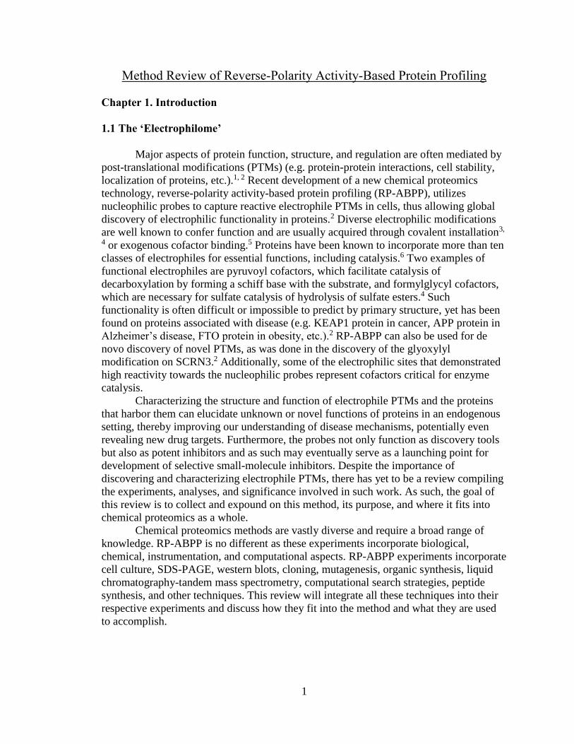
All RP-ABPP experiments utilize a chemical probe, which includes a nucleophilic
hydrazine warhead and an alkyne handle for detection and enrichment via ‘click’
chemistry (Cu(I)-catalyzed azide-alkyne cycloaddition (CuAAC)) to azide reporter tags.
In this ‘click’ reaction, azides and alkynes react to form triazoles, enabling probes
(alkyne) to bind to tags (azides) for further processing. For downstream analysis via mass
spectrometry, biotin-azide tags are used as they have a very high affinity for streptavidin,
allowing for detection, isolation, and purification of probe-bound proteins and peptides as
depicted in the top portion of Figure 2. For gel experiments, fluorophore tags (e.g.
rhodamine) are used to enable visual detection of probe-bound proteins via SDS-PAGE
gel as depicted in the bottom portion of Figure 2.
Figure 2. Schematic of utility of tags conjugated to alkyne probe. For mass spectrometry
(MS) experiments, biotin tags are used to enable enrichment of probe-bound targets on
streptavidin resin and conversely, for gel-based detection, a rhodamine-azide tag is used to
visualize the bound electrophile using in-gel fluorescence.
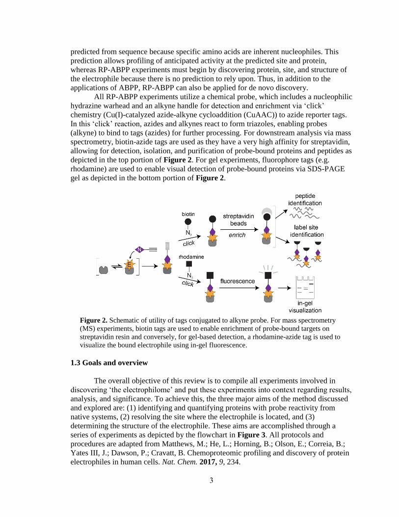
1.3 Goals and overview
The overall objective of this review is to compile all experiments involved in
discovering ‘the electrophilome’ and put these experiments into context regarding results,
analysis, and significance. To achieve this, the three major aims of the method discussed
and explored are: (1) identifying and quantifying proteins with probe reactivity from
native systems, (2) resolving the site where the electrophile is located, and (3)
determining the structure of the electrophile. These aims are accomplished through a
series of experiments as depicted by the flowchart in Figure 3. All protocols and
procedures are adapted from Matthews, M.; He, L.; Horning, B.; Olson, E.; Correia, B.;
Yates III, J.; Dawson, P.; Cravatt, B. Chemoproteomic profiling and discovery of protein
electrophiles in human cells. Nat. Chem. 2017, 9, 234.
4
Figure 3. Schematic flowchart of RP-ABPP experiments. (A) Target identification and
quantification is performed using SILAC RP-ABPP and targets are validated by gel RP-
ABPP western blots and RP-ABPP data for hydrazine probe-treated transfected cells
expressing a protein target.2 The first lane corresponds to a control transfection (‘mock’)
with the appropriate empty expression vector. (B) The site of probe-labeling is
characterized using IsoTOP ABPP experiments to determine co-eluting isotopically-
differentiated peptide pairs and de novo sequencing the ions to resolve the modified site.2
The site of probe-labeling is validated through comparison of mutation and wild-type (WT)
probe-labeling and expression profiles.2 (C) The electrophile is determined, confirmed, and
stoichiometrically quantified by inferring the electrophile and coelution of heavy-Arg/Lys-
labeled transfected cells treated with probe (followed by processing by isoTOP-ABPP)
with light amino acid-labeled synthetic standards.2
5
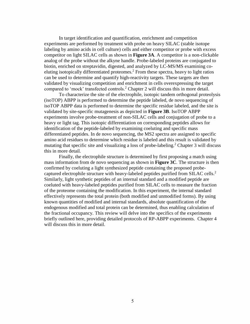
In target identification and quantification, enrichment and competition
experiments are performed by treatment with probe on heavy SILAC (stable isotope
labeling by amino acids in cell culture) cells and either competitor or probe with excess
competitor on light SILAC cells as shown in Figure 3A. A competitor is a non-clickable
analog of the probe without the alkyne handle. Probe-labeled proteins are conjugated to
biotin, enriched on streptavidin, digested, and analyzed by LC-MS/MS examining co-
eluting isotopically differentiated proteomes.2 From these spectra, heavy to light ratios
can be used to determine and quantify high-reactivity targets. These targets are then
validated by visualizing competition and enrichment in cells overexpressing the target
compared to ‘mock’ transfected controls.2 Chapter 2 will discuss this in more detail.
To characterize the site of the electrophile, isotopic tandem orthogonal proteolysis
(isoTOP) ABPP is performed to determine the peptide labeled, de novo sequencing of
isoTOP ABPP data is performed to determine the specific residue labeled, and the site is
validated by site-specific mutagenesis as depicted in Figure 3B. IsoTOP ABPP
experiments involve probe-treatment of non-SILAC cells and conjugation of probe to a
heavy or light tag. This isotopic differentiation on corresponding peptides allows for
identification of the peptide-labeled by examining coeluting and specific mass
differentiated peptides. In de novo sequencing, the MS2 spectra are assigned to specific
amino acid residues to determine which residue is labeled and this result is validated by
mutating that specific site and visualizing a loss of probe-labeling.2 Chapter 3 will discuss
this in more detail.
Finally, the electrophile structure is determined by first proposing a match using
mass information from de novo sequencing as shown in Figure 3C. The structure is then
confirmed by coeluting a light synthesized peptide containing the proposed probe-
captured electrophile structure with heavy-labeled peptides purified from SILAC cells.2
Similarly, light synthetic peptides of an internal standard and a modified peptide are
coeluted with heavy-labeled peptides purified from SILAC cells to measure the fraction
of the proteome containing the modification. In this experiment, the internal standard
effectively represents the total protein (both modified and unmodified forms). By using
known quantities of modified and internal standards, absolute quantification of the
endogenous modified and total protein can be determined, thus enabling calculation of
the fractional occupancy. This review will delve into the specifics of the experiments
briefly outlined here, providing detailed protocols of RP-ABPP experiments. Chapter 4
will discuss this in more detail.

6
Chapter 2. Target identification and quantification
The first aim of RP-ABPP is to determine probe-reactive proteins and quantify
their reactivity. Probe-reactive proteins are identified and quantified by performing
labeling, purification, and mass spectrometry (MS) analysis by stable isotope labeling by
amino acids in cell culture (SILAC) RP-ABPP, and validation via recombinant
expression protocols. Upon identification and quantification of these proteins, high-
reactivity targets can be further interrogated. Identification is an essential first step to RP-
ABPP because proteins harboring electrophilic PTMs are generally unknown and cannot
be predicted based on sequence. Quantification of this reactivity allows for exclusion of
weakly reactive or low stoichiometry electrophiles to bias toward functional sites. Thus
allowing further pursuit of highly reactive target proteins containing functional
electrophiles.
2.1 Labeling and preparation
RP-ABPP experiments begin with probe, competitor, or probe and competitor
mixture treatment and proteome harvesting and separation. These steps generate labeled
soluble and membrane proteomes at a known concentration from cell culture. These
proteomes are then used as the starting material for the rest of the RP-ABPP experiments.
Below, sample procedures are described for in situ labeling (Section 2.1.1) and proteome
preparation (Section 2.1.2).
2.1.1 In-situ labeling
To begin RP-ABPP, working stock solutions (~0.2−3 M) of probe and competitor
compounds are prepared in water from the hydrazinium chloride salts and the pH is
titrated to ~6.5−7 with concentrated sodium hydroxide. These solutions can then be
stored in aliquots at –80 °C. After solution preparation, low-passage human adherent cell
lines are grown at 37 °C in a humidified 5% CO2 atmosphere and expanded in media
containing high-glucose, L-glutamine, and pyruvate supplemented with 10% (v/v) fetal
bovine serum (FBS), and 1% Penicillin/Streptomycin antibiotic/antimycotic.13 For gel
experiments, one 6 cm cell culture plate can be used and for MS samples, two 10 cm cell
culture plates should be used.2
The treatment begins once the cells have reached near complete confluence, cells
are washed with cold phosphate buffered saline (PBS, pH 7.4) and replenished with
serum-free media (~15% of normal passage volume, e.g. a 10 cm plate normally
passaged with 10 mL media would receive 1.5 mL) supplemented with 10 mM Na-
HEPES buffer (pH 7.5). Then, cells are incubated with probe in the absence or presence
of 10-fold excess non-clickable analogs (competitors) for 30 minutes at 37 °C. Probe and
competitor should be premixed before co-administering to the cells when applicable.
Following treatment, cells are washed with cold PBS to remove the probe-containing
media, harvested by scraping, collected by centrifugation (1,400 g, 3 minutes, 4 °C), and
washed again by resuspension in cold PBS. Unless proceeding directly to proteome
preparation, cells should be pelleted and frozen at –80 °C.
7
2.1.2 Proteome preparation
To prepare the proteome for use in mass spectrometry-based and gel-based
experiments, cell pellets are resuspended on ice in PBS (100–500 μL) and lysed by a
Branson Sonifier probe sonicator (2 × 6–10 pulses, 50% duty cycle, output setting = 3–5).
For this step, resuspension volume and sonication power should be adjusted accordingly
for cell pellet yield. To separate soluble and membrane proteomes, samples are
ultracentrifuged (100,000 g, 30–45 minutes) and the protein concentrations for each
fraction are determined using the DC protein assay, which is similar the Lowry assay, and
read on a microplate reader.
2.2 SILAC RP-ABPP
SILAC was initially developed in 2002, and is a widely applicable tool for
quantitative proteomics.14 In this method, two groups of cells are used: (1) cells grown in
‘light’ media which includes all natural abundance amino acids and (2) cells gown in
‘heavy’ media which contains one or more heavier isotope amino acids (e.g. amino
acid(s) containing 2H vs 1H, 13C vs 12C, 15N vs 14N, etc.).15 This method is a simple and
easy way to isotopically differentiate samples because as the proteins incorporate the
amino acids from the culture media, peptides will contain a known mass shift. This is
sampler than other methods of isotopic differentiation used (e.g. reductive dimethylation
(ReDiMe)) because it allows the heavy and light samples to be mixed prior to all major
downstream processing. As such, this essentially allows the samples to halved when
compared to other methods in which heavy and light samples must be processed in
parallel and isotopic differentiation is incorporated later in the process, reducing
experimental variation between the samples.
The defined mass shift created using SILAC media can then be analyzed via mass
spectrometry to quantify by ratiometric comparison differences in abundance of peptides
in the respective proteomes. Coelution of isotopically differentiated peptides is a common
strategy in RP-ABPP as will be demonstrated throughout this review. This strategy is
used with experimental samples in SILAC RP-ABPP and isoTOP ABPP as depicted in
Figure 4. In Figure 4A, heavy and light cells, proteomes, and peptides are depicted in
blue and red, respectively. As shown in Figure 4A, isotopic differentiation is utilized in
SILAC RP-ABPP as protein targets are determined based on their heavy to light ratios. In
Figure 4B, heavy and light tagged proteins and peptides are depicted in dark and light
green , respectively. As is shown in Figure 4B, the isotopic differentiation strategy is
utilized in isoTOP ABPP to determine the peptide labeled by finding coeluting peptides
with a specific mass differential.

8
Figure 4. (A) Schematic for MS-based quantitative (SILAC) proteomics experiments
(enrichment and competition) as described in the text. Heavy (H) and light (L) cells,
proteomes, and peptides are depicted in blue and red, respectively. Biotin tags and
streptavidin resin are depicted in black and gray, respectively. (B) Characterization of probe
labeled peptides using the isoTOP-ABPP method as described in the text. Heavy and light
tagged proteins and peptides are depicted in dark and light green, respectively. Biotin-TEV
tags and streptavidin resin are depicted in green and gray respectively.
SILAC can be utilized in RP-ABPP enrichment16 and competition17 experiments,
as shown in Figure 4A.2, 18 In enrichment experiments, heavy cells are treated with probe
and light cells with competitor and in competition experiments, heavy cells are treated
with probe and light cells with probe and an excess of competitor. Enrichment and
competition can also be shown using gel experiments. For MS-based analysis, labeled
proteomes are conjugated to biotin tags via click chemistry and enriched on streptavidin
beads to remove unlabeled proteins. Proteins are then digested and analyzed via LC-
MS/MS. Proteins with high heavy to light ratios for competition and enrichment are
deemed to be high-reactivity targets and the reactivity is quantified using these ratios.
The experimental steps and analysis described for SILAC RP-ABPP are pictorially
represented in Figure 4A. Below, sample procedures are described for MS-based
analysis (Section 2.2.1), liquid chromatography-tandem MS (Section 2.2.2), and
determination of high-reactivity targets (Section 2.2.3). Profiling experiments were
initially adapted from those applied to other probes.19-22
2.2.1 MS-based analysis of probe-labeled proteins
For SILAC experiments, the in situ labeling protocol (Section 2.1.1) is followed,
with the alteration of passaging each cell line a minimum of six times in SILAC media
(lysine- and arginine-free) containing dialyzed FBS supplemented with either isotopically
enriched L-[13C615N2]lysine hydrochloride and L- [13C6
15N4]arginine hydrochloride or

9
natural abundance isotopologues (100 μg/mL each, 550 μM and 475 μM, respectively).
This allows the cells to incorporate the heavy or light amino acids. For both enrichment
and competition experiments, isotopically heavy cells are treated with the probe. For
enrichment experiments, isotopically light cells are treated with non-clickable analog (at
the same concentration used for the probe in the heavy cells) and for competition
experiments, probe in the presence of 10-fold excess non-clickable analog as a
competitor are used. This ultimately allows for high-reactivity targets to bind to probe in
heavy cells and bind to competitor in light cells, ensuring high heavy to light ratios in
high-reactivity targets.
After labeling, isotopically heavy and light whole cell lysates are mixed in equal
proportions and the proteome preparation protocol (Section 2.1.2) is followed as
described above. Following the determination of the protein concentration, the
fractionated equimolar mixture of heavy and light proteomes (~1−1.5 mg) is diluted to 1
mL in PBS. To conjugate the protein-bound probes to biotin tags, 110 μL of a freshly
prepared ‘click’ reagent mixture containing 0.1 mM tris(benzyltriazolylmethyl)amine
(TBTA) (60 μL/sample, 1.7 mM in 4:1 DMSO:t-BuOH), 1 mM CuSO4 (20 μL/sample,
50 mM in H2O), 100 μM biotin-azide (10 μL/sample, 10 mM in DMSO), and freshly
prepared 1 mM tris(2-carboxyethyl)phosphine (TCEP) (20 μL/sample, 50 mM in PBS or
H2O) is added to each sample (1 mL) and the mixture is vortexed. The ‘click’ reaction is
allowed to proceed for 1 hour at ambient temperature on a rotator. After the reaction
mixture has rotated for 1 hour, the ‘click’ reaction is quenched with sequential addition of
and mixture with pre-chilled methanol (MeOH, 2 mL), chloroform (CHCl3, 0.5 mL), and
PBS (1 mL) on ice. To fractionate the interphase protein from the organic and aqueous
solvent layers, the precipitated proteome is centrifuged (5,000 g, 10 min, 4 °C) and to
ensure the ‘click’ reagents are efficiently removed, the protein pellet is washed with cold
1:1 MeOH:CHCl3 (3 × 1 mL) followed by mild sonication in cold 4:1 MeOH:CHCl3 (2.5
mL).
Following conjugation to biotin tags, the samples are denatured, disulfides
reduced, and resulting thiols alkylated. This is performed by pelleting the remaining
precipitate by centrifugation (5,000 g, 10 min, 4 °C) and re-dissolving by mild sonication
in a freshly prepared solution of proteomics-grade urea (500 μL, 6M in PBS). Disulfides
are reduced with TCEP (9 mM) pre-neutralized with potassium carbonate (27 mM) for 30
minutes at 37 °C and the resulting thiols are alkylated with iodoacetamide (45 mM) for
30 minutes at ambient temperature protected from light. To ensure complete
denaturation, SDS [2% (w/v)] is added.
The final steps in the SILAC RP-ABPP MS-based analysis enrich samples on
streptavidin, digest samples with trypsin, and acidify with formic acid. To accomplish
this, first the solution is diluted to ~0.2% SDS with PBS (~5 mL) and incubated with pre-
equilibrated streptavidin agarose resin (50 μL column volume, 100 μL 1:1 slurry) for
~1.5−2 hours at ambient temperature on a rotator. Then, to remove unbound protein,
excess detergent, and small molecules, streptavidin beads are collected by centrifugation
(1,400 g, 1−2 min) and sequentially washed with 0.2% SDS in PBS (3 × ~10 mL),
detergent-free PBS (3 × ~10 mL) and H2O (3 × ~10 mL). To perform the digestion, the
resin is transferred to a Protein LoBind tube and bound proteins are digested on-bead
overnight at 37 °C in ~200 μL total volume containing sequencing grade porcine trypsin
(2 μg, Promega) in the presence of urea (2 M in PBS) and CaCl2 (1 mM). In the final
10
step, the proteolyzed supernatant is transferred to a fresh Protein LoBind tube and
acidified with formic acid (5%) to inactivate trypsin and stored at −80 °C.
2.2.2 Liquid chromatography-tandem mass spectrometry (LC/LC-MS/MS)
Numerous mass spectrometry protocols exist and historically MudPIT
(multidimensional protein identification technology) mass spectrometry protocols have
been used for ABPP and RP-ABPP.23-26 These protocols utilize loading acidified Trypsin
products onto a C18 silica and strong cation exchange (SCX) resin column and eluting
peptides with 5 salt ‘bumps’ followed by increasing acetonitrile (ACN). However, a more
efficient method without the salt ‘bumps’ has recently been incorporated into RP-ABPP. 27 While MudPIT protocols enable further fractionation, they require significantly more
time on the MS instrument. Utilizing another method, such as the sample protocol
described below, is preferred because significantly less instrument time is required while
obtaining equivalent results. In proteomics, the most limiting factor in almost all
experiments is instrument time, as such, any way to reduce instrument time without
compromising the quality of results is preferred.
Taking the digested acidic peptide mixture, C18 Stage Tips are used to desalt
samples. Desalted samples are concentrated under reduced pressure in an evacuated
centrifuge (SpeedVac) and re-dissolved in 10 µL of diluent (98% H2O, 2% acetonitrile,
0.1% formic acid) for nanoLC-MS/MS analysis. A 3–5 µL aliquot of this solution is
injected via a nano-LC system onto a 75 µm (inner diameter) fused-silica capillary
column hand-packed with C18 resin and laser-pulled tip in solvent A (0.1% formic acid
in H2O). The column is developed with a 60 minute gradient of 5%–100% solvent B
(20% H2O, 80% acetonitrile, 0.1% formic acid). Peptides are ionized in positive-ion
mode with a flow rate of 300 nL/min and an applied voltage of 2.3 kV. Spectra are
collected in a data dependent mode such that each scan cycle involves a single high
resolution (30,000) full MS spectrum of parent ions (MS1 scan from 400–1800 m/z)
collected in the orbitrap coupled to a 30 CID-induced fragmentation (MS2) scans in the
ion trap of the 30 most abundant parent ions from the MS1 scan. Dynamic exclusion is
enabled (repeat count of 1, exclusion duration of 20 s) as is monoisotopic precursor
selection. Parent ions with unassigned of +1 charge stated by the instrument are excluded
for fragmentation. All other parameters are left as default values.
2.2.3 Determination of high-reactivity targets
Once mass spectrometry data collection has been performed on the samples from
MS-based analysis (2.2.1), the MS2 spectra are extracted for all fragmented parent ions
(from .ms2 file) from each of the .raw files generated by the instrument (Xcalibur
software) using RAW Xtract or RawConverter28 with monoisotopic selection. After
extraction, each .ms2 file is searched using the ProLuCID algorithm against a reverse-
concatenated, nonredundant database of the human proteome and filtered using
DTASelect 2.0 within the Integrated Proteomics Pipeline (IP2) software. In this search, a
static modification on cysteine residues for carboxyamidomethylation (+57.02146 Da)
and up to one differential modification on methionine residues for oxidation (+15.9949
Da) are included. In the database search, peptides are also required to have at least one

11
tryptic terminus but allowed an unlimited number of missed cleavages. Additionally, the
exact mass shift of heavy atoms on specific amino acids from the SILAC media is
searched (e.g. if using 13C and 15N on Lysine and Arginine, K +8.0142 Da and R
+10.0082 Da). This can be done by performing a coupled ‘heavy’ search on each dataset
for both light and heavy isotopologues of the same peptide by specifying the mass shift of
heavy residues as static modifications on lysine (+8.0142 Da) and arginine (+10.0082
Da).
To further filter the data, the parent ion mass tolerance for a minimum envelope
of three isotopic peaks is set to 50 ppm, the minimum peptide length set to six residues,
the false positive rate set to 1%, and at least 2 peptides of a protein must be detected.
Heavy and light parent ion chromatograms associated with successfully identified
peptides are extracted and compared using CIMAGE software.25 Data is filtered so that at
least one ion of a co-eluting heavy-light pair must be accurately identified from a
fragmentation event that occurred within the retention time window (± 10 minutes) of
parent ion elution. Furthermore, to ensure that the correct pair of peaks is quantified,
chromatograms within a 10 ppm error tolerance of the predicted m/z, single-to-noise
ratios greater than 2.5, and ‘co-elution correlation scores’ and ‘envelope correlation
scores’ R2 values greater than or equal to 0.8 are extracted. To further eliminate false
positives and stochastic variability in the data, protein ratios are determined by median
peptide ratio derived from three or more unique qualified peptides. To provide final
values, protein ratios that comply with these criteria from a single experiment are
averaged with ratios acquired from at least three replicates.
2.3 Validation via recombinant expression
Once high-reactivity targets have been determined, targets can be validated by
demonstrating hydrazine reactivity as an intrinsic property of the protein targets that is
shared by both the endogenous and recombinant forms of these proteins. This validation
is performed by treating transfected cells with probe, followed by conjugation to a
fluorophore tag (e.g. rhodamine), and visualization of a strong fluorescent band at the
appropriate molecular weight. This band should be absent in ‘mock’ transfected cells and
in cells treated with excess non-clickable agents as this should block probe-labeling of
each protein. Additionally, recombinant expression of each protein (and lack of
expression in ‘mock’ transfected control cells) is confirmed by western blotting. In order
to perform these transfections, target genes need to be obtained in mammalian expression
vectors. These vectors can be purchased or made using cloning protocols.2 Sample
procedures for transfection and gel-based analysis (Section 2.3.1) are described below.
To begin the target validation experiment, standard growth conditions are
followed and cells are grown to ~40% confluence. The appropriate expression vector
[control cells (‘mock’) receive an equal amount of the appropriate empty vector] is added
as well as polyethyleneimine (PEI) ‘MAX’ (MW 40,000) as a transfection reagent under
standard transfection conditions [3:1 vector/PEI (w/w) ratio]. Cells are incubated with
transfection reagents for ~48 h before labeling in situ (2.1.1) and preparation of the
proteome (2.1.2).

12
2.3.1 Gel-based analysis of probe-labeled proteins
Following transfection (Section 2.3) and labeling (Section 2.1.1) and proteome
preparation (Section 2.1.2), proteomes from treated cells are diluted to 1 mg/mL. To
conjugate the fluorophore to probe-labeled proteins, 6 μL of a freshly prepared “click”
reagent mixture containing 0.1 mM TBTA (3 μL/sample, 1.7 mM in 4:1 DMSO:t-
BuOH), 1 mM CuSO4 (1 μL/sample, 50 mM in H2O), 25 μM azide-rhodamine (1
μL/sample, 1.25 mM in DMSO), and freshly prepared 1mM TCEP (1 μL/sample, 50 mM
in PBS or H2O) is added to each sample (50 μL). Immediately upon addition of the click
mixture, the samples are mixed by vortexing and the mixture is allowed to react at
ambient temperature for 1 hour. The ‘click’ reaction is quenched with SDS loading buffer
(4× stock, 17 μL) and proteins are resolved (~25 μg total protein loaded per gel lane) by
SDS-PAGE (10% acrylamide gels). Labeling is visualized using in-gel fluorescence
scanning on a flatbed fluorescence scanner (e.g. BioRad ChemiDoc MP). The same gel is
transferred to nitrocellulose membrane and western blotted using standard protocols.2
2.4 Conclusion
As seen above and will be evidenced throughout the review, most RP-ABPP
experiments build upon on another. For example, isoTOP ABPP experiments start with
in-situ labeling (Section 2.1.1) and proteome preparation (Section 2.1.2) and parts of MS-
based analysis (Section 2.2.1) are repeated as well. Additionally, mutagenic analysis
relies upon gel-based analysis (Section 2.3.1) and begins with in-situ labeling (Section
2.1.1) and proteome preparation (Section 2.1.2) as well. It is important to note that these
experiments are very rigorous. They require planning and attention to detail and have few
acceptable pause points. One of the most critical portions of target identification and
quantification is the equilibration and washing of the SILAC RP-ABPP samples on
streptavidin beads. This step is critical because if the beads are washed very vigorously,
some beads may be lost, causing low peptide signal. Conversely, if the beads are not
washed vigorously enough, too many contaminants (e.g. unbound protein, excess
detergent, small molecules, etc.) may be present, which decreases the ability to detect
proteins of interest. These risks could be mitigated through use of magnetic beads, which
are generally used for high-throughput applications. However, these beads are very
expensive and require additional equipment.
The results generated from target identification and quantification are quite
robust. When RP-ABPP was performed in two cell lines with two probes, eleven high-
reactivity targets were identified and only two of these proteins were previously known to
harbor an electrophile.2 Due to the promise of these initial results, performing
identification and quantification with other probes and cell lines is likely to widely
expand the knowledge of known functional electrophiles.
13
Chapter 3. Characterization of the probe-captured site
The second aim of RP-ABPP is to determine the site of electrophilic reactivity on
the protein. The peptide labeled is found using isoTOP-ABPP25, 29 experiments and the
specific amino-acid residue labeled is found using de novo sequencing. These results are
validated using site-specific mutagenic analysis. Characterization of the electrophilic site
yields information about where the reactivity is located within the greater context of the
protein. Once this site is determined, this paves the way for a large variety of downstream
experiments including monitoring the electrophile, examining its installation, and
potentially investigating its function. Additionally, after the site harboring the
electrophile is identified, this area can be further interrogated to determine the structure
of the electrophile.
3.1 IsoTOP ABPP
IsoTOP ABPP is used to determine the peptide labeled, which is depicted in
Figure 4B. Following probe treatment of non-SILAC cells (shown in gray in Figure 4B),
this method utilizes conjugation to heavy and light cleavable biotin-TEV tags (shown in
green in Figure 4B). After conjugation to heavy and light tags, labeled proteomes are
enriched on streptavidin beads and on bead digestion is performed. After digestion, all
unlabeled peptides are discarded as shown in Figure 4B. The remaining labeled peptides
are released from the beads through cleavage of the tag (by TEV protease), ultimately
generating probe-labeled peptides as mass differentiated pairs. Using the coeluting pair
with heavy and light tags (plotted in dark and light green, respectively, in Figure 4B), the
peptide harboring the labeled residue can be found. Determining which peptide is labeled
can give information about where the reactivity is located in the sequence of the protein
(e.g. active site, N-terminus, etc.). These searches enable differentiation between probe-
labeled peptides and other peptides in the sample regardless of identity or mass. A sample
isoTOP-ABPP procedure (Sections 3.1.1-3.1.2) is described below.
3.1.1 IsoTOP ABPP sample preparation to isolate probe-captured peptides
IsoTOP ABPP begins with probe treatment of wild-type or transfected cells in-
situ (Section 2.1.1) and proteome preparation (Section 2.1.2). After performing these
protocols, soluble proteomes (2 mg total protein) are diluted to 1 mL in PBS. To
conjugate half of the proteome (0.5 mL) to the light TEV tag and the other half to the
heavy TEV tag, click reactions are scaled accordingly to maintain final concentrations of
0.1 mM TBTA, 1 mM CuSO4, 100 μM of light or heavy biotin-TEV-azide (5 mM in
DMSO) and 1 mM TCEP. The mixture is vortexed and placed on a rotator at ambient
temperature. Once the mixture has rotated for 1 hour, it is centrifuged (16,000 g, 5
minutes, 4 °C) and resulting pellets are mildly sonicated in ice-cold methanol (0.5 mL).
The light- and heavy-labeled proteomes are combined and centrifuged once more. To
solubilize the proteomes, 1.2% SDS (1 mL in PBS) is added and samples are stored at –
80 °C overnight.
After leaving the samples in –80 °C overnight, samples are diluted to ~0.2% SDS
with PBS (~5 mL) and incubated with pre-equilibrated streptavidin agarose resin (100 μL

14
1:1 slurry) for ~2–3 hours at ambient temperature. To remove un-labeled proteins, resin
is washed as described above in the SILAC RP-ABPP MS-based analysis procedure
(Section 2.2.1), and after the washes, the resin is transferred to clean Eppendorf tubes and
resuspended in urea (500 μL, 6M in PBS). Cysteines are then reduced and alkylated with
TCEP and iodoacetamide, respectively, as described above in the SILAC RP-ABPP MS-
based analysis procedure (Section 2.2.1). To remove reagents, resin is washed once with
PBS and bound proteins are digested with sequencing grade porcine trypsin (Promega, 2
μg) for 8–12 h at 37 °C in the presence of 2 M urea (200 μL, in PBS) and CaCl2 (1 mM).
After trypsin digestion, sequential washes with PBS (5 × 0.5 mL) and H2O (5 ×
0.5 mL) are performed to remove unmodified peptides, urea, and trypsin. The resin is
transferred to fresh tubes and equilibrated with TEV buffer (50 mM Tris, pH 8). To
release the remaining immobilized peptides, TEV protease is added (~1–2 μM in ~200
μL TEV buffer at 30 °C for 3–5 hours). Heavy- and light-labeled peptides are collected
and recovered from the resin with H2O (2 × 50 μL). Samples are stored at –80 °C and
must be analyzed within several days.
3.1.2 Characterization of probe-labeled peptides by isoTOP ABPP
To analyze the samples generated from the isoTOP preparation, data is collected
from isolated probe-captured peptides using mass spectrometry protocols as described
above in the SILAC RP-ABPP procedure (Section 2.2.2). Then, the data is searched on
the MS1 level for paired spectra of mass differentiated coeluting peaks as shown on the
right in Figure 5. Every recorded monoisotopic precursor mass is searched 6.0138 Da (±
5 ppm) upstream and downstream in each total ion (MS1) spectrum for a possible
isotopic partner taking into account +2 and +3 charge states (3.0069 and 2.0046 Da,
respectively). The relative intensity of the monoisotopic peaks is required to be greater
than or equal to 5% of the base peak of each spectrum and each isotope profile (envelope)
must have at least three peaks. Additionally, the Euclidean distance between two isotope
profiles must be greater than or equal to 0.2. Pairs with the same m/z values (± 5 ppm)
and retention times (± 10 minutes) are grouped to eliminate duplicates. Pairs of parent
ions from transfected versus mock-transfected cells are analyzed and pairs of parent ions
from three biological replicates should be analyzed as well.
3.2 Fragmentation spectra assignment by de novo sequencing
To further resolve the site labeled and the mass of the probe-captured PTM, the
MS2 spectra from the isoTOP experiments is assigned via de novo sequencing by
extraction of b and y ions and assignment spectra to their respective amino acids. The
residue containing the modification can be identified as it will contain the known mass
shift of the fragmented heavy/light tag, containing covalently bound probe and
conjugated biotin-TEV tag. After all peaks are identified as either an amino acid or amino
acid with fragmented tag, the mass of the bound electrophile can be calculated. A sample
procedure is described below.
15
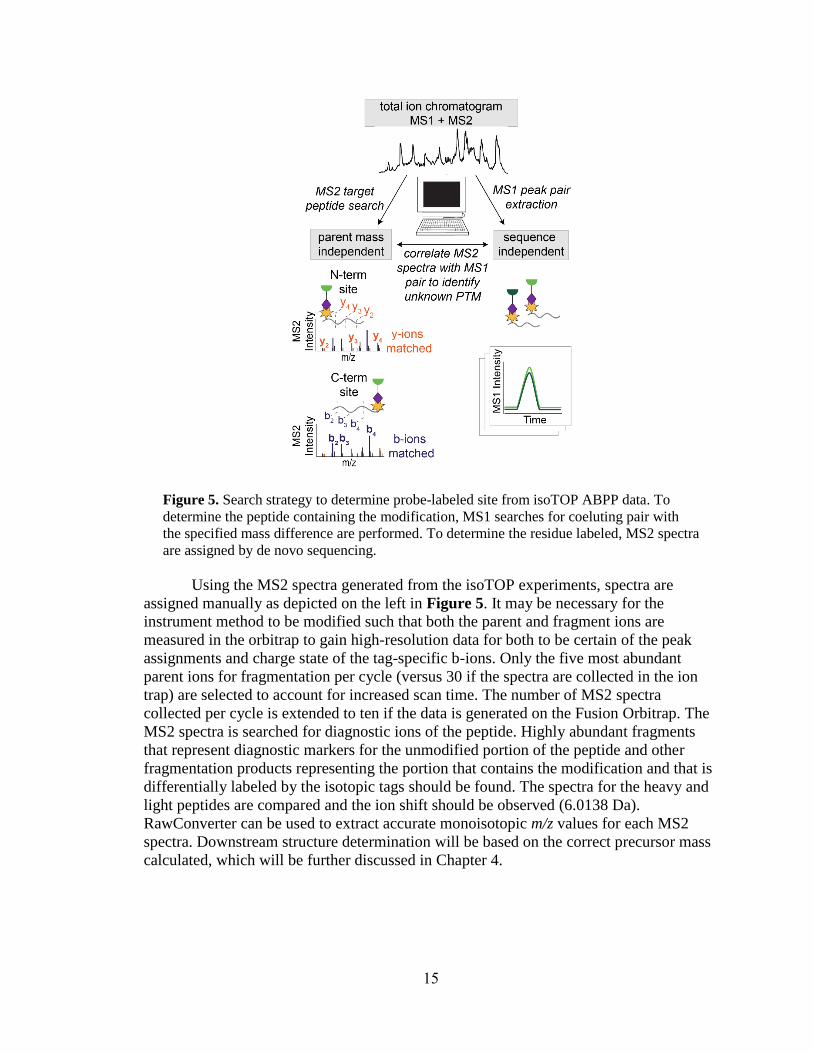
Figure 5. Search strategy to determine probe-labeled site from isoTOP ABPP data. To
determine the peptide containing the modification, MS1 searches for coeluting pair with
the specified mass difference are performed. To determine the residue labeled, MS2 spectra
are assigned by de novo sequencing.
Using the MS2 spectra generated from the isoTOP experiments, spectra are
assigned manually as depicted on the left in Figure 5. It may be necessary for the
instrument method to be modified such that both the parent and fragment ions are
measured in the orbitrap to gain high-resolution data for both to be certain of the peak
assignments and charge state of the tag-specific b-ions. Only the five most abundant
parent ions for fragmentation per cycle (versus 30 if the spectra are collected in the ion
trap) are selected to account for increased scan time. The number of MS2 spectra
collected per cycle is extended to ten if the data is generated on the Fusion Orbitrap. The
MS2 spectra is searched for diagnostic ions of the peptide. Highly abundant fragments
that represent diagnostic markers for the unmodified portion of the peptide and other
fragmentation products representing the portion that contains the modification and that is
differentially labeled by the isotopic tags should be found. The spectra for the heavy and
light peptides are compared and the ion shift should be observed (6.0138 Da).
RawConverter can be used to extract accurate monoisotopic m/z values for each MS2
spectra. Downstream structure determination will be based on the correct precursor mass
calculated, which will be further discussed in Chapter 4.
16
3.3 Mutagenic analysis
After the amino acid containing the reactive electrophile has been identified, the
next step is validation and confirmation of the location by mutation of the specific residue
labeled. The site is confirmed if mutation of the site enables visualization of a loss of
probe labeling. Additionally, expression should be present in both the wild-type and
mutant protein and confirmed via western blot. To start this experiment, mutants are
generated from the plasmids used in the recombinant expression procedure using
QuikChange30 site-directed mutagenesis with primers containing the desired mutations
and their respective complements. Once the mutation is confirmed, cells are transfected
(Section 2.3), proteomes are labeled (Section 2.1.1) and prepared (Section 2.1.2), and gel-
based analysis (Section 2.3.1) is performed as described above.
3.4 Conclusion
Characterizing the site labeled encompasses very difficult experiments. The
isoTOP ABPP sample preparation is particularly difficult and requires extensive planning
as the samples are very sensitive and should be analyzed quickly after performing the
preparation. Furthermore, as was the case in identification and quantification, incubation
and washes of the samples with streptavidin resin is a critical step. In addition to the
reasons mentioned above, excess urea not removed during washes can impact TEV
protease activity, thereby causing difficulty in releasing immobilized peptides.29 Further
evidence of this issue is that two common sources of problems in these experiments are
incomplete trypsin digestion and incomplete TEV digestion.29 Urea is used as a
denaturing agent in these experiments, but high urea concentrations can reduce trypsin
and TEV protease is sensitive to even trace amounts of urea, as such, these steps
represent critical points in this method.29 Additionally, the search strategys used to
determine the residue labeled are quite complex, but are designed with intent. The
strengths in these searches, laid out in Figure 5, are that for the MS1 search, no sequence
information is required to make the determination and for the MS2 search, no parent
mass information is required to make the determination. Once the site has been
determined and mutations at the electrophilic site have been generated, this allows for
extensive further experimentation. In downstream experiments, the wild-type protein
harboring the modification and the mutant that cannot contain the modification can be
used to interrogate installation, function, and other aspects of the modification.
17
Chapter 4. Structure determination of probe-captured electrophile
The third and final goal of RP-ABPP is to determine the structure of the
electrophile PTM. A structure match is initially proposed using the mass information
about the PTM from de novo sequencing and chemical intuition regarding the residue and
chemical reactivity of probe used. Information about enzyme class or similar motifs may
also be useful. Peptides are coeluted with synthetic standards to: (1) confirm the proposed
structure as well another confirmation of the site of labeling and (2) quantify the fraction
of protein bearing the modification.
4.1 Validation of proposed structure by coelution with synthetic standard
Validation of electrophilic site and structure utilizes coelution of isotopically
differentiated samples, light synthesized peptide with proposed probe-captured
electrophile PTM and probe-labeled heavy SILAC cells. A sample procedure (Sections
4.1.1-4.1.2) is described below.
4.1.1 Synthetic standard
To generate the synthetic standard, solid phase peptide synthesis is used to
prepare the peptide labeled and identified by isoTOP ABPP, including the proposed
electrophile PTM bound to probe. The obtained synthetic peptides are conjugated to
already cleaved tags, in contrast to the isoTOP sample preparation in which the tags are
conjugated prior to cleavage. To cleave the tags, stocks of the heavy and light biotin-
azide tags (0.25 μmol of each) are diluted to 250 μM with 1 mL 50 mM Tris, pH 8
supplemented with 1 mM DTT in the presence of 0.4 μM TEV protease and the reaction
is incubated overnight at 30 °C. Once the reaction has incubated overnight, the reaction is
concentrated to ~200 μL and the protease is precipitated with an equal volume of
acetonitrile and pelleted. The supernatants containing the cleaved tags are then purified
by RP-HPLC. To conjugate the synthetic hydrazone alkyne peptide to the cleaved heavy
and light tags, a ‘click’ reaction is performed with addition of TBTA (0.1 mM), CuCl2 (1
mM), and TCEP (1 mM) to a 0.5 mL mixture of the hydrazone alkyne peptide (~0.3 mM,
~0.15 μmol) and either form of cleaved tag (~60 μM, ~0.03 μmol of each) in phosphate
buffer (60 mM, pH 7). The reactions are incubated at ambient temperature for
approximately 2 hours and purified by RP-HPLC. The product is then neutralized with
phosphate buffer (25 mM, pH 7), lyophilized, and aliquots frozen at –80 °C.
4.1.2 Coelution with synthetic standard
The samples used for coelution are generated using the isoTOP sample
preparation procedure in Section 3.1.1, with the exception of growing cells in standard
SILAC media. Once probe-labeled peptide pairs conjugated to tags have been isolated,
the standard is diluted in water, the concentration is verified spectrophotometrically, and
0.5 pmol is added to the digested sample just prior to column loading for analysis. If the
proposed structure is correct, the peptides should co-elute and appear identical in the data
analysis with the only exception being the previously defined mass shift.

18
4.2 Determination of absolute stoichiometry with synthetic isotopolgues
The fraction of modified protein can be determined by coeluting (light) synthetic
modified and internal standards with (heavy) endogenous modified and internal peptides
as depicted in Figure 6. In these experiments, the internal peptide represents the total
protein in the cell (both modified and unmodified protein). Both the endogenous
modified and internal peptides absolute quantities are able to be calculated using the
known amount of respective standards added to the sample and the relative peak areas of
the standard to the endogenous peptides as shown by fractional occupancy in Figure 6. A
sample procedure for these experiments is described below.
Figure 6. Scheme of stoichiometry measurement for modified peptide. Using a known
quantity of synthetic standards of modified and internal peptides enables absolute
quantification of the endogenous modified and total protein, allowing for calculation of
fractional occupancy.
To generate the endogenous peptides in stoichiometric quantification, cells grown
in heavy SILAC media are transfected and probe treated (Section 2.1.1) using procedures
described above. The plasmids transfected must contain genes for the protein and FLAG
tag for downstream purification. Cells are lysed in PBS (pH 7.4), fractionated by
ultracentrifugation (100,000 g, 30–45 minutes), and the samples diluted to 1 mL with 50
mM Na-HEPES buffer (pH 7.5) supplemented with 500 mM NaCl and 1% Triton X-100.
Unsolubilized protein remaining in the sample is pelleted by centrifugation and denatured
with a small volume 10% SDS for 1 hour at 37 °C. The completely resolubilized sample
is incubated at 4 °C overnight by rotation with anti-FLAG resin. The resin is washed by
19
resuspension and centrifugation with the same buffer supplemented with 500 mM NaCl
(5 × 1 mL) followed by 100 mM NaCl (2 × 1 mL). The bound protein is eluted by
incubating the beads for ~1 h at 37 °C in PBS containing 8 M urea (2 × 50 μL). Cysteines
are reduced with TCEP (10 mM pre-neutralized with 30 mM potassium carbonate for 30
minutes at 37 °C) and alkylated with iodoacetamide (20 mM under the same conditions
but protected from light). The samples are diluted to 2M urea with PBS and digested at
37 °C overnight with 2 μg trypsin supplemented with 1 mM CaCl2. Trypsin is inactivated
with 5% formic acid. The natural abundance probe-labeled peptide standard generated
above is diluted in PBS and the digested protein sample with the modified standard as
well as an internal peptide that represents the total protein (5–50 pmol of each) are doped
in just prior to analysis by the same method used for proteomic profiling. Absolute
amounts of standards should be adjusted to achieve nearly comparable peak intensities
for quantitation.
4.3 Conclusion
Throughout RP-ABPP, coelution of isotopically differentiated samples is
thoroughly utilized. This principle is used for SILAC, isoTOP, and synthetic standard
experiments. RP-ABPP has been used for de novo discovery of a new previously
unknown modification2 and there is the potential that other human proteins may harbor
previously unknown electrophilic modifications. As such, electrophilic structure
determination experiments could yield novel and far-reaching results.

20
Chapter 5. Conclusions
5.1 Previous results and significance
Electrophilic modifications are not easily predicted by sequence and sequence
predictions do not yield information about function or activity. Initial results from RP-
ABPP have yielded eleven targets and these targets are largely functional and strongly
implicated in disease.2 Additionally, nine of the eleven targets were not previously known
to harbor an electrophile and on one of these targets, SCRN3, a novel previously
undiscovered modification, the glyoxylyl, was discovered.2 The results of the RP-ABPP
experiments used to discover the glyoxylyl are depicted in Figure 7. Figure 7A depicts
the probe reaction with the gyloxylyl. Identification and quantification of SCRN3 as a
target is shown in Figure 7B with extracted parent ion chromatograms and corresponding
heavy to light ratios for tryptic peptides of SCRN3 protein probe-treated cells quantified
in enrichment and competition (left) and quadrant plot of average competition versus
enrichment SILAC ratios from quantitative proteomics experiments (right).2 In Figure
7C, isoTOP ABPP MS1 ion chromatograms (left) and isotopic envelopes (right)
demonstrate coelution and specific mass differentiation of the labeled SCRN3 peptide.2
Figure 7D depicts de novo sequencing to determine the residue labeled and the
mutagenesis gel and western blot validate the result that both C6 and D7 must be present
for labeling to occur, because the y-ions resolve the modified site (Figure 7*) to the N-
terminal cysteine and/or adjacent aspartate and mutation profiles of Cys6-to-Ala6 (C6A)
and Asp7-to-Phe7 (D7F) mutant SCRN3 proteins compared to wild-type (WT) SCRN3
show a lack of probe labeling.2 In Figure 7E, heavy-Arg/Lys-labeled SCRN3-transfected
cells treated with probe, followed by processing by isoTOP-ABPP, yields an isotopically
differentiated probe-labeled SCRN3 peptide pair (light and dark green), which co-elutes
with a light amino acid-labeled probe-Glyoxylyl6-Arg20 standard (also an isotopically
differentiated peptide pair; light and dark gray).2 Inset chromatogram shows all four
traces scaled to the same intensity to show co-elution of endogenous and standard probe-
Glyoxylyl6-Arg20 SCRN3 peptides.2 The discovery of the glyoxylyl is significant
because it demonstrates the utility of RP-ABPP in discovering new unknown
electrophilic modifications. The results of these initial experiments indicate that other
human proteins may harbor electrophilic groups and that there are other unknown
modifications to be discovered. As such, the advent of this method is critical due to RP-
ABPP being the only global unbiased approach to discover electrophilic cofactors.

21
Figure 7. Sample data from RP-ABPP experiments. Adapted from Matthews, M.; He, L.;
Horning, B.; Olson, E.; Correia, B.; Yates III, J.; Dawson, P.; Cravatt, B. Chemoproteomic
profiling and discovery of protein electrophiles in human cells. Nat. Chem. 2017, 9, 234. (A)
Reaction scheme of probe captured glyoxylyl on SCRN3. (B) SCRN3 data from SILAC RP-
ABPP identification and quantification experiments. (C) Extracted MS1 ion chromatograms
(left) and corresponding isotopic envelopes (right) for co-eluting heavy- and light-tagged
peptides labeled by probe (in dark and light green, respectively). (D) Comparison of high-
resolution MS2 spectra generated from light- versus heavy-tagged parent ions. Through de
novo sequencing, the modified (*) site is determined and is confirmed by examining probe-
labeling and expression profiles of mutant SCRN3 proteins compared to wild-type (WT)
SCRN3. (E) The glyoxylyl structure and site were confirmed by coelution with synthetic
standards as described in the main text.
5.2 Future applications
RP-ABPP is a recently developed method, and there are many potential future
applications. These experiments are not limited to cell culture, they could easily be
adapted for use in bacteria, pathogens, tissue, or in living animals. This method opens up
a new area, the entire other half of the reactive proteome, the ‘electrophilome.’ Based on

22
targets generated from only two probes in two human cell lines,2 there are more
electrophile-bound proteins to be discovered and disease-relationships to be explored. In
addition to discovery of protein targets and electrophilic modifications, the tools in this
method can also be used to further investigate various aspects of the proteins and
modifications discovered. For example, discovery of installation mechanisms and
functions of these electrophiles could yield new information about the proteins and
mechanisms in these cells. Additionally, as many of the protein targets discovered have
strong associations to poorly understood disease mechanisms (e.g. APP – Alzheimer’s
Disease, FTO – Obesity, etc.) these modifications’ installation and function could play an
important role in or yield information about the pathogenesis of these diseases. Due to
hydrazines’ ability to covalent inhibit enzymes through reaction with their electrophilic
cofactors, these experiments could ultimately be used to discover inhibitors and launch
drug trials.
23
References
(1) Walsh, C. T.; Garneau‐Tsodikova, S.; Gatto Jr, G. J. Protein posttranslational
modifications: the chemistry of proteome diversifications. 2005, 44 (45), 7342-7372.
(2) Matthews, M. L.; He, L.; Horning, B. D.; Olson, E. J.; Correia, B. E.; Yates, J. R.;
Dawson, P. E.; Cravatt, B. F. Chemoproteomic profiling and discovery of protein
electrophiles in human cells. Nat. Chem. 2017, 9 (3), 234-243.
(3) Klinman, J. P.; Bonnot, F. Intrigues and Intricacies of the Biosynthetic Pathways for
the Enzymatic Quinocofactors: PQQ, TTQ, CTQ, TPQ, and LTQ. Chemical Reviews
2014, 114 (8), 4343-4365.
(4) Okeley, N. M.; Van Der Donk, W. A. Novel cofactors via post-translational
modifications of enzyme active sites. 2000, 7 (7), R159-R171.
(5) Phillips, R. S. Chemistry and diversity of pyridoxal-5′-phosphate dependent enzymes.
BBA - Proteins and Proteomics 2015, 1854 (9), 1167-1174.
(6) Walsh, C. Posttranslational modification of proteins: expanding nature's inventory.
Roberts and Company Publishers: 2006.
(7) Cravatt, B. F.; Wright, A. T.; Kozarich, J. W. Activity-based protein profiling: From
enzyme chemistry. Annual Review of Biochemistry 2008, 77, 383-414.
(8) Shantz, L. M.; Stanley, B. A.; Secrist III, J. A.; Pegg, A. E. Purification of human S-
adenosylmethionine decarboxylase expressed in Escherichia coli and use of this
protein to investigate the mechanism of inhibition by the irreversible inhibitors, 5'-
deoxy-5'-[(3-hydrazinopropyl) methylamino] adenosine and 5'-{[(Z)-4-amino-2-
butenyl] methylamino}-5'-deoxyadenosine. Biochemistry 1992, 31 (29), 6848-6855.
(9) Klaene, J. J.; Ni, W.; Alfaro, J. F.; Zhou, Z. S. Detection and Quantitation of
Succinimide in Intact Protein via Hydrazine Trapping and Chemical Derivatization.
Journal of Pharmaceutical Sciences 2014, 103 (10), 3033-3042.
(10) Carlson, B. L.; Ballister, E. R.; Skordalakes, E.; King, D. S.; Breidenbach, M. A.;
Gilmore, S. A.; Berger, J. M.; Bertozzi, C. R. Function and structure of a prokaryotic
formylglycine-generating enzyme. Journal of Biological Chemistry 2008, 283 (29),
20117-20125.
(11) Augusto, O.; Kunze, K. L.; de Montellano, P. O. N-Phenylprotoporphyrin IX
formation in the hemoglobin-phenylhydrazine reaction. Evidence for a protein-
stabilized iron-phenyl intermediate. Journal of Biological Chemistry 1982, 257 (11),
6231-6241.
(12) Binda, C.; Wang, J.; Li, M.; Hubalek, F.; Mattevi, A.; Edmondson, D. E.
Structural and mechanistic studies of arylalkylhydrazine inhibition of human
monoamine oxidases A and B. Biochemistry 2008, 47 (20), 5616-5625.
(13) Paul, J. Cell and tissue culture. 1970, (4th Edition).
(14) Ong, S. E.; Blagoev, B.; Kratchmarova, I.; Kristensen, D. B.; Steen, H.; Pandey,
A.; Mann, M. Stable isotope labeling by amino acids in cell culture, SILAC, as a
simple and accurate approach to expression proteomics. Molecular & Cellular
Proteomics 2002, 1 (5), 376-386.
(15) Mann, M. Functional and quantitative proteomics using SILAC. Nature Reviews
Molecular Cell Biology 2006, 7 (12), 952-958.
(16) Lannine, B. R.; Whitby, L. R.; Dix, M. M.; Douhan, J.; Gilbert, A. M.; Hett, E.
C.; Johnson, T.; Joslynl, C.; Kath, J. C.; Niessen, S.; Roberts, L. R.; Schnute, M.
24
E.; Wang, C.; Hulce, J. J.; Wei, B. X.; Whiteley, L. O.; Hayward, M. M.; Cravatt,
B. F. A road map to evaluate the proteome-wide selectivity of covalent kinase
inhibitors. Nature Chemical Biology 2014, 10 (9), 760-767.
(17) Ong, S.-E.; Schenone, M.; Margolin, A. A.; Li, X.; Do, K.; Doud, M. K.; Mani,
D.; Kuai, L.; Wang, X.; Wood, J. L. J. P. o. t. N. A. o. S. Identifying the proteins to
which small-molecule probes and drugs bind in cells. 2009, 106 (12), 4617-4622.
(18) Chen, X.; Wong, Y. K.; Wang, J. G.; Zhang, J. B.; Lee, Y. M.; Shen, H. M.; Lin,
Q. S.; Hua, Z. C. Target identification with quantitative activity based protein profiling
(ABPP). Proteomics 2017, 17 (3-4).
(19) Adibekian, A.; Martin, B. R.; Wang, C.; Hsu, K.-L.; Bachovchin, D. A.; Niessen,
S.; Hoover, H.; Cravatt, B. F. Click-generated triazole ureas as ultrapotent in vivo-
active serine hydrolase inhibitors. Nature Chemical Biology 2011, 7 (7), 469-478.
(20) Martin, B. R.; Wang, C.; Adibekian, A.; Tully, S. E.; Cravatt, B. F. Global
profiling of dynamic protein palmitoylation. Nature methods 2012, 9 (1), 84.
(21) Hulce, J. J.; Cognetta, A. B.; Niphakis, M. J.; Tully, S. E.; Cravatt, B. F. Proteome-
wide mapping of cholesterol-interacting proteins in mammalian cells. Nature methods
2013, 10 (3), 259.
(22) Niphakis, M. J.; Lum, K. M.; Cognetta III, A. B.; Correia, B. E.; Ichu, T.-A.;
Olucha, J.; Brown, S. J.; Kundu, S.; Piscitelli, F.; Rosen, H. A global map of lipid-
binding proteins and their ligandability in cells. Cell (Online) 2015, 161 (7), 1668-
1680.
(23) Kline, K. G.; Wu, C. C. MudPIT analysis: application to human heart tissue.
Membrane Proteomics 2009, 281-293.
(24) Galmozzi, A.; Dominguez, E.; Cravatt, B. F.; Saez, E. Application of Activity-
Based Protein Profiling to Study Enzyme Function in Adipocytes. Methods in
Emzymology 2014, 538, 151-169.
(25) Weerapana, E.; Wang, C.; Simon, G. M.; Richter, F.; Khare, S.; Dillon, M. B. D.;
Bachovchin, D. A.; Mowen, K.; Baker, D.; Cravatt, B. F. Quantitative reactivity
profiling predicts functional cysteines in proteomes. Nature 2010, 468 (7325), 790-
U79.
(26) Washburn, M. P.; Wolters, D.; Yates III, J. R. Large-scale analysis of the yeast
proteome by multidimensional protein identification technology. Nature biotechnology
2001, 19 (3), 242.
(27) Blaesi, E. J.; Palowitch, G. M.; Hu, K.; Kim, A. J.; Rose, H. R.; Alapati, R.;
Lougee, M. G.; Kim, H. J.; Taguchi, A. T.; Tan, K. O.; Laremore, T. N.; Griffin, R.
G.; Krebs, C.; Matthews, M. L.; Silakov, A.; Bollinger, J. M.; Allen, B. D.; Boal,
A. K. Metal-free class Ie ribonucleotide reductase from pathogens initiates catalysis
with a tyrosine-derived dihydroxyphenylalanine radical. Proceedings of the National
Academy of Sciences 2018, 115 (40), 10022-10027.
(28) He, L.; Diedrich, J.; Chu, Y.-Y.; Yates III, J. R. J. A. c. Extracting accurate
precursor information for tandem mass spectra by RawConverter. 2015, 87 (22),
11361-11367.
(29) Weerapana, E.; Speers, A. E.; Cravatt, B. F. Tandem orthogonal proteolysis-
activity-based protein profiling (TOP-ABPP) - a general method for mapping sites of
probe modification in proteomes. Nature Protocols 2007, 2 (6), 1414-1425.

25
(30) Wang, W. Y.; Malcolm, B. A. Two-stage PCR protocol allowing introduction of
multiple mutations, deletions and insertions using QuikChange (TM) site-directed
mutagenesis. Biotechniques 1999, 26 (4), 680-682.
26
Appendix A: Acronyms and Abbreviations
ABPP = Activity-Based Protein Profiling
ACN = Acetonitrile
APP = Amyloid Precursor Protein
CuAAC = Cu(I)-catalyzed azide-alkyne cycloaddition (‘click’ reaction)
DC Assay = Detergent Compatible Assay
DDA = Data Dependent Acquisition
DTT = Dithiothreitol
dFBS = dialyzed Fetal Bovine Serum
FBS = Fetal Bovine Serum
FTO = Fat mass and obesity-associated protein
HEPES = 4-(2-hydroxyethyl)-1-piperazineethanesulfonic acid
IAA = Iodoacetamide
IP2 = Integrated Proteomics Pipeline
IsoTOP = Isotopic Tandem Orthogonal Proteolysis
LC-MS/MS = Liquid Chromatography – Tandem Mass Spectrometry
KEAP1 = Kelch-like ECH-Associated Protein 1
MS = Mass Spectrometry
MS/MS = Tandem Mass Spectrometry
MudPIT = Multi-dimensional Protein Identification Technology
nLC = nano Liquid Chromatography
PBS = Phosphate Buffered Saline
PEI = Polyethyleneimine
PTM = Post-Translational Modification
ReDiMe = Reductive Dimethylation
RP-HPLC = Reversed Phase High-Performance Liquid Chromatography
SCRN3 = Secernin-3
SCX = Strong Cation Exchange
SDS = Sodium Dodecyl Sulfate
SDS-PAGE = Sodium Dodecyl Sulfate Polyacrylamide Gel Electrophoresis
SILAC = Stable Isotope Labelling by Amino Acids in Cell Culture
TBTA = Tris[(1-benzyl-1H-1,2,3-triazol-4-yl)methyl]amine
TCEP = Tris(2-carboxyethyl)phosphine
TEV = Tobacco Etch Virus
WT = Wild-Type

27
Top Related