


Languages
Pages
Legal


Linköping University Medical dissertations, No. 1247
Response to mechanical loading
in healing tendons
Pernilla Eliasson
Division of Orthopaedics
Department of Clinical and Experimental Medicine
Linköping University
Linköping, Sweden
Linköping 2011

© Pernilla Eliasson 2011
Cover picture by Per Aspenberg
All previously published papers were reproduced with permission from the
publishers.
Printed by LiU-tryck, Linköping 2011
During the course of the research underlying this thesis, Pernilla Eliasson was
enrolled in Forum Scientium, a multidisciplinary graduate school at Linköping
University, Sweden.
ISBN: 978-91-7393-166-3
ISSN: 0345-0082

“The more you know, the more you realize you know nothing.” – Socrates

Supervisor
Per Aspenberg
Department of Clinical and Experimental Medicine, Division of Orthopaedics,
Linköping University
Co-supervisor
Anna Fahlgren
Department of Clinical and Experimental Medicine, Division of Orthopaedics,
Linköping University
Faculty opponent
Michael Kjaer
Institute of Sports Medicine Copenhagen, Bispebjerg Hospital, University of
Copenhagen, Denmark
Committee board
Torbjörn Ledin
Department of Clinical and Experimental Medicine, Division of Oto-Rhino-
Laryngologi, Linköping University
Tomas Movin
Department of Clinical Science, Intervention and Technology, Division of
Orthopaedics, Karolinska Institutet
Folke Sjöberg
Department of Clinical and Experimental Medicine, Burn Unit, Linköping
University

TABLE OF CONTENTS
Abstract ............................................................................................ 7
Populärvetenskaplig sammanfattning ............................................. 9
List of papers .................................................................................. 11
Abbrevations .................................................................................. 13
Introduction ................................................................................... 15
Bundles, cross-links and a few cells ........................................................................................16
Tendons heal by three steps .................................................................................................... 17
The strength of the tendon comes from parallel collagen fibres .............................................19
Forces are transferred to the cells though matrix-cytoskeleton connections .........................21
Tendons are dynamic tissues and adapt to loading and unloading ....................................... 24
Aims of the thesis .......................................................................... 35
General ................................................................................................................................... 35
Specific ................................................................................................................................... 35
Materials and methods ................................................................... 37
Study designs .......................................................................................................................... 37
Achilles tendon transection model (all studies) ......................................................................41
Unloading and reloading .........................................................................................................41
Mechanical testing (study I-III, V and VI) ............................................................................. 42
Gene expression analyses (study III-VI) ................................................................................ 43
Histology (study II) ................................................................................................................ 45
Immunohistochemistry (Study V) ......................................................................................... 45
Alkaline phosphatase and luciferase reporter gene assays (study V) .................................... 46
Statistics ................................................................................................................................. 47
Results in brief .............................................................................. 49
Unloading by botulinium toxin reduced the strength of the healing tendon dramatically… 49

...but short loading episodes increased the strength of the unloaded tendon calluses.......... 50
The loading episodes increased the amount of bleeding in the callus tissue ......................... 52
Healing tendons with continuous loading had less expression of inflammatory genes and
more of ECM and tendon specific genes than unloaded ........................................................ 53
Similar but different effect on gene expression by one single loading episode ..................... 54
The gene expression is regulated up to 24 hours after one single loading episode ............... 55
Unloading by botulinium toxin for 5 days did not have much impact on intact tendons ..... 56
The difference between the healing and the intact tendons, however, comprised more than
just strength............................................................................................................................ 56
Myostatin stimulates proliferation ......................................................................................... 57
Discussion ..................................................................................... 59
Rest between loadings might allow the tendon callus to contract ......................................... 59
Loading generates more matrix but not necessarily of better quality ................................... 60
What is optimal loading? ........................................................................................................61
Inflammation: good or bad? ................................................................................................... 63
All research has limitations .................................................................................................... 65
Conclusions and future research ................................................... 69
What’s next? ........................................................................................................................... 70
Take-home message: ............................................................................................................... 71
Acknowledgements ......................................................................... 73
References ...................................................................................... 75

7
ABSTRACT
Ruptured tendons heal faster if they are exposed to mechanical loading.
Loading creates deformation of the extracellular matrix and cells, which give
rise to intracellular signalling, increased gene expression and protein
synthesis. The effects of loading have been extensively studied in vitro, and in
intact tendons in vivo. However, the response to loading in healing tendons is
less known.
The general aim of this thesis was to understand more about the response
to mechanical loading during tendon healing. The specific aims were to find
out how short daily loading episodes could influence tendon healing, and to
understand more about genes involved in tendon healing.
The studies were performed using rat models. Unloading of healing
tendons resulted in a weaker callus tissue. This could be reversed to some
extent by short daily loading episodes. Loading induced more matrix
production, making the tendons thicker and stronger, but there was no
improvement in the material properties of the matrix. Lengthening is one
potential adversity with early loading, during tendon healing in patients. This
was also seen with continuous loading in the rat models. However, short
loading episodes did not result in any lengthening, not even when loading was
applied during the inflammatory phase of healing. It also appeared as loading
once daily was enough to make healing tendons stronger, while loading twice
daily with 8 hours interval did not give any additional effect. The strongest
gene expression response to one loading episode was seen after 3 hours. The
gene expression changes persisted 12 hours after the loading episode but had
disappeared by 24 hours. Loading appeared to regulate genes involved in
inflammation, wound healing and coagulation, angiogenesis, and production
of reactive oxygen species. Inflammation-associated genes were regulated both
by continuous loading and by one short loading episode. Inflammation is an
important part of the healing response, but too much can be harmful. Loading
might therefore have a role in fine-tuning the inflammatory response during
healing.
In conclusion, these studies show that short daily loading episodes during
early tendon healing could potentially be beneficial for rehabilitation. Loading
might have a role in regulating the inflammatory response during healing.


9
POPULÄRVETENSKAPLIG SAMMANFATTNING
Folk i medelåldern motionerar allt mer, till följd av ett ökat hälsotänkande.
Med detta ökar problemen med smärtande eller avslitna senor. Brustna
hälsenor sys vanligen ihop, varefter benet gipsas. Därefter följer en lång och
besvärlig rehabilitering. Gipsningen leder till att senan avlastas från de
dragkrafter som normalt utvecklas av musklerna vid rörelser. Detta anses
nödvändigt för att inte senan ska förlängas eller gå av igen. Däremot visar
många experimentella studier, både på celler och djur, att dragkrafter
stimulerar senläkning. Rådande klinisk praxis går alltså inte i linje med den
prekliniska forskningen på området. Problemet är att det krävs ytterligare
kunskap för att man ska kunna utnyttja vad vi vet från den experimentella
forskningen för att stimulera läkningen i praktiken.
Syftet med denna avhandling har därför varit att öka förståelsen om hur
belastningen påverkar den läkande senan, dels genom att studera hur man
kan använda korta belastningar som en del av rehabiliteringen, och dels
genom att lära sig mer om vad som händer inuti senan vid belastningen.
Vi har studerat hur belastning påverkar senans läkning i en djurmodell. Vi
har sett att korta dagliga belastningar på 15 min kan påskynda läkningen och
ge en starkare sena. Dessa korta belastningar kunde utföras utan att senan
förlängdes. Vi såg också att belastning oftare än en gång per dag inte gav en
ytterligare förbättrad läkning. Svaret i senan fanns kvar i mer är 12 timmar
efter belastningen, men var borta efter 24 timmar. Vanligtvis anser man att
belastning bara stimulerar de senare läkningsfaserna, men vi har funnit att
även den tidigaste läkningsfasen påverkas gynnsamt, bl a genom att
belastningen inverkar på inflammationen.
Slutsatsen är därför att korta dagliga belastningar kan ge en starkare
läkande sena utan att nödvändigtvis riskera komplikationer såsom förlängning
av senan. Detta kan potentiellt användas inom rehabiliteringen av senskador
för att få en kortare rehabiliteringstid och minskade vårdkostnader.


11
LIST OF PAPERS
This thesis is based on the following original papers:
I. Andersson T, Eliasson P, Aspenberg P.
Tissue memory in healing tendons: short loading episodes stimulate
healing.
J Appl Physiol. 2009 Aug; 107(2):417-21
II. Eliasson P*, Andersson T*, Aspenberg P.
Achilles tendon healing in rats is improved by intermittent mechanical
loading during the inflammatory phase.
Accepted in J Orthop Res
III. Eliasson P, Andersson T, Aspenberg P.
Rat Achilles tendon healing: mechanical loading and gene expression.
J Appl Physiol. 2009 Aug; 107(2):399-407
IV. Eliasson P, Fahlgren A, Aspenberg P.
Mechanical load and BMP signaling during tendon repair: a role for
follistatin?
Clin Orthop Relat Res. 2008 Jul;466(7):1592-7.
V. Eliasson P, Andersson T, Kulas J, Seemann P, Aspenberg P.
Myostatin in tendon maintenance and repair.
Growth Factors. 2009 Aug;27(4):247-54.
VI. Eliasson P, Andersson T, Aspenberg P.
Influence of a single loading episode on gene expression in healing rat
Achilles tendons.
Submitted to J Appl Physiol.
*Equal contribution


13
ABBREVATIONS
ACTR Activin receptor
ALP Alkaline phosphatase
ATP Adenosine triphosphate
BMP Bone morphogenetic protein
cAMP Cyclic adenosine monophosphate
cGMP Cyclic guanosine monophosphate
COMP Cartilage oligomeric matrix protein
COX Cyclooxygenase
CTGF Connective tissue growth factor
DAG Diacylglycerol
ECM Extracellular matrix
EGF Epidermal growth factor
FGF Fibroblast growth factor
GDF Growth differentiation factor
IGF Insulin-like growth factor
IGFBP Insulin-like growth factor binding protein
IL Interleukin
IP3 Inositol trisphosphate
JAK/STAT Janus kinase/signal transducer and activator of transcription
JNK c-Jun N-terminal kinase
MAPK Mitogen-activated protein kinase
MEKK Mitogen-activated protein/ERK kinase kinase
MMP Matrix metalloproteinase
NOS Nitric oxide synthase
OP-1 Osteogenic protein-1
PCR Polymerase chain reaction
PDGF Platelet-derived growth factor
PGE2 Prostaglandin E2
PINP Procollagen type I N-terminal propeptide
ROS Reactive oxygen species
RTK Receptor tyrosine kinase
TBST Tris-buffered saline and tween-20
TGF Transforming growth factor

ABBREVATIONS
14
TIMP Tissue inhibitor of metalloproteinase
UTP Uridine 5´-triphosphate
VEGF Vascular endothelial growth factor

15
INTRODUCTION
Sore or painful tendons due to overloading and degeneration are a common
cause of morbidity in the general population. The etiology includes lifestyle,
loading pattern, biological variables (genetics, age, sex) as well as different
pharmacological agents (51). A painful tendon can heal, turn into a chronic
degenerative condition or rupture. Tendon ruptures are often preceded by
degeneration, even though there are rarely prodromal symptoms (56).
The Achilles tendon has to withstand forces up to 12.5 times the body
weight during running (122). Achilles tendon ruptures consequently occur
quite frequently. By age and by inactivity, the tendon becomes weaker and may
therefore rupture at high loads (51). This is particularly common among
middle-aged men who combine a sedentary lifestyle with occasionally intense
sporting activities like floorball, badminton, squash etc. Other tendons prone
to injuries due to degeneration or high loads are the rotator cuff tendons and
the ligaments and tendons in the knee. The population of older individuals is
growing, and there is an increased interest in recreational exercise, therefore
the incidence of tendon pathology will most likely even continue to increase in
the future.
Tendons heal poorly after injuries compared to other connective tissues
like skin, muscles and bones. Unloading or immobilization during tendon
healing has been shown to be detrimental for the healing process in animal
studies (33, 35, 89, 95). A few clinical studies have also shown that early
loading can improve the rehabilitation after rupture (26, 59, 88, 110). The
effect of mechanical loading in tendon healing is still not fully understood, and
the management of tendon injuries is still challenging. Very few studies have
investigated the mechanisms behind the improved healing after loading. The
purpose of this thesis was therefore to study the mechanism behind the
response to loading during tendon healing.

INTRODUCTION
16
Bundles, cross-links and a few cells
The two main components of the tendon extracellular matrix (ECM) are
collagens and proteoglycans (58). The collagen of the tendon is organised in a
parallel manner according to the direction of force transmission. The collagen
molecules are structurally arranged into fibrils, in an imbricate pattern with
cross-links in-between. The cross-links reduce the strain at failure and increase
the elastic modulus (122). The fibrils form fibres which creates a strong
structure. Fibre bundles are surrounded by a connective tissue, the endotenon,
which provides the tendon with blood supply and innervation (58).
Proteoglycans are important for retaining water inside the tendon, but
also for the creation of collagen fibrils (61). The water and the proteoglycans
may also be important for lubrication and spacing of the tendon. The
proportions and the amount of matrix are important for the mechanical
properties of the tissue as well as the direction of matrix alignment.
The cells, tenocytes are connected to each other through the matrix and
can communicate via gap-junctions (120). The cells are also connected to the
ECM and can therefore detect mechanical changes in the surrounding and
respond to this. Recent studies have also shown that tendons contain a small
amount of tendon stem cells (18). Tenocytes together with tendon stem cells
and cells from the surrounding are involved in the healing response in
different ways.

INTRODUCTION
17
Tendons heal by three steps
Tendon healing after rupture is believed to occur through three somewhat
overlapping phases (34, 105). First the injury causes bleeding, platelet
activation and haematoma formation, which is followed by infiltration of
inflammatory cells (e.g. neutrophils and macrophages) (Figure 1). The
macrophages remove damaged necrotic tissue and the neutrophils release
chemotactic and vasoactive factors. These factors will increase vascular
permeability, stimulate angiogenesis, tenocyte proliferation and further
recruitment of inflammatory cells. The tendon healing occurs by both extrinsic
cells from the blood supply and intrinsic cells from the ruptured tendon and
paratenon (57).
The first initial cellular response is followed by a more proliferatory
response together with an increased protein synthesis, where fibroblasts
proliferate and starts to form new ECM (34, 105). This ECM is of quite poor
quality in the beginning, consisting of mainly type III collagen, proteoglycans
and water. The callus size increases, and thereby also the mechanical strength
of the tissue.
The last phase is dominated by remodelling of the tissue (34, 105). The
poor quality matrix is replaced by more organised, better quality matrix,
mainly type I collagen. Loading is generally believed to be important during
this phase when the thick tendon callus can withstand high strain due to the
amount of matrix. The collagen is structurally arranged according to the
direction of the forces, and cross-linking increases in the collagen. The tendon
callus thereby reduces its size and cellularity, and the material properties start
to improve. However, the tendon will most likely never regain the exact same
properties as before the injury (122).
Figure 1. Tendons heal by
three overlapping phases:
the inflammatory phase
with cell infiltration (1), the
proliferatory phase with
matrix production (2), and
the remodelling phase for
which loading is generally
believed to be important
(3).

INTRODUCTION
18
Numerous growth factors synthesized and secreted by cells in the callus are
thought to be important during tendon healing. These growth factors include
bone morphogenetic proteins (BMPs), connective tissue growth factor (CTGF),
epidermal growth factor (EGF), fibroblast growth factors (FGFs), insulin-like
growth factors (IGFs), platelet-derived growth factors (PDGFs) and
transforming growth factor (TGF)-β (55, 106). Some of these growth factors
might promote scar-free healing more than others, but this is an ongoing
debate.

INTRODUCTION
19
The strength of the tendon comes from parallel
collagen fibres
The tendon fibre bundles are arranged in a crimp pattern in the resting
tendons, which protects the fibres from rupture during loading (122). This
crimp pattern is stretched out by loading, creating a toe region in a force-
distension or stress-strain curve during mechanical testing (Figure 2) (81). The
fibres of the tendon can subsequently be stretched out further. Still, the
elasticity is limited and at roughly 4% strain of the tendon, micro ruptures
starts to occur in the tendon fibres (122). At approximately 8% strain, the
tendon ruptures completely. The stretching ability of the tendon differs
between species and different tendons (15, 114). The properties of the tendon
can be described by a number of mechanical parameters: Force, stiffness,
strain, stress, elastic modulus and energy uptake (81). Stiffness describes the
relationship between the force and the deformation of the tendon. Strain
describes the deformation of the tendon and is depended on the length of the
tendon. Stress is the force of the tendon divided by the cross-sectional area and
thereby describes the material properties. Also describing the material
properties is the elastic modulus, this is the stress divided by the stain. Energy
uptake describes how much energy the tendon can store, and is calculated by
the area under the force-distension curve.
Figure 2. Tendon force–distension curve (left) and stress–strain curve (right). The curves show the mechanical properties of the tendon, force and stiffness, and the material properties of the tendon, stress and elastic modulus.

INTRODUCTION
20
Tendons also have viscoelastic properties, allowing them to be more
deformable at low stain rates (122). The tendons can thereby absorb more
energy but transfer less load at low strain rates compared to high strain rates
(122). The viscoelastic properties are important for dynamic interactions
between the tendon and the muscle and for energy storage (83). Viscoelasticity
is defined by hysteresis, creep and stress-relaxation.

INTRODUCTION
21
Forces are transferred to the cells though matrix-
cytoskeleton connections
Forces generate deformation of the tendon matrix and cells. This initiates an
intracellular response with increased transcription of genes and protein
synthesis. The response is called mechanotransduction. Cells in different
tissues detects load in a similar fashion, however the outcome is depending on
the cell type and the mechanical demands of the tissue (16). Stress in the ECM
is transferred to the cytoplasm and the cytoskeleton via ion-channels,
integrins, receptor tyrosine kinase (RTK), g-proteins, second messengers,
mitogen-activated protein kinases (MAPK), janus kinase/signal transducer
and activator of transcription (JAK/STAT) and mitogen-activated
protein/ERK kinase kinase (MEKK) 3/6 cascades (16, 101, 122, 128). It
initiates both rearrangements of the cytoskeleton and intracellular signalling
pathways.
The most rapid response to loading probably involves ion-channels (16).
These ion-channels can be coupled to the cytoskeleton. Loading activates
stress sensitive ion-channels and thereby influx of extracellular Ca2+ and
release of intracellular Ca2+ storage to the cytoplasm (54, 119). Deformation of
a cell membrane, by for example indentation, shear stress or tension, can
induce a rapid increase in intracellular Ca2+ (16). This response also probably
involves an increase in inositol trisphosphate (IP3) and diacylglycerol (DAG),
which are intracellular messengers involved in calcium signalling. The
calcium-signalling can be transferred to the surrounding cells (in a 7-10 cells
radius) via gap-junctions and IP3 transfer.
Integrins and cadherins in the cell membrane are linked to both
intracellular proteins as well as the ECM and they regulate intracellular
signalling pathways (101, 122). Focal adhesion points are clusters of matrix-
integrin-cytoskeletal components which contain multiple proteins like focal
adhesion kinase, c-Src (a mechanosensitive kinase) and different members of
the cytoskeleton (16, 101). These focal adhesions are usually concentrated at
cell adherence sites, and loading can rearrange both the shape and distribution
of them (16, 98, 101). Conformational changes in the focal adhesions and
especially in the kinases of these complexes can induce autophosphorylation
and a rapid signalling cascade initiation via for example c-Jun N-terminal
kinase (JNK) (16, 101). This cascade can be regulated by modulators like g-
proteins, inhibitors or phosphatases (16, 122). Inhibition of the kinases in the
INTRODUCTION
22
focal adhesion points blocks down-stream events like MAPK activation and cell
cycle progression (101). The exact functions of the focal adhesion proteins are
not entirely known. However, different types of mechanical stimuli are
believed to trigger these proteins. Cadherins might also have a role in
mechanotransduction, by adhering cells to each other and to the ECM, and
activation of intracellular signalling. There is also probably much more to
know about the role of cadherins in mechanotransduction.
The cells respond to loading in everything from rapid changes
(milliseconds) to longer changes (minutes-hours-days) (16). The rapid changes
include responses like activation of ion channels (Ca2+, Na+, K+, H+), second
messengers (IP3, cAMP, cGMP, prostaglandin-E2, DAG), kinases (RTKs,
NRTKs), g-proteins etc. The subsequent response includes kinase signalling
(SHC, SOS, GRB2, raf-ras, MEK, ERK), transcription (c-fos, jun other
transcription factors), translation (fos, jun, other transcription factors, cyclins,
CDKs), cytoskeletal changes and rearrangement of focal adhesions. The more
long term changes have mainly an effect on the basal stress state, cell division,
apoptosis, migration etc.
Cells are also connected to the ECM by tight- and adherens-junctions and
they can communicate with each other via gap-junctions (62). The adherence
junctions consists of cadherins and catenins (α- and β-) and they are
connected to the actin in the cytoskeleton. The gap-junctions are suggested to
be involved in the mechanotransduction (79). They mainly consist of
connexins and they allow the cells in a tissue to respond in a syncronized way
to both chemical and electrical signals (119). Different connexins may have
diverse roles. Gap-junctions with connexin 43 have been shown to mediate
inhibition of collagen synthesis, while gap-junctions with connexion 32 had a
stimulatory role (117). Gap-junctions are co-localised with the actin filaments
in the cytoskeleton and the number of connections appear to be regulated by
loading (79, 120). The permeability of the gap-junctions can also be regulated
by loading with a reduced permeability (79). This indicates that gap-junctions
have an important role in the response to loading.
Each tendon cell has also a single primary cilium, a sensory organelle
consisting of microtubule (71). The primary cilium is thought to be involved in
the mechanotransduction by sensing mechanical signals in the ECM and
converting them to changes in gene expression. The exact function of the
primary cilium is not entirely known but the length of the cilium as wells as its
angle to the cell surface appears to be regulated by changes in loading (43, 71).

INTRODUCTION
23
The response to loading and thereby the adaption of the ECM is also
regulated by growth factors and hormones (22). These induce intracellular
signalling together with the integrins and the cytoskeleton. Growth factors like
BMPs, FGF, IGF-1, interleukins (IL-1 and IL-6), nitric oxide (NO), PDGF,
prostaglandin E2 (PGE2), TGF-β and vascular endothelial growth factor
(VEGF) have all been shown to induce changes in fibroblasts, in vitro and in
vivo, in both animals and humans. The effect of growth factors can be
modulated by mechanical loading, and vice versa (15, 16, 38). For example
addition of PDGF and/or IGF-1 together with load increases the
phosphorylation of protein tyrosines in avian flexor cells (15).
Figure 3. Mechanotransduction in tendon cells. Loading generates deformation of the extracellular matrix. The deformation is transferred to the cytoskeleton and the cytoplasm via ion-channels, integrins, second messengers, receptor tyrosine kinase (RTK) etc. This initiates a response with increased transcription of genes and protein synthesis. The most rapid response involves Ca2+ influx through ion-channels coupled to the cytoskeleton. This change in intracellular Ca2+ levels can be transferred to the surrounding cells via gap-junctions and IP3 transfer. Integrins in the cell membrane are linked to intracellular proteins in focal adhesion points (FA). FA are clusters of matrix-integrin-cytoskeletal components with multiple proteins. Loading can rearrange both the shape and distribution of them, induce autophosphorylation and initiate a rapid signalling cascade. The response to loading can also be co-regulated by growth factors which induce intracellular signalling. Each tendon cell has also a primary cilium. This is believed to sense mechanical signals in the extracellular matrix and convert them to changes in gene expression.

INTRODUCTION
24
Tendons are dynamic tissues and adapt to loading
and unloading
Like other connective tissues (e.g. bone and muscles), tendons adapt to altered
levels of load or physical activity. The mechanical stimulus is crucial for cell
survival, growth and tissue specific functions. There are a number of studies on
the effect of loading in intact and healing tendons (20, 27, 63, 64, 95, 104, 125).
These studies show that tendons can change its mechanical properties and
cross-sectional area due to altered loading conditions. Several studies show
that the tendon tissue responds to exercise in an anabolic way by an increased
collagen production (12, 23, 25, 46, 48, 49, 69, 70, 86, 87, 93, 94, 100, 118).
However, some studies also show that tendon tissue can respond to loading in
a catabolic way by stimulating the release matrix degrading enzymes like
matrix metalloproteinases (MMPs) (12, 48, 65, 78). Overloading is believed to
cause tendon micro-damage and disorders like tendinosis and tendinopathy
(10). On the other hand, tendon healing is promoted by motion and loading
(21, 26, 59, 88, 95, 110, 125). The effect of loading has mostly been studied in
vitro in tendon cells or explants, or in vivo in intact tendons. There are only a
few studies on the response to loading in healing tendons.
The understanding that prolonged immobilization of the tissue can delay
the recovery and influence the surrounding tissues, has lead to a great
improvement in the promotion of musculoskeletal tissue healing. However,
early motion is not without risks of adverse effects. Loading might create
excessive damage to the repair tissue leading to failure of the healing process
and scar formation. It is therefore important to understand the interplay
between loading and tendon healing.

INTRODUCTION
25
Mechanical stress on tendon cells in vitro have given a lot of indications about the response
Tenocyts are subjected to mechanical loads. They detect and respond to fluid
flow, strain and shear stimuli by activating different mechano-transduction
pathways. In vitro experiments can tightly control different loading parameters
and it is therefore possible to study the effect of each parameter separately. In
vitro studies have investigated the effect of different frequencies, strain
magnitudes and duration of loading. However, these studies are only on the
response of a few genes and they do not say much about the in vivo situation.
The response to mechanical stimuli also differs between cells from different
anatomical locations and different species (119).
Intracellular adenosine triphosphate (ATP) is a known energy source for
cells, but it also functions in the extracellular space. ATP and UTP (uridine 5´-
triphosphate) acts as signal transducers via cell surface receptors when
released in the extracellular space (44). ATP is released by tenocytes in vitro
after loading and is believed to modulate the load response (116). Extracellular
signalling by ATP is thought to be regulated by two mechanisms: one is by the
ecto-nucleotidases families (ENTPD- and ENPP-family) expressed in
tenocytes. These can regulate the ATP levels and signalling by limiting the
availability of extracellular ATP in tendons in response to loading (115).
In vitro experiments have shown that proliferation of human tendon
fibroblasts increases after stretching (129, 132), but also apoptosis can be
induced by stretching (109). Strain, fluid flow and vibrations can all induce
mechanical deformation of cells and lead to increased intracellular Ca2+ levels
(54, 119). Numerous in vitro studies have evaluated how loading regulates the
gene expression and protein levels of PGE2, cyclooxygenase (COX)-1 and -2 as
well as collagen-1 and -3 (Table 1). Most of these studies have shown that the
levels of collagens and PGE2 are increased by loading (3, 4, 31, 36, 52, 53, 60,
78, 97, 123, 129, 130). Also MMPs and growth factors like TGF-β and VEGF
have been shown to be increased by loading, however not in all studies (6, 7,
32, 37, 77, 78, 81, 96, 97, 114, 116, 128, 130). Other growth factors like PDGFs
and FGFs have a more diverse response to loading, depending on the model
(37, 77, 107, 108).

INTRODUCTION
26
Table 1. Changes in gene expression or protein levels in response to altered levels of loading/unloading in different in vitro models of tendon or ligaments. ↑ means increased levels, ↓ means decreased levels and – means unchanged levels by loading or unloading. Cell type Loading/unloading Response Ref
ACL (rat) Static: 6 N, 0.5-2 h ↑ exp. of Coll 1 at 1h,
↓ exp. of Coll1 at 2h
(52)
ACL & MCL
fib. (H)
Stretch: 0.05-0.075 strain,
1 Hz, 0.5-24 h
ACL: ↑ exp. of Coll 1, Coll 3 (0.05)
↓ exp. of Coll 3 (0.075)
MCL: ↑ exp. of Coll 3
↓ exp. of Coll 1
(53)
ACL fib. (H) Stretch: 10%, 0.17 Hz, 24 h ↑ exp. of Coll 1, Coll 3 and levels of TGF-β1 (60)
ACL & MCL
fib. (canine)
Fluid flow: 25 dynes/cm2,
1 min
↑ intracellular Ca2+
(54)
ACL fib. (rab) Stretch: 4%, 0.1 Hz,
4 h/day, 3 days
↑ activation of c-jun, ATF-2, SAPK (128)
AT fib. (rab) Stretch: 5%, 0.33 Hz, 6 h ↑ exp. of MMP-3
- exp. of MMP-1, COX-2, Coll 1 unchanged
(6)
AT fib. (rab) Fluid flow: 1 dyn/cm2, 6 h ↑ exp. of IL-1β, COX-2, MMP-1, MMP-3 (7)
AT fib. (rat) Stretch: 8%, 0.5-1 Hz, 24 h ↑ levels of VEGF and HIF-1α (96)
AT & SST (rat) Stress deprivation or
Cyclic compression: 1 MPa,
0.5 Hz, 1 min/15 min, 4 h
SD: ↑ exp. of MMP-3, MMP-13, TIMP-2
CC: ↑ exp. of MMP-13 in SST.
(114)
AT (M) Fluid flow: 0-0.6 dyne/cm2 ↑ levels of scleraxis, p-smad2 and release of
active TGF-β1
(80)
Fetal tendon
fib. (M)
Fluid flow: 0.1 dyne/cm2,
14 h
↑ exp. of genes related to stress response,
transport, transcription
↓ exp. of genes related to ECM, apoptosis,
cell division, cell signalling
(76)
FDP tendon
(C)
Stretch: 3-12 MPa, 1 Hz,
1 day or 2 h/day for 12 days
↑ collagenase activity, GAG content and PGE2
production
- collagen content unchanged
(31)
FDP tendon
(C)
Stretch: 0.25-12 MPa, 1 Hz,
4-24 h
↑ levels of PGE2, NO (36)
FDP fib. (C) Stretch: 75 millistrain, 1 Hz,
8 h/day, 4 days
↑ levels of n-cadherin, vinculin, tropomyosin
- levels of actin unchanged
(98)
FDP fib. (H) Stretch: 3.5%, 1 Hz,
5-120 min
↑ secretion of ATP and ATPase activity (115)
FDP fib. (H) Stretch: 3.5%, 1 Hz,
1 h/day, 1-5 days.
↑ exp. of Coll 1, biglycan, fibronectin, TGF-β,
COX-2, MMP-27, ADAMTS-5
(97)
FDP fib. (H) Stretch: 3.5%, 1 Hz, 2 h ↑ exp. of IL-1, COX-2, MMP-3 and ATP
secretion
- exp. of MMP-1 unchanged
(116)
Flexor fib. (rat) Fluid shear stress: 0.41 Pa,
6-12 h.
↑ exp. of TGF-β1, MMPs, BMPs, VEGF
↓ exp. of collagens, TGF-βs, IGFs, FGFs,
PDGFs, TIMPs
(37)

INTRODUCTION
27
Continuation of Table 1: Cell type Loading/unloading Response Ref
PT fib. (canine) Stretch:1-9%, 0.5-3 Hz,
15-120 min
↑ activation of JNK1, JNK2 (12)
PT fib. (H) Stretch: 4-12%, 0.5 Hz, 4 h ↑levels of LTB4
- levels of 5-LO unchanged
(75)
PT fib. (H) Stretch: 5%, 1 Hz,
15-60 min
↑ activation of JNK1, JNK2 (109)
PT fib. (H) Stretch: 5%, 1 Hz,
15-60 min
↑ production of NO (121)
PT fib. (H) Stretch: 8%, 0.1-1 Hz, 4 h ↑ levels of PLA2, COX-1, COX-2, PGE2 (124)
PT fib. (H) Stretch: 4-12%, 0.5 Hz,
4-24 h
↑ levels of PGE2, COX-1, COX-2 (123)
PT fib. (H) Stretch: 4-8%, 0.5 Hz, 4 h ↑ PGE2 production and exp. of COX-2,
MMP-1
(130)
PT fib. (H) Stretch: 4-8%, 0.5 Hz, 4 h ↑ levels of Coll 1 and TGF-β1
- exp. of Coll 3 unchanged
(129)
SDF tendon (E) Stretch: 5%, 1 Hz, 24 h ↑ levels and activity of MMP-2 MMP-9 and
release of degraded COMP
(32)
Tendon fib. (H) Stretch: 0.25 strain,
0.17-1 Hz, 3 h
↑ secretion of PGE2
- levels of LTB4, LDH unchanged
(3)
Tendon fib. (H) Stretch: 5%, 1 Hz,
15-60 min
↑ secretion of IL-6
- levels of TGF-β, PDGF, bFGF unchanged
(107,
108)
Tendon fib. (H) Stretch: 0.25 strain, 1 Hz,
12 h
↑ secretion of PGE2, IL-6
- levels of IL-1 unchanged, LTB4 undetected (4)
TT (rat) Stress deprived, 24 h ↑ exp. and levels of MMP-1 (11,
72)
TT (rat) Stress deprived 1-3 days
Stretch: 1-6%, 0.17 Hz, 24 h
SD: ↓ TIMP-1/MMP-13 ratio
Stretch: ↑ TIMP-1/MMP-13 ratio
(42)
TTfsc (rat) Stress deprived, 0.5-48 h ↑ exp. of Coll 1, decorin, CatK (early),
MMP-2, MMP-3, MMP-13
↓ exp. of Coll 1, decorin, CatK (late)
(74)
TTfsc (rat) Stretch: 3% (+2% static
strain), 1 Hz, 1-24 h
↑ exp. of VEGF, FGF, TGF-β1, COX-2,
transcription & translation genes
↓ exp. of MMPs, ADAMTS, PGs,
inflammation & apoptosis genes
(77)
TTfsc (rat) Stretch: 3%, (+2% static
strain), 1 Hz, 10 min-24 h
↑ exp. of Coll 3, MMP-3, TGF-β
↓ exp. of MMP-13, decorin
- exp. of Coll 1, biglycan unchanged
(78)
TTfsc (rat) Static: 1 N, 10 min-1 h ↑ exp. of connexin 43
↓ levels of connexin 43
- levels and exp. of connexin 26 unchanged
(79)
ACL – Anterior cruciate ligament, AT – Achilles tendon, FDP – Flexor digitorum profundus,
MCL – Medial cruciate ligament, PT – Patellar tendon, SDF – superficial digital flexor tendon,
SST – Supraspinatus tendon, TT – Tail tendon, TTfsc – tail tendon fasicles, C – Chicken, E – Equine,
H – Human, M – Mouse, rab – Rabbit, GAG – glucose amino glycan, LDH – lactate dehydrogenase,
PCIP – procollagen I C-terminal propeptide, PIIINP – procollagen III N-terminal propeptide,
PLA2 – phospholipase A2
INTRODUCTION
28
The next step is animal models where the tissue is in its normal surrounding
It is easy to select and control different loading parameters during in vitro
studies, this is harder in vivo. Most studies on the effect of mechanical loading
in different animal models are performed in intact tendons. The response to
unloading does not appear to be the exact opposite to the response to loading.
The response also differs depending on the loading model, tendon type and for
how long the loading has been performed. Some studies have pronounced
effects of loading, while others have shown very modest response.
Intact tendons
Most studies on intact tendons have been performed with unloading or
different overloading models, thereby studying the response after several
weeks of unloading or loading. The gene expression of collagen-1 and -3 have
been extensively studied in intact tendons (Table 2). Unloading appears to
decrease the collagen levels but not always, the levels are sometimes unaltered
(49, 80, 118). The response to loading usually shows the opposite pattern with
increased collagen expression (8, 28, 48, 94). Growth factors like CTGF, IGFs,
TGF-β and VEGF have also been studied, but with diverse results (9, 48-50, 73,
92, 94, 102). The expression of TGF-β and CTGF are sometimes elevated but
this is not a clear-cut response and needs to be further investigated (48, 92).
The IGF-system with agonists and binding proteins appears to be
mechanosensitive and regulated by both unloading and loading (9, 49, 50, 94,
102).

INTRODUCTION
29
Table 2. Changes in gene expression or protein levels in response to altered levels of loading/unloading in intact tendons, in vivo. ↑ means increased levels, ↓ means decreased levels and – means unchanged levels by loading/unloading. Model Loading/unloading Response Ref.
ACl &
MCL (rab)
Knee immobilization,
1-12 weeks
↑ levels of integrin β1, α5, α6, αv subunits
(2)
ACl &
MCL (rab)
Knee immobilization,
9-12 weeks
↑ levels of integrin β1, α5, fibronectin (1)
AT (rab) Chronic loading: 1.25 Hz,
2 h/day, 3 day/week, 11 weeks
↑ exp. of IL-1β, Coll 3
↓ exp. of IGF-2
(8)
AT (rab) Overloading by kicking, 2 h/day,
1-3 weeks
↑ levels of substance P (14)
AT (C) Treadmill running,
30-60 min/day, 5 days/week,
8 weeks
↑ rate of collagen deposition
↓ levels of cross-links
- levels of GAGs and Collagen unchanged
(28)
AT (rat) Increased loading, 1-28 days or
Treadmill running,
20-60 min/day, 5 days or
Decreased load by muscle
transection 1-28 days
IL: ↑ levels of IGF-1
TR: ↑ levels of IGF-1
DL: ↓ levels of IGF-1
(45)
AT (rat) Hindlimb susp. 7-14 days
Reload 2-16 days
HS: ↑ exp. of IGF-1Ea, MGF
RL: ↑ exp. of Coll 1, Coll 3
- exp. of TGF-β, CTGF, myostatin
unchanged
(49)
AT (rat) Concentric, eccentric or
isometric training, 4 days,
2-4 sets/day of 10x2 s stim.
↑ exp. of TGF-β1, Coll 1, Coll 3, LOX,
MMP-2, TIMP-1, TIMP-2
- exp. of CTGF unchanged
(48)
AT (rat) Concentric, eccentric or
isometric training, 4 days,
2-4 sets/day of 10x2 s stim.
↑ exp. of IGF-1Ea, MGF
- exp. of myostatin unchanged
(50)
AT (rat) Running, strength or vibration
strength training, 12 weeks
↑ exp. of TIMP-1 (run.)
- exp. of TGF-β, CTGF, Coll 1, Coll 3,
MMP-2 unchanged
(73)
AT (M) Unloading by Botox, 1-4 weeks ↓ exp. of scleraxis, Coll 1, COMP (80)
FDP (rab) Electrical stimulation, 2 h/day, 3
day/week, in total 80 h
↑ levels of VEGF, VEGFR-1, CTGF (92)
PlT (rat) Increased loading by removal of
calf muscles and parts of the AT
↑ exp. of procoll 1, procoll 3, MGF, IGF-1,
IGFBP-4
↓ exp. of IGFBP-5
(94)
PT (rat) Hindlimb suspension, 28 days ↓ levels of collagen and PGs (118)
SST (rat) Treadmill overloading,
1-4 weeks, 1 h/day, 5 days/week
↑ exp. of cartilage genes
↓ exp. of tendon genes
(9)
SST (rat) Downhill running, 1 h/day,
4-16 weeks
↑ levels of IGF-1, PCNA and
IRS-1 phosphorylation
(102)
ACL – Anterior cruciate ligament, AT – Achilles tendon, FDP – flexor digitorum profundus tendon,
MCL – Medial cruciate ligament, PlT – plantaris tendon, SST – supra spinatus tendon,
rab – rabbit, C – chicken, M – mouse, PGs – proteoglycans, PCNA – proliferating cell nuclear antigen

INTRODUCTION
30
Healing tendons
The response to loading or unloading on the molecular level has been less
studied in healing tendons compared to intact tendons. The majority of the
studies done with loading/unloading and healing tendons have focused on the
outcome, stronger, stiffer tendons or more organized collagen (33, 89, 95, 125).
There are also a few studies on the effect of loading on tendon to bone healing
(17, 113), however the response to loading most likely differs between the
tendon and the bone-tendon-junction, because this is a very specialised tissue.
Most studies on the response to loading or unloading in healing tendons have
focused on the expression of ECM molecules like collagens and proteoglycans.
There are also a few studies on different neuropeptides or nerve marker (Table
3). It appears as the response to unloading, in collagen and proteoglycan
expression, during tendon healing varies in a time-dependent matter (13, 21,
84, 85, 100). There is sometimes an up-regulation and sometimes a down-
regulation of these genes. The expression of different neuropeptides appears to
be dependent on the type of loading or unloading (19-21, 29).

INTRODUCTION
31
Table 3. Changes in gene expression or protein levels in response to altered levels of loading/unloading in healing tendons and ligaments, in vivo. ↑ means increased levels, ↓ means decreased levels and – means unchanged levels by loading/unloading. Model Loading/unloading Response Ref.
ACL rupture
(rab)
Complete vs partial
rupture (with more
loading), 1-6 weeks
↑ exp. of Coll 1, Coll 3 (6w), α-SMA, MMP-1 ↓ exp. of Coll 1, Coll 3 (2w)
(13)
AT rupture
(rat)
Cast, 8 and 17 days ↓ exp. of bFGF, BDNF, COX-1, iNOS, HIF-1α
- exp. of NGF, IGF-1, COX-2 unchanged
(19)
AT rupture
(rat)
Cast, 8 and 17 days ↓ exp. of Coll 1, Coll 3, versican, decorin,
biglycan, NK-1, CRLR, RAMP-1
(21)
AT rupture
(rat)
Cast, 4 weeks
Running, 4 weeks
Cast: - levels of CGRP unchanged
Run: ↓ levels of CGRP
(20)
AT rupture
(rat)
IPC, 45-52 mmHg,
1 h/day, 2-4 weeks
↑ levels of substance P, CGRP (29)
AT rupture
(rat)
Cast, 14 days ± IPC
45-52 mmHg, 1h/day
UL: ↓ levels of Coll 3
IPC: ↑ levels of Coll 3
(100)
MCL rupture
(rab),
in vitro
Hydrostatic pressure or
tensile stress,
1 MPa, 0.5 Hz,
1 min/15 min for 4 h
↑ exp. of Aggrecan, Coll 2 (only HP)
↓ exp. of Collagenase
- exp. of Coll 1, Coll 3, biglycan, decorin,
fibromodulin, versican, c-fos, c-jun unchanged
(84)
MCL rupture
(rat)
Hindlimb unloading,
3-7 weeks
↑ exp. of fibronectin, biglycan, decorin (7w),
TIMP-1 (7w)
↓ exp. of Coll 1, Coll 3, Coll 5, decorin (3w),
MMP-2, TIMP-1 (3w)
(85)
ACL – anterior cruciate ligament, AT – Achilles tendon, MCL – Medial cruciate ligament, rab – rabbit
IPC –intermittent pneumatic compression, α-SMA – α smooth muscle actin,
BDNF – brain-derived neurotrophic factor, NGF – nerve growth factor, NK-1 – neurokinin-1,
CRLR – calcitonin-receptor-like-receptor, RAMP-1 – receptor activity modifying protein-1,
CGRP – calcitonin gene related peptide,

INTRODUCTION
32
The ultimate goal is studies in humans, however it is usually more limited
Studies in humans are usually more restricted due to ethical concerns,
sampling procedures and sample sizes. It has been shown that mechanical
loading can increase the tendon cross-sectional area and also stiffness of the
tendon can be altered by loading (47). This indicates that collagen synthesis
and cross-linking could be influenced by loading. There are a few studies
where tendon biopsies have been collected after loading or unloading (30, 86,
87, 111). However the introduction of the microdialysis technique in the
peritendinous space opened up for more studies on the effect of mechanical
loading in tendons. This technique is done in the close proximity to the tendon,
and therefore likely reflects what is going on in the tendon, even though there
might be some discrepancy. Microdialysis has mostly been done on healthy
men, and there is as far as I know no data on healing tendons in humans. Most
of these studies have looked at the collagen synthesis rate after loading and a
few studies have studied the growth factor response (Table 4). Collagen
production appears to increase after exercise, but not in all studies (23-25, 30,
46, 67, 69, 70, 86, 87, 93, 111). The increase might be dependent on the
duration, magnitude or type of exercise. Insulin-like growth factor binding
proteins (IGFBPs), IL-6, MMPs, PGE2, tissue inhibitors of metalloproteinase
(TIMPs) and TGF-β have all been showed to have altered levels in humans
after exercise (24, 46, 65, 66, 68, 70, 87, 93, 111, 113).

INTRODUCTION
33
Table 4. Changes in gene expression or protein levels in response to altered levels of loading/unloading in humans. ↑ means increased levels, ↓ means decreased levels and – means unchanged levels by loading or unloading. Model Loading/unloading Response Ref
MD Peritend. space (AT) Immobilization, 2 weeks
Remobilization, 2 weeks
Im: - levels of PINP unchanged
Rem: ↑ levels of PINP
(25)
MD Peritend. space (AT) Immobilization, 6-10 weeks
Remobilization, ~7 weeks
Im: ↑ levels of PINP, ICTP
Rem: ↓ levels of PINP
(24)
MD Peritend. space (AT)
blood samples
Uphill running, 1 h ↑ levels of TGF-β1 (blood), PICP
- levels of ICTP unchanged
(46)
MD Peritend. space (AT) Uphill running, 1 h ↑ levels of MMP-9, TIMP-1,
TIMP-2, lactoferrin
↓ levels of Pro-MMP-2
(65)
MD Peritend. space (AT) Eccentric training, twice/day,
12 weeks
↑ levels of PICP (Tendinosis pat.)
- levels of ICTP, PICP (controls)
unchanged
(67)
MD Peritend. space (AT) Intermittent static plantar
flexion, 30 min
↑ levels of PGE2 (66)
MD Peritend. space (AT) Running, 36 km (3 h) ↑ levels of IL-6 (68)
MD Peritend. space (AT) Training, 2-4 h/day,
4-11 weeks
↑ levels of PICP, ICTP (69)
MD Peritend. space (AT) Running, 36 km (3 h) ↑ levels of PICP, PGE2
↓ levels of ICTP
(70)
MD Peritend. space (AT) Running, 36 km (3 h) ↑ levels of PICP, IGFB-1, IGBP-4
- levels of ICTP, IGF-1, IGFBP-2,
IGFBP-3 unchanged
(93)
Biopsies (PT) Unloading, 10-23 days ↓ collagen synthesis rate (30)
MD Peritend. space (PT) Running, 36 km (3 h) ↑ levels of PINP
- levels of PGE2 unchanged
(23)
MD Peritend. space and
biopsies (PT)
One legged kicking exercise,
1 h (Women)
↑ PINP
- collagen synthesis rate unchanged
(86)
MD Peritend. space and
biopsies (PT)
One legged kicking exercise,
1 h (Men)
↑ collagen synthesis rate
↓ levels of PINP
- levels of IGF-1, IGFBP-3,
IGFBP-4 unchanged
(87)
Biopsies (PT) Knee extension, total 40
repetitions
↓ exp. of Coll 1, Coll 3, MMP-2
- exp. of MMP-9, MMP-3,
TIMP-1, proteoglycans unchanged
(111)
MD – Microdialysis, AT – Achilles tendon, PT – patellar tendon, blood – blood samples,
PIC/NP – procollagen I C/N-terminal propeptide, ICTP – C-terminal telopeptide of type I collagen.


35
AIMS OF THE THESIS
General
The general aim of this thesis was to understand more about the response to
mechanical loading during Achilles tendon healing.
Specific
Study I: To find out if short daily loading episodes could improve the healing
of otherwise unloaded tendons.
Study II: To find out if four short loading episodes were enough to stimulate
tendon healing, and if the response to loading differed between the early
inflammatory phase and the later proliferative phase of healing.
Study III: To investigate if unloading influenced the expression of specific
genes associated with inflammation, ECM and tendon specificity, but also to
study the gene expression pattern in healing and intact tendons.
Study IV: To study how the gene expression of the BMP-signalling system
was altered during different phases of tendon healing and if unloading
influenced this expression.
Study V: To study the gene expression of myostatin and its receptors in intact
and healing tendons, with or without mechanical loading, and to study if
myostatin administration during tendon healing could stimulate the repair.
Study VI: To investigate how a single bout of loading influenced gene
expression in otherwise unloaded tendons and to find out how long this
response lasted.


37
MATERIALS AND METHODS
A short summary of the materials and methods used in the thesis is presented
below. Please see the papers in the end for more details
Study designs
Short summary of the study designs.
Study I: The effect of short loading episodes on healing tendons
Study one was divided into three experiments (Figure 4). The right Achilles
tendon was transected and the rats were either unloaded by tail suspension or
kept in normal cages with free activity during the entire experiment. Tail-
suspended rats were unloaded for the entire experiment or released from
suspension once or twice daily for treadmill walking. The rats were sacrificed
14 days after surgery for mechanical evaluation.
Figure 4. Experimental setup for study I. The study consisted of 3 experiments with separate research questions. The box below illustrates the daily loading.

MATERIALS AND METHODS
38
Study II: Early vs late: when is it important to start loading the tendon?
The right Achilles tendon was transected and the rats were unloaded by tail
suspension. Half of the rats were unloaded the entire experiment and the other
half were released from the suspension for treadmill walking 30 min/day (day
2-5 or day 8-11, Figure 5). The rats were sacrificed on day 5 for histology and
day 8 or 14 for mechanical evaluation. We used ten rats in each group.
Figure 5. Experimental setup for study II, where we compared the effect of loading during early and late healing.
Study III: Unloading and tendon healing: inflammation, ECM and tendon specificity
This study consisted of two parts, mechanical evaluation and gene expression
analysis. Half of the rats received Botox into the right calf muscles for
unloading, the other half remained loaded (Figure 6). The right Achilles
tendon was transected and the healing tendons were analysed after 3, 8, 14 and
21 days of healing with mechanical evaluation or for gene expression levels.
Intact tendons were also analysed (loaded and unloaded tendons). Only the
right tendon was used for analyses, never the contralateral limb. We used five
rats in each group of healing tendons and 10 rats in each group for intact
tendons.
Study IV: BMP-signalling in tendons: mechanical loading and healing
The animals for the gene expression analysis in study III were also used for
study IV, except that only ten intact tendons were analysed (five loaded and
five unloaded).

MATERIALS AND METHODS
39
Study V: The role of myostatin in tendon healing
The animals for the gene expression analysis in study III and IV were also used
for study V. The same setup as for the gene expression analysis was used for
immunohistochemistry staining (n=3 in each group). Healing tendons were
also treated with myostatin and analysed by mechanical evaluation. Cell
cultures were used for affinity studies in the alkaline phosphatase (ALP) and
luciferace assays.
Figure 6. Experimental setup for the gene expression analyses in study III - V as well as the immunohistochemistry in study V and mechanical testing in study III. Both intact and healing tendons were studied.
Study VI: How long does the response last after one single loading episode?
The last study investigated the effect of one single loading episode by gene
expression analyses, microarray and real-time polymerase chain reaction
(PCR) and mechanical evaluation. The right Achilles tendon was transected in
all rats, followed by unloading by tail suspension. All animals, except the
unloaded control groups, were released from the suspension day 5 after tendon
transection to walk on a treadmill for 30 minutes before they were unloaded
again. The animals for microarray analysis were sacrificed 3, 12, 24 and 48
hours after loading was finished, together with continuously unloaded controls
at day 5 and 7 (Figure 7). Animals for real-time PCR were sacrificed 1, 3, and 12
hours after the loading was finished, together with continuously unloaded
controls day 5. Finally, animals for mechanical evaluation were sacrificed 3
and 7 days after loading was finished (day 8 and 12 after tendon transection)
together with continuously unloaded controls at the same time-points.

MATERIALS AND METHODS
40
Figure 7. Experimental setup for study VI. Animals were either completely unloaded or unloaded with one exception on day 5 when they were allowed to walk on a treadmill for 30 minutes before they were unloaded again. Animals were killed between day 5 and day 12 for gene expression analyses and mechanical evaluation.

MATERIALS AND METHODS
41
Achilles tendon transection model (all studies)
The rats were anesthetised with isoflurane gas and given preoperative
subcutaneous injections of antibiotics and analgesics. The right hind limb was
shaved and washed, and a skin incision was made lateral to the Achilles
tendon. The plantaris tendon was removed. The Achilles tendon was sharply
transected, and a 3 mm segment was removed. The skin was sutured, while the
Achilles tendon was left unsutured. In study V, myostatin (10µg/rat) was
applied onto a collagen sponge and placed in the tendon defected before skin
suturing. Untreated controls and controls with collagen sponges without
protein were also used.
Unloading and reloading
Unloading by botulinium toxin injections (study III-V)
Irreversible unloading of the Achilles tendon was achieved by botulinium toxin
(Botox®) injections into the calf muscles in study III-V. Botox was injected into
the gastrocnemius lateralis, medianus and the soleus muscles under
anaesthesia at a dose of 1 U per muscle, 5 days before transection. The
botulinium toxin is a specific blocker of acetylcholine release from the
presynaptic endings of the motor neurons, and it induces a gradual weakness.
Unloading by tail suspension (study I, II and VI)
To be able to easily reverse the unloading and apply short controlled loading
episodes, we used tail suspension in study I, II and VI. Unloading was carried
out in special cages with an over-head system, allowing the rats to move in all
directions. An adhesive tape was secured to the tail, which was connected to
the over-head system. The hind limbs were lifted just above the cage floor,
ensuring that no unwanted loading occurred during the experiment.
Reloading (study I, II and VI)
Treadmill walking (9 m/min) was used to apply short controlled loading
episodes in study I, II and VI. The walking episodes lasted for 15-60 min/day
in the different studies. In study I, loading was also applied as unrestricted
cage activity for 15 min/day, where the rats were allowed to move around in
the cage on their four limbs. Both types of loading were monitored so that the
rats were moving throughout the entire training sessions.

MATERIALS AND METHODS
42
Mechanical testing (study I-III, V and VI)
Mechanical testing was used in all studies except study IV. Following
euthanasia by CO2 inhalation, the healing tendon was dissected free from the
surrounding soft-tissue and harvested together with the calcaneal bone and
parts of the calf muscles. The callus’s sagittal and transverse diameters were
measured with a slide calliper as well as the distance between the old tendon
stumps (gap-distance). The cross-sectional area was calculated, assuming an
elliptical geometry. The muscle was removed and the tendon was fixed
between two metal clamps with the bone in 30◦ dorsiflexion relative to the
direction of traction. The distance between the top metal clamp and the bone
was measured (length). The clamps were fixed in a materials testing machine
(Figure 8) which pulled at constant speed of 0.1 mm/s until failure. Peak force,
stiffness and energy uptake were calculated by the software of the testing
machine. Peak stress and elastic modulus were calculated afterwards. In study
III, intact Achilles tendons were also tested mechanically according to the
same procedure, after removal of the plantaris tendon.
Figure 8. Materials testing machine

MATERIALS AND METHODS
43
Gene expression analyses (study III-VI)
Animals for gene expression analyses (microarray and real-time PCR) were
anesthetised with isoflurane gas (study III-V) or subcutaneous injections of
dexmedetomidine and ketamine (study VI). The subcutaneous injections were
used in study VI to sedate the rats while suspended, so that the hind limbs
were unloaded throughout the whole harvesting procedure. The tendon callus
tissue was then harvested under full anaesthesia. The skin on the right hind
limb was shaved and washed, and the callus tissue was dissected free from all
extraneous soft tissue. A 5-8mm segment of newly formed callus tissue was
harvested, quickly rinsed in saline, snap-frozen in liquid nitrogen and stored at
-80°C until RNA extraction. For gene expression analyses in study III-V, intact
loaded and unloaded tendons were also harvested according to the same
procedure. The plantaris tendon was not included in these analyses. The right
limb was always used for tendon healing. All rats were killed after harvesting
by an overdose of pentobarbital sodium.
RNA extraction (study III-VI)
Total RNA was extracted (in study III-VI) from intact tendons and callus
tissues by a combination of the TRIzol method and RNeasy Total RNA Kit. The
tendons were pulverised one by one in a liquid nitrogen-cooled vessel using a
Retsch Mixer Mill. TRIzol Reagent was added, followed by incubation in room
temperature. Chloroform was added to the samples, followed by
centrifugation. The top aqueous layer was transferred to ethanol, and RNA was
further purified using the RNeasy Total RNA Kit according to the
manufacturer’s instructions. Potential DNA contamination was eliminated by
DNase, and the RNA was stored at -80°C. RNA yield and integrity was
analysed by Nanodrop and Agilent bioanalyzer.
Microarray (study VI)
In study VI, samples were analysed by rat microarrays, Gene 1.0 ST array
system (Affymetrix). The microarray analysis was carried out by the
Bioinformatics and expression analysis (BEA) core facility, Karolinska
institute, Sweden. Genes with less fold change than 1.5 or p≥0.05 were
regarded as unchanged by loading.
MATERIALS AND METHODS
44
Reverse transcription and real-time PCR (study III-VI)
Both intact and healing tendons were analysed in study III-V, and 100 ng of
total RNA was converted into cDNA using a high-capacity cDNA reverse
transcription kit. In study VI, only healing tendons were analysed, which
yielded more RNA. Therefore 500 ng were transcribed into cDNA. Primers for
all genes except growth differentiation factor (GDF)-5 were bought from
Applied Biosystems. The primers for GDF-5 were designed using
PrimerExpress 2.0 software. BLASTn ensured gene specificity of the primers.
Amplification was performed in 15-µl reactions using TaqMan Fast PCR
MasterMix. Each sample was analysed in duplicate, and samples where the
cycle threshold (CT) values differed more than 0.5 were reanalysed. Real-time
PCR reactions were conducted using a standard curve method to quantitate the
specific gene targets of interest. The standard curves were made with rat
spleen RNA or embryonic rat RNA depending on the gene. Each sample in
study III-V was normalised to 18S rRNA. In study VI, each gene was
normalised to a ratio of three house-keeping genes (18S rRNA, Cyclophilin A
and Ubiquitin C). Reactions with no reverse transcription and no template
were added as negative controls.

MATERIALS AND METHODS
45
Histology (study II)
Hematoxylin-eosin staining was performed on early callus tissue in study II, to
estimate the amount of bleeding after loading. The hind limbs were therefore
kept unloaded during euthanasia to ensure that the results only came from
treadmill walking. Tendon calluses were fixed in 4% phosphate buffered
formaldehyde, imbedded in paraffin and sectioned parallel to the longitudinal
axis of the tendon. One slide per specimen, comprising the full length of the
tendon callus, was stained with Ehrlich hematoxylin-eosin. The slides were
analysed in a light microscope and bleeding was defined as large
accumulations of erythrocytes in the tissue.
Immunohistochemistry (Study V)
Immunohistochemistry for follistatin was performed in study V. The tissues
were fixed in 4% phosphate buffered formaldehyde, imbedded in paraffin and
sectioned transverse to the longitudinal axis of the tendon. One slide from the
mid part of the tendon callus, representing the entire cross-sectional area of
the sample, was used for immunohistochemistry. Sections were deparaffinized
and rehydrated. Endogenous peroxidase activity was blocked in H2O2 followed
by wash in tris-buffered saline with tween 20 (TBST). Non-specific bindings
were blocked with bovine serum albumin followed by incubation with primary
antibody (goat anti follistatin antibody), in antibody diluent. The slides were
rinsed in TBST and incubated with a secondary anti-goat antibody. Staining
was visualised by incubation with avidin-biotin-peroxidase complex followed
by diaminobenzidine (DAB) incubation. The sections were counterstained with
hematoxylin. Normal goat IgG was used as negative control.

MATERIALS AND METHODS
46
Alkaline phosphatase and luciferase reporter gene
assays (study V) The alkaline phosphatase assay was performed on C2C12 cells which stably
express the Bmpr1b. The cells were treated with recombinant proteins (GDF-5,
OP-1, noggin and follistatin) for 3 days and the induced ALP activity was
thereafter determined spectrophotometrically. The amount of p-NP released
from the substrate p-NPP was recorded at 405 nm and used to calculate ALP
activity. The luciferase reportergene assay was performed on HepG2 which
were co-transfected with a Smad Binding Element luciferase construct and a
normalisation vector pRL-Tk using Turbofect. The cells were seeded and
stimulated with recombinant proteins (noggin, follistatin and myostatin) 1 day
later. Luciferase activity was determined using the Dual-Glo Luciferase
Reporter Assay System.
Table 5. Overview of methods used in the different studies Study: I II III IV V VI
Tendon transection X X X X X X
Botox injections X X X
Tail suspension X X X
Treadmill walking X X X
Mechanical testing X X X X X
Real-time PCR, intact tendons X X X
Real-time PCR, healing tendons X X X X
Microarray X
Histology X
Immunohistochemistry X
ALP & luciferase reporter gene assays X

MATERIALS AND METHODS
47
Statistics
We have mainly used 10 rats in each group for mechanical testing. This group
size allows us to detect a change in peak force by 1.25 standard deviations with
a power of 80%. This means that we had sufficient power to detect changes
about 25-30%.
Study I
Each experiment in study one was analysed by a one-way ANOVA and
Bonferroni-Dunn for post hoc comparisons. A regression analysis including 15,
30, and 60 min of treadmill walking was made to evaluate the influence of the
duration of a loading episode.
Study II
The results from the mechanical testing were analysed by a two-way Anova
with time and loading status as independent variables. Student’s t-test was
used at each time-point (early and late) to test if loading had an effect on
strength at the different time-points.
Study III
Mechanical data were analysed by a two-way ANOVA, with time and loading
status as independent variables. Gene expression data were ln-transformed,
and intact and healing tendons were analysed separately. Intact tendons were
analysed by Student’s t-test. Healing tendons were analysed by a two-way
ANOVA. A significant ANOVA was followed by separate Student’s t-tests and
Bonferroni’s correction for multiplicity of time points but not for multiplicity
of genes. Gene expression in healing tendons, which showed inhomogeneity in
variance by Levene’s test, were analysed by nonparametric statistics with the
same principle as the parametric test (Kruskal-Wallis test was followed by
separately Mann-Whitney tests). Differences between intact and healing
tendons were tested by a two-way ANOVA with loading status and type of
tendon (intact or healing) as independent variables.
Study IV
Intact tendons and healing tendons were analysed separately. Intact tendons
were analysed by Student’s t-test. Healing tendons were analysed by a two-way
ANOVA followed by Bonferroni’s correction for multiple testing.

MATERIALS AND METHODS
48
Study V
Results of the luciferase and ALP assays were analysed by Students’s t-test.
Gene expression in intact tendons and healing tendons were analysed
separately. Intact tendons were analysed with Student’s t-test and healing
tendons were analysed by a two-way ANOVA. Results from the mechanical
testing after myostatin treatment were analysed by a two-way ANOVA with
time and treatment as independent variables.
Study VI
Results from the microarray analysis were regarded as descriptive. However,
p-values from Student’s t-test were used together with fold-change to identify
regulated genes. Results from the PCR study (3 and 12h) were seen as
confirmatory and thus, for each gene and time-point, where the microarray
suggested regulation, the PCR result was tested with Student’s t-test. PCR data
points at 3 and 12h (unregulated in the array) were not analysed. We also
analysed the PCR results for 1 hour after loading with Student’s t-test. Results
from the mechanical evaluation were analysed by Students t-test at each time-
point.

49
RESULTS IN BRIEF
Here follows a short summary of interesting results in the six studies.
Unloading by botulinium toxin reduced the
strength of the healing tendon dramatically…
The peak force of the healing tendons was influenced by mechanical loading.
Loaded tendon calluses were already stronger after 8 days of healing compared
to tendon calluses unloaded by botulinium toxin injections (study III, figure 9).
The peak force continued to increase with time in both the loaded and the
unloaded group but the difference between them became even more evident
with time. The greatest difference was found by day 14 when the loaded
calluses were 215% stronger. The stiffness showed a similar pattern as the peak
force. Loading mainly influenced the amount of tissue produced (i. e. increased
cross-sectional area) and did not improve the material properties (peak stress
and elastic modulus). In fact, by day 21 of healing, stress and modulus were
even lower in the loaded calluses. There was also a significant difference in
lengthening between the loaded tendon calluses and the unloaded, where the
loaded calluses had a larger gap distance (table 6). This was evident already
after 8 days of healing and sustained until 21 days; however, the length begun
to decrease between 14 and 21 days in the loaded samples.
Figure 9. Peak force of intact and healing tendons with continuous loading or unloaded by Botox injections. The horizontal axis shows intact tendons or time (3, 8, 14, and 21 days) after tendon transection. White dots are unloaded tendons, and grey dots are loaded; n = 5 in each group.

RESULTS IN BRIEF
50
...but short loading episodes increased the
strength of the unloaded tendon calluses
Short daily loading episodes on the treadmill increased the peak force of
the healing tendons. This was seen with daily loading episodes (15-60 min) for
11 days (Figure 10A, study I) but also with just four loading episodes of 30
min/day (Figure 10B, study II). Even as little as 30 min of treadmill walking
once, increased the peak force by 20% (figure 10C, study VI). The effect of one
loading episode was only apparent one week after the loading episode was
finished. The peak force was increased by 90% after 11 episodes of daily
loading (30 min/day) compared to 50-60% after four episodes of daily loading.
The response to four daily loading episodes did not differ between early
healing (during the inflammatory phase) and later healing (during the
proliferatory phase). An increase in the duration of each loading episodes from
15 to 60 minutes resulted in roughly 25% stronger tendon calluses. There was
no additional increase in callus strength by dividing the 30 minutes loading
episodes into two episodes of 15 minutes walking with 8 hours apart. Full-time
cage activity, resulted however in stronger calluses compared to all the
treadmill walking groups. In contrast, free cage activity for 15 minutes each
day did not increase the peak force of the healing calluses; only stiffness was
increased. Stiffness and energy uptake were also increased by daily treadmill
walking. This was seen after 11 and four daily episodes of loading but not after
one episode where only energy uptake, not stiffness, was improved.
The cross-sectional area was increased after 11 episodes of daily loading as
well as after one loading episode, but not after four episodes. This also reflects
on the material properties, peak stress and elastic modulus. These remained
unaffected by loading after 11 episodes and after one loading episode i.e. the
increased peak force and stiffness were an outcome by the increased cross-
sectional area. However, this differed in study II with four daily loading
episodes, where peak stress was increased.
The lengthening of the calluses that was seen in the continuously loaded
tendons compared to the botulinium toxin unloaded ones did not occur in any
of the groups with short daily loading episodes (Table 6).

RESULTS IN BRIEF
51
Figure 10. Peak force of the healing tendons after short daily loading episodes. White boxes represent completely unloaded tendons, and grey boxes are loaded. A: Study I, healing tendons with daily loading episodes (15-60 min) for 11 days. All animals were killed 14 days after tendon transection. B: Study II, healing tendons with four loading episodes (30 min each day) during the early or late phase of healing. Animals were killed 8 (early) or 14 (late) days after transection. C: Study VI, healing tendons with just one loading episode of 30 minutes, on day 5 after transection. Animals were killed day 8 or day 12.
A

RESULTS IN BRIEF
52
Table 6. Gap-distance (mm) of the healing tendons in study I, II, III and VI. The tendons were either unloaded by Botox injections or tail suspension or loaded with short daily loading episodes or continuous loading (free cage activity). Values are expressed as Mean±SD. Study Days of healing Unloaded Short loading episodes
(Total N of episodes x Time)
Continuous loading
III 3 days 7.1±0.2 (B) - 11±1.3
8 days 7.3±1.6 (B) - 12±1.9
14 days 6.2±1.3 (B) - 11±0.5
21 days 5.9±2.5 (B) - 9.3±1.3
I 14 days 9.7±1.1 (TS) 9.8±0.9 (11 x 15 min c.a.) 13±1.0
14 days 10±1.2 (TS) 8.7±0.9 (11 x 2 x 15 min)
9.7±1.1 (11 x 30 min)
12±1.4
14 days - 8.9±1.5 (11 x 15 min)
7.7±1.2 (11 x 60 min)
11±1.6
II 8 days 9.6± 0.9 (TS) 9.7±1.0 (4 x 30 min) -
14 days 9.4±1.3 (TS) 9.1±1.1 (4 x 30 min) -
IV 8 days 10±1.0 (TS) 9.9±0.9 (1 x 30 min) -
12 days 9.1±0.7 (TS) 9.5±1.2 (1 x 30 min) -
B means unloaded by Botox injections, TS means unloaded by tail suspension, 15 min c.a. means 15 minutes of daily free cage activity.
The loading episodes increased the amount of
bleeding in the callus tissue
Histology after four daily loading episodes during the early healing in study II
showed that signs of bleeding were apparent in both loaded and unloaded
calluses. The samples in the loaded group, however, had larger regions with
bleeding compared to the completely unloaded group.

RESULTS IN BRIEF
53
Healing tendons with continuous loading had less
expression of inflammatory genes and more of
ECM and tendon specific genes than unloaded
The mechanical properties of the healing tendons were regulated by loading. It
is therefore likely that there is also a change in gene expression pattern with
respect to loading. After 3 and 8 days of tendon healing with continuous
loading or unloading (by botulinium toxin) 10 genes out of the 24 studied (in
study III-V) were differently expressed (Table 7). Inflammation-associated
genes, as well as pro-collagens and BMP members like myostatin and
follistatin, all had a lower expression in the loaded calluses. Conversely, after
14 and 21 days of healing, most regulated genes had instead a higher
expression in the loaded calluses. These genes were mainly related to ECM
production and tendon cell specificity. The expression of follistatin and activin
receptor-2b (ACTR-2b) was however lower in the loaded samples compared to
the unloaded ones at 14 and 21 days of healing. COX-2, iNOS, IL-6, osteogenic
protein-1 (OP-1), GDF-6, GDF-7, BMPR-1b, BMPR-2 and ACTR-1b were not
differentially expressed in loaded and unloaded tendon calluses at any time-
point. The growth factor antagonist noggin was not expressed in any of the
tendon samples.
Table 7. Genes differentially expressed in continuously loaded and unloaded tendon calluses. ↓ means lower expression in the loaded calluses compared to unloaded calluses. ↑ means higher expression in the loaded calluses. No arrow means no difference between loaded and unloaded calluses. Loaded Tendons Compared With Unloaded
Day 3 Day 8 Day 14 Day 21
III
Inflammation
TNF-α ↓
IL-1 ↓
TGF-β ↓
ECM
Procoll. 1 ↓ ↓ ↑ ↑
Procoll. 3 ↓ ↓
LOX ↓
COMP ↑
Tenascin C ↑
Tendon specificity Tenomodulin ↑
Scleraxis ↓ ↑ ↑
IV
Growth
GDF-5 ↑
Follistatin ↓ ↓ ↓ ↓
V Myostatin ↓
ACTR-2b ↓ ↓

RESULTS IN BRIEF
54
Similar but different effect on gene expression by
one single loading episode
Inflammation-associated genes were regulated by one single loading episode as
well as by continuous loading. The most pronounced response of these genes
to one loading episode was seen 3 hours after loading. Nitric oxide synthesis as
well as PGE2 synthesis both appeared to be up-regulated by one loading
episode. This was not seen with continuous loading. The response in these
genes after one loading was rather short-lived (some hours). Several genes
belonging to the IL-1β family (agonist, antagonist and receptors) were also
regulated by one loading episode; both IL-1β and IL-1β receptor antagonist
were up-regulated by loading. This contrasts the expression with continuous
loading/unloading where IL-1β expression was down-regulated in loaded
calluses. Wound healing genes (e.g. CTGF, TGFβ-3, mdk etc.) as well as genes
involved in coagulation were also regulated by one loading episode. None of
the abundant tendon collagens (I and III) were regulated, however collagen
VIII and XI were both down-regulated 3 hours after the loading episode. Also
continuous loading down-regulated the collagen expression during the early
phase of healing (day 3 and 8). There were also other ECM related molecules
which were regulated by one single loading episode, proteoglycans were down-
regulated, while aggrecanases (ADAMTS) were up-regulated. The tendon
specific markers tenomodulin and scleraxis also appeared to be down-
regulated by loading. Down-regulation of scleraxis was also seen during the
early phases of tendon healing with continuous loading (day 3 and 8).
The microarray after one single loading episode did also highlight some
additional fields which appeared to be regulated by loading. Loading altered
the expression of several gene involved in angiogenesis, like angiopoietin,
angiopoietin like 1, angiomotin and VEGF. The majority of these genes were
down-regulated by loading but two of these genes, VEGF and inhibin were up-
regulated. Also genes involved in adipocyte differentiation or adipocyte
markers were regulated by loading, mostly through down-regulation. However
the confirmatory experiment, with real-time PCR did not show any changes in
the two selected genes. Finally, several genes associated with reactive oxygen
species (ROS) production and oxidative stress were influenced by loading. A
few genes were up-regulated, but the majority was down-regulated. This
included down-regulation of flavin containing monooxygenases and up-

RESULTS IN BRIEF
55
regulation of heme oxygenase 1, S100a9, and pregnancy-associated plasma
protein A. Two heat shock proteins were also up-regulated by loading.
The gene expression is regulated up to 24 hours
after one single loading episode
Loading twice each day with 8 hours apart (15 min each time) did not increase
the strength of the callus additionally compared to loading ones each day (30
min). When studying the gene expression profile after one single loading
episode we noticed that the strongest gene expression response (highest
number of regulated genes) was seen 3 hours after the loading episode was
finished (Figure 11). There was still a reasonably strong response 12 hours after
the loading episode but it was virtually gone by 24 hours. Some genes were
regulated at both 3 and 12 hours, but only seven genes were regulated during
the first 24 hours.
Figure 11. Number of regulated genes by microarray. To the left: Number of strongly regulated genes (compared to the pooled control group), at each time-point (3, 12, 24 and 48 hours after loading). Fold change ≥2 and p≤0.05. To the right: Number of strongly and moderately regulated genes, fold change ≥1.5 and p≤0.01, at each time-point (3, 12 and 24 hours) as well as the number of genes regulated at two or more time-points.
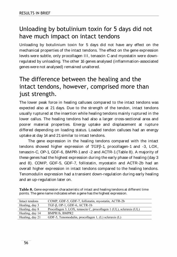
RESULTS IN BRIEF
56
Unloading by botulinium toxin for 5 days did not
have much impact on intact tendons
Unloading by botulinium toxin for 5 days did not have any effect on the
mechanical properties of the intact tendons. The effect on the gene expression
levels were subtle, only procollagen III, tenascin C and myostatin were down-
regulated by unloading. The other 16 genes analysed (inflammation-associated
genes were not analysed) remained unaltered.
The difference between the healing and the
intact tendons, however, comprised more than
just strength.
The lower peak force in healing calluses compared to the intact tendons was
expected also at 21 days. Due to the strength of the tendon, intact tendons
usually ruptured at the insertion while healing tendons mainly ruptured in the
lower callus. The healing tendons had also a larger cross-sectional area and
poorer material properties. Energy uptake and displacement at rupture
differed depending on loading status. Loaded tendon calluses had an energy
uptake at day 14 and 21 similar to intact tendons.
The gene expression in the healing tendons compared with the intact
tendons showed higher expression of TGFβ 1, procollagen-1 and -3, LOX,
tenascin-C, OP-1, GDF-6, BMPR-1 and -2 and ACTR-1 (Table 8). A majority of
these genes had the highest expression during the early phase of healing (day 3
and 8). COMP, GDF-5, GDF-7, follistatin, myostatin and ACTR-2b had an
overall higher expression in intact tendons compared to the healing tendons.
Tenomodulin expression had a transient down-regulation during early healing
and an up-regulation later on.
Table 8. Gene expression characteristic of intact and healing tendons at different time points. The gene name indicates when a gene has the highest expression. Intact tendons COMP, GDF-5, GDF-7, follistatin, myostatin, ACTR-2b
Healing, day 3 TGF-β, OP-1, GDF-6, ACTR-1b
Healing, day 8 Procollagen 3, LOX, tenascin C, procollagen 1 (UL), scleraxis (UL)
Healing, day 14 BMPR1b, BMPR2
Healing, day 21 GDF-5, Tenomodulin, procollagen 1, (L) scleraxis (L)
-
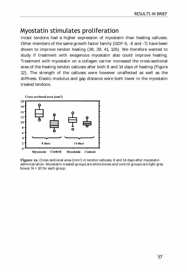
RESULTS IN BRIEF
57
Myostatin stimulates proliferation Intact tendons had a higher expression of myostatin than healing calluses.
Other members of the same growth factor family (GDF-5, -6 and -7) have been
shown to improve tendon healing (38, 39, 41, 126). We therefore wanted to
study if treatment with exogenous myostatin also could improve healing.
Treatment with myostatin on a collagen carrier increased the cross-sectional
area of the healing tendon calluses after both 8 and 14 days of healing (Figure
12). The strength of the calluses were however unaffected as well as the
stiffness. Elastic modulus and gap distance were both lower in the myostatin
treated tendons.
Figure 12. Cross-sectional area (mm2) in tendon calluses, 8 and 14 days after myostatin administration. Myostatin-treated groups are white boxes and control groups are light grey boxes. N = 10 for each group.
57


59
DISCUSSION
Unloading during tendon healing is detrimental for the mechanical properties
of the tendon callus. Botulinium toxin-induced paralysis of the calf muscles
resulted in a significantly weaker tendon callus already after 8 days of healing.
The detrimental effect of unloading can be partly reversed by short daily
loading episodes.
Rest between loadings might allow the tendon
callus to contract
Healing tendons with continuous loading were significantly longer than the
unloaded ones. Lengthening during tendon healing is one potential clinical
adversity after too much loading early on. However, healing tendons exposed
to short loading episodes (in all studies) did not show any lengthening
compared to the completely unloaded ones. This suggests that the resting time
between the loading episodes might give the callus tissue enough time to
contract. In fact, loading on intact tendons have been shown to increase the
amount of myofibroblasts in the tendon (112). Myofibroblasts can contract
tissues. Short loading episodes might therefore be beneficial for the
mechanical properties of healing tendons without risk of persistent
lengthening.

DISCUSSION
60
Loading generates more matrix but not
necessarily of better quality
The main response to loading, irrespective of it was continuous or short
episodes, was an increased cross-sectional area. This suggests an increased
production of matrix. The material properties (peak stress and elastic
modulus) appeared however to be unaltered by loading. This indicates that the
matrix produced early on after loading is not of any better quality than the
matrix produced in the unloaded tendons. Intact tendons have also been
shown to respond and adapt to loading by increased cross-sectional area and
not by altered material properties of the tendon (47). An exception to the lack
of improved material properties was the increased peak stress after four
loading episodes in study II. This was seen after loading during both early and
late healing. One discrepancy between this study and the first study, besides
the number of episodes, was the time between the final episode and
euthanasia. The animals with 11 loading episodes in study I were all killed one
day after the last loading episode, while the animals with four loading episodes
were unloaded for three more days after the last loading episode before they
were killed. We therefore unloaded the animals in study VI, with one loading
episode, for 3 days after the loading was finished. We wanted to study if the
recovery time after loading was the key for improved material properties.
However, these animals only differed in stiffness compared to the unloaded
animals. An increased peak force was first seen 7 days after the loading was
finished, but the peak stress in these tendons was not different from the peak
stress in the unloaded ones. However, because the response after only one
loading episode was weaker, it is possible that there was not sufficient power to
detect improved tissue qualities.

DISCUSSION
61
What is optimal loading?
Even though short loading episodes increased the peak force of the healing
tendons, the tendon calluses with continuous loading (i.e. free cage activity)
were both stronger and stiffer. A longer duration of each loading episode - 15
minutes compared to 60 minutes each day - only increased the peak force to
some extent. Therefore, a longer duration of loading in animals with free cage
activity, is presumably not the entire explanation for the discrepancy in peak
force. Rats with free cage activity have probably more opportunities to regulate
the amount of loading which they apply on the injured limb during the early
healing. The treadmill rats had perhaps less of a choice about how to load their
tendons and more monotonously loading pattern. Animals with free cage
activity could perhaps also apply loading of different frequencies and
magnitudes and thereby create deformations or fluid flows for more optimal
mechanotransduction.
There is also a possible threshold to get a response, because the animals
with only 15 minutes of free cage activity each day did not have stronger
tendons compared to the unloaded animals. Studies on intact tendons in
humans have shown that there appears to be some kind of threshold for the
stress or strain level to induce a response (47).
Frequency – magnitude – number of cycles
The increase in collagen synthesis after acute loading appears to be
independent of the magnitude of exercise (62). The effect of loading on COX-2
expression and PGE2 production in vitro was shown to be dependent on the
stretching frequency but not on the number of cycles (124). However, the
expression of scleraxis in bioartificial tendons containing mesenchymal stem
cells was dependent on the number of cycles, but it was also dependent on the
duration and strain (103). The same model showed that the expression of
collagen I was influenced by the strain magnitude, and short rests (10 s)
between each cycle increased the expression of both scleraxis and collagen I
even more. What is most important for the response to loading: the number of
cycles, duration, magnitude or frequency? The different factors most likely
create different effects in the tissue regarding fluid flow, strain and shear stress
which will affect mechanotransduction.
We have previously attempted to alter the frequency of the loading
stimulus by loading healing tendons on a vibration plate (unpublished data).
This loading stimulus did not increase the peak force of the healing tendons

DISCUSSION
62
compared to loaded tendons without vibrations. However it does not exclude
that frequency of the loading is important for the response: other frequencies
or magnitudes might be better for mechanotransduction, although the chosen
ones are efficacious for bone. We therefore still do not know how the optimal
mechanical stimulus looks like.
DISCUSSION
63
Inflammation: good or bad?
Inflammation-associated genes, among them IL-1β, were regulated by both
continuous loading and by one single loading episode. Continuously loaded
tendon calluses had a lower expression of IL-1β than unloaded tendons, while
one single loading episode induced a higher expression of IL-1β. Other
members of the IL-1β family were also regulated by one loading episode,
including its antagonist and receptors. This makes it difficult to determine the
net effect of IL-1β expression, and its role in tendon healing. However, our
results show that IL-1β appears to be mechanosensitive, which is in line with
results from previous in vitro and in vivo experiments (6, 8, 116).
The role of IL-1β in tendon healing and adaption to loading appear to
be complex. Too much IL-1β could be detrimental, but a small amount appears
to be good. Stretched tendon fibroblasts in vitro can produce a potentially
harmful response, by an increased expression of TGF-β, COX-2, MMP-27 and
ADAMTS-5 (97). This induced gene expression could be reversed by low doses
of exogenous IL-1β (100 pM). However, administration of 10 pM of IL-1β to
unstretched tendon fibroblasts also induced an increased expression of COX-2
and MMP-1 and PGE2 production (130). This IL-1β mediated gene expression
could be reversed by a 4% stretching of the cells, but an 8% stretching instead
increased the levels additionally. IL-1β and loading therefore appears to
regulate each other. We have attempted to inhibit IL-1β during tendon healing
but have so far not seen any effects on the mechanical properties of the callus
(unpublished). The role of IL-1β during tendon healing therefore needs to be
further investigated.
IL-6 is an other interleukin which is potentially involved in tendon
adaption to loading. IL-6 production has been shown to increase in the
peritendinous space after loading (68) and IL-6 infusion can increase the
collagen production in tendons (with and without loading) (5). IL-6 might
therefore play a role in tendon adaption and perhaps also in tendon healing.
Healing, loading, inflammation and tendinosis are all conditions which
appear to regulate or be regulated by the gene expression and protein levels for
PGE2 (COX-2) and NO. This has been shown in vitro and in vivo in both
animals and humans. Continuous loading in our model did not significantly
regulate the gene expression of iNOS and COX-2. These areas (NO and
prostaglandin synthesis) were however regulated after a single loading
episode. It is not known whether these proteins are good or bad for the tendon.
PGE2 is increased after a long duration of running, and this response could

DISCUSSION
64
both indicate injury by the exercise but it could also indicate that PGE2 is
involved in the adaption to loading. Inhibition of PGE2 during early tendon
healing results in a weaker tendon callus, but inhibition later on in the healing
process will improve the material properties (40, 127). Exercise-induced PGE2
can be inhibited by COX-2-inhibitors, and this also inhibits the increased
blood flow seen after exercise (66). Inhibition of COX-1 and COX-2 have also
been shown to inhibit the increase in procollagen type I N-terminal propeptide
(PINP) levels in the peritendinous space of the Achilles tendon after exercise
(23). In vitro studies have shown that inhibition of loading induced PGE2
results in increased levels of LTB4 (3). The role of leukotrienes in tendon
healing and adaption to loading has been even less investigated, and the
connection between prostaglandins and leukotrienes could be important.
Addition of exogenous NO has been shown to improve tendon healing
(90, 91, 131). However, NO is also mentioned as a potentially harmful
molecule, involved in oxidative stress etc. It appears as a lot of inflammation-
associated molecules have dual functions. They are needed in a small amount
in the beginning of the healing process, but prolonged exposure or too high
levels can be detrimental. This needs to be further investigated.
The general opinion is that exercise should be avoided during the
inflammatory phase of healing to minimize disruption of the healing process
(122). Our data indicates conversely that the healing process is not disrupted
but promoted. Four short loading episodes during the inflammatory phase
increased the peak force of the tissue. However, the amount of bleeding was
also increased. This could be a result from micro-traumas in the tissue and
might explain the up-regulation of wound healing and coagulation genes after
one loading episode. An alternative to mechanically induced activation of
genes involved in healing or regeneration could therefore be that microinjuries
in the healing tissue keep the healing response more active.

DISCUSSION
65
All research has limitations
Is a rat model for tendon healing relevant for the situation in humans?
The transferability of research from animals to humans can always be
questioned. The knowledge from animals is quite often transferable, but not
always, and we don’t know when it is. Animal models are reasonably fast and
the studies can usually be more detailed due to substrate availability.
Compared to cell cultures, they also include, all the mechanisms, like the
proper environment, circulation and immune system etc. We have used rats in
these studies, which are quadrupeds while humans are not, this could of course
result in discrepancies in the response to loading. Rats are for example able to
more easily regulate the amount of loading put on each limb. However, cells in
humans and animals most likely experience mechanotransduction by similar
or identical mechanisms, and it is likely that the response is at least similar.
The size of the Achilles tendons in the rats is small and the distance between
the old tendon stumps after transection is a few millimetres. This is roughly
the same distance as each fibre bundle has in ruptured human Achilles tendon
and we therefore guess that the response to healing with regards to cell
migration, capillary growth requirements etc. will be quite similar in rats and
humans.
Can we extrapolate the research on the Achilles tendon to other tendons?
We have used the Achilles tendon as a model for tendon healing. This tendon is
the largest tendon in the body and it is therefore easy to handle in small
animals. It is also prone to injuries. The Achilles tendon heals reasonably well
compared to other tendons, partly because there is a rich blood supply in close
proximity in the surrounding tissues. Other subcutaneous tendons (like the
extensor tendons in the hand) are similar to the Achilles tendon in this respect.
It is therefore likely that we can extrapolate the results from the Achilles
tendon to these types of tendons. However, tendons and ligaments in synovial
fluid are different from the Achilles tendon, because they have poorer nutrition
due to limited blood supply and they also have adherence problems during
healing. The results from the Achilles tendon will probably not resemble the
situation in these tendons. Still, early mobilization is even more important in
these tendons to avoid the adherence problems.

DISCUSSION
66
Frayed ends or sharp transection
The sharp transection of an otherwise healthy tendon like we do is quite
different from the ruptured Achilles tendon in patients. The ruptured tendons
almost always have frayed ends and a degenerative background. However,
when using an animal model, it is important to have an easily reproducible
method. This allows us to measure the effect of an intervention by mechanical
testing, without involvement of the surgical technique. We can therefore use
reasonably small groups and still have enough power in the analyses.
Short term vs long term effects on healing
We have only studied the healing up to day 21 after transection. This is partly
because it is difficult to get true measurements of the peak force in the healing
tendons at later time-points. The mechanical testing has some limitations. If
the tendon becomes too strong (like intact tendons) the failure will not occur in
the mid-substance of the tendon, but at the insertion to the calcaneal bone or
by the upper clamp. We have therefore used shorter time-points (mostly 8-14
days of healing) to measure improvements of tendon healing. Furthermore,
rats also spontaneously develop cartilage and bone formation in the healing
tendon after some weeks. This makes it unsuitable to use this model for a
longer follow up. A more long term follow up could perhaps give us more
information whether the loaded callus tissue results in more resemble to the
original tendon tissue or if loading has created more scar-tissue formation.
The potential sex differences
A limitation in our animal model is that we have only used female rats. Human
intact tendons respond to loading differently depending on sex, with regards to
collagen synthesis rate and tendon cross-sectional area (82). The mechanical
properties also appear to differ between men and women. The fascicles from
men are stronger then the fascicles from women. We have previously
investigated the role of oestrogen on tendon healing by comparing
ovariectomised rats to normal controls, and found no difference in healing
properties (unpublished). This however, does not rule out any difference in
healing properties between male and female rats. We have chosen female rats
because they tend to grow slower compared to male rats, and it might
influence the healing process.

DISCUSSION
67
Botox, tail suspension or cast
There are always advantages and disadvantages with different unloading
methods. Unloading by botulinium toxin is a quick and simple procedure
which allows us to do fairly large studies easily. The animals are also quite
unaffected by this method. Botox unloading has also a clear effect on tendon
healing. However, it does not resemble the human situation, where the
patient’s foot is immobilized and unloaded in a cast. Also, we can not entirely
exclude that any loading occurs. This could on the other hand also occur in
patients, where some patients, immobilised by a cast, seem to load their
tendons more than others (unpublished).
Tail suspension is an appropriate method when you want to reverse the
unloading for just short periods each day. This model is also quite different
from the situation in patients, with immobilised joints. However, tail
suspension allows us to study the effect of loading per se, and not just ankle
movements without loading. It is important to discriminate between
movements and loading when we want to increase the understanding for
treatment strategies (e.g. passive mobilization vs loading with forces). During
tail suspension, forces in the Achilles tendon are limited by the antagonistic
action of the dorsal flexors. These muscles are weak and have a short lever
arm. Therefore the Achilles tendon can only be exposed to weak forces, with
possible exception for when the rats scratch themselves.
Discrepancy between genes and proteins
We have so far only studied gene expression levels in our experiments (except
for the staining for follistatin with immunohistochemistry). Regulations at the
gene expression level do not always correspond to changes in protein
expression and secretion. The findings in these studies would therefore need to
be confirmed at the protein level before any solid conclusion could be drawn.
Real-time PCR is a fast way to study responses, but it does not say anything
about where in the tissue the genes are expressed (e.g. in the centre or in the
periphery) or by which cells. All cells in a tissue do not respond equally to
loading/unloading (114). If only a fraction of the cells have a higher expression,
this will probably go without finding when using real-time PCR, because it will
be clouded by the lower expression the rest of the cells. In situ hybridization or
immunohistochemistry can be used to avoid this problem.
When analysing with real-time PCR you need to use reliable house-
keeping genes that are stably expressed and this can sometime be hard to find.

DISCUSSION
68
We used 18S in the first three studies. We found it to be stably expressed
among the groups. However in the last study, we used a combination of three
different house-keeping genes (which were all stably expressed). By using
three genes, the risk of house-keeping gen errors was diminished.
The number of animals is the limit - RRR
We have used the same material for the gene expression analysis in study III-
V. This could mean that the effects seen in these studies can only be observed
in this particular group of animals. However, when using animals in research
you are always limited to the number of animals you can use, due to ethical
reasons (RRR reduce, refine and replace). This is a limiting factor, and animal
experiments are not often repeated. This is also why there are no mechanistic
studies done in study I-II.

69
CONCLUSIONS AND FUTURE RESEARCH
Short daily loading episodes during tendon healing improve the strength of the
tendon callus. Loading has an effect already during early tendon healing
(during the inflammatory phase) although it is generally believed that loading
mainly has an effect first later on, during remodelling. It appears as the loading
episodes don’t have to be long, but they need to be applied on a daily basis to
sustain a response (at least on the gene expression level). Loading more
frequently than once per day, does not seem to improve the healing response
additionally. It also appears as short loading episodes do not produce any
persisting lengthening of the tendon callus. This observation opens up for new
possibilities to use short daily loading episodes as a part of the rehabilitation
after tendon rupture.
Continuous loading only altered a few of the chosen genes but not
dramatically as for the effect on the peak force and cross-sectional area. The
reason for this could be that the response to loading is transient and instead of
continuous loading for several days, the loading needs to be better defined.
There were several genes which were regulated over time during tendon
healing, like ECM components and different members of the BMP-family. The
different BMP-members might also have diverse roles during the three phases
of tendon healing. A few genes appeared even to be more important in the
maintenance of the intact tendon rather than having a role during tendon
healing. Genes that are responsible for the improved healing by loading are not
easy to point-out. Inflammation definitively appears to be regulated by
loading, but the exact role for this during tendon healing needs to be further
investigated.

CONCLUSIONS AND FUTURE RESEARCH
70
What’s next?
We began this project by describing which loading parameters that were
important for an improved healing response. We managed to diminish loading
until we could use just one episode. This opened up a new possibility to study
gene expression: with only one episode. We could see which genes were
regulated by loading and mechanotransduction, rather than by the fact that
loading had stimulated a better healing response. The effect of one single
loading episode was studied 3 hours after loading. This is a fairly long time
after loading, which probably results in activation of secondary signalling
pathways in addition to the purely mechano-driven pathways. It would
therefore be appealing to study the effect of loading at an earlier time-point to
find the very first responsive pathways after loading in vivo. It would also be
interesting to use this model to study the response to loading in intact tendons.
This is probably more difficult, since intact tendons are less responsive to
loading.
Inflammation or inflammation-associated proteins like interleukins,
NO and eicosanoids are important during tendon healing. They are also
involved in the response to loading in both intact and healing tendons. Still,
there is much more to know about inflammation and its role in tendons and
tendon healing. Inflammation is most likely necessary for tendon healing, but
it can also probably do harm. The same factors which are involved in the
healing response and adaption to loading are also thought to have a role in
tendon overloading and injuries. We need to know how these factors are
regulated during tendon healing and also what their role is in intact overloaded
and degenerated tendons.
The microarray experiment revealed angiogenesis and production of
ROS as two potential mechanisms regulated by loading. A lot of the genes
involved in these mechanisms have a variety of functions. It would therefore be
interesting to find out more about the role of angiogenesis and ROS production
during tendon healing and the potential regulation by loading.
These experiments and a lot of other studies on tendon healing have
mainly measured the peak force of the healing tendons shortly after
transection. A strong tendon early on probably speeds up the rehabilitation
and thereby reduces the health care costs. Early mechanical properties of the
tendon have also been shown to correlate with late function (99). However, the
question remains whether an initially strong tendon have more resemblance to
the original tendon or less. A study on the long term effects of early loading

CONCLUSIONS AND FUTURE RESEARCH
71
would be useful for a greater understanding of the benefits and response to
loading. Does loading drive the tendon towards scar-formation or
regeneration? Tendon markers like scleraxis and tenomodulin could perhaps
give some clue whether the healing is directed towards scar or tendon tissue.
Before we can really distinguish between scar-tissue, tendon-tissue and callus-
tissue, we have to understand more about which markers to use. This could be
used to understand if the healing progress is heading in the right direction.
Take-home message:
15 minutes of daily loading already at an early stage of tendon healing might
improve the rehabilitation after tendon rupture.


73
ACKNOWLEDGEMENTS
I would like to thank everyone that has been involved in me and my thesis over
the years, at work and outside work, but most of all I would like to thank:
Therese Andersson, for almost everything =). All the weekends and
evenings spent in the animal department over the years. I couldn’t have done
this thesis without you. Half of this thesis belongs to you!
Per Aspenberg, my supervisor, for your enthusiasm, encouragement,
interesting discussions, support and help. But most of all for believing in me
and giving me the freedom to develop.
Anna Fahlgren, for all the support and help during the years.
Björn Pasternak, for introducing me to research in general and in the lab in
particular, and for the nice talks during my first years in the lab.
Fredrik Agholme, for the nice company inside and outside the lab.
Anna Nilsson, for your energy and fruitful discussions.
Anna-Carin Lundin, for all your kind words and all the laughs.
Olof Sandberg, for your positive attitude.
Bibbi Mårdh, for the assistance over the years.
Mats Christensson, for manufacturing the tail suspension cages
Tony Andersson, Olena Virchenko, Maria Bolin, Paul Ackermann
and Daniel Bring, for all the work you did before I started this thesis.
Maria Jenmalm, for all answers to my questions regrding different
molecular biology methods.

REFERENCES
74
Piiha-Lotta Jerevall, for all your help with my thesis. =)
Cissi, Nettan, Anders, Linda, Andreas, Jonas, Lena, Jan and Danne
at the animal department, for your assistance over the years
The members of Forum Scientium, for all the fun times at travels and
conferences.
Ulf Eliasson, Britt Tessem and Mikael Eliasson, for being the best
family one can ask for and for your support and love.
Stina, Lotta and Anna, and the rest of my friends, for giving me a great
time in living in Linköping.
Lars Johansson, for the nice and quite place where I could write my thesis.
Alexander Nordström, my love and soon to be husband. Thank you for
always being there and also for your help with the animals during weekends
and evenings =)
And finally in memory of my mum who unfortunately passed away
too early in my life, but has given me a lot of good characteristics
necessary for this thesis…
This thesis was supported by the Swedish National Centre for Sports Research,
the Strategic Research Project “Materials in medicine”, the Swedish Research
Council, the King Gustaf V and Queen Victoria Free Mason Foundation and the
County Council of Östergötland.

75
REFERENCES
1. AbiEzzi SS, Foulk RA, Harwood FL, Akeson WH, and Amiel D. Decrease in
fibronectin occurs coincident with the increased expression of its integrin
receptor alpha5beta1 in stress-deprived ligaments. Iowa Orthop J 17: 102-109,
1997.
2. AbiEzzi SS, Gesink DS, Schreck PJ, Amiel D, Akeson WH, and Woods VL, Jr.
Increased expression of the beta 1, alpha 5, and alpha v integrin adhesion
receptor subunits occurs coincident with remodeling of stress-deprived rabbit
anterior cruciate and medial collateral ligaments. J Orthop Res 13: 594-601,
1995.
3. Almekinders LC, Banes AJ, and Ballenger CA. Effects of repetitive motion on
human fibroblasts. Med Sci Sports Exerc 25: 603-607, 1993.
4. Almekinders LC, Baynes AJ, and Bracey LW. An in vitro investigation into the
effects of repetitive motion and nonsteroidal antiinflammatory medication on
human tendon fibroblasts. Am J Sports Med 23: 119-123, 1995.
5. Andersen MB, Pingel J, Kjaer M, and Langberg H. Interleukin-6: a growth
factor stimulating collagen synthesis in human tendon. J Appl Physiol 110:
1549-1554.
6. Archambault J, Tsuzaki M, Herzog W, and Banes AJ. Stretch and interleukin-
1beta induce matrix metalloproteinases in rabbit tendon cells in vitro. J Orthop
Res 20: 36-39, 2002.
7. Archambault JM, Elfervig-Wall MK, Tsuzaki M, Herzog W, and Banes AJ.
Rabbit tendon cells produce MMP-3 in response to fluid flow without
significant calcium transients. J Biomech 35: 303-309, 2002.
8. Archambault JM, Hart DA, and Herzog W. Response of rabbit Achilles tendon
to chronic repetitive loading. Connect Tissue Res 42: 13-23, 2001.
9. Archambault JM, Jelinsky SA, Lake SP, Hill AA, Glaser DL, and Soslowsky LJ.
Rat supraspinatus tendon expresses cartilage markers with overuse. J Orthop
Res 25: 617-624, 2007.
10. Arnoczky SP, Lavagnino M, and Egerbacher M. The mechanobiological
aetiopathogenesis of tendinopathy: is it the over-stimulation or the under-
stimulation of tendon cells? Int J Exp Pathol 88: 217-226, 2007.

REFERENCES
76
11. Arnoczky SP, Tian T, Lavagnino M, and Gardner K. Ex vivo static tensile
loading inhibits MMP-1 expression in rat tail tendon cells through a
cytoskeletally based mechanotransduction mechanism. J Orthop Res 22: 328-
333, 2004.
12. Arnoczky SP, Tian T, Lavagnino M, Gardner K, Schuler P, and Morse P.
Activation of stress-activated protein kinases (SAPK) in tendon cells following
cyclic strain: the effects of strain frequency, strain magnitude, and cytosolic
calcium. J Orthop Res 20: 947-952, 2002.
13. Attia E, Brown H, Henshaw R, George S, and Hannafin JA. Patterns of gene
expression in a rabbit partial anterior cruciate ligament transection model: the
potential role of mechanical forces. Am J Sports Med 38: 348-356.
14. Backman LJ, Andersson G, Wennstig G, Forsgren S, and Danielson P.
Endogenous substance P production in the Achilles tendon increases with
loading in an in vivo model of tendinopathy - peptidergic elevation preceding
tendinosis-like tissue changes. J Musculoskelet Neuronal Interact 11: 133-140.
15. Banes AJ, Tsuzaki M, Hu P, Brigman B, Brown T, Almekinders L, Lawrence
WT, and Fischer T. PDGF-BB, IGF-I and mechanical load stimulate DNA
synthesis in avian tendon fibroblasts in vitro. J Biomech 28: 1505-1513, 1995.
16. Banes AJ, Tsuzaki M, Yamamoto J, Fischer T, Brigman B, Brown T, and Miller
L. Mechanoreception at the cellular level: the detection, interpretation, and
diversity of responses to mechanical signals. Biochem Cell Biol 73: 349-365,
1995.
17. Bedi A, Kovacevic D, Fox AJ, Imhauser CW, Stasiak M, Packer J, Brophy RH,
Deng XH, and Rodeo SA. Effect of early and delayed mechanical loading on
tendon-to-bone healing after anterior cruciate ligament reconstruction. J Bone
Joint Surg Am 92: 2387-2401.
18. Bi Y, Ehirchiou D, Kilts TM, Inkson CA, Embree MC, Sonoyama W, Li L, Leet
AI, Seo BM, Zhang L, Shi S, and Young MF. Identification of tendon
stem/progenitor cells and the role of the extracellular matrix in their niche.
Nat Med 13: 1219-1227, 2007.
19. Bring D, Reno C, Renstrom P, Salo P, Hart D, and Ackermann P. Prolonged
immobilization compromises up-regulation of repair genes after tendon
rupture in a rat model. Scand J Med Sci Sports 20: 411-417.
20. Bring DK, Kreicbergs A, Renstrom PA, and Ackermann PW. Physical activity
modulates nerve plasticity and stimulates repair after Achilles tendon rupture.
J Orthop Res 25: 164-172, 2007.
21. Bring DK, Reno C, Renstrom P, Salo P, Hart DA, and Ackermann PW. Joint
immobilization reduces the expression of sensory neuropeptide receptors and
impairs healing after tendon rupture in a rat model. J Orthop Res 27: 274-280,
2009.
REFERENCES
77
22. Chiquet M. Regulation of extracellular matrix gene expression by mechanical
stress. Matrix Biol 18: 417-426, 1999.
23. Christensen B, Dandanell S, Kjaer M, and Langberg H. Effect of anti-
inflammatory medication on the running-induced rise in patella tendon
collagen synthesis in humans. J Appl Physiol 110: 137-141.
24. Christensen B, Dyrberg E, Aagaard P, Enehjelm S, Krogsgaard M, Kjaer M,
and Langberg H. Effects of long-term immobilization and recovery on human
triceps surae and collagen turnover in the Achilles tendon in patients with
healing ankle fracture. J Appl Physiol 105: 420-426, 2008.
25. Christensen B, Dyrberg E, Aagaard P, Kjaer M, and Langberg H. Short-term
immobilization and recovery affect skeletal muscle but not collagen tissue
turnover in humans. J Appl Physiol 105: 1845-1851, 2008.
26. Costa ML, MacMillan K, Halliday D, Chester R, Shepstone L, Robinson AH,
and Donell ST. Randomised controlled trials of immediate weight-bearing
mobilisation for rupture of the tendo Achillis. J Bone Joint Surg Br 88: 69-77,
2006.
27. Couppe C, Kongsgaard M, Aagaard P, Hansen P, Bojsen-Moller J, Kjaer M, and
Magnusson SP. Habitual loading results in tendon hypertrophy and increased
stiffness of the human patellar tendon. J Appl Physiol 105: 805-810, 2008.
28. Curwin SL, Vailas AC, and Wood J. Immature tendon adaptation to strenuous
exercise. J Appl Physiol 65: 2297-2301, 1988.
29. Dahl J, Li J, Bring DK, Renstrom P, and Ackermann PW. Intermittent
pneumatic compression enhances neurovascular ingrowth and tissue
proliferation during connective tissue healing: a study in the rat. J Orthop Res
25: 1185-1192, 2007.
30. de Boer MD, Selby A, Atherton P, Smith K, Seynnes OR, Maganaris CN,
Maffulli N, Movin T, Narici MV, and Rennie MJ. The temporal responses of
protein synthesis, gene expression and cell signalling in human quadriceps
muscle and patellar tendon to disuse. J Physiol 585: 241-251, 2007.
31. Devkota AC, Tsuzaki M, Almekinders LC, Banes AJ, and Weinhold PS.
Distributing a fixed amount of cyclic loading to tendon explants over longer
periods induces greater cellular and mechanical responses. J Orthop Res 25:
1078-1086, 2007.
32. Dudhia J, Scott CM, Draper ER, Heinegard D, Pitsillides AA, and Smith RK.
Aging enhances a mechanically-induced reduction in tendon strength by an
active process involving matrix metalloproteinase activity. Aging Cell 6: 547-
556, 2007.

REFERENCES
78
33. Enwemeka CS. Functional loading augments the initial tensile strength and
energy absorption capacity of regenerating rabbit Achilles tendons. Am J Phys
Med Rehabil 71: 31-38, 1992.
34. Enwemeka CS. Inflammation, cellularity, and fibrillogenesis in regenerating
tendon: implications for tendon rehabilitation. Phys Ther 69: 816-825, 1989.
35. Enwemeka CS, Spielholz NI, and Nelson AJ. The effect of early functional
activities on experimentally tenotomized Achilles tendons in rats. Am J Phys
Med Rehabil 67: 264-269, 1988.
36. Flick J, Devkota A, Tsuzaki M, Almekinders L, and Weinhold P. Cyclic loading
alters biomechanical properties and secretion of PGE2 and NO from tendon
explants. Clin Biomech (Bristol, Avon) 21: 99-106, 2006.
37. Fong KD, Trindade MC, Wang Z, Nacamuli RP, Pham H, Fang TD, Song HM,
Smith RL, Longaker MT, and Chang J. Microarray analysis of mechanical
shear effects on flexor tendon cells. Plast Reconstr Surg 116: 1393-1404;
discussion 1405-1396, 2005.
38. Forslund C and Aspenberg P. CDMP-2 induces bone or tendon-like tissue
depending on mechanical stimulation. J Orthop Res 20: 1170-1174, 2002.
39. Forslund C and Aspenberg P. Tendon healing stimulated by injected CDMP-2.
Med Sci Sports Exerc 33: 685-687, 2001.
40. Forslund C, Bylander B, and Aspenberg P. Indomethacin and celecoxib
improve tendon healing in rats. Acta Orthop Scand 74: 465-469, 2003.
41. Forslund C, Rueger D, and Aspenberg P. A comparative dose-response study of
cartilage-derived morphogenetic protein (CDMP)-1, -2 and -3 for tendon
healing in rats. J Orthop Res 21: 617-621, 2003.
42. Gardner K, Arnoczky SP, Caballero O, and Lavagnino M. The effect of stress-
deprivation and cyclic loading on the TIMP/MMP ratio in tendon cells: an in
vitro experimental study. Disabil Rehabil 30: 1523-1529, 2008.
43. Gardner K, Arnoczky SP, and Lavagnino M. Effect of in vitro stress-
deprivation and cyclic loading on the length of tendon cell cilia in situ. J
Orthop Res 29: 582-587.
44. Hansen M, Boitano S, Dirksen ER, and Sanderson MJ. Intercellular calcium
signaling induced by extracellular adenosine 5'-triphosphate and mechanical
stimulation in airway epithelial cells. J Cell Sci 106 ( Pt 4): 995-1004, 1993.
45. Hansson HA, Engstrom AM, Holm S, and Rosenqvist AL. Somatomedin C
immunoreactivity in the Achilles tendon varies in a dynamic manner with the
mechanical load. Acta Physiol Scand 134: 199-208, 1988.

REFERENCES
79
46. Heinemeier K, Langberg H, Olesen JL, and Kjaer M. Role of TGF-beta1 in
relation to exercise-induced type I collagen synthesis in human tendinous
tissue. J Appl Physiol 95: 2390-2397, 2003.
47. Heinemeier KM and Kjaer M. In vivo investigation of tendon responses to
mechanical loading. J Musculoskelet Neuronal Interact 11: 115-123.
48. Heinemeier KM, Olesen JL, Haddad F, Langberg H, Kjaer M, Baldwin KM,
and Schjerling P. Expression of collagen and related growth factors in rat
tendon and skeletal muscle in response to specific contraction types. J Physiol
582: 1303-1316, 2007.
49. Heinemeier KM, Olesen JL, Haddad F, Schjerling P, Baldwin KM, and Kjaer
M. Effect of unloading followed by reloading on expression of collagen and
related growth factors in rat tendon and muscle. J Appl Physiol 106: 178-186,
2009.
50. Heinemeier KM, Olesen JL, Schjerling P, Haddad F, Langberg H, Baldwin KM,
and Kjaer M. Short-term strength training and the expression of myostatin
and IGF-I isoforms in rat muscle and tendon: differential effects of specific
contraction types. J Appl Physiol 102: 573-581, 2007.
51. Hess GW. Achilles tendon rupture: a review of etiology, population, anatomy,
risk factors, and injury prevention. Foot Ankle Spec 3: 29-32.
52. Hsieh AH, Sah RL, and Paul Sung KL. Biomechanical regulation of type I
collagen gene expression in ACLs in organ culture. J Orthop Res 20: 325-331,
2002.
53. Hsieh AH, Tsai CM, Ma QJ, Lin T, Banes AJ, Villarreal FJ, Akeson WH, and
Sung KL. Time-dependent increases in type-III collagen gene expression in
medical collateral ligament fibroblasts under cyclic strains. J Orthop Res 18:
220-227, 2000.
54. Hung CT, Allen FD, Pollack SR, Attia ET, Hannafin JA, and Torzilli PA.
Intracellular calcium response of ACL and MCL ligament fibroblasts to fluid-
induced shear stress. Cell Signal 9: 587-594, 1997.
55. James R, Kesturu G, Balian G, and Chhabra AB. Tendon: biology,
biomechanics, repair, growth factors, and evolving treatment options. J Hand
Surg Am 33: 102-112, 2008.
56. Jozsa L and Kannus P. Histopathological findings in spontaneous tendon
ruptures. Scand J Med Sci Sports 7: 113-118, 1997.
57. Kajikawa Y, Morihara T, Watanabe N, Sakamoto H, Matsuda K, Kobayashi M,
Oshima Y, Yoshida A, Kawata M, and Kubo T. GFP chimeric models exhibited
a biphasic pattern of mesenchymal cell invasion in tendon healing. J Cell
Physiol 210: 684-691, 2007.

REFERENCES
80
58. Kannus P. Structure of the tendon connective tissue. Scand J Med Sci Sports
10: 312-320, 2000.
59. Kerkhoffs GM, Struijs PA, Raaymakers EL, and Marti RK. Functional
treatment after surgical repair of acute Achilles tendon rupture: wrap vs
walking cast. Arch Orthop Trauma Surg 122: 102-105, 2002.
60. Kim SG, Akaike T, Sasagaw T, Atomi Y, and Kurosawa H. Gene expression of
type I and type III collagen by mechanical stretch in anterior cruciate ligament
cells. Cell Struct Funct 27: 139-144, 2002.
61. Kjaer M. Role of extracellular matrix in adaptation of tendon and skeletal
muscle to mechanical loading. Physiol Rev 84: 649-698, 2004.
62. Kjaer M, Langberg H, Heinemeier K, Bayer ML, Hansen M, Holm L, Doessing
S, Kongsgaard M, Krogsgaard MR, and Magnusson SP. From mechanical
loading to collagen synthesis, structural changes and function in human
tendon. Scand J Med Sci Sports 19: 500-510, 2009.
63. Kongsgaard M, Aagaard P, Kjaer M, and Magnusson SP. Structural Achilles
tendon properties in athletes subjected to different exercise modes and in
Achilles tendon rupture patients. J Appl Physiol 99: 1965-1971, 2005.
64. Kongsgaard M, Reitelseder S, Pedersen TG, Holm L, Aagaard P, Kjaer M, and
Magnusson SP. Region specific patellar tendon hypertrophy in humans
following resistance training. Acta Physiol (Oxf) 191: 111-121, 2007.
65. Koskinen SO, Heinemeier KM, Olesen JL, Langberg H, and Kjaer M. Physical
exercise can influence local levels of matrix metalloproteinases and their
inhibitors in tendon-related connective tissue. J Appl Physiol 96: 861-864,
2004.
66. Langberg H, Boushel R, Skovgaard D, Risum N, and Kjaer M. Cyclo-
oxygenase-2 mediated prostaglandin release regulates blood flow in connective
tissue during mechanical loading in humans. J Physiol 551: 683-689, 2003.
67. Langberg H, Ellingsgaard H, Madsen T, Jansson J, Magnusson SP, Aagaard P,
and Kjaer M. Eccentric rehabilitation exercise increases peritendinous type I
collagen synthesis in humans with Achilles tendinosis. Scand J Med Sci Sports
17: 61-66, 2007.
68. Langberg H, Olesen JL, Gemmer C, and Kjaer M. Substantial elevation of
interleukin-6 concentration in peritendinous tissue, in contrast to muscle,
following prolonged exercise in humans. J Physiol 542: 985-990, 2002.
69. Langberg H, Rosendal L, and Kjaer M. Training-induced changes in
peritendinous type I collagen turnover determined by microdialysis in
humans. J Physiol 534: 297-302, 2001.

REFERENCES
81
70. Langberg H, Skovgaard D, Petersen LJ, Bulow J, and Kjaer M. Type I collagen
synthesis and degradation in peritendinous tissue after exercise determined by
microdialysis in humans. J Physiol 521 Pt 1: 299-306, 1999.
71. Lavagnino M, Arnoczky SP, and Gardner K. In situ deflection of tendon cell-
cilia in response to tensile loading: an in vitro study. J Orthop Res 29: 925-
930.
72. Lavagnino M, Arnoczky SP, Tian T, and Vaupel Z. Effect of amplitude and
frequency of cyclic tensile strain on the inhibition of MMP-1 mRNA expression
in tendon cells: an in vitro study. Connect Tissue Res 44: 181-187, 2003.
73. Legerlotz K, Schjerling P, Langberg H, Bruggemann GP, and Niehoff A. The
effect of running, strength, and vibration strength training on the mechanical,
morphological, and biochemical properties of the Achilles tendon in rats. J
Appl Physiol 102: 564-572, 2007.
74. Leigh DR, Abreu EL, and Derwin KA. Changes in gene expression of individual
matrix metalloproteinases differ in response to mechanical unloading of
tendon fascicles in explant culture. J Orthop Res 26: 1306-1312, 2008.
75. Li Z, Yang G, Khan M, Stone D, Woo SL, and Wang JH. Inflammatory
response of human tendon fibroblasts to cyclic mechanical stretching. Am J
Sports Med 32: 435-440, 2004.
76. Mackley JR, Ando J, Herzyk P, and Winder SJ. Phenotypic responses to
mechanical stress in fibroblasts from tendon, cornea and skin. Biochem J 396:
307-316, 2006.
77. Maeda E, Fleischmann C, Mein CA, Shelton JC, Bader DL, and Lee DA.
Functional analysis of tenocytes gene expression in tendon fascicles subjected
to cyclic tensile strain. Connect Tissue Res 51: 434-444.
78. Maeda E, Shelton JC, Bader DL, and Lee DA. Differential regulation of gene
expression in isolated tendon fascicles exposed to cyclic tensile strain in vitro.
J Appl Physiol 106: 506-512, 2009.
79. Maeda E, Ye S, Wang W, Bader DL, Knight MM, and Lee DA. Gap junction
permeability between tenocytes within tendon fascicles is suppressed by
tensile loading. Biomech Model Mechanobiol.
80. Maeda T, Sakabe T, Sunaga A, Sakai K, Rivera AL, Keene DR, Sasaki T,
Stavnezer E, Iannotti J, Schweitzer R, Ilic D, Baskaran H, and Sakai T.
Conversion of Mechanical Force into TGF-beta-Mediated Biochemical Signals.
Curr Biol 21: 933-941.
81. Maganaris CN, Narici MV, and Maffulli N. Biomechanics of the Achilles
tendon. Disabil Rehabil 30: 1542-1547, 2008.

REFERENCES
82
82. Magnusson SP, Hansen M, Langberg H, Miller B, Haraldsson B, Westh EK,
Koskinen S, Aagaard P, and Kjaer M. The adaptability of tendon to loading
differs in men and women. Int J Exp Pathol 88: 237-240, 2007.
83. Magnusson SP, Narici MV, Maganaris CN, and Kjaer M. Human tendon
behaviour and adaptation, in vivo. J Physiol 586: 71-81, 2008.
84. Majima T, Marchuk LL, Sciore P, Shrive NG, Frank CB, and Hart DA.
Compressive compared with tensile loading of medial collateral ligament scar
in vitro uniquely influences mRNA levels for aggrecan, collagen type II, and
collagenase. J Orthop Res 18: 524-531, 2000.
85. Martinez DA, Vailas AC, Vanderby R, Jr., and Grindeland RE. Temporal
extracellular matrix adaptations in ligament during wound healing and
hindlimb unloading. Am J Physiol Regul Integr Comp Physiol 293: R1552-
1560, 2007.
86. Miller BF, Hansen M, Olesen JL, Schwarz P, Babraj JA, Smith K, Rennie MJ,
and Kjaer M. Tendon collagen synthesis at rest and after exercise in women. J
Appl Physiol 102: 541-546, 2007.
87. Miller BF, Olesen JL, Hansen M, Dossing S, Crameri RM, Welling RJ,
Langberg H, Flyvbjerg A, Kjaer M, Babraj JA, Smith K, and Rennie MJ.
Coordinated collagen and muscle protein synthesis in human patella tendon
and quadriceps muscle after exercise. J Physiol 567: 1021-1033, 2005.
88. Mortensen HM, Skov O, and Jensen PE. Early motion of the ankle after
operative treatment of a rupture of the Achilles tendon. A prospective,
randomized clinical and radiographic study. J Bone Joint Surg Am 81: 983-
990, 1999.
89. Murrell GA, Lilly EG, 3rd, Goldner RD, Seaber AV, and Best TM. Effects of
immobilization on Achilles tendon healing in a rat model. J Orthop Res 12:
582-591, 1994.
90. Murrell GA, Szabo C, Hannafin JA, Jang D, Dolan MM, Deng XH, Murrell DF,
and Warren RF. Modulation of tendon healing by nitric oxide. Inflamm Res
46: 19-27, 1997.
91. Murrell GA, Tang G, Appleyard RC, del Soldato P, and Wang MX. Addition of
nitric oxide through nitric oxide-paracetamol enhances healing rat achilles
tendon. Clin Orthop Relat Res 466: 1618-1624, 2008.
92. Nakama LH, King KB, Abrahamsson S, and Rempel DM. VEGF, VEGFR-1, and
CTGF cell densities in tendon are increased with cyclical loading: An in vivo
tendinopathy model. J Orthop Res 24: 393-400, 2006.
93. Olesen JL, Heinemeier KM, Gemmer C, Kjaer M, Flyvbjerg A, and Langberg
H. Exercise-dependent IGF-I, IGFBPs, and type I collagen changes in human

REFERENCES
83
peritendinous connective tissue determined by microdialysis. J Appl Physiol
102: 214-220, 2007.
94. Olesen JL, Heinemeier KM, Haddad F, Langberg H, Flyvbjerg A, Kjaer M, and
Baldwin KM. Expression of insulin-like growth factor I, insulin-like growth
factor binding proteins, and collagen mRNA in mechanically loaded plantaris
tendon. J Appl Physiol 101: 183-188, 2006.
95. Palmes D, Spiegel HU, Schneider TO, Langer M, Stratmann U, Budny T, and
Probst A. Achilles tendon healing: long-term biomechanical effects of
postoperative mobilization and immobilization in a new mouse model. J
Orthop Res 20: 939-946, 2002.
96. Petersen W, Varoga D, Zantop T, Hassenpflug J, Mentlein R, and Pufe T.
Cyclic strain influences the expression of the vascular endothelial growth factor
(VEGF) and the hypoxia inducible factor 1 alpha (HIF-1alpha) in tendon
fibroblasts. J Orthop Res 22: 847-853, 2004.
97. Qi J, Chi L, Bynum D, and Banes AJ. Gap junctions in IL-1{beta}-mediated cell
survival response to strain. J Appl Physiol 110: 1425-1431.
98. Ralphs JR, Waggett AD, and Benjamin M. Actin stress fibres and cell-cell
adhesion molecules in tendons: organisation in vivo and response to
mechanical loading of tendon cells in vitro. Matrix Biol 21: 67-74, 2002.
99. Schepull T, Kvist J, and Aspenberg P. Early E-modulus of healing Achilles
tendons correlates with late function: Similar results with or without surgery.
Scand J Med Sci Sports.
100. Schizas N, Li J, Andersson T, Fahlgren A, Aspenberg P, Ahmed M, and
Ackermann PW. Compression therapy promotes proliferative repair during rat
Achilles tendon immobilization. J Orthop Res 28: 852-858.
101. Schwartz MA and DeSimone DW. Cell adhesion receptors in
mechanotransduction. Curr Opin Cell Biol 20: 551-556, 2008.
102. Scott A, Cook JL, Hart DA, Walker DC, Duronio V, and Khan KM. Tenocyte
responses to mechanical loading in vivo: a role for local insulin-like growth
factor 1 signaling in early tendinosis in rats. Arthritis Rheum 56: 871-881,
2007.
103. Scott A, Danielson P, Abraham T, Fong G, Sampaio AV, and Underhill TM.
Mechanical force modulates scleraxis expression in bioartificial tendons. J
Musculoskelet Neuronal Interact 11: 124-132.
104. Seynnes OR, Erskine RM, Maganaris CN, Longo S, Simoneau EM, Grosset JF,
and Narici MV. Training-induced changes in structural and mechanical
properties of the patellar tendon are related to muscle hypertrophy but not to
strength gains. J Appl Physiol 107: 523-530, 2009.

REFERENCES
84
105. Sharma P and Maffulli N. Biology of tendon injury: healing, modeling and
remodeling. J Musculoskelet Neuronal Interact 6: 181-190, 2006.
106. Sharma P and Maffulli N. Tendinopathy and tendon injury: the future. Disabil
Rehabil 30: 1733-1745, 2008.
107. Skutek M, van Griensven M, Zeichen J, Brauer N, and Bosch U. Cyclic
mechanical stretching enhances secretion of Interleukin 6 in human tendon
fibroblasts. Knee Surg Sports Traumatol Arthrosc 9: 322-326, 2001.
108. Skutek M, van Griensven M, Zeichen J, Brauer N, and Bosch U. Cyclic
mechanical stretching modulates secretion pattern of growth factors in human
tendon fibroblasts. Eur J Appl Physiol 86: 48-52, 2001.
109. Skutek M, van Griensven M, Zeichen J, Brauer N, and Bosch U. Cyclic
mechanical stretching of human patellar tendon fibroblasts: activation of JNK
and modulation of apoptosis. Knee Surg Sports Traumatol Arthrosc 11: 122-
129, 2003.
110. Speck M and Klaue K. Early full weightbearing and functional treatment after
surgical repair of acute achilles tendon rupture. Am J Sports Med 26: 789-793,
1998.
111. Sullivan BE, Carroll CC, Jemiolo B, Trappe SW, Magnusson SP, Dossing S,
Kjaer M, and Trappe TA. Effect of acute resistance exercise and sex on human
patellar tendon structural and regulatory mRNA expression. J Appl Physiol
106: 468-475, 2009.
112. Szczodry M, Zhang J, Lim C, Davitt HL, Yeager T, Fu FH, and Wang JH.
Treadmill running exercise results in the presence of numerous myofibroblasts
in mouse patellar tendons. J Orthop Res 27: 1373-1378, 2009.
113. Thomopoulos S, Zampiakis E, Das R, Silva MJ, and Gelberman RH. The effect
of muscle loading on flexor tendon-to-bone healing in a canine model. J
Orthop Res 26: 1611-1617, 2008.
114. Thornton GM, Shao X, Chung M, Sciore P, Boorman RS, Hart DA, and Lo IK.
Changes in mechanical loading lead to tendonspecific alterations in MMP and
TIMP expression: influence of stress deprivation and intermittent cyclic
hydrostatic compression on rat supraspinatus and Achilles tendons. Br J
Sports Med 44: 698-703.
115. Tsuzaki M, Bynum D, Almekinders L, Faber J, and Banes AJ. Mechanical
loading stimulates ecto-ATPase activity in human tendon cells. J Cell Biochem
96: 117-125, 2005.
116. Tsuzaki M, Bynum D, Almekinders L, Yang X, Faber J, and Banes AJ. ATP
modulates load-inducible IL-1beta, COX 2, and MMP-3 gene expression in
human tendon cells. J Cell Biochem 89: 556-562, 2003.

REFERENCES
85
117. Waggett AD, Benjamin M, and Ralphs JR. Connexin 32 and 43 gap junctions
differentially modulate tenocyte response to cyclic mechanical load. Eur J Cell
Biol 85: 1145-1154, 2006.
118. Vailas AC, Deluna DM, Lewis LL, Curwin SL, Roy RR, and Alford EK.
Adaptation of bone and tendon to prolonged hindlimb suspension in rats. J
Appl Physiol 65: 373-376, 1988.
119. Wall ME and Banes AJ. Early responses to mechanical load in tendon: role for
calcium signaling, gap junctions and intercellular communication. J
Musculoskelet Neuronal Interact 5: 70-84, 2005.
120. Wall ME, Otey C, Qi J, and Banes AJ. Connexin 43 is localized with actin in
tenocytes. Cell Motil Cytoskeleton 64: 121-130, 2007.
121. van Griensven M, Zeichen J, Skutek M, Barkhausen T, Krettek C, and Bosch U.
Cyclic mechanical strain induces NO production in human patellar tendon
fibroblasts--a possible role for remodelling and pathological transformation.
Exp Toxicol Pathol 54: 335-338, 2003.
122. Wang JH. Mechanobiology of tendon. J Biomech 39: 1563-1582, 2006.
123. Wang JH, Jia F, Yang G, Yang S, Campbell BH, Stone D, and Woo SL. Cyclic
mechanical stretching of human tendon fibroblasts increases the production of
prostaglandin E2 and levels of cyclooxygenase expression: a novel in vitro
model study. Connect Tissue Res 44: 128-133, 2003.
124. Wang JH, Li Z, Yang G, and Khan M. Repetitively stretched tendon fibroblasts
produce inflammatory mediators. Clin Orthop Relat Res: 243-250, 2004.
125. Virchenko O and Aspenberg P. How can one platelet injection after tendon
injury lead to a stronger tendon after 4 weeks? Interplay between early
regeneration and mechanical stimulation. Acta Orthop 77: 806-812, 2006.
126. Virchenko O, Fahlgren A, Skoglund B, and Aspenberg P. CDMP-2 injection
improves early tendon healing in a rabbit model for surgical repair. Scand J
Med Sci Sports 15: 260-264, 2005.
127. Virchenko O, Skoglund B, and Aspenberg P. Parecoxib impairs early tendon
repair but improves later remodeling. Am J Sports Med 32: 1743-1747, 2004.
128. Wright V, Attia E, Bohnert K, Brown H, Bhargava M, and Hannafin JA.
Activation of MKK3/6, SAPK, and ATF-2/c-jun in ACL fibroblasts grown in 3
dimension collagen gels in response to application of cyclic strain. J Orthop
Res 29: 397-402.
129. Yang G, Crawford RC, and Wang JH. Proliferation and collagen production of
human patellar tendon fibroblasts in response to cyclic uniaxial stretching in
serum-free conditions. J Biomech 37: 1543-1550, 2004.
REFERENCES
86
130. Yang G, Im HJ, and Wang JH. Repetitive mechanical stretching modulates IL-
1beta induced COX-2, MMP-1 expression, and PGE2 production in human
patellar tendon fibroblasts. Gene 363: 166-172, 2005.
131. Yuan J, Murrell GA, Wei AQ, Appleyard RC, Del Soldato P, and Wang MX.
Addition of nitric oxide via nitroflurbiprofen enhances the material properties
of early healing of young rat Achilles tendons. Inflamm Res 52: 230-237, 2003.
132. Zeichen J, van Griensven M, and Bosch U. The proliferative response of
isolated human tendon fibroblasts to cyclic biaxial mechanical strain. Am J
Sports Med 28: 888-892, 2000.
Top Related