


Languages
Pages
Legal

413JOP. Journal of the Pancreas–http://www.serena.unina.it/index.php/jop–Vol. 15 No. 5 – Sep 2014. [ISSN 1590-8577]
REVIEW ARTICLE
JOP. J Pancreas (Online) 2014 Sep 28; 15(5):413-426
Received May 11th, 2014 – Accepted September 2nd, 2014Key words Pancreatitis, Acute Necrotizing; RecurrenceCorrespondence Vishal Khurana Department of Gastroenterology, Sarvodaya Hospital and Research CentreYMCA Road, Sector-8, Faridabad, Haryana, India-121005Phone: 09654340305E-mail [email protected]
Recurrent Acute Pancreatitis
Vishal Khurana1, Ishita Ganguly2
1Department of Gastroenterology, Sarvodaya Hospital and Research Centre, Faridabad, Haryana, India2Department of Zoology, Ravenshaw University, Cuttack, Orrisa, India
IntroductionInflammation of pancreas and/or peripancreatic tissue leads to acute pancreatitis (AP). Repeated episode of AP leads to recurrent acute pancreatitis (RAP) which has propensity to develop into chronic pancreatitis (CP). Development in understanding of RAP is hindered by lack of proper long term prospective follow-up, variability in period between attacks of pancreatitis, lack of standard guideline/algorithmic approach. Purpose of this review is to highlight present knowledge on RAP.
Relevant AnatomyPancreas, a vital gland derived from the foregut, having both exocrine and endocrine functions. In over 90% of individuals, dorsal pancreatic duct fuses with ventral pancreatic duct forming main pancreatic duct, thus pancreatic secretion drain through duct of Wirsung into duodenum by major duodenal papilla. However, 5-10% of healthy individuals have pancreatic divisum (PD) which is the most common pancreaticobiliary malformation [1,2]. In PD there is patent dorsal pancreatic duct, duct of Santorini, which opens into duodenum proximal to major papilla by minor duodenal papilla [3].
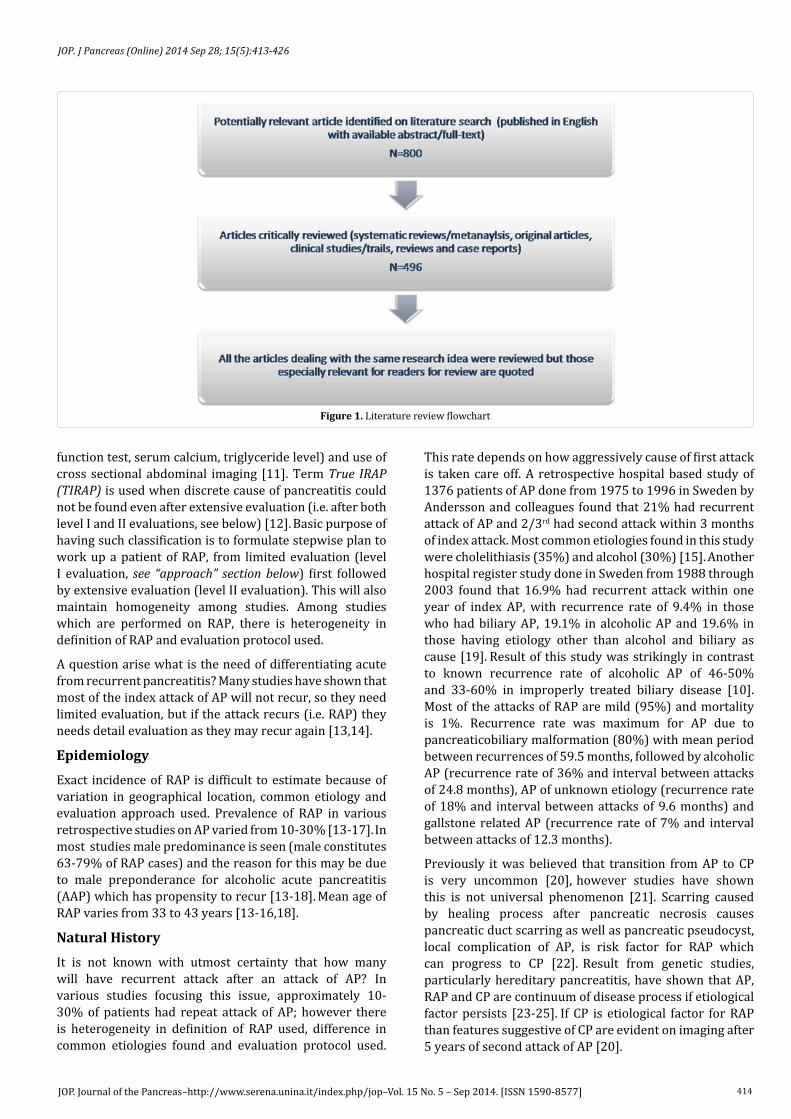
Literature SearchLiterature review was performed by PubMed search (1948 through 30 Apr, 2014) for following terms: “recurrent acute pancreatitis,” “acute recurrent pancreatitis,” “recurrent pancreatitis” and “relapsing acute pancreatitis” in title/abstract. A total of 800 relevant articles dealing human
species published in English language identified, out of which 496 articles were selected for critical review (Figure 1). All the articles dealing with the same research ideas were reviewed but those especially relevant to readers for review are quoted.
Definitions The term “recurrent acute pancreatitis” was first used in medical literature by Henry Doubilet in 1948 [4], but the nomenclature was accepted during Marseilles symposium in 1963 [5]. It has been 50 years since the term accepted but still few is known about the entity. AP is an inflammatory process of pancreas with or without involvement of peripancreatic tissue or distant site. AP is clinically defined as the presence of two of the following features: pancreatic type pain, elevated serum lipase (or amylase) more than three times upper limit of normal and/or findings of AP with absence of changes characteristic of CP on cross-sectional abdominal imaging (contrast enhanced computed tomography or ultrasonography/magnetic resonance imaging) [6]. CP is said to be present when there is evidence of pancreatic duct change, pancreatic stone, fibrosis or calcification. There may be evidence of exocrine & endocrine insufficiency [7].
RAP is defined as more than one well documented & separate attacks of pancreatitis that completely or nearly completely resolved with more than three months in between the attacks [8-11]. Three months between the attacks is important because most of the sequel of previous attack of AP is present by the time. If the patient of AP redevelops pain abdomen with elevated pancreatic enzyme within three months, it may be due to some complication of first attack of AP and not RAP. If the pain recurs with initiation of feeding, it will be called as relapse of acute pancreatitis not RAP. Idiopathic RAP (IRAP) is defined as failure to disclose the discrete etiology of pancreatitis despite thorough history, routine laboratory, investigations (liver
ABSTRACTRecurrent acute pancreatitis (RAP) is commonly encountered, but less commonly understood clinical entity, especially idiopathic RAP, with propensity to lead to repeated attacks and may be chronic pancreatitis if attacks continue to recur. A great number of studies have been published on acute pancreatitis, but few have focused on RAP. Analysing the results of clinical studies focusing specifically on RAP is problematic in view due to lack of standard definitions, randomised clinical trials, standard evaluation protocol used and less post intervention follow-up duration. With the availability of newer investigation modalities less number of etiologies will remains undiagnosed. This review particularly is focused on the present knowledge in understanding of RAP.
414JOP. Journal of the Pancreas–http://www.serena.unina.it/index.php/jop–Vol. 15 No. 5 – Sep 2014. [ISSN 1590-8577]
JOP. J Pancreas (Online) 2014 Sep 28; 15(5):413-426
This rate depends on how aggressively cause of first attack is taken care off. A retrospective hospital based study of 1376 patients of AP done from 1975 to 1996 in Sweden by Andersson and colleagues found that 21% had recurrent attack of AP and 2/3rd had second attack within 3 months of index attack. Most common etiologies found in this study were cholelithiasis (35%) and alcohol (30%) [15]. Another hospital register study done in Sweden from 1988 through 2003 found that 16.9% had recurrent attack within one year of index AP, with recurrence rate of 9.4% in those who had biliary AP, 19.1% in alcoholic AP and 19.6% in those having etiology other than alcohol and biliary as cause [19]. Result of this study was strikingly in contrast to known recurrence rate of alcoholic AP of 46-50% and 33-60% in improperly treated biliary disease [10]. Most of the attacks of RAP are mild (95%) and mortality is 1%. Recurrence rate was maximum for AP due to pancreaticobiliary malformation (80%) with mean period between recurrences of 59.5 months, followed by alcoholic AP (recurrence rate of 36% and interval between attacks of 24.8 months), AP of unknown etiology (recurrence rate of 18% and interval between attacks of 9.6 months) and gallstone related AP (recurrence rate of 7% and interval between attacks of 12.3 months).
Previously it was believed that transition from AP to CP is very uncommon [20], however studies have shown this is not universal phenomenon [21]. Scarring caused by healing process after pancreatic necrosis causes pancreatic duct scarring as well as pancreatic pseudocyst, local complication of AP, is risk factor for RAP which can progress to CP [22]. Result from genetic studies, particularly hereditary pancreatitis, have shown that AP, RAP and CP are continuum of disease process if etiological factor persists [23-25]. If CP is etiological factor for RAP than features suggestive of CP are evident on imaging after 5 years of second attack of AP [20].
function test, serum calcium, triglyceride level) and use of cross sectional abdominal imaging [11]. Term True IRAP (TIRAP) is used when discrete cause of pancreatitis could not be found even after extensive evaluation (i.e. after both level I and II evaluations, see below) [12]. Basic purpose of having such classification is to formulate stepwise plan to work up a patient of RAP, from limited evaluation (level I evaluation, see “approach” section below) first followed by extensive evaluation (level II evaluation). This will also maintain homogeneity among studies. Among studies which are performed on RAP, there is heterogeneity in definition of RAP and evaluation protocol used.
A question arise what is the need of differentiating acute from recurrent pancreatitis? Many studies have shown that most of the index attack of AP will not recur, so they need limited evaluation, but if the attack recurs (i.e. RAP) they needs detail evaluation as they may recur again [13,14].
EpidemiologyExact incidence of RAP is difficult to estimate because of variation in geographical location, common etiology and evaluation approach used. Prevalence of RAP in various retrospective studies on AP varied from 10-30% [13-17]. In most studies male predominance is seen (male constitutes 63-79% of RAP cases) and the reason for this may be due to male preponderance for alcoholic acute pancreatitis (AAP) which has propensity to recur [13-18]. Mean age of RAP varies from 33 to 43 years [13-16,18].
Natural HistoryIt is not known with utmost certainty that how many will have recurrent attack after an attack of AP? In various studies focusing this issue, approximately 10-30% of patients had repeat attack of AP; however there is heterogeneity in definition of RAP used, difference in common etiologies found and evaluation protocol used.
Figure 1. Literature review flowchart
415JOP. Journal of the Pancreas–http://www.serena.unina.it/index.php/jop–Vol. 15 No. 5 – Sep 2014. [ISSN 1590-8577]
JOP. J Pancreas (Online) 2014 Sep 28; 15(5):413-426
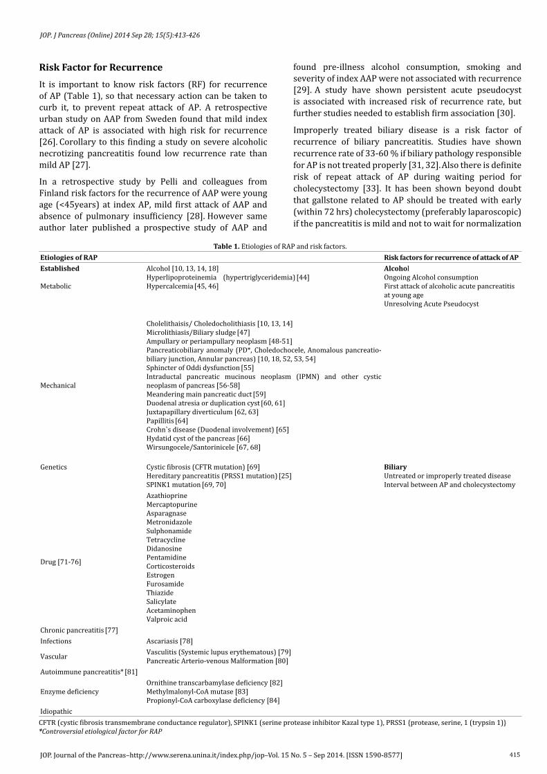
Risk Factor for RecurrenceIt is important to know risk factors (RF) for recurrence of AP (Table 1), so that necessary action can be taken to curb it, to prevent repeat attack of AP. A retrospective urban study on AAP from Sweden found that mild index attack of AP is associated with high risk for recurrence [26]. Corollary to this finding a study on severe alcoholic necrotizing pancreatitis found low recurrence rate than mild AP [27].
In a retrospective study by Pelli and colleagues from Finland risk factors for the recurrence of AAP were young age (<45years) at index AP, mild first attack of AAP and absence of pulmonary insufficiency [28]. However same author later published a prospective study of AAP and
found pre-illness alcohol consumption, smoking and severity of index AAP were not associated with recurrence [29]. A study have shown persistent acute pseudocyst is associated with increased risk of recurrence rate, but further studies needed to establish firm association [30].
Improperly treated biliary disease is a risk factor of recurrence of biliary pancreatitis. Studies have shown recurrence rate of 33-60 % if biliary pathology responsible for AP is not treated properly [31, 32]. Also there is definite risk of repeat attack of AP during waiting period for cholecystectomy [33]. It has been shown beyond doubt that gallstone related to AP should be treated with early (within 72 hrs) cholecystectomy (preferably laparoscopic) if the pancreatitis is mild and not to wait for normalization
CFTR (cystic fibrosis transmembrane conductance regulator), SPINK1 (serine protease inhibitor Kazal type 1), PRSS1 {protease, serine, 1 (trypsin 1)} *Controversial etiological factor for RAP
Etiologies of RAP Risk factors for recurrence of attack of APEstablished
Metabolic
Alcohol [10, 13, 14, 18]Hyperlipoproteinemia (hypertriglyceridemia) [44]Hypercalcemia [45, 46]
AlcoholOngoing Alcohol consumptionFirst attack of alcoholic acute pancreatitis at young ageUnresolving Acute Pseudocyst
Mechanical
Cholelithaisis/ Choledocholithiasis [10, 13, 14]Microlithiasis/Biliary sludge [47]Ampullary or periampullary neoplasm [48-51]Pancreaticobiliary anomaly (PD*, Choledochocele, Anomalous pancreatio-biliary junction, Annular pancreas) [10, 18, 52, 53, 54]Sphincter of Oddi dysfunction [55]Intraductal pancreatic mucinous neoplasm (IPMN) and other cystic neoplasm of pancreas [56-58]Meandering main pancreatic duct [59]Duodenal atresia or duplication cyst [60, 61]Juxtapapillary diverticulum [62, 63]Papillitis [64]Crohn`s disease (Duodenal involvement) [65]Hydatid cyst of the pancreas [66]Wirsungocele/Santorinicele [67, 68]
Genetics Cystic fibrosis (CFTR mutation) [69]Hereditary pancreatitis (PRSS1 mutation) [25]SPINK1 mutation [69, 70]
BiliaryUntreated or improperly treated diseaseInterval between AP and cholecystectomy
Drug [71-76]
AzathioprineMercaptopurineAsparagnaseMetronidazole SulphonamideTetracyclineDidanosinePentamidineCorticosteroidsEstrogenFurosamideThiazideSalicylateAcetaminophen Valproic acid
Chronic pancreatitis [77]Infections Ascariasis [78]
Vascular Vasculitis (Systemic lupus erythematous) [79]Pancreatic Arterio-venous Malformation [80]
Autoimmune pancreatitis* [81]
Enzyme deficiencyOrnithine transcarbamylase deficiency [82]Methylmalonyl-CoA mutase [83]Propionyl-CoA carboxylase deficiency [84]
Idiopathic
Table 1. Etiologies of RAP and risk factors.
416JOP. Journal of the Pancreas–http://www.serena.unina.it/index.php/jop–Vol. 15 No. 5 – Sep 2014. [ISSN 1590-8577]
JOP. J Pancreas (Online) 2014 Sep 28; 15(5):413-426
of serum pancreatic enzyme level or resolution of systemic inflammatory response syndrome (SIRS) [34,35]. In mild acute biliary pancreatitisearly cholecystectomy prevents the recurrence without increased operative difficulty compared to late cholecystectomy [36-38]. In case of severe acute gallstone pancreatitis cholecystectomy to be done once patient is stable to undergo surgery or acute attack is over. Those who are at high risk for surgery can undergo endoscopic sphincterotomy [39]. If choledocholithiasis is the etiology of AP, then urgent ERCP to be done if there is cholangitis or impacted stone [40], otherwise patient will be subjected ERCP once acute attack has subsided followed by cholecystectomy if gallstone is also there.
A retrospective study of 245 patients of AP identified 77 patients of RAP, with most common etiology being biliary (48 patient) [41]. In an univariate analysis of 245 patients authors found the presence of obstructive jaundice, liver enzyme derangement & local complication is significantly associated with recurrent AP with p value of <0.05 for each. However, on multivariate analysis none of the factor evaluated has shown to be associated with recurrence of AP. Limitation of this study is that they included patient who develop recurrence of the pain abdomen after refeeding as attack of RAP.
Recurrence in patients with AP due to other etiologies depends on how effectively the etiology has been treated. Surgical treatment of hypercalcemia in case of primary hyperparathyroidism prevents recurrence in AP but not in CP [42]. Recurrence of AP in hypertriglyceridemia can be prevented if serum triglyceride level is reduced to normal with diet and drugs [43].
Etiology Various causes of RAP are described in Table 1. After reviewing thorough history (especially alcohol intake, trauma, family history & drug history), routine laboratory investigation & cross sectional imaging (level I evaluation) cause of RAP can be found in 70-90% [85]. Those who remain undiagnosed (IRAP; 10-30%) need extensive evaluation by level II investigation because it has been shown more than half of the patients of IRAP with continue to experience repeated attacks of pancreatitis and RAP is harbinger of CP [20].
As per North American Pancreatitis Study2 (NAPS2) study five or more drinks (in USA one drink is alcohol of 14 gms) per day for more than 5 years is considered as risk factor for CP [86]. There are more than 100 drugs implicated as cause of AP, continuous use may be associated with RAP [87]. A drug may be suspected as cause of pancreatitis if there is consistence latency between initiation of drug and onset of cause pancreatitis. Positive rechallenge may be a coincidental finding and is not always proof of association between drug and pancreatitis, recurrence of rechallenge may be due to microlithiasis or IRAP associated recurrent attack.
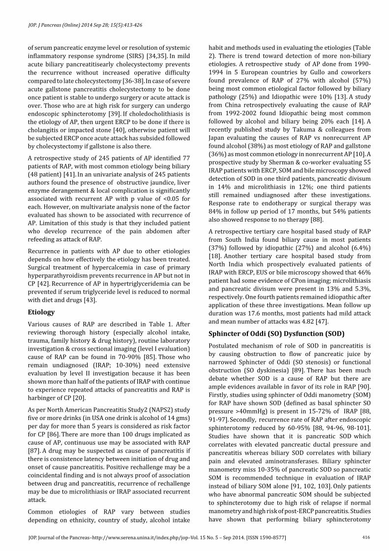
Common etiologies of RAP vary between studies depending on ethnicity, country of study, alcohol intake
habit and methods used in evaluating the etiologies (Table 2). There is trend toward detection of more non-biliary etiologies. A retrospective study of AP done from 1990-1994 in 5 European countries by Gullo and coworkers found prevalence of RAP of 27% with alcohol (57%) being most common etiological factor followed by biliary pathology (25%) and Idiopathic were 10% [13]. A study from China retrospectively evaluating the cause of RAP from 1992-2002 found Idiopathic being most common followed by alcohol and biliary being 20% each [14]. A recently published study by Takuma & colleagues from Japan evaluating the causes of RAP vs nonrecurrent AP found alcohol (38%) as most etiology of RAP and gallstone (36%) as most common etiology in nonrecurrent AP [10]. A prospective study by Sherman & co-worker evaluating 55 IRAP patients with ERCP, SOM and bile microscopy showed detection of SOD in one third patients, pancreatic divisum in 14% and microlithiasis in 12%; one third patients still remained undiagnosed after these investigations. Response rate to endotherapy or surgical therapy was 84% in follow up period of 17 months, but 54% patients also showed response to no therapy [88].
A retrospective tertiary care hospital based study of RAP from South India found biliary cause in most patients (37%) followed by idiopathic (27%) and alcohol (6.4%) [18]. Another tertiary care hospital based study from North India which prospectively evaluated patients of IRAP with ERCP, EUS or bile microscopy showed that 46% patient had some evidence of CPon imaging; microlithiasis and pancreatic divisum were present in 13% and 5.3%, respectively. One fourth patients remained idiopathic after application of these three investigations. Mean follow up duration was 17.6 months, most patients had mild attack and mean number of attacks was 4.82 [47].
Sphincter of Oddi (SO) Dysfunction (SOD)Postulated mechanism of role of SOD in pancreatitis is by causing obstruction to flow of pancreatic juice by narrowed Sphincter of Oddi (SO stenosis) or functional obstruction (SO dyskinesia) [89]. There has been much debate whether SOD is a cause of RAP but there are ample evidences available in favor of its role in RAP [90]. Firstly, studies using sphincter of Oddi manometry (SOM) for RAP have shown SOD (defined as basal sphincter SO pressure >40mmHg) is present in 15-72% of IRAP [88, 91-97]. Secondly, recurrence rate of RAP after endoscopic sphinterotomy reduced by 60-95% [88, 94-96, 98-101]. Studies have shown that it is pancreatic SOD which correlates with elevated pancreatic ductal pressure and pancreatitis whereas biliary SOD correlates with biliary pain and elevated aminotransferases. Biliary sphincter manometry miss 10-35% of pancreatic SOD so pancreatic SOM is recommended technique in evaluation of IRAP instead of biliary SOM alone [91, 102, 103]. Only patients who have abnormal pancreatic SOM should be subjectedto sphincterotomy due to high risk of relapse if normal manometry and high risk of post-ERCP pancreatitis. Studies have shown that performing biliary sphincterotomy
417JOP. Journal of the Pancreas–http://www.serena.unina.it/index.php/jop–Vol. 15 No. 5 – Sep 2014. [ISSN 1590-8577]
JOP. J Pancreas (Online) 2014 Sep 28; 15(5):413-426
do decrease recurrence rate of RAP, this is mainly due to cutting sphincter of common channel which will also reduced residual pancreatic sphincter length thereby reducing basal pancreatic sphincter pressure. A long term (≥10 years) follow up study by Wehrmann prospectively evaluated patients of RAP with SOD who underwent endoscopic sphincterectomy and found that 14% patients had recurrence of pancreatitis in first two year of follow up, however they responded to dual endoscopic sphincterotomy or surgical pancreaticojejunostomy [101].
Pancreatic Division (PD) PD is the most congenital anomaly of pancreas present in 5-10% of healthy population [1]. There are two schools of thought regarding its role in RAP: proponents argue that PD is causative factor for RAP by causing functional obstruction to pancreatic secretion [104, 105]. Evidences in favor of PD`s role in RAP are many. Firstly, Studies have shown elevated pancreatic ductal pressure in patients with PD versus that don’t have PD [106]. Secondly, widening of minor papilla by endoscopic pancreatic sphincterotomy or surgical sphincteroplasty have shown to decrease risk of recurrence of AP [107, 108]. Thirdly, retrospective studies have shown statistically significant high prevalence of PD in RAP cases [109]. Fourthly, a cross sectional study by Gonoi and colleagues found unbiased statistically higher prevalence of PD in IRAP cases (33%) compared to community dwelling subjects (2.6%) by MRCP [110].
However newer studies are revealing that PD itself doesn’t cause RAP. Most of studies on PD in RAP are retrospective. Some authors argue that detection of PD in RAP may be coincidental finding [111, 112]. A recent study by Bertin and co-workers evaluated the frequency of PD by MRCP in subjects of IARP and simultaneously evaluated for genetic mutation, found no increase prevalence of PD in IRAP as compared to healthy subjects or patients with alcohol-related pancreatitis however the prevalence of PD increased in patients with CFTR gene mutations [113]. Other studies have also found high prevalence of genetic mutation in patient of RAP who also had PD. So new school thought argues that it is genetic mutation act as cofactor in patient with PD to cause RAP.
Genetic Factors in RAPSince the description by Whitcomb regarding genetic mutation in pancreatitis multiple studies have shown the association of various genetic mutations in susceptible
genes & modifier genes as mechanism of RAP [114-116]. Prevalence of genetic mutations in children with IRAP is high [115-118]. Common genetic mutations shown to have role in RAP are CFTR (cystic fibrosis transmembrane conductance regulator), SPINK1 (serine protease inhibitor Kazal type 1) and PRSS1 (cationic trypsinogen). These mutations make pancreas susceptible to RAP by retarding protection against insult. These mutations are also seen in patients with anatomic variants (PD) and metabolic disorders (hypertriglyceridemia and hyperparathyroidism) which predispose a person to RAP, suggesting a major role of these mutations plays in RAP. Patients usually present in young age with RAP and subsequently have features to CP after many years depending on mutation. A retrospective study from Italy by Lucidi and coworkers evaluated etiological cause in 78 children with IRAP by ERCP, sweat chloride analysis and mutation analysis found positive family history in 20.5%, abnormal sweat chloride analysis in 10.3%, biliary lithiasis in 6.5% and mutation in susceptibility genes in 51.2% (CFTR 39.6%, SPINK1 7.1% and PRSS1 in 4.5%) [118]. As of now testing for genetic mutation in clinical practice is limited for PRSS1 gene in hereditary pancreatitis as it is autosomal dominant with high penetrence and predispose to pancreatic malignancy, rest of predisposing genes are tested for research purpose only. A review suggested to perform PRSS1 testing in IRAP first, then to test CFTR if PRSS1 negative and SPINK1 if both PRSS1 and CFTR negative [119, 120]. Genetic testing will be beneficial especially in patients of younger age and RAP, but it will not help in planning treatment but just help establishing cause-effect relationship and prognosticating patient for likely development of CP/pancreatic adenocarcinoma in future. Lack of long term follow-up studies hinders proper understanding of role genetic factors plays as first attack of pancreatitis may be just an attack of AP (genetic or non-genetic) or first presentation of CP, and attacks of RAP may be seperated by months to years of pain free period.
Microlithiasis and Biliary SludgeMicroliths are defined as stone <3mm in size made of whereas biliary sludge is mixture of calcium carbonate microspheroliths, calcium bilirubinate granules and amorphous cholesterol monohydrate crystals suspended in layer of mucus, glycoprotein and cellular debris [121].Microliths, by virtue of its size and ability to travel from
Table 2. Etiological studies of RAP over 10 years.Gullo et al. [13] (2002) Gao et al. [14] (2006) Sajith et al. [18] (2010) Takuma et al. [10] (2012)
Number of patients=RAP/AP 288/1068 157 /1471 188/_ 74/381 RAP % 27% 10.7% NA 19.42% Study country 5 European countries China India (CMC-V) Japan Study period Jan 1990-Dec 1994 1992-2002 2002-2007 Jan 1975-dec 2010 Study design Retrospective Retrospective Retrospective Retrospective Male % 73.3% 63% 70.2% 62% Mean age ± SD (range) 43 yr (16-9yrs) 41 (13-82yr) 33 yrs 50.1 ± 18.5 yr Most common etiology Alcohol (57%) Idiopathic (27%) Biliary (37%) Alcohol (38%) 2nd Most common etiology Biliary (25%) Biliary (20%) Idiopathic (32%) Idiopathic (26%) 3rd Most common etiology Idiopathic (10%) Alcohol (20%) PD (8.5%) Alcohol(6.4%) Biliary (11%)
418JOP. Journal of the Pancreas–http://www.serena.unina.it/index.php/jop–Vol. 15 No. 5 – Sep 2014. [ISSN 1590-8577]
JOP. J Pancreas (Online) 2014 Sep 28; 15(5):413-426
gallbladder to duodenum with ease, can obstruct the ampulla leading to AP and repeated attacks leads to RAP; also as result of repeated trauma to papilla by stone there is papillary stenosis and development of SOD. Many evidences in support of its role in RAP exist. Firstly, it occurs with increased prevalence in RAP (60-73% prevalence in IRAP patients using bile microscopy), particularly in noncholecystectomisedpatientscompared to healthy individuals [88, 196, 122-124]. Secondly, recurrent attack of AP almost eliminated post—cholecystectomy [122-124]. Sensitivity for diagnosis of microliths and biliary sludge is 50-60% by US [122, 125, 126], 65-90% by microscopic bile examination (MBE) [126-131], and 92.6% by EUS [132-134]. It was a common practice to assume the presence of bile crystal by bile microscopy as microlithiasis but both are different entity. Bile crystal themselves does not cause pancreatitis. Previous studies have used MBE as gold standard for diagnosis of occult gallstone disease but this has been largely replaced by EUS which has equivalent accuracy for diagnosis as well as ability to diagnose of other etiologies. Recent studies on IRAP which have used EUS instead of bile microscopy have reported low prevalence (13%) of microlithiasis as a cause of IRAP then what was reported for positive bile microscopy [47]. Ideal situation to label microlithiasis as cause of RPA would be detection of microliths by EUS and presence of elevated
aminotransferases within first 24 hrs of development of pancreatitis. Another point to be noted also that patients who have long history of RAP and detected to be microliths, microlithiasis is hard to be attributed as cause of RAP as microlithiasis will not remain micro for years. Microliths is not an attributable factor for RAP in cholecystectomised patients [96,98].
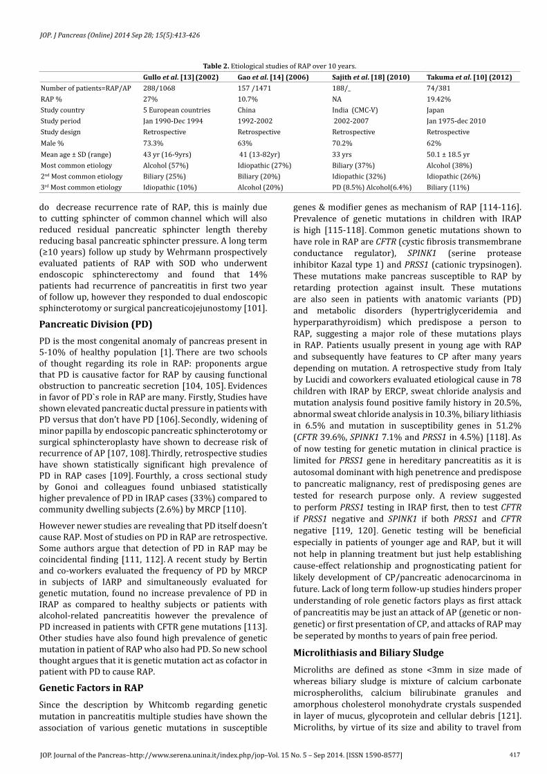
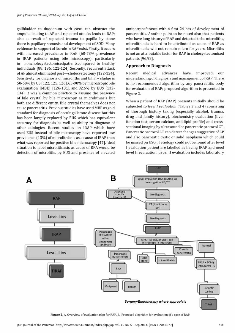
Approach to DiagnosisRecent medical advances have improved our understanding of diagnosis and management of RAP. There is no recommended algorithm by any pancreatitis body for evaluation of RAP; proposed algorithm is presented in Figure 2.
When a patient of RAP (RAP) presents initially should be subjected to level I evaluation (Tables 3 and 4) consisting of thorough history taking (especially alcohol, trauma, drug and family history), biochemistry evaluation (liver function test, serum calcium, and lipid profile) and cross-sectional imaging by ultrasound or pancreatic protocol CT. Pancreatic protocol CT can detect changes suggestive of CP and also pancreatic cystic or solid neoplasm which could be missed on USG. If etiology could not be found after level I evaluation patient are labelled as having IRAP and need level II evaluation. Level II evaluation includes laboratory
RAP
Level l inv
IRAP
Level ll inv
TIRAP
RAP
No diagnosis
CT (if not doneyet)
No diagnosis
IRAP
Normal
Pancreaticdivisum or
othercongenitalanomaly
FNA
Malignant Benign
CBDstone microlithaisis
Chronicpancreatitis
ERCP + SOM+intraductal US
Genetictesting
TIRAP
Pancreaticduct stricture
MRCP (S) and/or EUS+ bilemicroscopy (if intact GB)
Diagnosis(specific
treatment)
Level evaluation (HO, routine labinvestigation, US/CT
Surgery/Endotherapy where approplate
A B
-
-
Figure 2. A. Overview of evaluation plan for RAP, B. Proposed algorithm for evaluation of a case of RAP.
419JOP. Journal of the Pancreas–http://www.serena.unina.it/index.php/jop–Vol. 15 No. 5 – Sep 2014. [ISSN 1590-8577]
JOP. J Pancreas (Online) 2014 Sep 28; 15(5):413-426
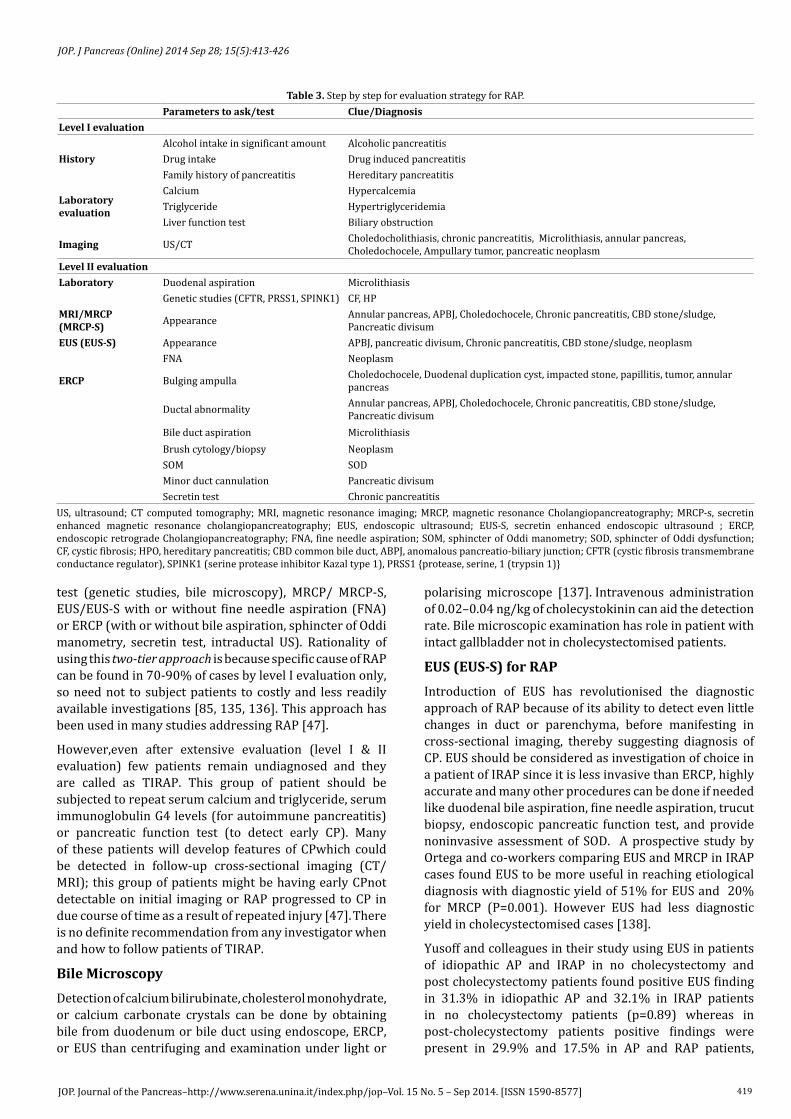
test (genetic studies, bile microscopy), MRCP/ MRCP-S, EUS/EUS-S with or without fine needle aspiration (FNA) or ERCP (with or without bile aspiration, sphincter of Oddi manometry, secretin test, intraductal US). Rationality of using this two-tier approach is because specific cause of RAP can be found in 70-90% of cases by level I evaluation only, so need not to subject patients to costly and less readily available investigations [85, 135, 136]. This approach has been used in many studies addressing RAP [47].
However,even after extensive evaluation (level I & II evaluation) few patients remain undiagnosed and they are called as TIRAP. This group of patient should be subjected to repeat serum calcium and triglyceride, serum immunoglobulin G4 levels (for autoimmune pancreatitis) or pancreatic function test (to detect early CP). Many of these patients will develop features of CPwhich could be detected in follow-up cross-sectional imaging (CT/MRI); this group of patients might be having early CPnot detectable on initial imaging or RAP progressed to CP in due course of time as a result of repeated injury [47]. There is no definite recommendation from any investigator when and how to follow patients of TIRAP.
Bile MicroscopyDetection of calcium bilirubinate, cholesterol monohydrate, or calcium carbonate crystals can be done by obtaining bile from duodenum or bile duct using endoscope, ERCP, or EUS than centrifuging and examination under light or
polarising microscope [137]. Intravenous administration of 0.02–0.04 ng/kg of cholecystokinin can aid the detection rate. Bile microscopic examination has role in patient with intact gallbladder not in cholecystectomised patients.
EUS (EUS-S) for RAPIntroduction of EUS has revolutionised the diagnostic approach of RAP because of its ability to detect even little changes in duct or parenchyma, before manifesting in cross-sectional imaging, thereby suggesting diagnosis of CP. EUS should be considered as investigation of choice in a patient of IRAP since it is less invasive than ERCP, highly accurate and many other procedures can be done if needed like duodenal bile aspiration, fine needle aspiration, trucut biopsy, endoscopic pancreatic function test, and provide noninvasive assessment of SOD. A prospective study by Ortega and co-workers comparing EUS and MRCP in IRAP cases found EUS to be more useful in reaching etiological diagnosis with diagnostic yield of 51% for EUS and 20% for MRCP (P=0.001). However EUS had less diagnostic yield in cholecystectomised cases [138].
Yusoff and colleagues in their study using EUS in patients of idiopathic AP and IRAP in no cholecystectomy and post cholecystectomy patients found positive EUS finding in 31.3% in idiopathic AP and 32.1% in IRAP patients in no cholecystectomy patients (p=0.89) whereas in post-cholecystectomy patients positive findings were present in 29.9% and 17.5% in AP and RAP patients,
Table 3. Step by step for evaluation strategy for RAP.Parameters to ask/test Clue/Diagnosis
Level I evaluation
History Alcohol intake in significant amount Alcoholic pancreatitis Drug intake Drug induced pancreatitis Family history of pancreatitis Hereditary pancreatitis
Laboratory evaluation
Calcium Hypercalcemia Triglyceride Hypertriglyceridemia Liver function test Biliary obstruction
Imaging US/CT Choledocholithiasis, chronic pancreatitis, Microlithiasis, annular pancreas, Choledochocele, Ampullary tumor, pancreatic neoplasm
Level II evaluationLaboratory Duodenal aspiration Microlithiasis
Genetic studies (CFTR, PRSS1, SPINK1) CF, HP MRI/MRCP (MRCP-S) Appearance Annular pancreas, APBJ, Choledochocele, Chronic pancreatitis, CBD stone/sludge,
Pancreatic divisum EUS (EUS-S) Appearance APBJ, pancreatic divisum, Chronic pancreatitis, CBD stone/sludge, neoplasm
FNA Neoplasm
ERCP Bulging ampulla Choledochocele, Duodenal duplication cyst, impacted stone, papillitis, tumor, annular pancreas
Ductal abnormality Annular pancreas, APBJ, Choledochocele, Chronic pancreatitis, CBD stone/sludge, Pancreatic divisum
Bile duct aspiration Microlithiasis Brush cytology/biopsy Neoplasm SOM SOD Minor duct cannulation Pancreatic divisum Secretin test Chronic pancreatitis
US, ultrasound; CT computed tomography; MRI, magnetic resonance imaging; MRCP, magnetic resonance Cholangiopancreatography; MRCP-s, secretin enhanced magnetic resonance cholangiopancreatography; EUS, endoscopic ultrasound; EUS-S, secretin enhanced endoscopic ultrasound ; ERCP, endoscopic retrograde Cholangiopancreatography; FNA, fine needle aspiration; SOM, sphincter of Oddi manometry; SOD, sphincter of Oddi dysfunction; CF, cystic fibrosis; HPO, hereditary pancreatitis; CBD common bile duct, ABPJ, anomalous pancreatio-biliary junction; CFTR (cystic fibrosis transmembrane conductance regulator), SPINK1 (serine protease inhibitor Kazal type 1), PRSS1 {protease, serine, 1 (trypsin 1)}
420JOP. Journal of the Pancreas–http://www.serena.unina.it/index.php/jop–Vol. 15 No. 5 – Sep 2014. [ISSN 1590-8577]
JOP. J Pancreas (Online) 2014 Sep 28; 15(5):413-426
respectively (p=0.15). Addition of bile microscopy, in no cholecystectomy group if normal EUS finding, yielded positive finding in 20/43 (46.5%) RAP patients [139].
Secretin-stimulated EUS (EUS-S), done after 1 IU/kg i.v. bolus of secretin injection, enhances pancreatic duct morphology which is especially useful in nondilated system. Also, addition of secretin, which increases pancreatic secretion leading to transient dilatation of pancreatic duct in normal subjects, help in real time assessment of pancreatic flow dynamics, so functional impairment can also be seen which is regarded as surrogate noninvasive marker of SOD [140].
A study evaluating the correlation between EUS finding and pancreatic function test by endoscopic collection of duodenal sample for bicarbonate levels in patient with suspected early CP changes, found moderate negative correlation and 76% concordance rate between the finding on EUS and peak bicarbonate level [141].
MRCP (MRCP-S)MRCP is an excellent tool for assessment of ductal morphology. Secretin stimulated MRCP (MRCP-S) increase the diagnostic yield by better delineating ductal morphology in otherwise nondilated pancreatic ducts and ability to detect pancreatic functional outflow obstruction [142]. It is performed by intravenous administration of 1 IU/kg of secretin, and persistence of main pancreatic duct dilatation of >1mm between baseline and 15 minutes is taken as noninvasive marker of SOD. Mariani and co-workers compared MRCP-S and SOM for evaluation of SO function in patients with IRAP and found concordance rate of 86.7% between both tests, and agreed positive and negative diagnoses in 81.8% and 100%, respectively [143]. Another study comparing MRCP-S versus ERCP with SOM in 37 IRAP patients with non-cholecystectomised nondilated pancreatic ductal system found sensitivity, specificity, positive predictive and negative predictive value for pancreatic outflow obstruction were 57.1%,
100%, 100%, and 64%, respectively [144]. MRCP-S also help in quantitative assessment of pancreatitis reserve exocrine function and it correlate significantly with fecal elastase-1 levels, which is marker of pancreatic exocrine function, thereby it can help in diagnosis of early CPeven if there is no structural change suggestive of CPon imaging [145].
ERCP in RAPWith the advances in pancreaticobiliary imaging and availability of EUS, ERCP is rarely used now-a-days for diagnostic purpose only except for sphincter of Oddi manometry (SOM) and intraductal US. Main advantage of ERCP over MRCP or EUS is the ability to perform therapeutic measures in the same session of procedure if abnormality detected. Coyle and colleagues in their study in 66 patient of IRAP by EUS and ERCP with bile microscopy and SOM found positive yield in 79% cases, SOD being the most common etiology (30%) [95]. A prospective study by Kim and co-workers in 31 IRAP patients with normal ERCP findings found possible cause of RAP in 42% patients by addition of intraductal US [146]. Finding being microlithiasis (16.1%), biliary sludge (9.7%), features of CP(9.7%) and distal pancreatic duct polypoidal lesions (6.5%). A prospective study compared EUS-S, MRCP-S and ERCP in evaluation of 44 consecutive IRAP patients with non-dilated ducts and found highest diagnostic yield for EUS-S i.e. 79.6% followed by MRCP-S 65.9% and ERCP 62.8% [147].
Pancreatic Function Testing (PFT)Many patients of IRAP have CPeither on evaluation by EUS/ MRCP/ERCP or they may develop evidence of CP in follow up thereby indicating that these patients might be have subtle evidence of CP from the vary beginning which might be responsible for recurrent attacks of pancreatitis or they may develop CP as a result of repeated insult to pancreas by RAP [47]. In PFT, duodenal aspirate is collected for estimation of bicarbonate concentration
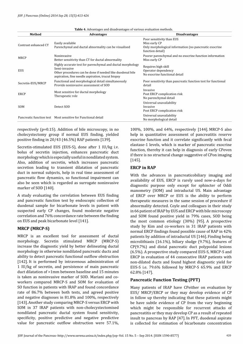
Method Advantages Disadvantages
Contrast enhanced CT Easily availableParenchymal and ductal abnormality can be visualised
Poor sensitivity than EUSMiss early CPOnly morphological information (no pancreatic exocrine function detail)
MRCP NoninvasiveBetter sensitivity than CT for ductal abnormality
Poorer parenchymal and no exocrine function informationMiss early CP
EUS
Highly accurate test for parenchymal and ductal morphologySemi-invasiveOther procedures can be done if needed like duodenal bile aspiration, fine needle aspiration, trucut biopsy
Requires high skillOperator dependencyNo exocrine functional detail
Secretin-EUS/MRCP Functional and morphological detail simultaneously Provide noninvasive assessment of SOD
Poor sensitivity than pancreatic function test for functional detail
ERCP Most sensitive for ductal morphologyTherapeutic role
Invasive Post ERCP complication riskNo parenchymal detail
SOM Detect SODUniversal unavailability Invasive Post ERCP complication risk
Pancreatic function test Most sensitive for Functional detail Universal unavailabilityNo morphological detail
Table 4. Advantages and disadvantages of various evaluation methods.
421JOP. Journal of the Pancreas–http://www.serena.unina.it/index.php/jop–Vol. 15 No. 5 – Sep 2014. [ISSN 1590-8577]
JOP. J Pancreas (Online) 2014 Sep 28; 15(5):413-426
after intravenous secretin injection [148]. PFT is the most sensitive test for evaluation of early evidence of CP. So it will be prudent to subject the patients of IRAP to EUS followed by pancreatic function testing (if EUS is normal), so that CP will be detected early. Combination EUS with PFT give 100% sensitivity for diagnosis of CP [149].
Treatment Management of acute attack of RAP is similar to standard treatment guidelines of AP with nil per mouth, intravenous hydration, adequate analgesia, correction of electrolyte or metabolic abnormalities and proper treatment of complications of AP. If a specific cause pertinent to RAP is ascertained than specific therapy is directed to that etiology: cessation of alcohol intake and smoking [150], cholecystectomy/ES/ERCP, stoppage intake of offender drug, parathyroidectomy and hypolipidemic drugs if alcohol, gallstone/choledocholithiasis, drug, hypercalcemia due to primary hyperparathyroidism and hypertriglyceridemia are responsible factor for RAP, respectively.
Role of endotherapy for patient of RAP with pancreatic divisum (PD) is still controversial. Studies which had shown role of endotherapy (minor papilla sphincterotomy or stenting) or surgery (sphincteroplasty) are mainly retrospective with less mean follow up period [108,113,151-162]. In a systematic review on endotherapy and surgical treatment for PD found not statistically different pooled response rate of 79.2 and 83.2% for endotherapy and surgery, respectively [163]. Recent study by Rustagi and co-worker shown response rate of 94% in patient of PD with RAP [164]. It has always been argued whether the benefit of ES in PD is true phenomenon or placebo effect as the interval between attacks of RAP can vary from months to many years [165,166]. In carefully selected patients with PD, endoscopic minor papilla sphincterotomy and/or stent insertion (for short term unflanged stent preferred to aid spontaneous stent migration) can relieve the obstruction to pancreatic juice flow.
Most of the studies on RAP with SOD have recommended dual sphinterotomy as treatment of choice [91,100,101,102], however these studies were nonrandomised and all study except Wehrmann had follow up of less than 2 years. Recent randomised control trial by Cote and coworkers have shown similar efficacy for biliary endoscopic sphinterotomy and dual endoscopic sphinterotomy with recurrence rate of 47% and 49% for biliary and dual endoscopic sphinterotomy, respectively, during follow up of 1-10 years [11].
Patient of microlithiasis should be subjected to laparoscopic cholecystectomy if good operative candidate or ES and/or UDCA can be used as alternative for elderly patients, poor operative candidates or unwillingness for surgery [85,123,167,168]. In study conducted by Saraswat and colleague showed positive bile microscopy in 74% patients of IRAP and all patients who underwent cholecystectomy or ES remained asymptomatic for mean
follow up of 23months (range 6–48 months) and 90% patients on UDCA remain asymptomatic for more than 9 months [168].
There is no validated therapy for TIRAP patients. Treatment usually offered to TIRAP patients includes laparoscopic cholecystectomy, ES or UDCA. However recent study has raised doubt on this practice [169]. A study by Trna and colleagues evaluated the role of cholecystectomy in idiopathic pancreatitis or presumed gallstone related pancreatitis revealed that absence of elevation of liver enzymes on day 1 of AP or absence of gallstone/sludge on US were associated with increased risk of recurrence of AP [170].
Antioxidants have no beneficial effect in RAP. Morris-Stiff GJ and colleagues compared the levels of trace elements (selenium, copper, zinc), vitamins A and E, and carotenoids (alpha-carotene, beta-carotene, xanthine, beta-cryptoxanthine, lycopene) among normal, RAP and CP found that levels of these antioxidants was statistically different between normal and CP but not in normal versus RAP [171].
Conclusion RAP is common but still a misunderstood disease. Interpretation of results of available studies is hampered by lack of standard definition and evaluation protocol used. Also analysing the results of endotherapy/surgery for various etiology of IRAP is also hurdled by lack of significant follow up period. With the advancement and availability of investigations to diagnoses less common causes of pancreatitis, fewer patients are left idiopathic.
Source of Support/FundNo funding source was involved in the preparation of this paper or in the decision to submit it for publication.
Author ContributionsVishal Khurana did (a) searching the literature, (b) conception and design or analysis and interpretation of the data, (c) the drafting of the article or critical revision for important intellectual content, and (d) final approval of the version to be published. Vishal khurana and Ishita Ganguly was involved in writing and editing of this manuscript.
Conflicting Interest The authors have no conflicts of interest
References
1. Cotton PB. Congenital anomaly of pancreas divisum as cause ofobstructive pain and pancreatitis. Gut. 1980; 21:105-114. [PMID:7380331]
2. Bernard JP, Sahel J, Giovannini M, Sarles H. Pancreas divisum is aprobable cause of acute pancreatitis: a report of 137 cases. Pancreas. 1990; 5: 248-254. [PMID:2343039]
3. Lehman GA, Sherman S. Pancreas divisum. Diagnosis, clinicalsignificance, and management alternatives. Gastrointest Endosc Clin N Am. 1995; 5:145-170. [PMID:7728342]
4. Doubilet H, Mulholland JH. Recurrent Acute Pancreatitis: Observations on Etiology and Surgical Treatment. Ann Surg. 1948; 128:609–636. [PMID:17859221]
422JOP. Journal of the Pancreas–http://www.serena.unina.it/index.php/jop–Vol. 15 No. 5 – Sep 2014. [ISSN 1590-8577]
JOP. J Pancreas (Online) 2014 Sep 28; 15(5):413-426
5. Sarles H, Sarles JC, Camatte R, et al. Observations on 205 confirmedcases of acute pancreatitis, recurring pancreatitis, and chronic pancreatitis. Gut. 1965; 6:545-559. [PMID:5857891]
6. Banks PA, Bollen TL, Dervenis C, et al. Classification of acutepancreatitis--2012: revision of the Atlanta classification and definitions by international consensus. Gut. 2013; 62:102-11. [PMID:23100216]
7. Sarner M, Cotton PB. Classification of pancreatitis. Gut. 1984;25:756-759. [PMID:6735257]
8. Al-Haddad M, Wallace MB. Diagnostic approach to patients with acute idiopathic and recurrent pancreatitis, what should be done? World J Gastroenterol. 2008; 14:1007–1010. [PMID:18286679]
9. Romagnuolo J, Guda N, Freeman M, et al. Preferred designs, outcomes, and analysis strategies for treatment trials in idio-pathic recurrent acute pancreatitis. Gastrointest Endosc. 2008; 68:966 –974. [PMID:18725158]
10. Takuma K, Kamisawa T, Hara S, et al. Etiology of recurrent acutepancreatitis, with special emphasis on pancreaticobiliary malformation. Adv Med Sci. 2012; 57:244-250. [PMID:23183766]
11. Coté GA, Imperiale TF, Schmidt SE, et al. Similar efficacies of biliary,with or without pancreatic, sphincterotomy in treatment of idiopathic recurrent acute pancreatitis. Gastroenterology. 2012; 143:1502-1509 .e1. [PMID:22982183]
12. Lara LF, Levy MJ. Idiopathic Recurrent Acute Pancreatitis. MedGenMed. 2004; 6:10. [PMID:15775837]
13. Gullo L, Migliori M, Pezzilli R, et al. An update on recurrent acutepancreatitis: data from five European countries. Am J Gastroenterol. 2002; 97:1959-1962. [PMID:12190160]
14. Gao YJ, Li YQ, Wang Q, et al. Analysis of the clinical features ofrecurrent acute pancreatitis in China. J Gastroenterol. 2006; 41:681-685. [PMID:16933006]
15. Andersson R, Andersson B, Haraldsen P, Drewsen G, Eckerwall G.Incidence, management and recurrence rate of acute pancreatitis. Scand J Gastroenterol. 2004; 39:891-894. [PMID:15513389]
16. Zhang W, Shan HC, Gu Y. Recurrent acute pancreatitis and its relativefactors. World J Gastroenterol. 2005; 11:3002-3004. [PMID:15902746]
17. Corfield AP, Cooper MJ, Williamson RC. Acute pancreatitis: a lethaldisease of increasing incidence. Gut. 1985; 26:724-729. [PMID:4018637]
18. Sajith KG, Chacko A, Dutta AK. Recurrent acute pancreatitis: clinicalprofile and an approach to diagnosis. Dig Dis Sci. 2010; 55:3610-3616. [PMID:20232145]
19. Sandzén B, Rosenmüller M, Haapamäki MM, Nilsson E, StenlundHC, Oman M. First attack of acute pancreatitis in Sweden 1988 - 2003: incidence, aetiological classification, procedures and mortality - a register study. BMC Gastroenterol. 2009; 9:18. [PMID:19265519]
20. Mariani A, Testoni PA. Is acute recurrent pancreatitis a chronicdisease? World J Gastroenterol. 2008; 14:995-998. [PMID:18286677]
21. Riaz C, Ochi K, Tanaka J, Harada H, Ichimura M, Miki H. Does recurrent acute pancreatitis lead to chronic pancreatitis? Sequential morphological and biochemical studies. Pancreas. 1997; 14:334-341. [PMID:9163778]
22. Whitcomb DC. Mechanism of disease: advances in understanding the mechanism leading to chronic pancreatitis. Nat Clin Pract Gastroenterol Hepatol. 2004; 1:46-52. [PMID:16265044]
23. Kloppel G, Maillet B. Pseudocyst in chronic pancreatitis: amorphological analysis of 57 resection specimens and 9 autopsy pancreata. Pancreas. 1991; 6:266-274. [PMID:1862065]
24. Gorry MC, Gabbaizedeh D, Furey W, et al. Mutations in the cationictrypsinogen gene are associated with recurrent acute and chronic pancreatitis. Gastroenterology. 1997; 113:1063-1068. [PMID:9322498]
25. Whitcomb DC, Gorry MC, Preston RA, et al. Hereditary pancreatitis is caused by a mutation in the cationic trypsinogen gene. Nat Genet. 1996; 14:141-145. [PMID:8841182]
26. Appelros S, Borgström A. Incidence, etiology and mortality rate ofacute pancreatitis over 10 years in a defined urban population in sweden. Br J Surg. 1999; 86:465–470. [PMID:10215815]
27. Tzovaras G, Parks RW, Diamond T, Rowlands BJ. Early and long termresults of surgery for severe necrotising pancreatitis. Dig Surg. 2004; 21:41–7.[PMID:14707392]
28. Pelli H, Sand J, Laippala P, Nordback I. Long-term follow-up after thefirst episode of acute alcoholic pancreatitis: time course and risk factors for recurrence. Scand J Gastroenterol. 2000; 35:552-555. [PMID:10868461]
29. Pelli H, Lappalainen-Lehto R, Piironen A, Sand J, Nordback I. Riskfactors for recurrent acute alcohol-associated pancreatitis: a prospective analysis. Scand J Gastroenterol. 2008; 43:614-621. [PMID:18415757]
30. Pelli H, Lappalainen-Lehto R, Piironen A, Järvinen S, Sand J, Nordback I. Pancreatic damage after the first episode of acute alcoholic pancreatitis and its association with the later recurrence rate. Pancreatology. 2009; 9:245-251. [PMID:19407478]
31. Goodman A, Neoptolemos JP, Carr Locke DL, Finlay DB, Fossard DP.Detection of gallstones after acute pancreatitis. Gut. 1985; 26:125–132. [PMID:2578422]
32. Frei G, Frei VT, Thirlby RC, McClelland RN. Biliary pancreatitis: clinical presentation and surgical management. Am J Surg. 1986; 151:170–175. [PMID:2418700]
33. van Baal MC, Besselink MG, Bakker OJ, et al. Timing of cholecystectomy after mild biliary pancreatitis: a systematic review. Ann Surg. 2012; 255:860-866. [PMID:22470079]
34. Bouwense SA, Besselink MG, van Brunschot S, et al. Pancreatitisof biliary origin, optimal timing of cholecystectomy (PONCHO trial): study protocol for a randomized controlled trial. Trials. 2012; 13:225. [PMID:23181667]
35. Nebiker CA, Frey DM, Hamel CT, Oertli D, Kettelhack C. Early versusdelayed cholecystectomy in patients with biliary acute pancreatitis. Surgery. 2009; 145:260-264. [PMID:19231577]
36. Prabhu RY, Irpatgire R, Naranje B, Kantharia CV, Bapat RD, Supe AN.Influence of timing on performance of laparoscopic cholecystectomy for acute biliary pancreatitis. Trop Gastroenterol 2009; 30:113-115. [PMID:19231577]
37. van Geenen EJ, van der Peet DL, Mulder CJ, Cuesta MA, BrunoMJ. Recurrent acute biliary pancreatitis: the protective role of cholecystectomy and endoscopic sphincterotomy. Surg Endosc. 2009; 23:950-956. [PMID:19266236]
38. Bakker OJ, van Santvoort HC, Hagenaars JC, et al. Timing ofcholecystectomy after mild biliary pancreatitis. Br J Surg. 2011; 98:1446-1454. [PMID:21710664]
39. Hernandez V, Pascual I, Almela P, et al. Recurrence of acutegallstone pancreatitis and relationship with cholecystectomy or endoscopic sphincterotomy. Am J Gastroenterol. 2004; 99:2417-2423. [PMID:15571590]
40. Attasaranya S, Fogel EL, Lehman GA. Choledocholithiasis, ascendingcholangitis, and gallstone pancreatitis. Med Clin North Am. 2008; 92:925-960. [PMID:18570948]
41. Zhang W, Shan HC, Gu Y. Recurrent acute pancreatitis and its relativefactors. World J Gastroenterol. 2005; 11:3002-3004. [PMID:15902746]
42. Carnaille B, Oudar C, Pattou F, Combemale F, Rocha J, Proye C.Pancreatitis and primary hyperparathyroidism: forty cases. Aust N Z J Surg. 1998; 68:117-119. [PMID:9494002]
43. Athyros VG, Giouleme OI, Nikolaidis NL, et al. Long-term follow-upof patients with acute hypertriglyceridemia-induced pancreatitis. J Clin Gastroenterol. 2002; 34:472-475. [PMID:11907366]
44. Kota SK, Kota SK, Jammula S, Krishna SV, Modi KD.Hypertriglyceridemia-induced recurrent acute pancreatitis: A case-based review. Indian J Endocrinol Metab. 2012; 16:141-143. [PMID:22276267]
45. Lanitis S, Sivakumar S, Zaman N, Westerland O, Al Mufti R, Hadjiminas DJ. Recurrent acute pancreatitis as the first and sole presentation of undiagnosed primary hyperparathyroidism. Ann R Coll Surg Engl. 2010; 92:W29-31. [PMID:20353632]
46. Misgar RA, Mathew V, Pandit K, Chowdhury S. Primaryhyperparathyroidism presenting as recurrent acute pancreatitis: A case report and review of literature. Indian J Endocrinol Metab. 2011; 15:54-56. [PMID:21584170]
423JOP. Journal of the Pancreas–http://www.serena.unina.it/index.php/jop–Vol. 15 No. 5 – Sep 2014. [ISSN 1590-8577]
JOP. J Pancreas (Online) 2014 Sep 28; 15(5):413-426
47. Garg PK, Tandon RK, Madan K. Is Biliary Microlithiasis a Significant Cause of Idiopathic Recurrent Acute Pancreatitis? A Long-term Follow-up Study. Clin Gastroenterol Hepatol. 2007; 5:75–79. [PMID:16931169]
48. Tsai MJ, Liao KS, Shih PM, et al. Relapsed acute pancreatitis as theinitial presentation of pancreatic cancer in a young man: a case report. Kaohsiung J Med Sci. 2010; 26:448-455. [PMID:20705257]
49. Petrou A, Bramis K, Williams T, Papalambros A, Mantonakis E,Felekouras E. Acute recurrent pancreatitis: a possible clinical manifestation of ampullary cancer. JOP. 2011; 12:593-597. [PMID:22072249]
50. Kantarcioglu M, Kilciler G, Turan I, et al. Solitary Peutz-Jeghers-type hamartomatous polyp as a cause of recurrent acute pancreatitis. Endoscopy. 2009; 41 Suppl 2:E117-118. [PMID:19544255]
51. Katsinelos P, Kountouras J, Zavos C, Chatzimavroudis G, ParoutoglouG. Recurrent acute pancreatitis caused by intra-ampullary carcinoid tumor. Gastrointest Endosc. 2009; 69:1387-1388. [PMID:19481661]
52. Hwang SS, Paik CN, Lee KM, Chung WC, Jang UI, Yang JM. Recurrentacute pancreatitis caused by an annular pancreas in a child. Gastrointest Endosc.2010; 72:848-849. [PMID:20605148]
53. Ohno Y, Kanematsu T. Annular pancreas causing localized recurrentpancreatitis in a child: report of a case. Surg Today. 2008; 38:1052-1055. [PMID:18958567]
54. Arulprakash S, Balamurali R, Pugazhendhi T, Kumar SJ. Pancreasdivisum and choledochal cyst. Indian J Med Sci. 2009; 63:198-201. [PMID:19584491]
55. Geenen JE, Nash JA. The role of sphincter of Oddi manometry andbiliary microscopy in evaluating idiopathic recurrent pancreatitis. Endoscopy. 1998; 30:A237-41. [PMID:9932788]
56. Asari S, Matsumoto I, Toyama H, et al. Repeating regional acutepancreatitis in the head of the pancreas caused by intraductal papillary mucinous neoplasms in the tail: report of a case. Surg Today. 2012; 42:398-402. [PMID:22327284]
57. Ozturk Y, Soylu OB, Gurcu B, Ortac R, Cakmakci H, Coker A. Solidpseudopapillary tumor of the pancreas as a cause of recurrent pancreatitis. Acta Gastroenterol Belg. 2008; 71:390-392. [PMID:19317280]
58. Paramhans D, Shukla S, Mathur RK. Mucinous cystadenoma of thepancreas associated with recurrent pancreatitis. Trop Gastroenterol. 2011; 32:76-78. [PMID:21922866]
59. Gonoi W, Akai H, Hagiwara K, et al. Meandering main pancreatic duct as a relevant factor to the onset of idiopathic recurrent acute pancreatitis. PLoS One. 2012; 7:e37652. [PMID:22655061]
60. Salemis NS, Liatsos C, Kolios M, Gourgiotis S. Recurrent acutepancreatitis secondary to a duodenal duplication cyst in an adult. A case report and literature review. Can J Gastroenterol. 2009; 23:749-752. [PMID:19893770]
61. Al-Qahtani HH. Duodenal duplication cyst communicating with themain pancreatic duct. A rare cause of recurrent acute pancreatitis. Saudi Med J. 2010; 31:1368-1370. [PMID:21136003]
62. Szabó M, Horváth OP. Acute pancreatitis caused by an intraluminalduodenal diverticulum. Magy Seb. 2009; 62:344-346. [PMID:19945936]
63. Reichert MC, Bittenbring JT, Fries P, Zimmer V, Lammert F, DauerM. Recurrent pancreatitis caused by a huge intraluminal duodenal diverticulum. J Gastrointestin Liver Dis. 2012; 21:126. [PMID:22720296]
64. Nardi GL, Acosta JM. Papillitis as a cause of pancreatitis and abdominal pain: role of evocative test, operative pancreatography and histologic evaluation. Ann Surg. 1966; 164:611–621. [PMID:5924783]
65. Moolsintong P, Loftus EV Jr, Chari ST, Egan LJ, Tremaine WJ,Sandborn WJ. Acute pancreatitis in patients with Crohn's disease: clinical features and outcomes. Inflamm Bowel Dis. 2005; 11:1080-1084. [PMID:16306770]
66. Pouget Y, Mucci S, O'Toole D, Lermite E, Aubé C, Hamy A. Recurrentacute pancreatitis revealing a hydatid cyst of the pancreas. Rev Med Interne. 2009; 30:358-360. [PMID:18818003]
67. Gupta R, Lakhtakia S, Tandan M, Santosh D, Rao GV, Reddy DN.Recurrent acute pancreatitis and Wirsungocele. A case report and review of literature. JOP. 2008; 9:531-533. [PMID:18648148]
68. Khan SA, Chawla T, Azami R. Recurrent acute pancreatitis due toa santorinicele in a young patient. Singapore Med J. 2009; 50:e163-5. [PMID:19495498]
69. Cavestro GM, Zuppardo RA, Bertolini S, et al. Connections betweengenetics and clinical data: Role of MCP-1, CFTR, and SPINK-1 in the setting of acute, acute recurrent, and chronic pancreatitis. Am J Gastroenterol. 2010; 105:199-206. [PMID:19844201]
70. Aoun E, Muddana V, Papachristou GI, Whitcomb DC. SPINK1 N34Sis strongly associated with recurrent acute pancreatitis but is not a risk factor for the first or sentinel acute pancreatitis event. Am J Gastroenterol. 2010; 105:446-451. [PMID:19888199]
71. Balani AR, Grendell JH. Drug-induced pancreatitis : incidence,management and prevention. Drug Saf. 2008; 31:823-837. [PMID:18759507]
72. Bermejo F, Lopez-Sanroman A, Taxonera C, et al. Acute pancreatitisin inflammatory bowel disease, with special reference to azathioprine-induced pancreatitis. Aliment Pharmacol Ther. 2008; 28:623-628. [PMID:18513380]
73. Halalsheh H, Bazzeh F, Alkayed K, Salami K, Madanat F.6-Mercaptopurine-induced Recurrent Acute Pancreatitis in Children With Acute Lymphoblastic Leukemia/Lymphoma. J Pediatr Hematol Oncol. 2013; 35: 470-472. [PMID:23138114]
74. O'Halloran E, Hogan A, Mealy K. Metronidazole-induced pancreatitis.HPB Surg. 2010. [PMID:20862338]
75. Park TY, Oh HC, Do JH. A case of recurrent pancreatitis induced bytrimethoprim-sulfamethoxazole re-exposure. Gut Liver. 2010; 4: 250-252. [PMID:20559530]
76. Igarashi H, Ito T, Yoshinaga M, Oono T, Sakai H, Takayanagi R.Acetaminophen-induced acute pancreatitis. A case report. JOP. 2009; 10:550-553. [PMID:19734636]
77. Bank S, Indaram A. Causes of acute and recurrent pancreatitis. Clinical considerations and clues to diagnosis. Gastroenterol Clin North Am. 1999; 28:571-589. [PMID:10503137]
78. Lee KH, Shelat VG, Low HC, Ho KY, Diddapur RK. Recurrent pancreatitis secondary to pancreatic ascariasis. Singapore Med J. 2009; 50:e218-9. [PMID:19551301]
79. Koga T, Miyashita T, Koga M, et al. A case of lupus-associatedpancreatitis with ruptured pseudoaneurysms. Mod Rheumatol. 2011; 21:428-431. [PMID:21308389]
80. Choi JK, Lee SH, Kwak MS, et al. A Case of Recurrent Acute Pancreatitis due to Pancreatic Arteriovenous Malformation. Gut Liver. 2010; 4:135-139. [PMID:20479928]
81. Takayama M, Hamano H, Ochi Y, et al. Recurrent attacks of autoimmune pancreatitis result in pancreatic stone formation. Am J Gastroenterol. 2004; 99:932-927. [PMID:15128363]
82. Prada CE, Kaul A, Hopkin RJ, et al. Recurrent pancreatitis in ornithine transcarbamylase deficiency. Mol Genet Metab. 2012; 106:482-484. [PMID:22728053]
83. Marquard J, El Scheich T, Klee D, et al. Chronic pancreatitis inbranched-chain organic acidurias--a case of methylmalonic aciduria and an overview of the literature. Eur J Pediatr. 2011; 170:241-245. [PMID:20924605]
84. Bultron G, Seashore MR, Pashankar DS, Husain SZ. Recurrent acutepancreatitis associated with propionic acidemia. J Pediatr Gastroenterol Nutr. 2008; 47:370-371. [PMID:18728537]
85. Levy MJ, Geenen JE. Idiopathic acute recurrent pancreatitis. Am JGastroenterol. 2001; 96:2540-2555. [PMID:11569674]
86. Yadav D, Hawes RH, Brand RE, et al. Alcohol consumption, cigarettesmoking, and the risk of recurrent acute and chronic pancreatitis. Arch Intern Med. 2009; 169:1035-1045. [PMID:19506173]
87. Badalov N, Baradarian R, Iswara K, Li J, Steinberg W, Tenner S. Drug-induced acute pancreatitis: an evidence-based review. Clin Gastroenterol Hepatol. 2007; 5:648-661. [PMID:17395548]
88. Sherman S, Jamidar P, Reber H. Idiopathic acute pancreatitis:endoscopic approach to diagnosis and treatment (abstr). Am J. Gastroenterol. 1993; 88:1541.
424JOP. Journal of the Pancreas–http://www.serena.unina.it/index.php/jop–Vol. 15 No. 5 – Sep 2014. [ISSN 1590-8577]
JOP. J Pancreas (Online) 2014 Sep 28; 15(5):413-426
89. Varadarajulu S, Hawes R. Key issues in sphincter of Oddi dysfunction. Gastrointest Endosc Clin N Am. 2003; 13:671-694. [PMID:14986793]
90. Steinberg WM. Controversies in clinical pancreatology: should thesphincter of Oddi be measured in patients with idiopathic recurrent acute pancreatitis, and should sphincterotomy be performed if the pressure is high? Pancreas. 2003; 27:118-121. [PMID:12883258]
91. Eversman D, Fogel EL, Rusche M, Sherman S, Lehman GA. Frequencyof abnormal pancreatic and biliary sphincter manometry compared with clinical suspicion of sphincter of Oddi dysfunction. Gastrointest Endosc. 1999; 50:637-641. [PMID:10536318]
92. Gregg JA, Carr-Locke DL. Endoscopic pancreatic and biliarymanometry in pancreatic, biliary, and papillary disease, and after endoscopic sphincterotomy and surgical sphincteroplasty. Gut. 1984; 25:1247-1254. [PMID:6500363]
93. Toouli J, Roberts-Thomson IC, Dent J, Lee J. Sphincter of Oddi motility disorders in patients with idiopathic recurrent pancreatitis. Br J Surg. 1985; 72:859-863. [PMID:4063750]
94. Venu RP, Geenen JE, Hogan W, Stone J, Johnson GK, Soergel K. Idiopathic recurrent pancreatitis. An approach to diagnosis and treatment. Dig Dis Sci. 1989; 34:56-60. [PMID:2631687]
95. Coyle WJ, Pineau BC, Tarnasky PR, et al. Evaluation of unexplainedacute and acute recurrent pancreatitis using endoscopic retrograde cholangiopancreatography, sphincter of Oddi manometry and endoscopic ultrasound. Endoscopy. 2002; 34:617-623.
96. Kaw M, Brodmerkel GJ Jr. ERCP, biliary crystal analysis, and sphincter of Oddi manometry in idiopathic recurrent pancreatitis. Gastrointest Endosc. 2002; 55:157-162. [PMID:11818915]
97. Geenen JE, Hogan WJ, Dodds WJ, Toouli J, Venu RP. The efficacyof endoscopic sphincterotomy after cholecystectomy in patients with sphincter-of-Oddi dysfunction. N Engl J Med. 1989; 320:82–87. [PMID:2643038]
98. Elta GH. Sphincter of Oddi dysfunction and bile duct microlithiasisin acute idiopathic pancreatitis. World J Gastroenterol. 2008; 14:1023–1026. [PMID:18286682]
99. Sgouros SN, Pereira SP. Systematic review: sphincter of Oddidysfunction—non-invasive diagnostic methods and long-term outcome after endoscopic sphincterotomy. Aliment Pharmacol Ther. 2006; 24:237–246.
100. Freeman ML, Gill M, Overby C, Cen YY. Predictors of outcomes after biliary and pancreatic sphincterotomy for sphincter of Oddi dysfunction. J Clin Gastroenterol. 2007; 41:94–102. [PMID:17198071]
101. Wehrmann T. Long-term results (≥ 10 years) of endoscopic therapy for sphincter of Oddi dysfunction in patients with acute recurrent pancreatitis. Endoscopy. 2011; 43:202-207. [PMID:21108172]
102. Park SH, Watkins JL, Fogel EL, Sherman S, Lazzell L, Bucksot L, Lehman GA. Long-term outcome of endoscopic dual pancreatobiliary sphincterotomy in patients with manometry-documented sphincter of Oddi dysfunction and normal pancreatogram. Gastrointest Endosc. 2003; 57:483-491
103. Lehman GA. Acute recurrent pancreatitis. Can J Gastroenterol. 2003; 17:381-383. [PMID:12813605]
104. Warshaw AL, Simeone J, Schapiro RH, Hedberg SE, Mueller PE, Ferrucci JT. Objective evaluation of ampullary stenosis with ultrasonography and pancreatic stimulation. Am J Surg. 1985; 149:65-71. [PMID:3881057]
105. Gregg JA. Pancreas divisum: its association with pancreatitis. Am J Surg. 1977; 134:539-543. [PMID:920876]
106. Staritz M, Meyer zum Buschenfelde KH. Elevated pressure in the dorsal part of pancreas divisum: the cause of chronic pancreatitis. Pancreas. 1988; 3:108-110. [PMID:3362837]
107. Liao Z, Gao R, Wang W, et al. A systematic review on endoscopic detection rate, endotherapy, and surgery for pancreas divisum. Endoscopy. 2009; 41:439-444. [PMID:19337962]
108. Kwan V, Loh SM, Walsh PR, Williams SJ, Bourke MJ. Minor papilla sphincterotomy for pancreatitis due to pancreas divisum. ANZ J Surg. 2008; 78:257-261. [PMID:18366396]
109. Dhar A, Goenka MK, Kochhar R, Nagi B, Bhasin DK, Singh K. Pancrease divisum: five years' experience in a teaching hospital. Indian J Gastroenterol. 1996; 15:7-9. [PMID:8840617]
110. Gonoi W, Akai H, Hagiwara K, et al. Pancreas divisum as a predisposing factor for chronic and recurrent idiopathic pancreatitis: initial in vivo survey. Gut. 2011; 60:1103-1108. [PMID:21325173]
111. Delhaye M, Engelholm L, Cremer M. Pancreas divisum: congenital anatomic variant or anomaly?. Gastroenterology. 1985; 89:951-958. [PMID:4043675]
112. Tandon M, Topazian M. Endoscopic ultrasound in idiopathic acute pancreatitis. Am J Gastroenterol. 2001; 96:705 – 9. [PMID:11280538]
113. Bertin C, Pelletier AL, Vullierme MP, et al. Pancreas divisum is not a cause of pancreatitis by itself but acts as a partner of genetic mutations. Am J Gastroenterol. 2012; 107:311-7. [PMID:22158025]
114. Whitcomb DC. Value of genetic testing in management of pancreatitis. Gut. 2004; 53:1710-1717. [PMID:15479696]
115. Uomo G, Manes G, Rabitti PG. Role of hereditary pancreatitis and CFTR gene mutations in the aetiology of acute relapsing pancreatitis of unknown origin. How are they important? JOP. 2001; 2:368-372. [PMID:11880696]
116. Keim V. Role of genetic disorders in acute recurrent pancreatitis. World J Gastroenterol. 2008; 14:1011-1015. [PMID:18286680]
117. Sultan M, Werlin S, Venkatasubramani N. Genetic prevalence and characteristics in children with recurrent pancreatitis. J Pediatr Gastroenterol Nutr. 2012; 54:645-50. [PMID:22094894]
118. Lucidi V, Alghisi F, Dall'Oglio L, et al. The etiology of acute recurrent pancreatitis in children: a challenge for pediatricians. Pancreas. 2011; 40:517-521. [PMID:21499205]
119. Ellis I. Genetic counseling for hereditary pancreatitis the role of molecular genetics testing for the cationic trypsinogen gene, cystic fibrosis and serine protease inhibitor Kazal type 1. Gastroenterol Clin N Am. 2004; 33:839-854
120. Besselink M, van Santvoort H, Freeman M, et al. IAP/APA evidence-based guidelines for the management of acute pancreatitis. Working Group IAP/APA Acute Pancreatitis Guidelines. Pancreatology. 2013; 13:e1-15. [PMID:24054878]
121. Venneman NG, van Brummelen SE, van Berge-Henegouwen GP, van Erpecum KJ. Microlithiasis: an important cause of "idiopathic" acute pancreatitis? Ann Hepatol. 2003; 2:30-35. [PMID:15094703]
122. Lee SP, Nicholls JF, Park HZ. Biliary sludge as a cause of acute pancreatitis. N Engl J Med. 1992; 326:589–93. [PMID:1734248]
123. Ros E, Navarro S, Bru C, Garcia-Pugés A, Valderrama R. Occult microlithiasis in 'idiopathic' acute pancreatitis: prevention of relapses bycholecystectomy or ursodeoxycholic acid therapy. Gastroenterology. 1991; 101:1701–1709. [PMID:1955135]
124. Siegel JH, Veerappan A, Cohen SA, Kasmin FE. Endoscopic sphincterotomy for biliary pancreatitis: an alternative to cholecystectomy in high-risk patients. Gastrointest Endosc. 1994; 40:573-575. [PMID:7988821]
125. Ko CW, Sekijima JH, Lee SP. Biliary sludge. Ann Intern Med. 1999; 130:301-311. [PMID:10068389]
126. Venu RP, Geenen JE, Toouli J, Stewart E, Hogan WJ. Endoscopic retrograde cholangiopancreatography. Diagnosis of cholelithiasis in patients with normal gallbladder x-ray and ultrasound studies. JAMA. 1983; 249:758-761.
127. Neoptolemos JP, Davidson BR, Winder AF, Vallance D. Role of duodenal bile crystal analysis in the investigation of 'idiopathic' pancreatitis. Br J Surg. 1988; 75:450-453. [PMID:3390676]
128. Delchier JC, Benfredj P, Preaux AM, Metreau JM, Dhumeaux D. The usefulness of microscopic bile examination in patients with suspected microlithiasis: a prospective evaluation. Hepatology. 1986; 6:118-122. [PMID:3943777]
129. Moskovitz M, Min TC, Gavaler JS. The microscopic examina-tion of bile in patients with biliary pain and negative imaging tests. Am J Gastroenterol. 1986; 81:329-333. [PMID:3706246]
425JOP. Journal of the Pancreas–http://www.serena.unina.it/index.php/jop–Vol. 15 No. 5 – Sep 2014. [ISSN 1590-8577]
JOP. J Pancreas (Online) 2014 Sep 28; 15(5):413-426
130. Buscail L, Escourrou J, Delvaux M, et al. Microscopic examination of bile directly collected during endoscopic cannulation of the papilla. Utility in patients with suspected microlithiasis. Dig Dis Sci. 1992; 37:116-120.
131. Agarwal DK, Choudhuri G, Saraswat VA, Negi TS. Utility of biliary microcrystal analysis in predicting composition of common bile duct stones. Scand J Gastroenterol. 1994; 29:352-354. [PMID:8047811]
132. Dahan P, Andant C, Levy P, et al. Prospective evaluation of endoscopic ultrasonography and microscopic examination of duodenal bile in the diagnosis of cholecystolithiasis in 45 patients with normal conventional ultrasonography. Gut. 1996; 38:277-281.
133. Dill JE, Hill S, Callis J, et al. Combined endoscopic ultrasound and stimulated biliary drainage in cholecystitis and microlithiasis—diagnoses and outcomes. Endoscopy. 1995; 27:424-427. [PMID:8549438]
134. Ardengh JC, Malheiros CA, Rahal F, Pereira V, Ganc AJ. Microlithiasis of the gallbladder: role of endoscopic ultrasonography in patients with idiopathic acute pancreatitis. Revista da Associacao Medica Brasileira. 2010; 56; 27-31. [PMID:20339782]
135. Testoni PA. Aetiologies of recurrent acute pancreatitis: acute or chronic relapsing disease? JOP. 2001; 2:357-367. [PMID:11880695]
136. Baillie J. What should be done with idiopathic recurrent acute pancreatitis that remains idiopathic after standard investigation? JOP. 2001; 2:401-406. [PMID:11880699]
137. Delchier JC, Benfredj P, Preaux AM, Metreau JM, Dhumeaux D. The usefulness of microscopic bile examination in patients with suspected microlithiasis: a prospective evaluation. Hepatology. 1986; 6:118–122. [PMID:3943777]
138. Ortega AR, Gómez-Rodríguez R, Romero M, Fernández-Zapardiel S, Céspedes Mdel M, Carrobles JM. Prospective comparison of endoscopic ultrasonography and magnetic resonance cholangiopancreatography in the etiological diagnosis of "idiopathic" acute pancreatitis. Pancreas. 2011; 40:289-294.
139. Yusoff IF, Raymond G, Sahai AV. A prospective comparison of the yield of EUS in primary vs. recurrent idiopathic acute pancreatitis. Gastrointest Endosc. 2004; 60:673-678. [PMID:15557941]
140. Catalano MF, Lahoti S, Alcocer E, Geenen JE, Hogan WJ. Dynamic imaging of the pancreas using real-time endoscopic ultrasonography with secretin stimulation. Gastrointest Endosc. 1998; 48:580-587. [PMID:9852447]
141. Stevens T, Dumot JA, Parsi MA, Zuccaro G, Vargo JJ. Combined endoscopic ultrasound and secretin endoscopic pancreatic function test in patients evaluated for chronic pancreatitis. Dig Dis Sci. 2010; 55:2681-2687. [PMID:20101462]
142. Manfredi R, Costamagna G, Brizi MG, et al. Pancreas divisum and "santorinicele": diagnosis with dynamic MR cholangiopancreatography with secretin stimulation. Radiology. 2000; 217:403-408. [PMID:11058635]
143. Mariani A, Curioni S, Zanello A, et al. Secretin MRCP and endoscopic pancreatic manometry in the evaluation of sphincter of Oddi function: a comparative pilot study in patients with idiopathic recurrent pancreatitis. Gastrointest Endosc. 2003; 58:847-852.
144. Testoni PA, Mariani A, Curioni S, Zanello A, Masci E. MRCP-secretin test-guided management of idiopathic recurrent pancreatitis: long-term outcomes. Gastrointest Endosc. 2008; 67:1028-1034. [PMID:18179795]
145. Manfredi R, Perandini S, Mantovani W, Frulloni L, Faccioli N, Pozzi Mucelli R. Quantitative MRCP assessment of pancreatic exocrine reserve and its correlation with faecal elastase-1 in patients with chronic pancreatitis. Radiol Med. 2012; 117:282-292. [PMID:22231574]
146. Kim HS, Moon JH, Choi HJ, et al. The role of intraductal US in the management of idiopathic recurrent pancreatitis without a definite cause on ERCP. Gastrointest Endosc. 2011; 73:1148-1154. [PMID:21316049]
147. Mariani A, Arcidiacono PG, Curioni S, Giussani A, Testoni PA. Diagnostic yield of ERCP and secretin-enhanced MRCP and EUS in patients with acute recurrent pancreatitis of unknown aetiology. Dig Liver Dis. 2009; 41:753-758. [PMID:19278909]
148. Stevens T, Conwell DL, Zuccaro G Jr., et al. A prospective crossover study comparing secretin-stimulated endoscopic and Dreiling tube
pancreatic function testing in patients evaluated for chronic pancreatitis. Gastrointest Endosc. 2008; 67:458–446.
149. Albashir S, Bronner MP, Parsi MA, Walsh RM, Stevens T. Endoscopic ultrasound, secretin endoscopic pancreatic function test, and histology: correlation in chronic pancreatitis. Am J Gastroenterol. 2010; 105:2498–2503. [PMID:20606675]
150. Yadav D, Whitcomb DC. The role of alcohol and smoking in pancreatitis. Nat Rev Gastroenterol Hepatol. 2010; 7:131-145. [PMID:20125091]
151. Warshaw AL, Simeone JF, Schapiro RH, Flavin-Warshaw B. Evaluation and treatment of the dominant dorsal duct syndrome (pancreas divisum redefined). Am J Surg. 1990; 159:59–64. [PMID:2403764]
152. Heyries L, Barthet M, Delvasto C, Zamora C, Bernard JP, Sahel J. Long-term results of endoscopic management of pancreas divisum with recurrent acute pancreatitis. Gastrointest Endosc. 2002; 55:376–381. [PMID:11868012]
153. Lans JI, Geenen JE, Johanson JF, Hogan WJ. Endoscopic therapy in patients with pancreas divisum and acute pancreatitis: a prospective,randomized, controlled clinical trial. Gastrointest Endosc. 1992; 38:430–434 .
154. Delhaye M, Matos C, Arvanitakis M, Deviere J. Pancreatic ductal system obstruction and acute recurrent pancreatitis. World J Gastroenterol. 2008; 14:1027–1033. [PMID:18286683]
155. Coleman SD, Eisen GM, Troughton AB, Cotton PB. Endoscopic treatment in pancreas divisum. Am J Gastroenterol. 1994; 89:1152-1155. [PMID:8053426]
156. Jacob L, Geenen JE, Catalano MF, Johnson GK, Geenen DJ, Hogan WJ. Clinical presentation and short-term outcome of endoscopic therapy of patients with symptomatic incomplete pancreas divisum. Gastrointest Endosc. 1999; 49:53-57. [PMID:9869723]
157. Lehman GA, Sherman S, Nisi R, Hawes RH. Pancreas divisum: results of minor papilla sphincterotomy. Gastrointest Endosc. 1993; 39:1–8. [PMID:8454127]
158. Keith RG, Shapero TF, Saibil FG, Moore TL Dorsal duct sphincterotomy is effective long-term treatment of acute pancreatitis associated with pancreas divisum. Surgery. 1989; 106:660-6. [PMID:2799640]
159. Tzovaras G, Rowlands BJ. Santoriniplasty in the management of symptomatic pancreas divisum. Eur J Surg. 2000; 166:400–404. [PMID:10881953]
160. Siegel JH, Ben-Zvi JS, Pullano W, Cooperman A. Effectiveness of endoscopic drainage for pancreas divisum: endoscopic and surgical results in 31 patients. Endoscopy. 1990; 22:129-133. [PMID:2103724]
161. Kozarek RA, Ball TJ, Patterson DJ, Brandabur JJ, Raltz SL. Endoscopic approach to pancreas divisum. Dig Dis Sci. 1995; 40:1974-1981. [PMID:7555452]
162. Schlosser W, Rau BM, Poch B, Beger HG. Surgical treatment of pancreas divisum causing chronic pancreatitis: the outcome benefits of duodenum-preserving pancreatic head resection. J Gastrointest Surg. 2005; 9:710-715. [PMID:15862268]
163. Liao Z, Gao R, Wang W, et al. A systematic review on endoscopic detection rate, endotherapy, and surgery for pancreas divisum. Endoscopy. 2009; 41:439-444. [PMID:19337962]
164. Rustagi T, Golioto M. Diagnosis and therapy of pancreas divisum by ERCP: A single center experience. J Dig Dis. 2013; 14:93-99. [PMID:23134252]
165. Borak GD, Romagnuolo J, Alsolaiman M, Holt EW, Cotton PB. Long-term clinical outcomes after endoscopic minor papilla therapy in symptomatic patients with pancreas divisum. Pancreas. 2009; 38:903-906. [PMID:19672208]
166. Gerke H, Byrne MF, Stiffler HL, et al. Outcome of endoscopic minor papillotomy in patients with symptomatic pancreas divisum. JOP. 2004; 5:122-131. [PMID:15138333]
167. Testoni PA, Caporuscio S, Bagnolo F, Lella F. Idiopathic recurrent pancreatitis: long term result after ERCP, endoscopic sphincterectomy,
426JOP. Journal of the Pancreas–http://www.serena.unina.it/index.php/jop–Vol. 15 No. 5 – Sep 2014. [ISSN 1590-8577]
JOP. J Pancreas (Online) 2014 Sep 28; 15(5):413-426
or ursodeoxycholic acid treatment. Am J Gastroenterol. 2000; 95:1702-1707. [PMID:10925971]
168. Saraswat VA, Sharma BC, Agarwal DK, Kumar R, Negi TS, Tandon RK. Biliary microlithiasis in patients with idiopathic acute pancreatitis and unexplained biliary pain: response to therapy. J Gastroenterol Hepatol. 2004; 19:1206-1211. [PMID:15377301]
169. Evans WB, Draganov P. Is empiric cholecystectomy a reasonable treatment option for idiopathic acute pancreatitis?. Natur Clin Pract Gastroenterol Hepatol. 2006; 3:356–357. [PMID:16819478]
170. Trna J, Vege SS, Pribramska V, et al. Lack of significant liver enzyme elevation and gallstones and/or sludge on ultrasound on day 1 of acute pancreatitis is associated with recurrence after cholecystectomy: a population-based study. Surgery. 2012; 151:199-205. [PMID:21975288]
171. Morris-Stiff GJ, Bowrey DJ, Oleesky D, Davies M, Clark GW, Puntis MC. The antioxidant profiles of patients with recurrent acute and chronic pancreatitis. Am J Gastroenterol. 1999; 94:2135-2140. [PMID:10445540]
Top Related