


Languages
Pages
Legal

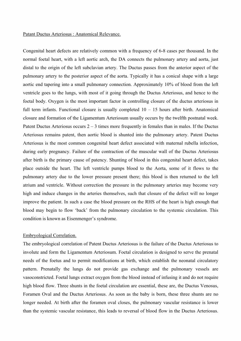
Patant Ductus Arteriosus : Anatomical Relevance.
Congenital heart defects are relatively common with a frequency of 6-8 cases per thousand. In the
normal foetal heart, with a left aortic arch, the DA connects the pulmonary artery and aorta, just
distal to the origin of the left subclavian artery. The Ductus passes from the anterior aspect of the
pulmonary artery to the posterior aspect of the aorta. Typically it has a conical shape with a large
aortic end tapering into a small pulmonary connection. Approximately 10% of blood from the left
ventricle goes to the lungs, with most of it going through the Ductus Arteriosus, and hence to the
foetal body. Oxygen is the most important factor in controlling closure of the ductus arteriosus in
full term infants. Functional closure is usually completed 10 – 15 hours after birth. Anatomical
closure and formation of the Ligamentum Arteriosum usually occurs by the twelfth postnatal week.
Patent Ductus Arteriosus occurs 2 – 3 times more frequently in females than in males. If the Ductus
Arteriosus remains patent, then aortic blood is shunted into the pulmonary artery. Patent Ductus
Arteriosus is the most common congenital heart defect associated with maternal rubella infection,
during early pregnancy. Failure of the contraction of the muscular wall of the Ductus Arteriosus
after birth is the primary cause of patency. Shunting of blood in this congenital heart defect, takes
place outside the heart. The left ventricle pumps blood to the Aorta, some of it flows to the
pulmonary artery due to the lower pressure present there; this blood is then returned to the left
atrium and ventricle. Without correction the pressure in the pulmonary arteries may become very
high and induce changes in the arteries themselves, such that closure of the defect will no longer
improve the patient. In such a case the blood pressure on the RHS of the heart is high enough that
blood may begin to flow ‘back’ from the pulmonary circulation to the systemic circulation. This
condition is known as Eisenmenger’s syndrome.
Embryological Correlation.
The embryological correlation of Patent Ductus Arteriosus is the failure of the Ductus Arteriosus to
involute and form the Ligamentum Arteriosum. Foetal circulation is designed to serve the prenatal
needs of the foetus and to permit modifications at birth, which establish the neonatal circulatory
pattern. Prenatally the lungs do not provide gas exchange and the pulmonary vessels are
vasoconstricted. Foetal lungs extract oxygen from the blood instead of infusing it and do not require
high blood flow. Three shunts in the foetal circulation are essential, these are, the Ductus Venosus,
Foramen Oval and the Ductus Arteriosus. As soon as the baby is born, these three shunts are no
longer needed. At birth after the foramen oval closes, the pulmonary vascular resistance is lower
than the systemic vascular resistance, this leads to reversal of blood flow in the Ductus Arteriosus.
Thus the flow of blood is now from the aorta to the pulmonary trunk. Often there is a small shunt of
blood from the aorta to the pulmonary system in healthy full term infants. The high level of oxygen,
which the Ductus Arteriosus is exposed to, causes it to close, in most cases within 24 hours.
Causes and Physiological Features.
Patent Ductus Arteriosus is much more common in premature babies, which may also have
problems arising from the lack of maturity of the lungs. Blood flows across the Patent Ductus
depending on the relative resistance in the pulmonary and systemic circulations. In the foetus
approximately 55 – 60 % of the systemic circulation passes from right to left across the ductus. The
DA is the predominant route of circulation for blood passing through the RV and Pulmonary
Artery. In the foetus, oxygen tension is relatively low. This coupled with a high level of circulating
prostaglandins, acts to keep the DA open. At birth the lungs expand, activating the major organ of
prostaglandins metabolism, oxygen tension in the blood markedly increases and pulmonary vascular
resistance decreases. These changes result in the contraction of the smooth muscle in the wall of the
Ductus. Thus a preferential shift of blood flow occurs, as blood moves away from the Ductus and
directly from RV to the lungs. Until functional closure is complete and PVR is lower than SVR
some left to right residual flow will occur from the aorta to the pulmonary arteries.
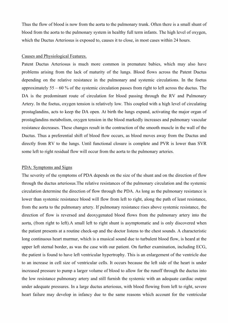
PDA: Symptoms and Signs
The severity of the symptoms of PDA depends on the size of the shunt and on the direction of flow
through the ductus arteriosus.The relative resistances of the pulmonary circulation and the systemic
circulation determine the direction of flow through the PDA. As long as the pulmonary resistance is
lower than systemic resistance blood will flow from left to right, along the path of least resistance,
from the aorta to the pulmonary artery. If pulmonary resistance rises above systemic resistance, the
direction of flow is reversed and deoxygenated blood flows from the pulmonary artery into the
aorta, (from right to left).A small left to right shunt is asymptomatic and is only discovered when
the patient presents at a routine check-up and the doctor listens to the chest sounds. A characteristic
long continuous heart murmur, which is a musical sound due to turbulent blood flow, is heard at the
upper left sternal border, as was the case with our patient. On further examination, including ECG,
the patient is found to have left ventricular hypertrophy. This is an enlargement of the ventricle due
to an increase in cell size of ventricular cells. It occurs because the left side of the heart is under
increased pressure to pump a larger volume of blood to allow for the runoff through the ductus into
the low resistance pulmonary artery and still furnish the systemic with an adequate cardiac output
under adequate pressures. In a large ductus arteriosus, with blood flowing from left to right, severe
heart failure may develop in infancy due to the same reasons which account for the ventricular
hypertrophy, i.e. much more demand on the heart to pump a larger volume of blood. In a more
moderate sized PDA, the child may be underweight with a reduced exercise tolerance and increased
tendency to chest infections. The heart is enlarged with a prominent left ventricle and a systolic
thrill, which is like a palpable murmur, can be felt in the pulmonary area on palpation of the chest.
This is due to the turbulence caused by the runoff of blood into the pulmonary artery. The diastolic
blood pressure is low, also due to the runoff of blood to the pulmonary circulation. The patient also
may experience cyanosis and breathlessness during increased exertion, as E.S. did. Cyanosis is a
dark or purple discolouration of the skin due to deficient oxygenation of the blood. This occurs
because the heart is unable to pump enough oxygenated blood to all the tissues
Pulmonary hypertension may develop as a result of a longstanding large shunt because of the
increased volume being pumped into the lungs. This causes the pulmonary resistance to rise and if it
rises sufficiently, reversal of the flow of blood occurs and deoxygenated blood from the pulmonary
artery flows into the aorta. As a result the child becomes mildly cyanosed, with clubbed fingers and
toes. Clubbing refers to thickening of the tissues at the bases of the finger and the toes so that the
angle between the nail and the digit is filled in. The digit end becomes bulbous like a club. On
examination the right ventricle is hypertrophic as the body tries to compensate for the lower levels
of oxygen in the blood by pumping a higher volume of blood to the lungs for oxygenation. Exercise
tolerance is considerably reduced with exertion causing dyspnea (shortness of breath).
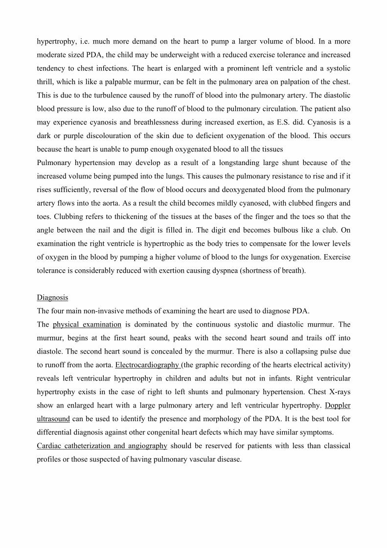
Diagnosis
The four main non-invasive methods of examining the heart are used to diagnose PDA.
The physical examination is dominated by the continuous systolic and diastolic murmur. The
murmur, begins at the first heart sound, peaks with the second heart sound and trails off into
diastole. The second heart sound is concealed by the murmur. There is also a collapsing pulse due
to runoff from the aorta. Electrocardiography (the graphic recording of the hearts electrical activity)
reveals left ventricular hypertrophy in children and adults but not in infants. Right ventricular
hypertrophy exists in the case of right to left shunts and pulmonary hypertension. Chest X-rays
show an enlarged heart with a large pulmonary artery and left ventricular hypertrophy. Doppler
ultrasound can be used to identify the presence and morphology of the PDA. It is the best tool for
differential diagnosis against other congenital heart defects which may have similar symptoms.
Cardiac catheterization and angiography should be reserved for patients with less than classical
profiles or those suspected of having pulmonary vascular disease.
Treatment
A Patent Ductus Arteriosus may close spontaneously, however if this does not happen, it may have
to be prompted to close pharmacologically or surgically ligated. If left untreated, a PDA can lead to
cardiac failure and pulmonary edema (where the lungs are filled with too much fluid), which
accounts for the cyanosis and breathlessness that the case patient E.S. experienced. Bacterial
endocarditis, infection of the endocardium, is also a common complication of PDA. Factors such as
age, overall health of the patient, medical history, extent of the disease, and tolerance for specific
medications can help specify which treatment is best for each patient.
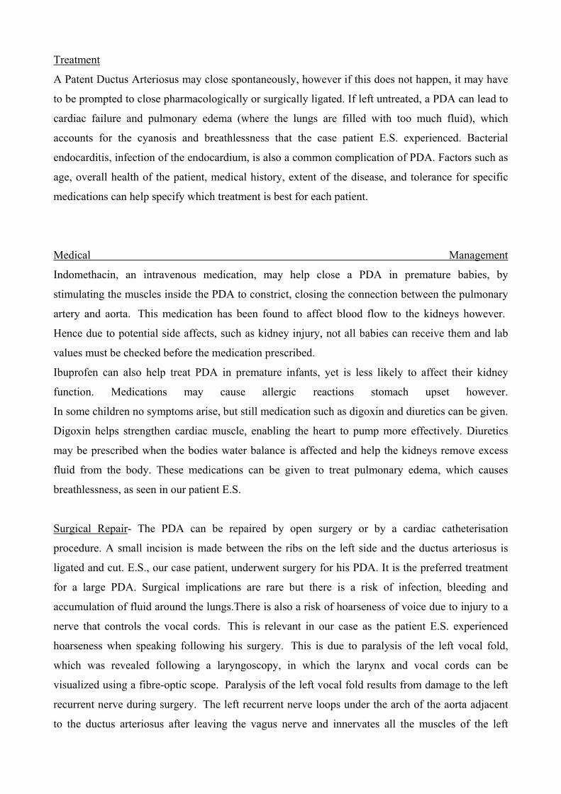
Medical Management
Indomethacin, an intravenous medication, may help close a PDA in premature babies, by
stimulating the muscles inside the PDA to constrict, closing the connection between the pulmonary
artery and aorta. This medication has been found to affect blood flow to the kidneys however.
Hence due to potential side affects, such as kidney injury, not all babies can receive them and lab
values must be checked before the medication prescribed.
Ibuprofen can also help treat PDA in premature infants, yet is less likely to affect their kidney
function. Medications may cause allergic reactions stomach upset however.
In some children no symptoms arise, but still medication such as digoxin and diuretics can be given.
Digoxin helps strengthen cardiac muscle, enabling the heart to pump more effectively. Diuretics
may be prescribed when the bodies water balance is affected and help the kidneys remove excess
fluid from the body. These medications can be given to treat pulmonary edema, which causes
breathlessness, as seen in our patient E.S.
Surgical Repair- The PDA can be repaired by open surgery or by a cardiac catheterisation
procedure. A small incision is made between the ribs on the left side and the ductus arteriosus is
ligated and cut. E.S., our case patient, underwent surgery for his PDA. It is the preferred treatment
for a large PDA. Surgical implications are rare but there is a risk of infection, bleeding and
accumulation of fluid around the lungs.There is also a risk of hoarseness of voice due to injury to a
nerve that controls the vocal cords. This is relevant in our case as the patient E.S. experienced
hoarseness when speaking following his surgery. This is due to paralysis of the left vocal fold,
which was revealed following a laryngoscopy, in which the larynx and vocal cords can be
visualized using a fibre-optic scope. Paralysis of the left vocal fold results from damage to the left
recurrent nerve during surgery. The left recurrent nerve loops under the arch of the aorta adjacent
to the ductus arteriosus after leaving the vagus nerve and innervates all the muscles of the left
larynx, with the exception of the left cricothyroid muscle (which is innervated by the superior
laryngeal branch of the vagus).
Cardiac catheterisation procedure-this can be an effective alternative to surgical intervention.
A catheter is inserted into a blood vessel in the groin and guided to the inside of the heart by the
cardiologist. When positioned in the PDA the coil is pushed through the catheter into the PDA.
The coil prevents blood flow through the vessel, in part by stimulating a blood clot at the site. This
simple procedure takes approximately only three hours to complete. Complete closure is achieved
in nearly 100% of small PDAs. There are few complications of cardiac catheterisation but include
bleeding, infection, and early dislodgement of the coil.
Case 2
Symptoms: Our case has presenting symptoms, which can help us make a preliminary diagnosis about her
condition and what is causing her such discomfort. We believe she is suffering from osteoporosis, which is
the thinning of the bones. Loss of bone density is reported as one of her symptoms. Solid bone mass reaches
an all time high in the body around age 35, from then on there is a decline. Calcium absorption is hindered by
age related decrease vit D in blood. Not sufficient calcium means it must be found elsewhere so is reabsorbed
from bones into blood stream. Weight bearing exercises such as walking yield high calcium in bones so
inactivity due to age may also hamper bone mass. Bones are left weak and brittle – so more susceptible to
damage.
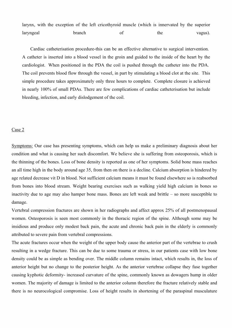
Vertebral compression fractures are shown in her radiographs and affect approx 25% of all postmenopausal
women. Osteoporosis is seen most commonly in the thoracic region of the spine. Although some may be
insidious and produce only modest back pain, the acute and chronic back pain in the elderly is commonly
attributed to severe pain from vertebral compressions.
The acute fractures occur when the weight of the upper body cause the anterior part of the vertebrae to crush
resulting in a wedge fracture. This can be due to some trauma or stress, in our patients case with low bone
density could be as simple as bending over. The middle column remains intact, which results in, the loss of
anterior height but no change to the posterior height. As the anterior vertebrae collapse they fuse together
causing kyphotic deformity- increased curvature of the spine, commonly known as dowagers hump in older
women. The majority of damage is limited to the anterior column therefore the fracture relatively stable and
there is no neurocological compromise. Loss of height results in shortening of the paraspinal musculature
requiring prolonged active contraction for the maintenance of posture. This causes pain from muscle fatigue,
standing and walking can exacerbate this pain.
This may explain our patients reported pain after long periods of sitting or standing. The loss of height of the
backbone and kyphotic curvature of the spine means the whole ribcage moves downwards and may catch
uncomfortably against the wings of hip and rim of pelvis. Nerve root pain occurs from the nipping of a nerve
by damaged vertebrae. She also reports episodes severe pain in the thoracic region of the back. We can
assume this is where her fractures are occurring. Damaged vertebrae easily squash nerves – when this occurs
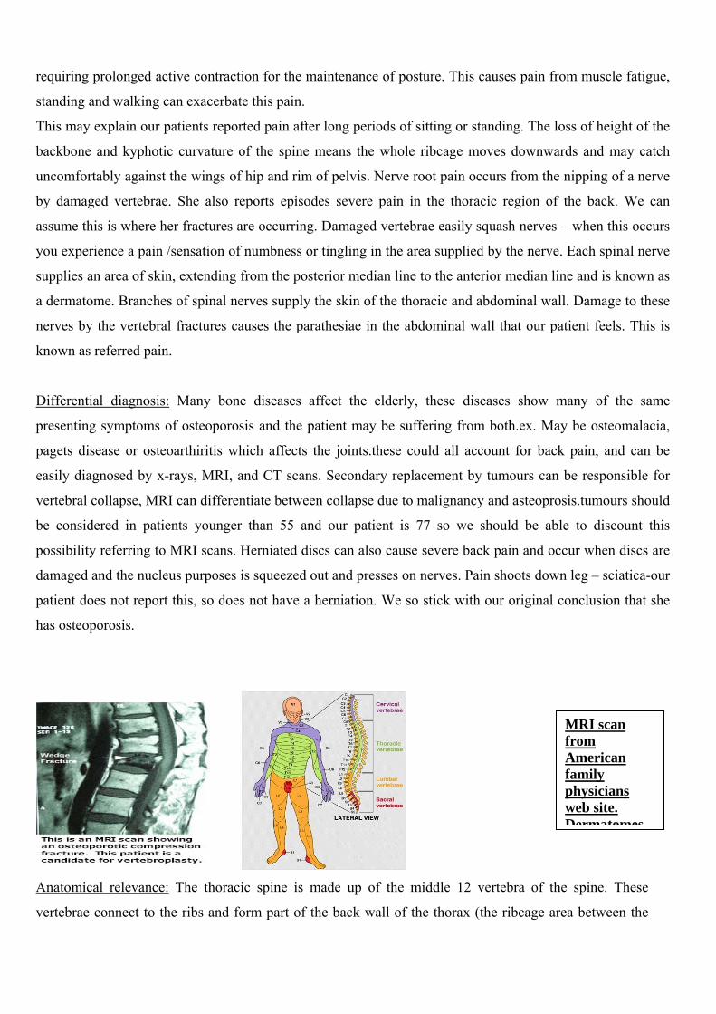
you experience a pain /sensation of numbness or tingling in the area supplied by the nerve. Each spinal nerve
supplies an area of skin, extending from the posterior median line to the anterior median line and is known as
a dermatome. Branches of spinal nerves supply the skin of the thoracic and abdominal wall. Damage to these
nerves by the vertebral fractures causes the parathesiae in the abdominal wall that our patient feels. This is
known as referred pain.
Differential diagnosis: Many bone diseases affect the elderly, these diseases show many of the same
presenting symptoms of osteoporosis and the patient may be suffering from both.ex. May be osteomalacia,
pagets disease or osteoarthiritis which affects the joints.these could all account for back pain, and can be
easily diagnosed by x-rays, MRI, and CT scans. Secondary replacement by tumours can be responsible for
vertebral collapse, MRI can differentiate between collapse due to malignancy and asteoprosis.tumours should
be considered in patients younger than 55 and our patient is 77 so we should be able to discount this
possibility referring to MRI scans. Herniated discs can also cause severe back pain and occur when discs are
damaged and the nucleus purposes is squeezed out and presses on nerves. Pain shoots down leg – sciatica-our
patient does not report this, so does not have a herniation. We so stick with our original conclusion that she
has osteoporosis.
Anatomical relevance: The thoracic spine is made up of the middle 12 vertebra of th
vertebrae connect to the ribs and form part of the back wall of the thorax (the ribcage ar
MRI scan from American family physicians web site. Dermatomes
e spine. These
ea between the
neck and the diaphragm). It’s provides stability and structural support to the upper back and allows very
little motion.
The thoracic spine's curve is called kyphotic because of its shape, which is a regular "C"-shaped curve
with the opening of the "C" in the front. The normal amount of curve in the thoracic spine is considered
to be from 20 to 40 degrees within the entire thoracic spine. There is a range because the amount of
"normal" curve varies from person to person. Though the thoracic spine is supposed to be curved, if the
curve in a person's thoracic spine is more than 40 degrees, it is considered abnormal - or a spinal
deformity.
The vertebrae of the thoracic spine has very narrow, thin intervertebral discs, in fact the thinnest
intervertebral discs is located in superior thoracic region, so there is much less movement allowed
between vertebrae than in the lumbar or cervical parts of the spine. It also has less space in the vertebral
canal for the spinal cord and smaller vertebral (neural) foramen than in cervical and lumbar region. The
transition from the relatively inflexible thoracic region to the much more mobile lumbar region occurs
abruptly, and that is the cause of T11 and T12 being the most commonly fractured vertebrae.
The nerves that branch out from the spinal cord in the thoracic spine are dorsal and ventral primary rami.
These nerves go to the chest and abdomen region and let us control the body’s part and the muscles.
Damage to the nerves can cause pain, tingling or numbness in the area where the nerve travels.
Erector spinae are the muscles that located at the back, next to the spine. They support the spine and are
the motor for movement of the spine. When any part of the spine is injured including: a disc, ligaments,
bones, or muscles, this erector spinae muscles automatically go into spasm (sudden contraction) to reduce
the motion around the area. However, when the muscles are in spasm they produce too much lactic acid
that causes a painful burning sensation.
Physical examination and investigation: Before the patient undergoes any physical examination, it is
important for the doctor to get the complete physical history of the patient’s condition. History taking is
very important to help the doctor understand when the back pain began, the patient’s lifestyle, physical
factors that might cause the back pain, and also if the patient’s family had a history of osteoporosis.
Then, the doctor will give a physical examination based upon the symptoms and information given by the
patient. For an osteoporosis patient, the typical examination will begin with the measurement of the
patient’s height. Next is to check the patient’s tenderness of certain areas, especially at the thoracic
region.
The doctor will also check the motion of the spine. This is done by looking at the flexibility of the patient
bending in certain directions and if there is any accompanying pain. Among the motions are lateral
bending, flexion, extension, and rotation of the hip.
Finally, the nerves will be tested by checking the patient’s sensation in specific areas of the feet and
hands, the patient’s reflexes by testing the tendon reflexes under the knee cap or under the Achilles
tendon of the ankle. The strength of the patient’s muscle will also be tested by asking the patient to lift
the arm, hand or leg when light resistance is put against them.
Depending on the outcome of the patient’s history, physical examination, and initial X-rays, other tests
may be ordered to look at specific aspects of the spine. The most common tests that are ordered are: the
Magnetic Resonance Imaging (MRI) scan; the Computer Assisted Tomography (CAT) scan and
Electromyogram nerve tests.
Bone Composition and Remodelling: Bone is composed of a combination of collagen and calcium
phosphate which allows it to withstand the stress exerted on it. The two types of bone in the body are
cortical and trabecular. Cortical forms the outer dense layer while trabecular bone makes up the interior
spongy structure.
Bone remodelling is the process by which bone is renewed and has two stages: resorption and formation.
Osteoclasts are the cells which break down bone and osteoblasts are involved in bone formation. The
function of these bone cells is directly related to each others activities and is regulated by several
hormones. After the age of 30-35, more bone is resorbed than formed. In the case of osteoporosis, bone
resorption takes place too quickly or the formation takes place too slowly. The genes involved in
osteoporotic risk factors have yet to be fully identified
Biochemical Features: Biochemistry is used in the testing and diagnosis of osteoporosis. Routine
biochemical and haematological screening should be carried out to screen for any secondary causes of
osteoporosis.
Bone turnover markers are carried out as a biochemical test measuring the serum and urine levels for
factors produced or released during bone remodelling. However, this test cannot as of yet be used as a
definite diagnosis as elevated levels are a non-specific finding in patients with other metabolic bone
diseases.
Bone Mineral Density (BMD) tests are also used to measure bone density. The most common test is the
Dual energy x-ray absorptiometry, involving a small x-ray detector scanning the spine and hip. Heel
ultrasounds and radiographic absorptiometry are also used but carry with them the disadvantage of
measuring less clinically important areas.
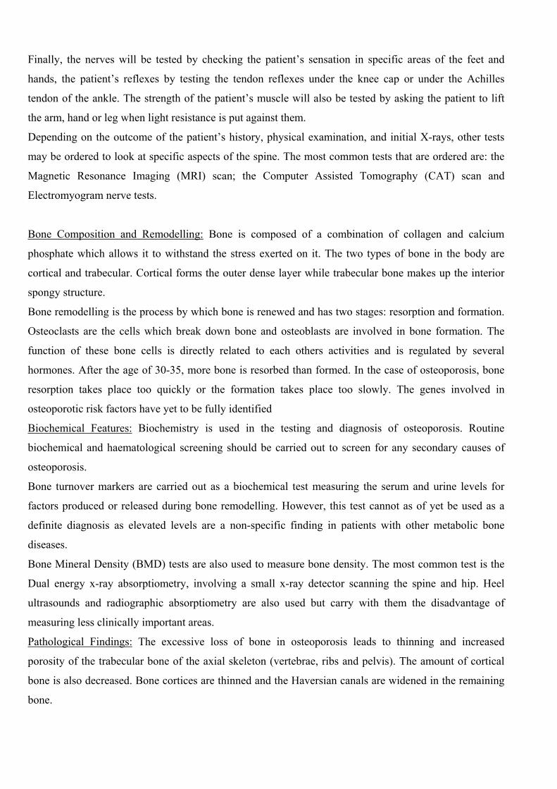
Pathological Findings: The excessive loss of bone in osteoporosis leads to thinning and increased
porosity of the trabecular bone of the axial skeleton (vertebrae, ribs and pelvis). The amount of cortical
bone is also decreased. Bone cortices are thinned and the Haversian canals are widened in the remaining
bone.

The vertebral bodies may be weakened by microfractures and collapse anteriorly. This results in
compression fractures, wedging of the vertebrae and a kyphotic deformity. These features would be likely
to be visible in our patient as her symptoms included an overall height decrease and wedging of the
vertebral bodies
Picture from: MEDLINEplus encyclopedia
Treatments Available: Alterations can be made to the diet/nutrition of a patient as well as the prescription
of medications. Among the medications used are: Estrogen Replacement Therapy; Alendronate (anti-
resportive therapy) and Calcitonin (anti-resportive therapy). Both of the anti-resportive therapies listed
could be considered for this patient.
Alendronate and Calcitonin cause a shift in bone mass towards bone formation. However, Calcitonin is a
naturally produced hormone which has been found to increase bone density mainly in the area of the
spine. As the 77 year old woman in question is experiencing problems primarily in her spine, Calcitonin
may be more suitable.
GRAND CLINICAL ROUND CASE 3 1. MOHD AFIFI, MOHD HAFIZ 2. MUHAMMAD NAWAWI, KHAIRUL NAJMI 3. ALIAS, RIDZWAN
Introduction
Rotator Cuff Tendonitis (RCT) is an inflammation of the tendon of Rotator Cuff (RC)
Known as bursitis or impingement syndrome
RC get irritated on under surface of acromion (supraspinatus)
Causes: 1. intrinsic
2. Extrinsic
1˚ (increase subacromial load)
2˚ (RC overload and muscle imbalance)
Combination of both in this case (an athlete)
Diagram. (Cannot be provided since it is hand drawing. reference can be made to any anatomy
textbook discussing supraspinatus or rotator cuff muscle)
Supraspinatus Anatomical Features and Relevance
Medial 2/3 of supraspinous fossa/ tendon pass through supraspinous outlet/ under acromial
process/courses over superior lip (glenohumeral joint)/ continue inferiorly and attach to superior
edge of greater tubercle of humerus
Abduction (initiate) - 45% strength
Dynamic stabilization of humeral head on glenoid/ prevent subluxation
Supraspinous outlet:
Upper rim- acromion/coracoacromial arch/acromioclavicular joint
Lower rim- humeral head/ glenoid
Accommodates passage and excursion of tendon
Abnormalities: results in impingement and RC injury
1. acromion size and shape
2. acromioclavicular osteophytes
3. coracoacromial ligament thickening
Impingement sites further compress when flexion/abduction/med. Rotation)
Non outlet:
1. loss of normal humeral head depression
2. hypertrophy of subacromial bursa and RC tendons
Pathology of the Disease
Causes of Tendonitis
Rotator cuff pathology can be caused by extrinsic (outside) or intrinsic (from within)
causes1. Extrinsic examples include a traumatic tear in the tendon(s) from a fall or accident.
Overuse injuries from repetitive lifting, pushing, pulling, or throwing are also extrinsic in nature. 1 http://www.orthoassociates.com/shoulderRCD.htm
Inflammation of the tendons of the shoulder muscles can occur in sports requiring the arm to be
moved over the head repeatedly as in tennis, baseball (particularly pitching), swimming, and lifting
weights over the head2.
Intrinsic factors include poor blood supply, normal attrition or degeneration with aging, and
calcific invasion of the tendon(s). The acromion morphology also can cause tendonitis; some people
are born with a "hooked" acromion that will predispose them to this problem3. The rotator cuff
weakness or imbalance can cause the humerus to ride up and pinch the cuff.
Symptoms
• Pain - Primarily on top and in the front of your shoulder. Sometimes you can have pain at
the side of your shoulder. Usually is worse with any overhead activity (reaching up above
the level of your shoulder)4.
• Weakness - mild to moderate weakness, especially worse with overhead activity such as
brushing hair, reaching for objects on shelves, etc5.
• Popping - sometimes bursitis that occurs with rotator cuff tendonitis can cause a mild
popping or crackling sensation in the shoulder
• Unable to sleep on the shoulder - most patients complain of difficulty sleeping on the
shoulder at night
The tendonitis will lead to several complications (if untreated) such as bursitis (subdeltoid or
subacromial bursitis), complete rotator cuff tear (due to chronic inflammation) and failure of
treatment to improve symptoms.
DIFFERENTIAL DIAGNOSIS There are other possibilities of injuries that closely related to supraspinatus tendonitis which are
calcific tendonitis and bursitis.
Calcific tendonitis occurs by the calcifications, situated in the tendon of supraspinatus,
infraspinatus, subscapularis and long biceps muscles. The tendons become harden because of the
deposit of calcium salts. The deposit of calcium salts probably because of poor blood supply or
inflammation. Bursitis on the other hand is the inflammation or irritation of the bursa. Bursa is a
2 http://www.nlm.nih.gov/medlineplus/ency/article/000438.htm 3 http://www.jointhealing.com/pages/shoulder/rotcuff_tend_1.html 4 Ibid. 5 http://www.nlm.nih.gov/medlineplus/ency/article/000438.htm
soft, fluid-filled sac that covers the movement between the bones, tendons and muscles near the
joint. Bursitis is caused by trauma, overuse to the joint from playing or working, incorrect posture at
work or rest and many more. The symptoms for these injuries more or less are the same as
supraspinatus tendonitis, which include pain, tenderness, restriction of motion and swelling in the
affected areas.
SHOULDER EXAMINATION
1. Inspection - The examiner will visualize the shoulder girdle and noting for the mass
muscles, bone asymmetry as well as any swelling.
2. Palpation – The examiner usually press over the patient’s shoulder including
acromioclavicular joint, clavicle, scapula, supraspinous fossa, infraspinous fossa and
proximal humerus. In this case, the patient felt pain at superior part of greater tubercle of
humerus (insertion of supraspinatus tendon). This indicates that the patient was having some
problems with her supraspinatus tendon.
3. Manual Muscle Testing –
i. Subscapularis lift-off test of Gerber and Krushell: arm internally rotated behind
the back with the elbow flexed. In normal condition, patient can maintain the arm
in a fully extended position with elbow flexed against resistance. If the patient is
unable to move his arm away from his back, the tear of subscapularis tendon
might occur.
ii. Supraspinatus Test: arm abducted at scapular plane and internally rotated so that
the thumb point towards the floor. Examiner applies a downward force while the
patient attempts to maintain the arm parallel to the floor. Any inability to do so
indicates the tear or inflammation of supraspinatus tendon. Shoulder abduction
against resistance also can be done. The patient’s shoulder is abducted against
resistance and if pain is felt between initial 350, the supraspinatus tendon could
be inflamed.
iii. Infraspinatus and teres minor test: Patient put his arm at the side with elbow
flexed. External rotation against resistance is applied.
4. Diagnostic Imaging –
i. X-Ray = visualize the anatomy of shoulder bone.
ii. MRI = accurate at indicating rotator cuff tears. Able to show the location, size
and retraction of the tear.
iii. Arthrography = Dye injected into the joint. Low cost, but risk of radiation
exposure.
iv. Ultrasonography = painless, inexpensive. But, reliability depends on
sonographer’s skill
v.
TREATMENT
Mild = for mild injury, a patient is usually given a period of rest as well as inflammatory
medication. The doctor may also give a cortisone injection into the bursa space in order to
relieve the pain. Some doctors might prescribe ultrasound therapy whereby a medicated
cream is applied to the patient’s shoulder. Then, a small device will be rubbed to the
shoulder and wave sound from that device will loosen any tightness. The most suitable
treatment for mild shoulder injury should be physical therapy. The goals are to relieve pain,
prevent muscle atrophy without exacerbating the pain and reestablish non-painful range of
motion. The therapy includes joint mobilization, strengthening exercises and neuromuscular
control exercises.
Severe = the patient might undergoes surgery if he or she is having severe injury of rotator
cuff. The surgery is usually done in three stages, which are removal of inflammed bursa, cut
of coracoacromial ligament and lastly the removal off small portion of acromion.
Case 4 Radial nerve palsy occurs when the radial nerve has been damaged. It results in temporary paralysis or loss of feeling. Anatomical Relevance ●The radial nerve is derived from the posterior cord of the brachial plexus. It supplies all the extensor muscles of the posterior compartment of the upper limb, skin on the posterior aspect of arm and forearm and skin on the dorsum of the hand. Therefore, if it is damaged, as in this case, the patient will be unable to extend the elbow and wrist properly. ●The radial nerve is formed in the axilla. In the axilla it gives off a branch called the posterior cutaneous nerve of the arm. This supplies cutaneous innervation to the skin on the back of the arm. In this case, the patient does not exhibit loss of sensation over the posterior aspect of the arm and so the radial nerve must not be damaged before the point where this branch is given off. ●The radial nerve runs posterolaterally from the axilla towards the arm. In doing so it crosses the posterior wall of the axilla anteriorly. The nerve then winds around the back of the arm from the medial side to the lateral side. It goes between the 3 heads of triceps, long, lateral and medial, supplying all three. The triceps is the chief extensor of the forearm. The nerve comes into direct contact with the shaft of the humerus at the radial groove. This is a key anatomical feature of this case. If the radial nerve is injured in the radial groove, the triceps is affected, though not completely paralysed. It is weakened as only the medial head of the triceps is affected. This is because the branches to the long and lateral heads are given off prior to the nerve entering the radial groove whereas the branch to the medial head is given off in the spiral groove. In his case the damage to the radial nerve occurs in the axilla and so all three heads of the triceps are affected. This explains the patient’s weakness in extending his forearm. ● It supplies the anconeus muscle and also gives off another branch, the posterior cutaneous nerve of the forearm. This branch perforates the lateral head of the triceps and descends along the
posterior aspect of the forearm to the wrist. It supplies the skin on the posterior surface of the forearm. In this case, the patient exhibits loss of sensation over the dorsum of the forearm implying that the radial nerve has been damaged at a point prior to this branch being given off ie.in the axilla. ●The radial nerve then comes to lie lateral to the brachialis muscle. It leaves the posterior compartment of the arm and pierces the lateral intermuscular septum in the lower arm. Here, in the anterior compartment of the arm, above the lateral epicondyle of the humerus, it gives branches to the brachioradialis, a flexor of the forearm and the extensor carpi radialis longus(ECRL), an extensor of the hand. This muscle extends the 2nd metacarpal of the hand. ●It then passes into the cubital fossa where it divides into deep and superficial branches. The deep branch pierces the supinator and enters the posterior aspect of the forearm where it continues as the posterior interosseous nerve. This nerve supplies extensor carpi radialis brevis (ECRB) which causes extension of the 3rd metacarpal at the wrist. It also supplies the pollicis group of muscles- abductor pollicis longus(extends and abducts thumb at carpometacarpal joint), extensor pollicis brevis(extends proximal phalanx of thumb) and extensor pollicis longus(extends distal phalanx of thumb). The patient in this case presents with loss of sensation over the posterior aspect of the 1st interdigital cleft implying that ECRL, and the pollicis group of muscles have been affected. The posterior interosseous branch also supplies extensor digitorum which causes extension of the medial 4 digits and extensor indicis which causes extension of the index finger. These muscles supplying the 2nd digit have also been affected. The patient will have difficulty extending the fingers and therefore in grasping objects. This is a major effect of damage to the radial nerve. ●The superficial branch of the radial nerve descends lateral to the radial artery and posterior to the brachioradialis. It enters the dorsum of the hand and supplies cutaneous innervation to the skin over the lateral two thirds of the dorsum of hand. This branch of the radial nerve has also been affected as the patient presents with loss of sensation over the posterior aspect of the first interdigital cleft as already mentioned. Key Physiological Feature The action of a muscle i.e. its contraction is due to a nerve conducting an action potential to the neuromuscular junction. If a nerve is compressed sufficiently, the nerve fibres stop conducting the action potentials along their axons and their function is impaired, an action potential (or depolarization) doesn’t arrive at the neuromuscular junction. The patient in this case has compressed his radial nerve due to improper use of a crutch and it no longer functions properly. This is why the muscles mentioned above don’t function and he has weakness in extending his left elbow and wrist Causes and symptoms of injury to the Radial Nerve Radial Nerve injury or compression may occur at any point along the anatomical course of the nerve. The four main areas where it can occur are in the Axilla, in the Radial Groove (Spiral Groove), in the Deep branch of the Radial Nerve or in the Superficial branch of the Radial Nerve. The most frequent site of compression is in the area of the Supinator muscle and involves the Posterior Interrosseous Branch. However, problems can occur proximally in relation to fracturesof the Humerus at the junction of the middle and proximal thirds, as well as distally on the radial aspect of the wrist. In the Axilla, the nerve can be injured by the pressure of the upper end of a badly fitting crutch pressing up into the armpit. This causes a type of palsy known as “Crutch Palsy”. This problem was present in our case where a middle aged man suffering from a leg injury had been using an Axillary
crutch. The crutch compressed the Radial Nerve in the Axilla and caused palsy in the arm, forearm and fingers, all of which are symptoms of “Crutch Palsy”. The Radial Nerve can also be damaged in the Axilla by improper positioning of the upper limb during sleeping. This may occur when a person falls asleep with their arm hanging over the back of a chair. The palsy which occurs in this case is known as “Saturday Night Palsy”. The Radial Nerve can also be damaged in the Axilla by fractures and dislocations of the proximal end of the Humerus. When the Humerus is displaced downward in dislocations of the shoulder, the Radial nerve, which is wrapped around the back of the shaft of the bone, is pulled downward, stretching the nerve in the Axilla excessively. When the Radial Nerve is injured in the Axilla, the Triceps are paralyzed. Both “Crutch Palsy” and “Saturday Night Palsy” cause wasting and loss of reflex. There is both wrist and finger drop due to weakness of the wrist and finger extensors, as well as weakness of Extensor Pollicis Longus and Abductor Pollicis Longus. There may be sensory impairment in the distribution of the Superficial Radial Nerve. All of the above symptoms were present in this case where the patient experienced difficulty in extending his left elbow and wrist and had loss of sensation over the dorsum of the forearm and the posterior aspect of the first interdigital cleft as a result of the pressure of the crutch on the Radial Nerve in the Axilla. It is important that the patient is treated as the wrist drop which he experiences as part of the palsy is very debilitating. In Wristdrop the hand is flexed as the extensor muscles on the back of the hand are not functioning. It is difficult to grasp objects strongly when the wrist is in a flexed position, therefore it is vital that the Wristdrop is treated. In the Spiral Groove of the Humerus, the Radial Nerve can be injured at the time of fracture of the shaft of the Humerus or subsequently during the formation of the callus. The pressure on the back of the arm on the edge of the operating table in an unconscious patient has been known to injure the nerve at this site. The prolonged application of a Tourniquet to the arm in a person with a slender triceps muscle is often followed by temporary Radial Palsy. The injury to the Radial Nerve occurs most commonly in the distal part of the groove beyond the origin of the Triceps and the Anconeus and beyond the origin of the Cutaneous Nerves. As a result, the patient is unable to extend the wrist and the fingers and there is Wristdrop. A variable small area of anesthesia is present over the dorsal surface of the hand and the dorsal surface of the roots of the lateral three and one-half fingers. The Deep Branch of the Radial Nerve is a motor nerve to the extensor muscles in the posterior compartment of the forearm. It can be damaged in fractures of the proximal end of the Radius or during dislocation of the Radial Head. The nerve supply to the Supinator and the Extensor Carpi Radialis Longus will be undamaged, and because the latter muscle is powerful, it will keep the wrist joint extended, and Wristdrop will not occur. There will be no sensory loss in this case since this is a motor nerve. Division of the Superficial Branch of the Radial Nerve, which is sensory, which may occur for example in a stab wound, results in a variable small area of anesthesia over the dorsum of the hand and the dorsal surface of the roots of the lateral three and one half fingers. Diagnosis, Physical Examinations & Investigations, Treatments And Preventions For Radial Nerve Palsy. History and a physical examination are often all that is needed to determine the level of injury and the suspected cause of radial nerve palsy/paralysis. Tests maybe ordered to help figure out the cause of the radial nerve injury. Tests that reveal nerve dysfunction may include EMG, nerve conduction tests, nerve biopsy, MRI and neuromuscular examinations. Radiographs should be obtained if a fracture, dislocation, or foreign body is suspected. EMG is basically recording of electrical activity in muscle. A nerve conduction velocity study may be done to determine the location of the nerve injury. This test involves attaching wires to the skin. Small shocks are used to stimulate the nerve and measure its function. . Patients with nerve palsy/paralysis that persists beyond 6 to 8 weeks should be examined with electrodiagnostic studies. By 12 weeks,
motor unit potentials will be present and will help to differentiate between recoverable injures and those that will require surgery. Blood tests or a nerve biopsy are sometimes needed in unusual cases. A biopsy is a procedure to remove a small piece of tissue from the body. A special tool or needle can be inserted through the skin and into the nerve. A small piece of the nerve can be removed with the tool. The piece can then be sent to the lab for further examination and testing. Magnetic resonance imaging should be obtained if a mass is suspected at any level along the course of the radial nerve. The use of MRI may rule out stroke. A neuromuscular examination of the arm, hand and wrist can identify radial nerve dysfunction. There may be weakness of the wrist and finger extension muscles (with decreased ability to extend the arm at the elbow); a minor decreased ability to rotate the arm outward (supination); and difficulty lifting the wrist or fingers (extensor muscle weakness). Wrist drop or finger drop may be present, or there may be atrophy (muscle loss) of some of the muscles of the forearm. A detailed patient history may be needed to determine the possible cause of the neuropathy. Rarely, radial nerve dysfunction may be difficult to differentiate from a stroke in the brain. After full investigation, it is confirmed that the patient in our case suffers from radial nerve palsy due to compression by the crutch. The treatment is aimed at maximizing the ability to use the hand and arm. The cause should be identified and treated as appropriate. In most cases, no treatment is required and recovery is spontaneous, eg. In the case of “Saturday night Palsy.” If there is no history of trauma to the area, conservative treatment is indicated by a sudden onset, minimal sensation changes and no difficulty in movement, and no test results indicating degeneration of the nerve axon. Surgical removal of lesions that press on the nerve may benefit some people. Basically there three types of treatments which are common. They are conservative treatment, physical therapy and surgical treatment. Conservative treatment refers to the use of splints and is relevant when there wrist drop. Initially the wrist and fingers are splinted in a position of extension at the wrist and M.P. joints by a 'Cock up' splint made of Plaster of Paris of aluminium applied on the volar aspect. This is to prevent overstretching of the paralysed muscles. This conventional wrist drop splint has the disadvantage of preventing activity in the unparalysed flexor muscles of the wrist and M.P.joints. The modern splint for Radial nerve palsy is a Dynamic or Lively splint, applied on the dorsal aspect which keeps the wrist and fingers extended by elastic bands or springs attached to it but allows active flexion of the fingers and wrist.Physical therapy exercises may be appropriate for some people to maintain muscle strength. Physical therapy is a form of treatment that employs physical methods to promote healing which includes the use of light, infra red, ultra violet, heat, electric current, massage, remedial exercise and hydrotherapy. Physical therapy is necessary to minimize the muscle wasting and maximize the muscle function during the recovery period.The most common type of radial nerve injury is when there is a fracture in the humerus. The fractured humerus damages the radial nerve and radial nerve palsy/paralysis is experienced. The appropriate treatment for this trauma would be surgical. In other cases however, surgical decompression might be necessary for prolonged radial palsy due to serious compression by an anatomical structure. Referring to the case, the treatment most suitable would be physical therapy while the usage of splints might be relevant. The need for surgery relies upon how severe the injury is. Basically, there is no need for surgery unless the compression of the radial nerve is very serious. Furthermore, fracture of the humerus never occurred. Among the best ways to prevent radial nerve palsy would simply be by using the proper technique when handling with an axillary crutch, occupational therapy (exercise) and the use of an alternative walking aid, a four-legged support.
Reference:
• Edmonson AS, Crenshaw AH: Peripheral nerve injuries. In: Campbell's Operative Orthopedics. 6th ed. 1980: 1678-9.
• Lubahn JD, Cermak MB: Uncommon nerve compression syndromes of the upper extremity. J Am Acad Orthop Surg 1998 Nov-Dec; 6(6): 378-86
• Ritts GD, Wood MB, Linscheid RL: Radial tunnel syndrome. A ten-year surgical experience. Clin Orthop 1987 Jun; (219): 201-5
• Seddon HJ: Surgical Disorders of the Peripheral Nerves. 1972: 66-88. • Spinner M, Spencer PS: Nerve compression lesions of the upper extremity. A clinical and
experimental review. Clin Orthop 1974 Oct; 0(104): 46-67 • Spinner M: Injuries to the Major Branches of Peripheral Nerves of the Forearm. 2nd ed.
1978: 234. • Sunderland S: Nerves and Nerve Injuries. 2nd ed. 1978: 127. • www.AllReferHealth.com • www.thirdage.com
CASE STUDY: 5 (COLLES’ FRACTURE)
BY: IZZATUL AINI, KHOR CHIN CHUAN, NURUL IZZA
ANATOMICAL RELEVANCE
When someone begins to fall, they almost always extend their hand to break the fall and to reduce
the force of hitting the ground as well. The sudden impact of their body weight on the hand may
cause the distal end of the radius to fracture just above the wrist when they fall on the outstretched
hand. This condition is known as a Colles’ Fracture or Transverse Wrist Fracture.
Forearm consists of radius and ulna, bound together by interosseous membrane. Radius is lateral
and shorter than ulna. At the proximal end, it has a short cylindrical head, neck and a medially
directed tuberosity. Head of the ulna lying in the ulnar notch of the radius. The radial styloid
process is larger than that of the ulnar styloid process. The distal end of the radius is marked by
lateral two of the proximal row of the carpal bones which are scaphoid and lunate bones. Here, it
forms a direct articulation between the forearm and carpal bones. Because of this, the carpal bones
move with the radius. When forces applied to the hand, they are transmitted to the radius through
these carpal bones which is then subjected to injury.
First joint involves is the radioulnar joint. The articulation is between the rounded head of the ulna
and the ulnar notch of the radius. This is of synovial pivot joint. The articular disc is triangular and
composed of fibrocartilage. It shuts off the distal radioulnar joint from the wrist and strongly unites
the radius to the ulna. Dislocation at this joint may happen as a result of fracture at the distal end of
the radius. Anterior and posterior interosseous nerves innervate this joint. Blood supplies here are
anterior and posterior interosseous arteries. Movements allowed at this type of joint are pronation
and supination. Pronation is performed by pronator quadratus and teres and flexor carpi radialis
which are innervated by median nerve. Supination is performed by biceps brachii muscle which is
innervated by musculocutaneous nerve and brachioradialis and supinator are innervated by radial
nerve.
The second joint is radiocarpal (wrist). Articulation is between the distal end of the radius, the
articular disc above and the scaphoid and lunate bones below. This is of condyloid type of synovial
joint. Weak anterior and posterior ligaments strengthen the fibrous capsule which encloses the
joint. Medial ligament is attached to the ulnar styloid process and triquetral bone. Strong lateral
ligament is attached to the radial styloid process and scaphoid. This joint receives blood supplies
from branches of the dorsal and palmar carpal arches. Anterior and posterior interosseous nerves,
dorsal and deep branches of the ulnar nerve innervate it. For flexion, median nerve innervates the
flexor carpi radialis, flexor pollicis longus, palmaris longus, flexor digitorum superficialis and half
of the profundus. Ulnar nerve innervates flexor carpi ulnaris. For extension, extensor carpi radialis
longus and brevis, extensor carpi ulnaris, extensor digitorum, indicis, digiti minimi and pollicis
longus are innervated by radial nerve. Muscles involve in abduction are extensor carpi radialis
longus and brevis and abductor pollicis longus. Those are innervated by radial nerve. Median nerve
innervates flexor carpi radialis. Adduction is performed by flexor carpi ulnaris, innervated by ulnar
nerve and extensor carpi ulnaris, innervated by radial nerve. Circumduction also occurs at this joint.
Normal range of wrist flexion is about 80º, 70º in extension, 30º in ulnar deviation and 20º in radial
deviation.
SYMPTOMS AND SIGN
Once the fracture of the wrist has occurred, the wrist joint is usually very painful. If patient does
not seek medical assistance, the wrist gradually swells and mobilization becomes painful. Bruising
can happens as a result of escape of blood from ruptured vessels underlying the skin. The patient is
unable to hold or lift object of any significant weight due to the affected wrist and distal radioulnar
areas. The most important clinical deformity is ‘dinner-fork’. This is produced by the backward,
outward and tilt of the distal fragment of the radius. The fracture results from dorsiflexion of the
hand. Normally the radial styloid process projects further distally than the ulnar styloid process,
consequently when a Colles’s fracture occurs, this relationship is reversed because of shortening of
the radius.
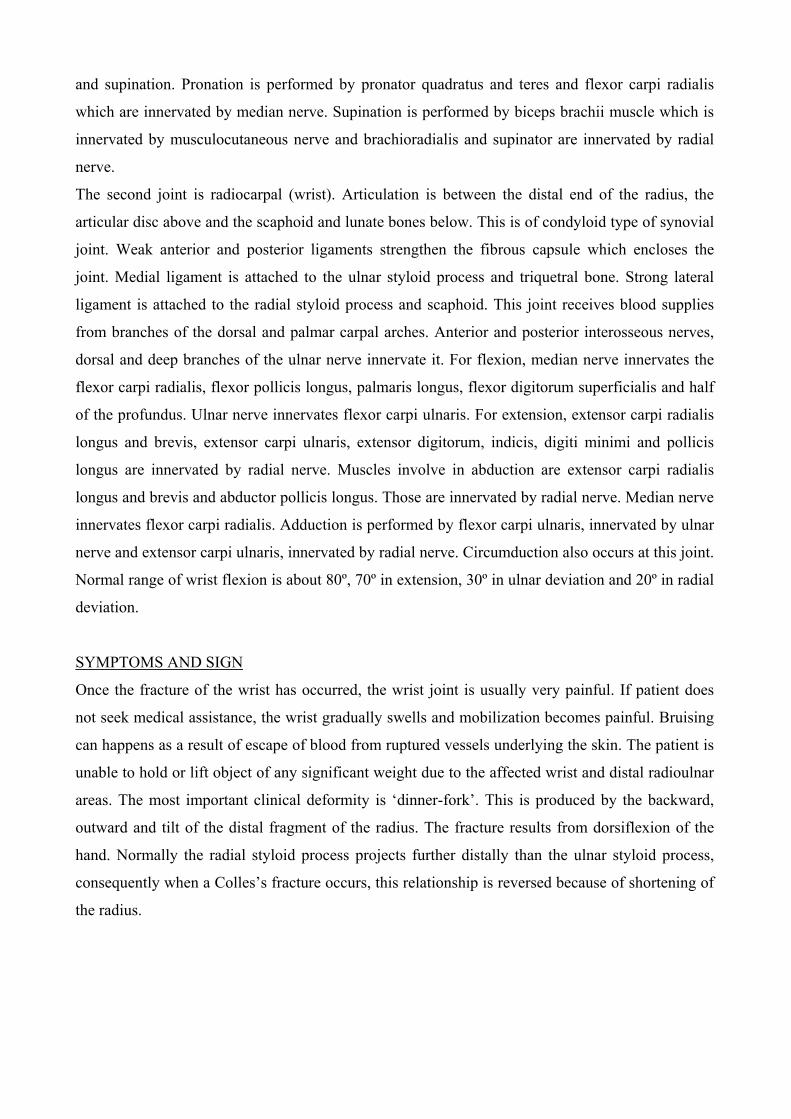
Colles’ fracture (From http://orthoinfo.aaos.org/fact/thr_report.cfm)
CAUSES
Colles’ fracture is an injury that is usually the result of trauma from a fall in which the person
attempts to break the fall using the hands and arms. An example of this is by throwing the hands
forward. The impact of the hand on the ground and the sudden uptake of body weight by the wrist
cause the ends of the radius and/or the ulna to buckle just above the wrist. In relation to the case, the
volleyball player fell on her right outstretched arm during a game, thus causing a Colles’ fracture.
This fracture can also happen due to a direct blow to the wrist. An example of this is a moving
object hitting the radio-ulnar region. Another factor that may contribute to Colles’ fracture is a
severe twist in the wrist, probably caused in a fight.
Colles’ fracture is frequently associated with sports such as rollerblading, skateboarding or any
other activity in which the hands may be called upon to prevent a foreword fall occurring at
relatively high speed. Our patient is a volleyball player and it is obvious that volleyball requires a
great event of motility and an ugly fall occurring in a game is highly probable. Colles’ fracture is
common among children and the elderly. Children’s bones are likely to buckle because they are still
growing and therefore are somewhat soft. Because bones become brittle with age, fractures are
common among the elderly. Osteoporosis increases the risk of a Colles’ fracture occurring due to
the fragility of the bones.
PHYSICAL EXAMINATION
The physician will firstly check for a deformity. In this case, the orthopedic surgeon has described
the deformity in her right wrist as similar to a “dinner fork”. X-ray seen from the lateral view shows
the dinner fork deformity clearly. It also shows the distal end of the radius that has tilted backwards
and radially. The physician will then check if there is tenderness in the distal radio-ulnar region. In
our case, the patient has already reported an immediate pain in her wrist. Furthermore, the physician
will also examine if there is any swelling in the area. Following this is to test the ability of the
patient to grip, whether there is any or none at all. If the patient is able to grip, a further check-up of
the grip strength will be carried out. Lastly, the physician will examine if there are any range of
movements and to what degree. However, in our case, all movements of the patient’s wrist is
painful, hence no movements are possible.
The test that is often carried out to confirm the Colles’ fracture and the exact position of the fracture
is X-ray. X-ray seen anteriorly shows a mild increase in density on the top side of the bone with a
slight irregularity in the surface rather than a smooth line and the radius becomes shortened. Severe
injuries will show evidence of a fracture through the entire bone. Although X-ray is the most
common method used to pinpoint the exact location of a fracture, MRI and CT scan also play
significant roles. MRI is used to detect hidden scaphoid fractures and CT scan is mainly for
detecting unusual small fractures of the wrist bone.
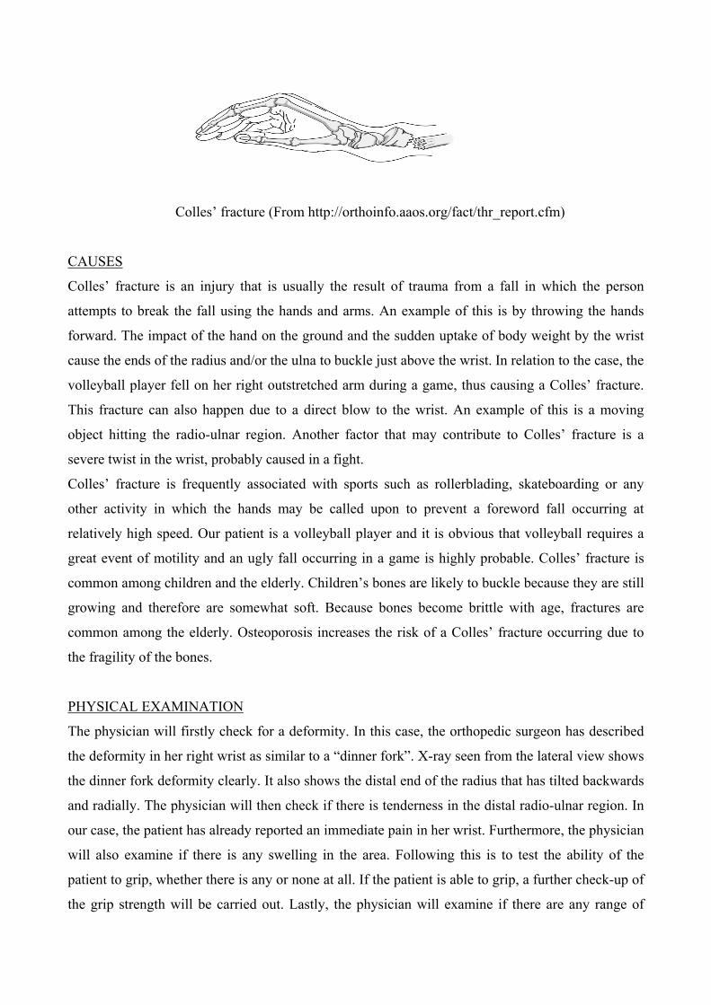
Wrist fracture (X-ray) seen from the side view to show "dinner fork" deformity
of Colle’s fracture
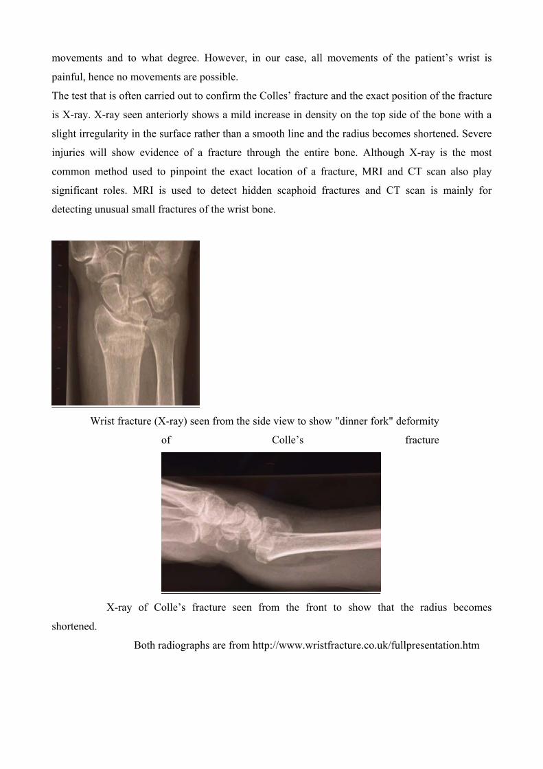
X-ray of Colle’s fracture seen from the front to show that the radius becomes
shortened.
Both radiographs are from http://www.wristfracture.co.uk/fullpresentation.htm
DIFFRENTIAL DAIGNOSIS
There are three types of bone fracture in the wrist that is related to Colles’ fracture, which are
Smith’s fracture, Barton’s fracture and Chauffeur’s fracture. Smith’s fracture is the fracture of the
distal end of radius with volar displacement (facing the palm) and angulation. It is due to backward
fall on palm of an outstretched hand causing the pronation of upper extremity while hands are fixed
to ground. Barton’s fracture is the fracture of distal radius associated with the dislocation of carpus.
The fractured distal radius is displaced either volarly or dorsally along with distal carpus. This is
different from Smith's or Colles' fracture in that clinically and on radiographs, the dislocation is the
most obvious abnormality, with the radial fracture noted secondarily. Next, Chauffeur’s fracture is
the fracture of the distal radial styloid commonly seen in the early 1900s when a car backfired on
starting, causing the crank to hit the distal radius.
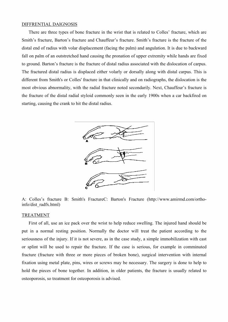
A: Colles’s fracture B: Smith's FractureC: Barton's Fracture (http://www.amirmd.com/ortho-info/dist_radfx.html)
TREATMENT
First of all, use an ice pack over the wrist to help reduce swelling. The injured hand should be
put in a normal resting position. Normally the doctor will treat the patient according to the
seriousness of the injury. If it is not severe, as in the case study, a simple immobilization with cast
or splint will be used to repair the fracture. If the case is serious, for example in comminuted
fracture (fracture with three or more pieces of broken bone), surgical intervention with internal
fixation using metal plate, pins, wires or screws may be necessary. The surgery is done to help to
hold the pieces of bone together. In addition, in older patients, the fracture is usually related to
osteoporosis, so treatment for osteoporosis is advised.
From the case study, we know that the fractured forearm was put in a cast for 6-8 weeks. After
that period, the patient is advised to undergo physical therapy to improve her movement around her
wrist.
COMPLICATION
Regardless of treatment, recovery takes a surprisingly long time, between six to twelve months
is typical. Pain, fatigability, and loss of grip strength are a nuisance in about half of people with this
type of injury. Older people with Colles' fractures often fail to regain full mobility of the wrist joint.
Chronic pain may result from injury to the ligaments or the joint surface of the wrist. Wrist arthritis
may occur due to the cartilage injury at the time of break or wear and tear from changes in the join
alignment after the bone is healed. Carpal tunnel syndrome may also occur as a late complication of
the injury. This syndrome, which causes numbness and tingling sensation in fingertips, is due to the
compression of median nerve. Last but not least, there will be a change in the contour of the back of
the wrist due to the bone healing in a tipped back position.
Case Study 6:
Carpal Tunnel Syndrome
Presented by: Tara Rigney, Presented on: Wednesday, October 20th 2004
Deirdre Kelly, Submitted: Friday October 22nd 2004
Deirdre Nally.
CARPAL TUNNEL SYNDROME
Carpal Tunnel syndrome is the most common entrapment mononeuropathy caused by compression
of the median nerve as it passes through the fibro osseous tunnel beneath the flexor retinaculum.
The carpal tunnel has both bony and fibrous components. The carpus is the collective term
for the 8 bones of the wrist and the concave curvature to the anterior of these bones constitutes the
carpal groove. The carpal groove is converted into a tunnel by the flexor retinaculum, a fibrous
band of fascia which attaches to the trapezium laterally and the pisiform and hook of the hamate
medially. In the wrist, the flexor retinaculum strengthens the carpus and augments flexor efficiency.
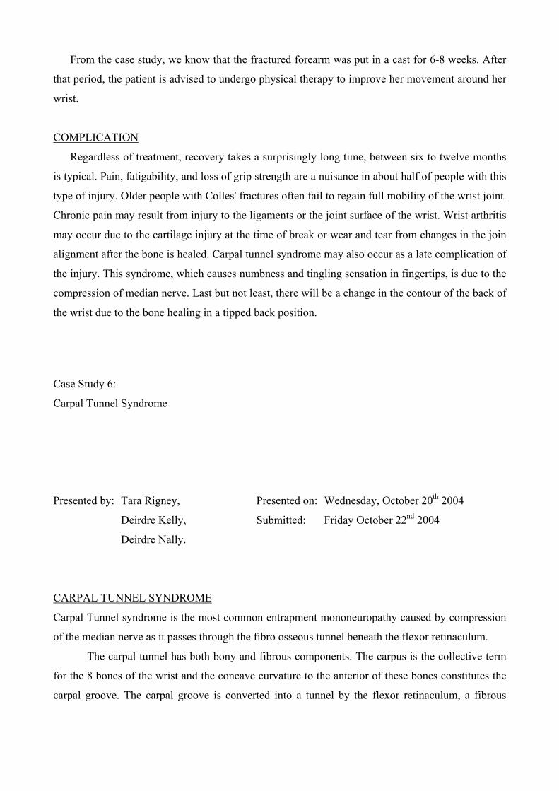
The tendons of the flexor muscles of on the anterior surface of the forearm pass through the
carpal tunnel to take attachment within the hand. Some tendons are enclosed in synovial sheaths to
reduce friction when these tendons contract: The flexor pollicis longus is surrounded by the radial
bursa and both the flexor digitorum superficialis and flexor digigtorum profundus are enclosed by
the ulnar bursa. The median nerve also enters the hand through this passageway. A terminal cord of
the brachial plexus, this nerve is formed in the axilla by the union of anterior divisions of C5 and
C6. Near the wrist, this nerve becomes superficial and in the carpal tunnel it lies lateral to the two
superficial tendons of the flexor digitorum superficialis, against the deep surface of the flexor
retiniculum.
Thus, the median nerve lies adjacent to flexor tendons in a limited diameter tunnel. Any
condition that causes a swelling of tissue or a change in position of tissue within the tunnel
generates pressure and is likely to irritate the median nerve producing symptoms similar to those
experienced by the dentist.
Symptoms:
Hypesthesia and parathesia, or diminished sensibility and a tingling sensation are explained
by the distribution of sensory nerve fibres of the median nerve within the hand. Essentially, the
median nerve sends cutaneous sensory fibres to the lateral three and a half digits. Sensory deficits
are therefore, localised in the thumb, index, middle and lateral side of the dentist’s ring finger.
Hypesthesia is demonstrated during clinical examinations by an impaired appreciation of light touch
and pin pricks.
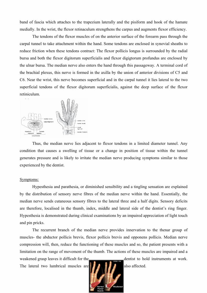
The recurrent branch of the median nerve provides innervation to the thenar group of
muscles- the abductor pollicis brevis, flexor pollicis brevis and opponens pollicis. Median nerve
compression will, then, reduce the functioning of these muscles and so, the patient presents with a
limitation on the range of movement of the thumb. The actions of these muscles are impaired and a
weakened grasp leaves it difficult for the dentist to hold instruments at work.
The lateral two lumbrical muscles are also affected.
Patients with Carpal tunnel syndrome frequently experience a more intense sensation at night, and
the dentist is no exception. This is related to the flexed wrist sleeping position and fluid
accumulation around the wrist while lying flat.
Over working tends to aggravate the condition since repeated and prolonged performance of
fine movements of the hand, as is required of dentists, can cause tendon inflammation. The tendon
bursae swell and since there is little room for expansion, the median nerve is affected, accentuating
our patient’s discomfort.
Palmar sensation however is unaffected. There is no sensory impairment of the palm since
the palmar cutaneous nerve arises from the median nerve proximal to the flexor retinaculum and is,
therefore untroubled by the lesion within the carpal tunnel.
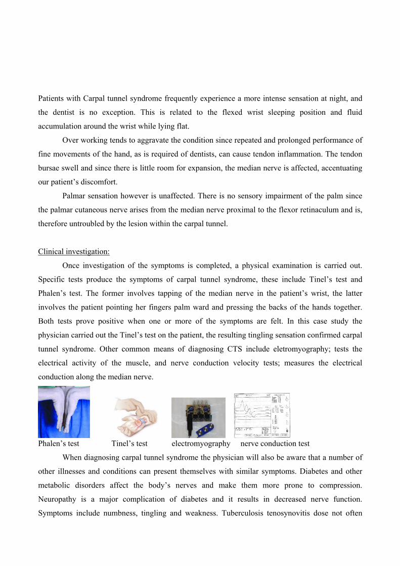
Clinical investigation:
Once investigation of the symptoms is completed, a physical examination is carried out.
Specific tests produce the symptoms of carpal tunnel syndrome, these include Tinel’s test and
Phalen’s test. The former involves tapping of the median nerve in the patient’s wrist, the latter
involves the patient pointing her fingers palm ward and pressing the backs of the hands together.
Both tests prove positive when one or more of the symptoms are felt. In this case study the
physician carried out the Tinel’s test on the patient, the resulting tingling sensation confirmed carpal
tunnel syndrome. Other common means of diagnosing CTS include eletromyography; tests the
electrical activity of the muscle, and nerve conduction velocity tests; measures the electrical
conduction along the median nerve.
Phalen’s test Tinel’s test electromyography nerve conduction test
When diagnosing carpal tunnel syndrome the physician will also be aware that a number of
other illnesses and conditions can present themselves with similar symptoms. Diabetes and other
metabolic disorders affect the body’s nerves and make them more prone to compression.
Neuropathy is a major complication of diabetes and it results in decreased nerve function.
Symptoms include numbness, tingling and weakness. Tuberculosis tenosynovitis dose not often
present itself as CTS but it should be kept in mind. In almost all cases the diagnosis is made post-op
after suspicious tenosynovial material was sent for and confirmed with biopsy. With auto-immunal
diseases the body’s system attacks its own tissue causing widespread inflammation, including in
many cases, the carpal tunnel of the hand. With regard to obesity, greater body mass appears to
reduce nerve flow speed into the hand. Individuals who undergo hemodialysis for chronic renal
damage often experience a build-up of a protein called 2-microglobulin which can also result in
similar symptoms. Patients with COPD (Chronic Obstructive Pulmonary Disease) and other
respiratory illnesses also may experience such symptoms which suggest oxygen supply is important
in nerve function to the hand.
It is apparent from the evidence above that there are a variety of diseases that physicians
must consider during the course of his/her diagnosis. With regard to this particular case study
differential diagnosis can be ruled out as there was no evidence to suggest that any of the above
illnesses was the cause of the patient‘s symptoms. Similarly there were no symptoms found that
would suggest anything other than carpal tunnel syndrome.
Treatment:
The choice of treatment for carpal tunnel syndrome depends on the severity of the symptoms and
any underlying disease which might be causing the symptoms. It is therefore, essential that
metabolic disorders such as diabetes and arthritis are treated first to reduce the susceptibility of
further compression on the median nerve. Initial treatment generally involves resting the affected
hand and wrist for at least two weeks, ideally avoiding activities that may worsen symptoms.
However, if our patient in this case cannot afford to take time off work, it would be recommend that
she wear a splint at night to immobilize the wrist, thus avoiding further damage from twisting or
bending. Physical therapy may also reduce symptoms. If there is no effect after a number of weeks
then our patient can be given a hydrocortisone injection into the carpal tunnel which may give
relief. Hydrocortisone is an anti-inflammatory steroid which, when injected into the carpal tunnel,
can relieve pressure on the median nerve and provide immediate, temporary relief. If there is no
change after 6 months then surgery is be required. Surgery basically involves severing the band of
tissue around the wrist to reduce pressure on the median nerve. There are two types of surgery:
• Open release surgery; consists of making an incision up to two inches in the wrist and then
cutting the carpal ligament to enlarge the carpal tunnel.
• Endoscopic surgery; whereby the surgeon makes two incisions, about ½ inches each, in the
wrist and palm, and using a camera cuts the carpal ligament. This is a more effective
technique as it reduces scarring and speeds up healing time.
Although symptoms may be relieved immediately after surgery, full recovery from carpal tunnel
syndrome can take months. Some patients may experience stiffness, nerve damage, pain at the scar
and occasionally the wrist loses strength because the carpal ligament is cut.
Long Term Effects:
The dominant hand is usually affected first, perhaps because it is used more frequently and more
vigorously. It is noticed that in CTS there is impaired sensation of 1st, 2nd, 3rd, and median side of
the 4th digit. Wasting and weakness of the Abductor Pollicis Breveis also occurs. The palmar
branch is spared since it does not pass through the carpal tunnel.
Salient Features:
Carpal tunnel syndrome is also called tardy median nerve palsy. It occurs most often in patients
aged between thirty and sixty years old. Women are five times more likely than men to develop
CTS, probably because the carpal tunnel is smaller in women. Also at risk are those who suffer
from metabolic disorders, such as hypothyroidism, over activity of the pituitary gland, diabetes and
rheumatoid arthritis (disorders that directly affect the body’s nerves and make them
more susceptible to compression). The risk of developing CTS is especially common in assembly-
line workers, office workers and others like our patient here where there is occupational strain and
overexertion of the hand and wrist. Carpal tunnel syndrome is often the result of a combination of
factors that increase pressure on the median nerve and tendons in the carpal tunnel, rather than a
problem with the nerve itself. One main cause of carpal tunnel syndrome is congenital
predisposition, where a person is simply born with a smaller carpal tunnel. Other factors include
injury to the wrist such as a sprain or fracture and fluid retention during pregnancy and menopause,
where there is increased swelling of the carpal tunnel and increased pressure on the median nerve.
The development of a tumour or cyst will also cause CTS as will trauma by repetitive hand
movements especially in patients whose work requires forceful finger and wrist flexion and
extension as in the case of our patient here.
References:
Clinically Oriented Anatomy (3rd edition), Keith L. Moore.
Grays Anatomy (38th edition), Henry Gray.
Lachman’s Case studies in Anatomy (4th edition), Donald R. Cahill.
Campall’s Operative Orthopaedics (8th edition), A.H. Crenshaw.
Peripheral Neuropathy (4th edition), Peter Dyck.
www.ninds.com www.medicinenet.com
www.bupa.co.uk www.arthroscopy.com
www.carpaltunnel.upmc.com www.neurologychannel.com
www.centerforcarpaltunnel.umpc.com
Pictures:
www.bupa.co.uk www.emedicine.com
Case 7- Part 2: Symptoms & Diagnosis By Seán King. The scaphoid is the most commonly fractured bone of the carpus: it accounts for 60% of carpal fractures. As the patient in this case reports pain in the region of the anatomical snuffbox, it is almost certainly his scaphoid that he’s fractured. Scaphoid fracture usually occurs in men of ages 20 years to 40 years, and is caused by a fall on an outstretched hand, so it’s a common sports injury, and also often results from a car accident. As one fall, one instinctively puts out a hand to break one’s fall: landing on this hand damages the wrist. The only fracture of the wrist more common than scaphoid fracture is Colle’s fracture, and it is the angle at which the wrist is bent that deter- -mines the nature of the injury. As a general rule, if the wrist is at an angle of less than 90 degrees, the radius breaks; if the wrist at an angle of ninety degrees or greater, the scaphoid breaks. It’s usually the middle or lower portion of the bone that’s fractured. In this case, the patient injured his wrist when he was thrown from a horse, but the scaphoid is often fractured in rollerblading or skateboarding accidents. Wrist guards are worn to prevent against it. Symptoms The symptoms of scaphoid fracture are: -Pain and tenderness on the thumb side of the wrist. -Motion may be painful (especially gripping). -There may be some swelling on the back and thumb side of the wrist. -The pain may subside and return as a deep, dull ache. -There is marked tenderness to pressure on the anatomical snuffbox. These symptoms are very similar to those of a sprain, which is simply a tearing of the ligaments, and- especially because scaphoid fracture causes no obvious deformity, and very little swelling- scaphoid fracture is often mistaken for a sprain. (In this case, the patient was diagnosed as having a sprain when he first went to the A&E). It’s important to distinguish a scaphoid fracture from a sprain as it’s a much more serious injury; a physician should treat a wrist injury as a fracture until a diagnosis is confirmed. Diagnosis It’s difficult to diagnose a scaphoid fracture because, unless the fracture is displaced (i.e., unless the two pieces of bone no longer even touch each other), the fracture will not be obvious on the first set of X-rays. The injury cannot be distinguished from a sprain on the basis of an interview and a physical examination. A second set of X-rays, taken a week to ten days after the injury should verify whether or not it is indeed a scaphoid fracture, as healing will have begun, making the fracture visible.
Other imaging techniques, such as MRI or a CT scan, may be used to evaluate the fracture for surgical treatment, but this is rare.
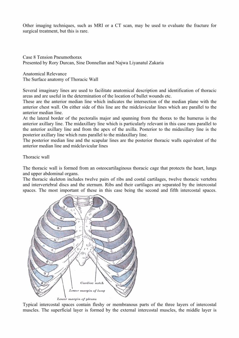
Case 8 Tension Pneumothorax Presented by Rory Durcan, Sine Donnellan and Najwa Liyanatul Zakaria Anatomical Relevance The Surface anatomy of Thoracic Wall Several imaginary lines are used to facilitate anatomical description and identification of thoracic areas and are useful in the determination of the location of bullet wounds etc. These are the anterior median line which indicates the intersection of the median plane with the anterior chest wall. On either side of this line are the midclavicular lines which are parallel to the anterior median line. At the lateral border of the pectoralis major and spanning from the thorax to the humerus is the anterior axillary line. The midaxillary line which is particularly relevant in this case runs parallel to the anterior axillary line and from the apex of the axilla. Posterior to the midaxillary line is the posterior axillary line which runs parallel to the midaxillary line. The posterior median line and the scapular lines are the posterior thoracic walls equivalent of the anterior median line and midclavicular lines Thoracic wall The thoracic wall is formed from an osteocartilaginous thoracic cage that protects the heart, lungs and upper abdominal organs. The thoracic skeleton includes twelve pairs of ribs and costal cartilages, twelve thoracic vertebra and intervertebral discs and the sternum. Ribs and their cartilages are separated by the intercostal spaces. The most important of these in this case being the second and fifth intercostal spaces.
Typical intercostal spaces contain fleshy or membranous parts of the three layers of intercostal muscles. The superficial layer is formed by the external intercostal muscles, the middle layer is
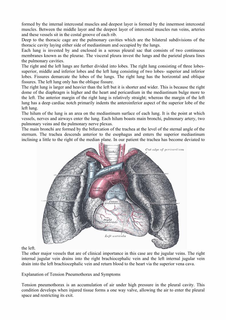
formed by the internal intercostal muscles and deepest layer is formed by the innermost intercostal muscles. Between the middle layer and the deepest layer of intercostal muscles run veins, arteries and these vessels sit in the costal groove of each rib. Deep to the thoracic cage are the pulmonary cavities which are the bilateral subdivisions of the thoracic cavity laying either side of mediastinum and occupied by the lungs. Each lung is invested by and enclosed in a serous pleural sac that consists of two continuous membranes known as the pleurae. The visceral pleura invest the lungs and the parietal pleura lines the pulmonary cavities. The right and the left lungs are further divided into lobes. The right lung consisting of three lobes- superior, middle and inferior lobes and the left lung consisting of two lobes- superior and inferior lobes. Fissures demarcate the lobes of the lungs. The right lung has the horizontal and oblique fissures. The left lung only has the oblique fissure. The right lung is larger and heavier than the left but it is shorter and wider. This is because the right dome of the diaphragm is higher and the heart and pericardium in the mediastinum bulge more to the left. The anterior margin of the right lung is relatively straight; whereas the margin of the left lung has a deep cardiac notch primarily indents the anteroinferior aspect of the superior lobe of the left lung. The hilum of the lung is an area on the mediastinum surface of each lung. It is the point at which vessels, nerves and airways enter the lung. Each hilum boasts main bronchi, pulmonary artery, two pulmonary veins and the pulmonary nerve plexus. The main bronchi are formed by the bifurcation of the trachea at the level of the sternal angle of the sternum. The trachea descends anterior to the esophagus and enters the superior mediastinum inclining a little to the right of the median plane. In our patient the trachea has become deviated to
the left. The other major vessels that are of clinical importance in this case are the jugular veins. The right internal jugular vein drains into the right brachiocephalic vein and the left internal jugular vein drain into the left brachiocephalic vein and return blood to the heart via the superior vena cava. Explanation of Tension Pneumothorax and Symptoms Tension pneumothorax is an accumulation of air under high pressure in the pleural cavity. This condition develops when injured tissue forms a one way valve, allowing the air to enter the pleural space and restricting its exit.
As we can see from this case a penetrating trauma such as a gunshot wound or a stabbing can lead to a tension pneumothorax. It may also begin as a simple pneumothorax and develops to become tension pneumothorax. Simple pneumothorax is referred to as a collapsed lung and occurs when air escapes from lung or else leaks through a cavity. People who are placed on ventilators and people who have had cardiopulmonary resuscitation performed on them are also prone to developing tension pneumothorax. Clinical interpretation of the presenting signs is crucial for immediate diagnosis and treatment is essential at early stages. This patient presented with a low B.P. 100mg compared with normal value of 200mg. As pressure is increasing with every breath he takes it results in the collapse of the lung on the right hand side. The lack of a breathing sound on the right indicates this. This allows the heart, thoracic aorta, vena cava and great vessels to be pulled toward opposite side. The trachea deviates away from the right lung and the jugular veins become distended. When the lung collapses there is a loss of ventilation capability to the affected lung. Therefore oxygen transfer to the lung becomes compromised. Physiologic shunting leads to hypoxia and subsequently to hypoxemia. The mediastinum shift may cause compression of opposite lung and it may also affect the flow of blood returning to the heart. This causes blood to pool in capacitance vessels following this cardiovascular collapse and death may occur unless treatment is given immediately. The palpable creptius which the patient is noted to have may be the result of chest and lung auscultation which leads to a crunching sound that is actually synchronous with systole. This sound is caused by air bubbles in subcutaneous tissue. Diagnosis If tension pneumothorax is suspected, treatment should not be delayed to get an x-ray, as it would prove life threatening to the patient. Tension pneumothorax is not an overly common condition in hospitals but it is vital that doctors are vigilant as it requires urgent diagnosis and treatment. Treatment of Tension Pneumothorax There are two ways of treatment, the first one is needle thoracocentesis and it is followed by tube thoracostomy or sometimes called chest tube insertion. Treatment should start before diagnostic tests are confirmed. If treatment is delayed the situation might become worse. Goal of these two treatments is to alleviate the high interpleural pressure. This is most quickly accomplished by performing the needle thoracocentesis. So in this case, we used the 14 gauge needle to be inserted in the right midclavicular line at the second intercostal space. The second intercostal space is located relatively superior compared to other intercostal spaces; it is easier for the air to escape through the tube since it is very light so it will move upward. However, if water accumulates in the pleural cavity instead of air, the needle will be inserted at the lower intercostal space in hydropneumothorax case. In an emergency condition, needle thoracocentesis is an immediate treatment and followed by chest tube insertion if tension pneumothorax recur or if it persistent. The needle is inserted into the skin superior to the rib into the second intercostal space after the patient has been anesthetised. The reason that the needle is inserted superior to the rib is to avoid any damage to veins, arteries and nerves at the costal groove located at the inferior border of the rib. This needle then punctures the parietal pleura. When the needle is removed from the catheter air escaping sound can be heard which tell us that air is being sucked out of the pleural cavity. It is very important to make a correct diagnosis and be sure that the patient really has pneumothorax, because if he is not and needle thoracocentesis are done on them, pneumothorax and lung damaged may occur as a result of puncturing their pleural cavity. One main risk using this treatment is injury to the vessels, at the inferior border of the rib which can lead to hemothorax. Hemothorax is an accumulation of blood in the pleural cavity.
The chest tube insertion is a definitive treatment for tension pneumothorax. The aim of this treatment is to remove air from the pleural space and allow lung to re-expand. A flexible chest tube is inserted into the fifth intercostal space. Insertion of the chest tube at the fifth intercostal space is to avoid possible damage to both intraabdominal and intrathoracic organ. It is important to insert your finger in the incision site on the chest. The other end of the chest tube is connected to a bottle or canister or known as chest drainage device to encourage rapid drainage. The chest tube passes through skin, fat, external intercostal muscle, internal intercostal muscle, endothoracic fascia and parietal pleura. The chest tube remains in place until the x-rays shows that all air has drained out of the chest and the lung has re-expanded. In this case, the patient was taped with an occlusive dressing, this is done to close the hole made by the bullet to prevent air from being sucked into the pleural cavity or forming the one way valve. Usually almost every patient receives 100% oxygen through a face mask. Once the tension pneumothorax is treated, the collapsed lung will return to normal size within 48 to 72 hours. Recovering from collapsed lung may take up to several weeks. Surgery is needed if lung does not re-expand after 5 days or if there is possible air leaking from the tube for more than 10 days. Once tension pneumothorax has healed, there is usually no long term effect. But up to 50% of people have another pneumothorax. To decrease the risk, the patient is advised to avoid a change in air pressure such as climbing and scuba driving. References www.allreferhealth.comImages taken from, Henry Gray 1918 Anatomy of the Human Body. www.bartleby.com/107/Essential Clinical Anatomy by Moore & Agur CASE 12 ALIAS, NOR AZALINA AYOB, ZURITA AZRINA ABDUL SHOKRI, AHMAD FAIRUZ The esophagus is a muscular tube approximately 25 cm long with an average diameter of 2 cm that extends from the pharynx to the stomach. The esophagus normally has four constrictions where adjacent structures produce impression. The first one is at its beginning, approximately 15 cm from the incisor teeth and caused by the cricopharyngeus muscle (upper esophageal sphincter), second is where it is crossed by the arch of aorta, 22.5 cm from the incisor teeth. While the third one is where it is crossed by the left main bronchus, 27.5 cm from the incisor teeth and the last is where it passes through the diaphragm, approximately 40 cm from the incisor teeth (lower esophageal sphinter. The short abdominal part of the esophagus extends from the diaphragm to the cardia (cardiac) orifice or the stomach.
The function of the esophagus is to convey food from the pharynx to stomach. The
esophagus follows the curve of the vertebral column as it descends through the neck and mediastinum and it passes through the elliptical esophageal hiatus in the muscular right crus of the diaphragm, just to the left of the median plane at the level of T10 vertebra. The esophagus terminates when it enters the stomach at the cardial orifice of the stomach to the left of the midline at the level of the 7th left costal cartilage and T12 vertebra.
The esophagus has internal circular and external longitudinal layers of muscle. In its
superior third, the external layer consist of skeletal muscle; the inferior third is composed of smooth muscle, and the middle third is made up of both types of muscle. Food passes rapidly through the esophagus rapidly because of the peristaltic of its musculature. The esophagus is attached to the margins of the esophageal hiatus in the diaphragm by the phrenicoesophageal ligament an extension of inferior diaphragmatic fascia. This ligament permits independent movement of the diaphragm and esophagus during respiration and swallowing.
The short, trumpet-shaped abdominal part of the esophagus-approximately 1.25 cm long-
passes from the esophageal hiatus in the right crus of the diaphragm to the stomach. The right border of the esophagus is continuous with the lesser curvature of the stomach; however its left border is separated from the fundus of the stomach by the cardial notch. The esophagogastric junction lies to the left of T11 vertebra on the horizontal plane that passes through to the tip of the xiphoid process. At this junction, the diaphragmatic musculature forming the esophageal hiatus function as a physiological esophageal sphincter that contracts and relaxes. Radiological studies show that food stops here momentarily and that the sphincter mechanism is normally efficient in preventing reflux of gastric contents into the esophagus. When one is not eating, the lumen is normally collapsed above this level to prevent food or stomach juices from regurgitating into the esophagus.
The stomach is the expanded part of the digestive tract between the esophagus and small
intestine. It acts as a food blender and reservoir; its chief function is enzymatic digestion. There are four parts of the stomach which are cardia, fundus, body and pyloric part. Cardia is the part surrounding the cardial orifice. Fundus is the dilated superior part that is related to the left dome of the diaphragm and is limited inferiorly by the horizontal plane of the cardial orifice. The superior part of the fundus usually reaches the level of the left 5th intercostals spaces. The cardial notch is between the esophagus and fundus. Body lies between the fundus and the pyloric antrum while the pyloric part is the funnel-shaped region of the stomach.
Pyrosis (heartburn) is the most common type of esophageal discomfort or substernal pain.
This burning sensation in the abdominal part of the esophagus is often accompanied by regurgitation of small amount of foods or gastric fluid into the esophagus. Pyrosis may be associated with hiatus hernia. Hiatus hernia is a protrusion of a part of the stomach into the mediastinum through th esophageal hiatus of the diaphragm.
There are two main types of hiatal hernia; sliding hiatus hernia and paraesophageal hiatus
hernia. In sliding hiatus hernia, the abdominal part of the esophagus, the cardia, and parts of the fundus of the stomach slide superiorly through the esophageal hiatus into the thorax, especially when the person lies down or bends over. Some regurgitation of stomach contents into the esophagus is possible because the clamping action of the right crus of the diaphragm on the inferior end of the esophagus is weak. In the less common paraesophageal hiatus hernia, the cardia remains in its normal position; however a pouch of peritoneum, often containing part of the fundus, extends through the esophageal hiatus anterior to the esophagus. In this case, usually no regurgitation of gastric contents occurs because the cardial orifice is in its normal position.
The cause of hiatus hernia may include poor muscle tone around the hiatus, increase fat
pushing open the hiatus and increase pressure in the abdomen pushing the stomach upwards through the hiatus.
Hiatus hernia usually does not show any symptoms, except when it has become large. The patient will usually experience an uncomfortable feeling of burning behind the breastbone which is
known as heartburn. It happens due to the reflux of a small amount of acid into the esophagus. Acid reflux occur more common after meals hence the patient will usually feel the heartburn after meal. Lying down and bending forward make the condition even worst because without the effect of gravity, the reflux can occur easier and the acid will return to the stomach slower. Next is chest pain that is felt anywhere along the front of the body between the neck and the upper abdomen. The pain spread from below the sternum to the neck, jaw and to the arms. The patient may also experience shortness in breath. The chest pain is caused by the twisting of part of the stomach which restricts blood flow to the stomach. Next symptom is difficulty in swallowing due to the inflammation of a part of the esophagus caused by the acid reflux. As a result, the tract become scarred and narrowed, hence make it difficult for the food to pass through into the stomach. Hiatus hernia will also cause the patient to belch up sour substance due to the reflux of the acid into the esophagus. Other than that, the patient will also have bloating, a condition when the abdomen feels full and tight which is caused by the twisting of part of the stomach that protrudes into the chest cavity. The backflow of the stomach acid can cause sour or bitter taste in the patient mouth. They also will have the feeling of food stuck in their esophagus. Some patient will experience regurgitate where some stomach content back flow into the throat.
There are several other conditions that show almost similar symptoms as the hiatus hernia such as esophageal spasm which involve irregular contractions of the muscle in the esophagus. The spasm does not propel food effectively into the stomach. Next is heart attack; pain as pressure, squeezing and tightness in the chest that radiates to the jaw, left arm or between the shoulder blades in the back. Other than that is angina which is the pain felt in chest and sometimes upper abdomen due to the temporary shortage of oxygen supply to the heart muscle. Indigestion; a pain in the upper abdomen or chest usually after meals also shares the same symptoms as hiatus hernia. Next is the gastroesophageal reflux disease (GERD) which occurs when the esophageal sphincter does not work properly causing the stomach content to leak back into the esophagus. The heartburn that is felt by our patient here may also caused by the side effect of certain drugs such as calcium channel blockers for high blood pressure or bisphosphonates for osteoporosis.
There are two tests that can be done to diagnose hiatus hernia. The first one is the Barium X-ray where the patient is asked to drink liquid containing barium which coat the lining of internal organs and outline the esophagus, stomach and the upper part of the small intestine so that they will shows up more clearly on the X-ray picture. The hiatus hernia will appear as a separate sac lying between the esophagus and the stomach. The sac is delineated by the lower esophageal sphincter above and diaphragm below. Next is the endoscopy or the gastrocopy. The patient will be given a sedative first. Then, the doctor will insert a thin flexible lighted tube called an endoscope down into the esophagus. The endoscope allows the doctor to look at the esophagus and check for abnormalities. Hiatus hernia will shows up as a bulge that is positioned between the esophagus and the stomach.
Complications resulting from untreated paraesophageal hiatal hernia is incarceration of the hernial sac which would eventually lead to the strangulation of the sac. The perforation of the sac would cause gastrointestinal haemorrhage. Meanwhile, untreated GERD will result into esophagitis as the esophagus becomes inflamed and irritated and this would then cause difficulty and pain to the individual when swallowing food. Apart from that, the damaged lining of the esophagus could be come thick and hardened, causing strictures which might interfere with drinking and eating as the strictures may prevent solids and liquids from reaching the stomach. The constant irritation would also cause the cells that line the esophagus to change from simple squamous to specialized columnar cell. This condition is referred to as the Barrett’s esophagus and individuals with this condition have a much higher tendency to develop cancer of the esophagus in comparison with normal individuals.
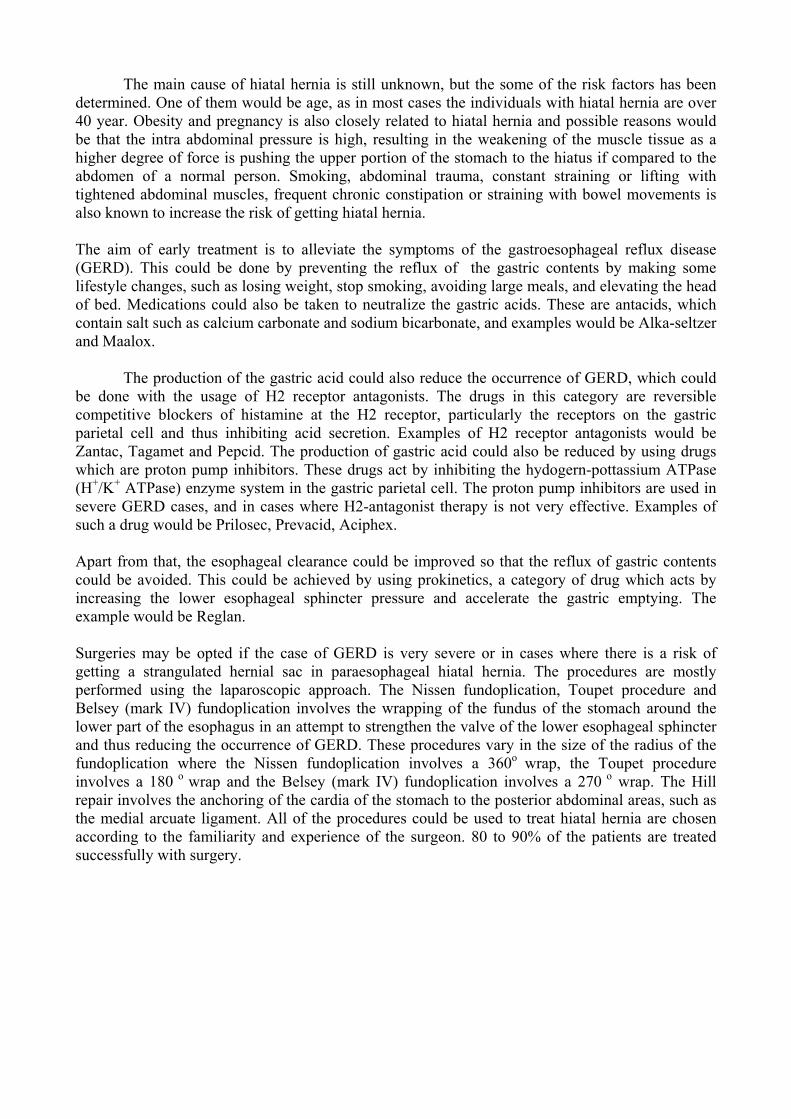
The main cause of hiatal hernia is still unknown, but the some of the risk factors has been determined. One of them would be age, as in most cases the individuals with hiatal hernia are over 40 year. Obesity and pregnancy is also closely related to hiatal hernia and possible reasons would be that the intra abdominal pressure is high, resulting in the weakening of the muscle tissue as a higher degree of force is pushing the upper portion of the stomach to the hiatus if compared to the abdomen of a normal person. Smoking, abdominal trauma, constant straining or lifting with tightened abdominal muscles, frequent chronic constipation or straining with bowel movements is also known to increase the risk of getting hiatal hernia. The aim of early treatment is to alleviate the symptoms of the gastroesophageal reflux disease (GERD). This could be done by preventing the reflux of the gastric contents by making some lifestyle changes, such as losing weight, stop smoking, avoiding large meals, and elevating the head of bed. Medications could also be taken to neutralize the gastric acids. These are antacids, which contain salt such as calcium carbonate and sodium bicarbonate, and examples would be Alka-seltzer and Maalox.
The production of the gastric acid could also reduce the occurrence of GERD, which could be done with the usage of H2 receptor antagonists. The drugs in this category are reversible competitive blockers of histamine at the H2 receptor, particularly the receptors on the gastric parietal cell and thus inhibiting acid secretion. Examples of H2 receptor antagonists would be Zantac, Tagamet and Pepcid. The production of gastric acid could also be reduced by using drugs which are proton pump inhibitors. These drugs act by inhibiting the hydogern-pottassium ATPase (H+/K+ ATPase) enzyme system in the gastric parietal cell. The proton pump inhibitors are used in severe GERD cases, and in cases where H2-antagonist therapy is not very effective. Examples of such a drug would be Prilosec, Prevacid, Aciphex. Apart from that, the esophageal clearance could be improved so that the reflux of gastric contents could be avoided. This could be achieved by using prokinetics, a category of drug which acts by increasing the lower esophageal sphincter pressure and accelerate the gastric emptying. The example would be Reglan. Surgeries may be opted if the case of GERD is very severe or in cases where there is a risk of getting a strangulated hernial sac in paraesophageal hiatal hernia. The procedures are mostly performed using the laparoscopic approach. The Nissen fundoplication, Toupet procedure and Belsey (mark IV) fundoplication involves the wrapping of the fundus of the stomach around the lower part of the esophagus in an attempt to strengthen the valve of the lower esophageal sphincter and thus reducing the occurrence of GERD. These procedures vary in the size of the radius of the fundoplication where the Nissen fundoplication involves a 360o wrap, the Toupet procedure involves a 180 o wrap and the Belsey (mark IV) fundoplication involves a 270 o wrap. The Hill repair involves the anchoring of the cardia of the stomach to the posterior abdominal areas, such as the medial arcuate ligament. All of the procedures could be used to treat hiatal hernia are chosen according to the familiarity and experience of the surgeon. 80 to 90% of the patients are treated successfully with surgery.
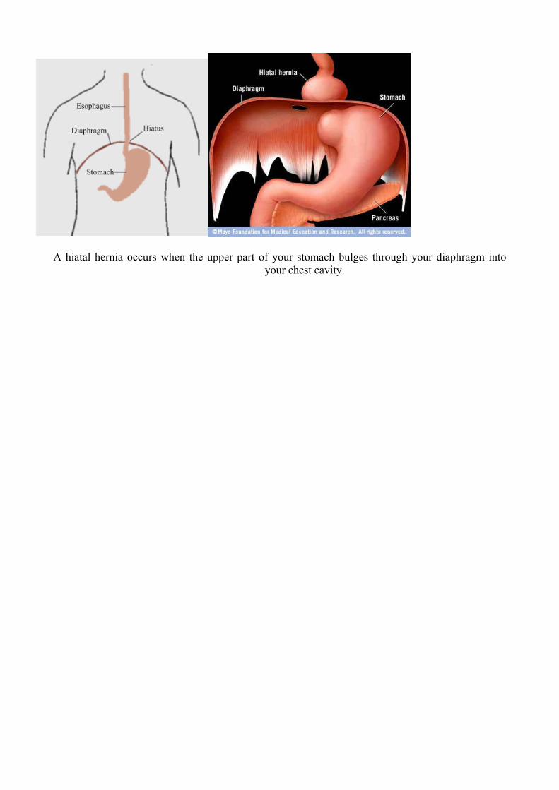
A hiatal hernia occurs when the upper part of your stomach bulges through your diaphragm into
your chest cavity.
Top Related