


Languages
Pages
Legal


Quality Assurance And Quality ControlIn Breast Cancer Screening Programme
Dr. Ruta Grigiene, Dr. Laima Grinyte
2016 10 02 – 10 07

“The greatest need we have today in the human cancer problem, except for a universal cure, is a method of detecting the presence of cancer before there are any clinical signs
of symptoms.”
- Sidney Farber, letter to Etta Rosensohn, November 1962 -(The Emperor of All Maladies, Siddhartha Mukherjee)
Sidney Farber (1903-1973)
Paediatric pathologist and “father” of modern chemotherapy.
The Dana-Farber Cancer Institute in Boston is partly named after him.
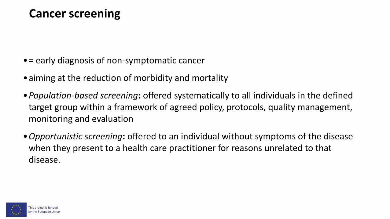
•= early diagnosis of non-symptomatic cancer
•aiming at the reduction of morbidity and mortality
•Population-based screening: offered systematically to all individuals in the defined target group within a framework of agreed policy, protocols, quality management, monitoring and evaluation
•Opportunistic screening: offered to an individual without symptoms of the disease when they present to a health care practitioner for reasons unrelated to that disease.
Cancer screening
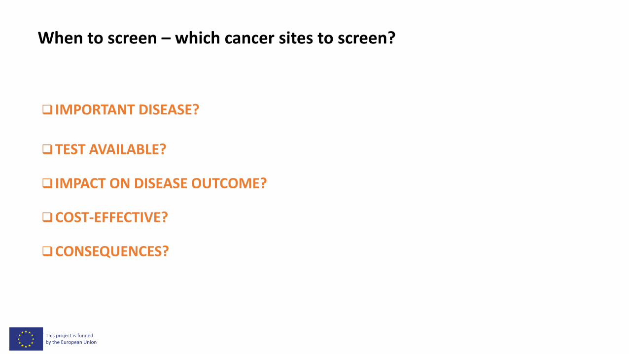
IMPORTANT DISEASE?
TEST AVAILABLE?
IMPACT ON DISEASE OUTCOME?
COST-EFFECTIVE?
CONSEQUENCES?
When to screen – which cancer sites to screen?

When to screen – which cancer sites to screen?
• Important health problem for the general population
•Natural history well known
•Accurate diagnostic assessment
•Effective treatment options
•Earlier treatment improves disease outcome/prognosis
IMPORTANT DISEASE?

When to screen – which cancer sites to screen?
IMPORTANT DISEASE?
Top 10 cancers in European men and women
WSR

When to screen – which cancer sites to screen?
• Acceptable to the population
• Test characteristics
• Cancer process:
• initation – promotion – abnormal growth – invasion – metastases
• symptoms
• diagnosis and treatment
• long interim period - window for screening
SUITABLE TEST?

When to screen – which cancer sites to screen?
Sensitivity:
• Ability of the test to identify positive results
• Proportion of actual positives which are correctly identified as such (i.e. the percentage of people with cancer
who are correctly identified as having cancer)
• TRUE POSITIVE rate
• Never 100%
Specificity
• Ability of the test to identify negative results
• Proportion of negatives which are correctly identified (i.e. the percentage of healthy people who are correctly
identified as not having cancer)
• TRUE NEGATIVE rate
TEST CHARACTERISTICS
When to screen – which cancer sites to screen?
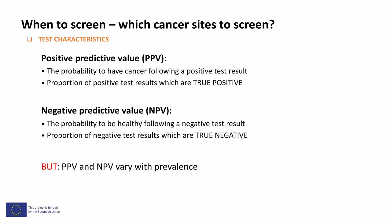
Positive predictive value (PPV):
• The probability to have cancer following a positive test result
• Proportion of positive test results which are TRUE POSITIVE
Negative predictive value (NPV):
• The probability to be healthy following a negative test result
• Proportion of negative test results which are TRUE NEGATIVE
BUT: PPV and NPV vary with prevalence
TEST CHARACTERISTICS
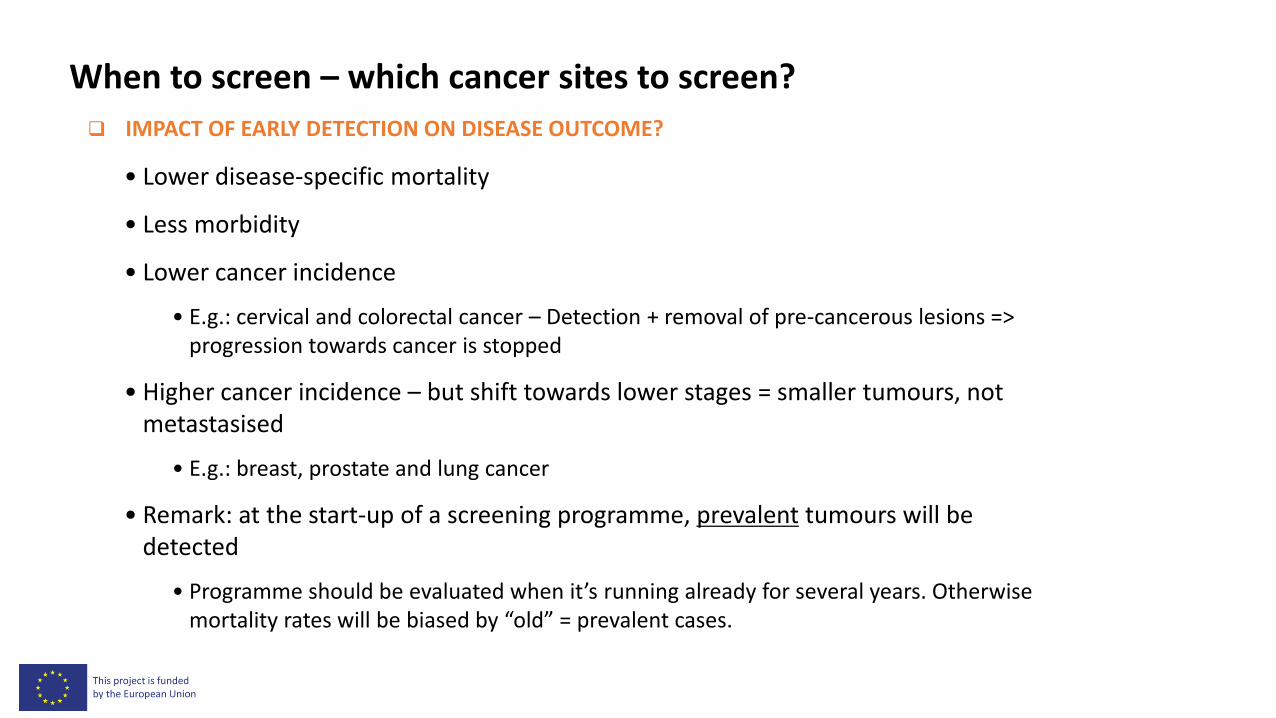
• Lower disease-specific mortality
• Less morbidity
• Lower cancer incidence
• E.g.: cervical and colorectal cancer – Detection + removal of pre-cancerous lesions => progression towards cancer is stopped
• Higher cancer incidence – but shift towards lower stages = smaller tumours, not metastasised
• E.g.: breast, prostate and lung cancer
• Remark: at the start-up of a screening programme, prevalent tumours will be detected
• Programme should be evaluated when it’s running already for several years. Otherwise mortality rates will be biased by “old” = prevalent cases.
When to screen – which cancer sites to screen?
IMPACT OF EARLY DETECTION ON DISEASE OUTCOME?
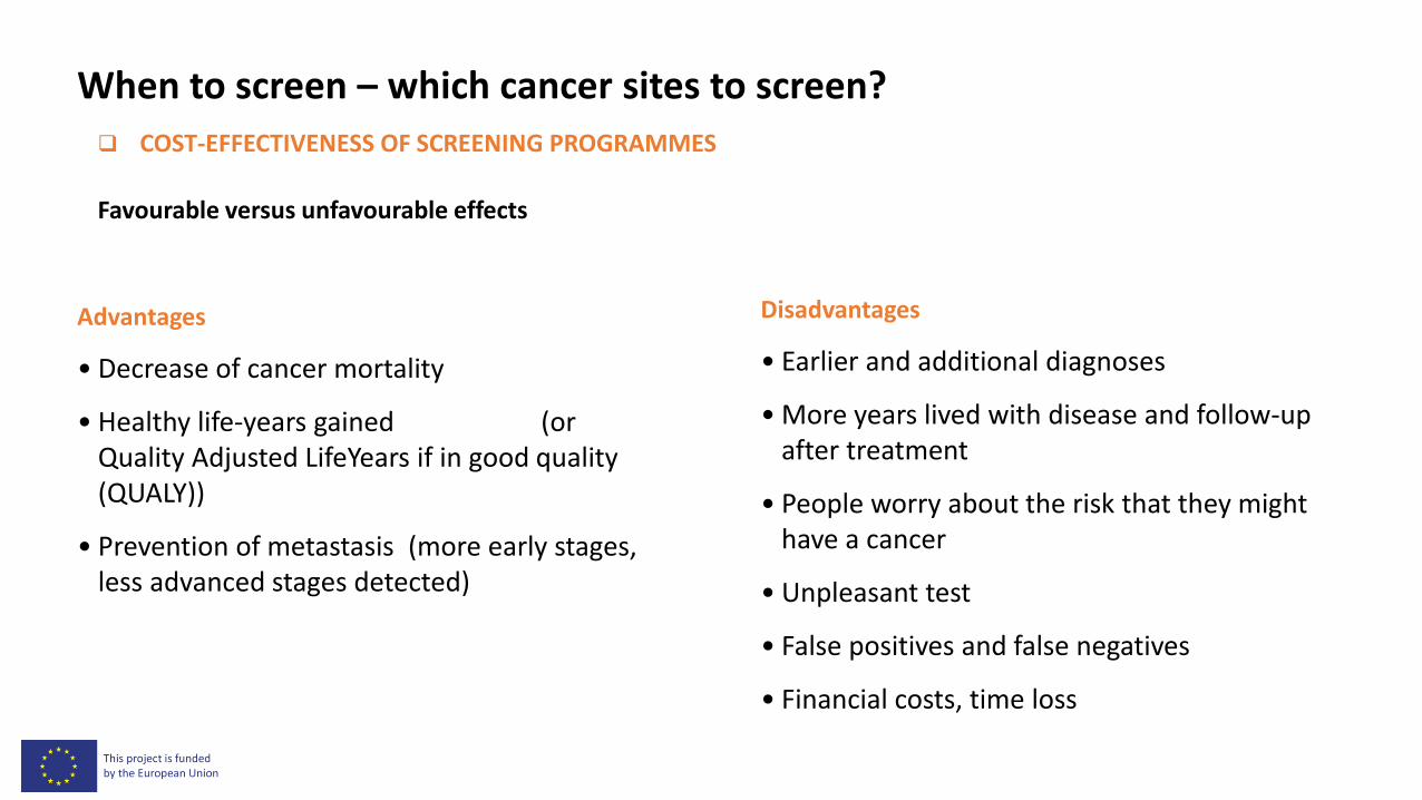
Favourable versus unfavourable effects
Advantages
• Decrease of cancer mortality
• Healthy life-years gained (or Quality Adjusted LifeYears if in good quality (QUALY))
• Prevention of metastasis (more early stages, less advanced stages detected)
Disadvantages
• Earlier and additional diagnoses
• More years lived with disease and follow-up after treatment
• People worry about the risk that they might have a cancer
• Unpleasant test
• False positives and false negatives
• Financial costs, time loss
When to screen – which cancer sites to screen?
COST-EFFECTIVENESS OF SCREENING PROGRAMMES

• A large benefit for a few, and relatively small unfavourable effects for many
• The main benefit - prevention of deaths, and the main harm - the over-detection, is not known to the individual participant
• On the other hand, individual participants are confronted with less serious harms - false positive and false negative test results.
• Screening programmes will always cause harm
• Physical harm: e.g. invasive interventions
• Psychological harm: e.g. anxiety, additional years of living with a disease,…
• Social harm: e.g. family relations, employment, insurance, financial implications,…
When to screen – which cancer sites to screen?
COST-EFFECTIVENESS OF SCREENING PROGRAMMES
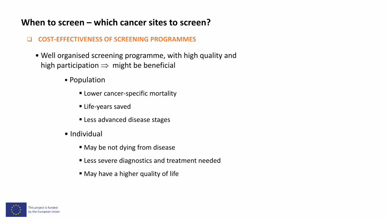
• Well organised screening programme, with high quality and high participation might be beneficial
• Population
Lower cancer-specific mortality
Life-years saved
Less advanced disease stages
• Individual
May be not dying from disease
Less severe diagnostics and treatment needed
May have a higher quality of life
When to screen – which cancer sites to screen?
COST-EFFECTIVENESS OF SCREENING PROGRAMMES
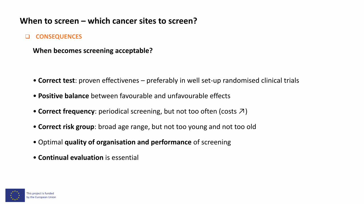
When becomes screening acceptable?
• Correct test: proven effectivenes – preferably in well set-up randomised clinical trials
• Positive balance between favourable and unfavourable effects
• Correct frequency: periodical screening, but not too often (costs ↗)
• Correct risk group: broad age range, but not too young and not too old
• Optimal quality of organisation and performance of screening
• Continual evaluation is essential
When to screen – which cancer sites to screen?
CONSEQUENCES

• Proven effectiveness and acceptable unfavourable side-effects
• => population-based screening more efficient than ad hoc screening of individual patients
• Screening always implicates negative effects
• => balanced information on both advantages and disadvantages is indispensable
• Population-based screening aims to improve public health.
• => This can collide with interests of individual participants
• Organising a screening programme is complex.
• Effects only visible in a long period
Summary

European recommendations
Breast cancer screening:
• 2-yearly Mammography screening for women aged 50 to 69 in accordance with European guidelines on
quality assurance in mammography.
• Minimum participation rate of 70% recommended
• Current issues:
•allowed rate of overdiagnosis (5%? 10%? 50%?)
•lower age limit? (40? 45?)
•upper age limit?
•dense breast tissue: mmx -> ultrasound?
http://eu-cancer.iarc.fr/ (2007)

Program goals
The main aim of breast cancer screening is to reduce mortality from thedisease without adversely affecting the health status of participants.
The objectives :
• To decrease breast cancer mortality
• To detect breast cancer at an early stage of the disease in up to 70 percent of all cases
• To achieve compliance rate of at least 70 percent of target population
• To increase the quality of life of patients suffering from breast cancer by early diagnosis and complex treatment.

Radiology screening units
• Mammography - the main method for population-based breast cancer screening
• Radiographer - the central player in producing high quality mammograms
• Radiologist - the prime responsible for mammographic image quality and diagnostic interpretation

Screening test
High quality mammography
• Cancer detection 1 - 3 years before its clinical manifestation
• Quality of requisites required for its performance and interpretationdetermines balance of sensitivity and specificity.
• Full-field digital mammography has multiple advantages • image manipulation and transmission,
• data display and other technological advantages.

Risks of Mammography
• False positive results• 11% abnormal, 3% Ca• Increase anxiety, fear, healthcare visits
• Overdiagnosis (ductal carcinoma in-situ)
• Pain
• Radiation: 10 yrs x 10,000 women=1 breast Ca
• False negative results (more common in young women)

Mammography examination
• Comparable high quality results for all centres participating in the mammography screening programme.
• Specific concern has to be paid on quality control of physical and technical aspects of mammography and the dosimetry:• images that have the best possible diagnostic information obtainable
• image quality is stable and consistent with other screening centers
• breast dose is As Low As Reasonably Achievable (ALARA)
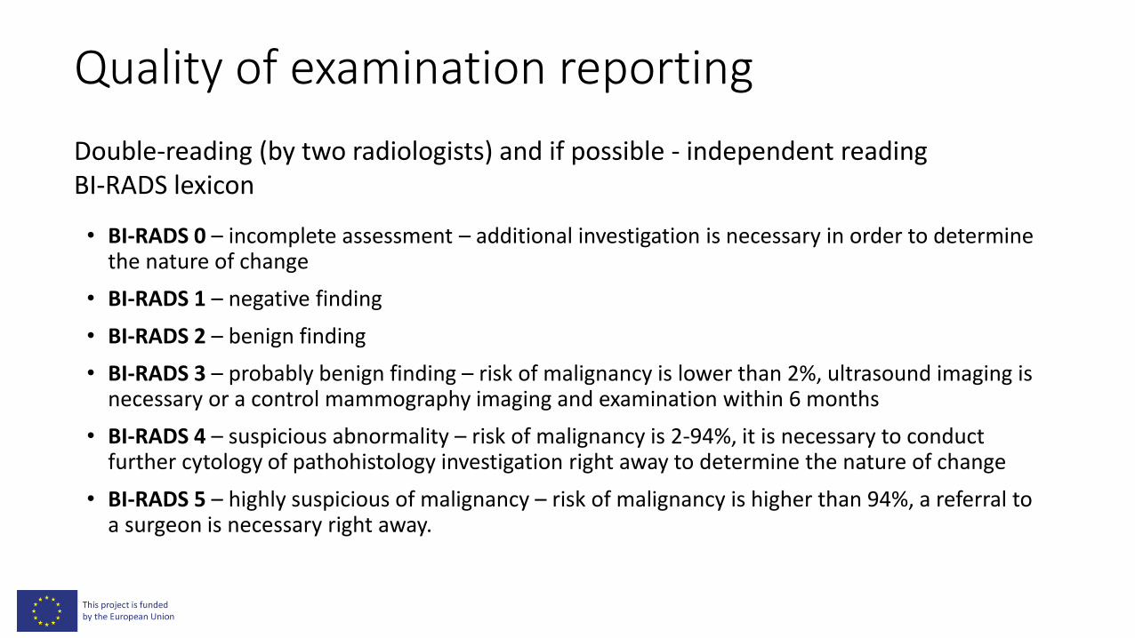
• BI-RADS 0 – incomplete assessment – additional investigation is necessary in order to determine the nature of change
• BI-RADS 1 – negative finding
• BI-RADS 2 – benign finding
• BI-RADS 3 – probably benign finding – risk of malignancy is lower than 2%, ultrasound imaging is necessary or a control mammography imaging and examination within 6 months
• BI-RADS 4 – suspicious abnormality – risk of malignancy is 2-94%, it is necessary to conduct further cytology of pathohistology investigation right away to determine the nature of change
• BI-RADS 5 – highly suspicious of malignancy – risk of malignancy is higher than 94%, a referral to a surgeon is necessary right away.
Double-reading (by two radiologists) and if possible - independent readingBI-RADS lexicon
Quality of examination reporting

Quality of examination reporting. Recomendations
• The conclusions BI-RADS 0, 3, 4 or 5 – further investigation is required.
• The conclusions BI-RADS 1 or 2 – next mammography screening test after two years.
• Women with BIRADS 4 or 5 have to be invited immediately to radiology unit not to delay the treatment in case of breast cancer diagnosis.

General/family medicine practitioners
• Patient education
• Formation of positive preventive attitude
• Individual risk assessment
• Motivation of women
• Monitoring the response of invited women
• Determining reasons for non-response

General/family medicine practitioners
• Close relations with Screening program coordination centre, Radiology screening unit
• Trained in communication
• Acquainted with the breast cancer screening organization scheme
• Introduced to IT system
• Have a deep knowledge in evaluation of screening mammography results (BIRADS system).
• Close relationship with breast cancer units timely addressing patients for necessary procedures.

Patronage services
• Through a screening IT system obtain a list of non-responding women for a particular region
• Additionally motivate those women
• Schedule appointment at the mammography screening unit
• Record not responders

Invitation of women
• Personalized letter
• Personal oral invitation
• Open non-personal invitation
• Combination of all three

PROGRAMME MONITORING AND QUALITY CONTROL
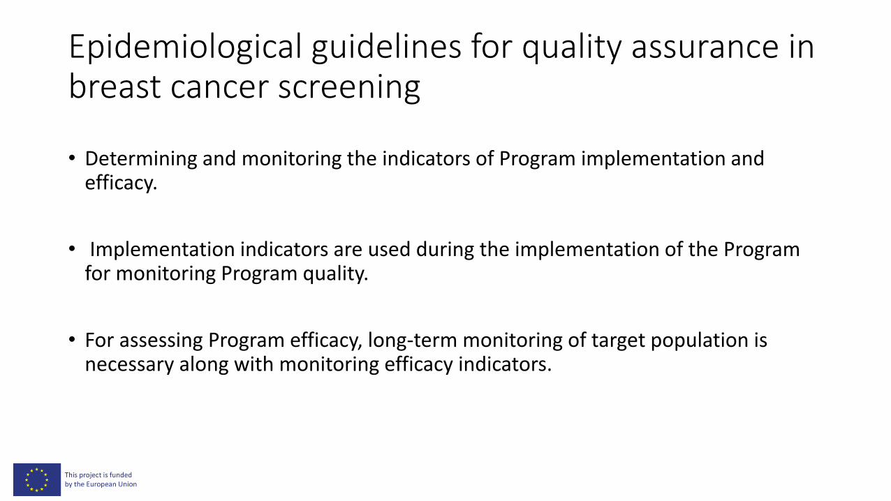
Epidemiological guidelines for quality assurance in breast cancer screening
• Determining and monitoring the indicators of Program implementation and efficacy.
• Implementation indicators are used during the implementation of the Program for monitoring Program quality.
• For assessing Program efficacy, long-term monitoring of target population is necessary along with monitoring efficacy indicators.

Implementation
Complete and accurate recording of:
• individual data,
• the screening test, its result,
• the decisions made and their eventual outcome in terms of diagnosis and treatment.
A fundamental concern at each step is the quality of the data collected.

Radiological quality control
• Setting of target standards and performance indicators, to comply with these wherever possible.
• Local quality assurance manuals based upon European or national documents.
• Regional and local organisations for QA, working at individual discipline level as well as in a multidisciplinary setting

Radiological quality control
• Digital techniques will have a significant impact on practice, analysis and performance of screening programmes.
• Centralization of mammography reading could enable better radiologic services, training and auditing possibilities as the part of quality control and assurance system.
• Teleradiology service is as an option for quality control, higher effectiveness, and cost savings.
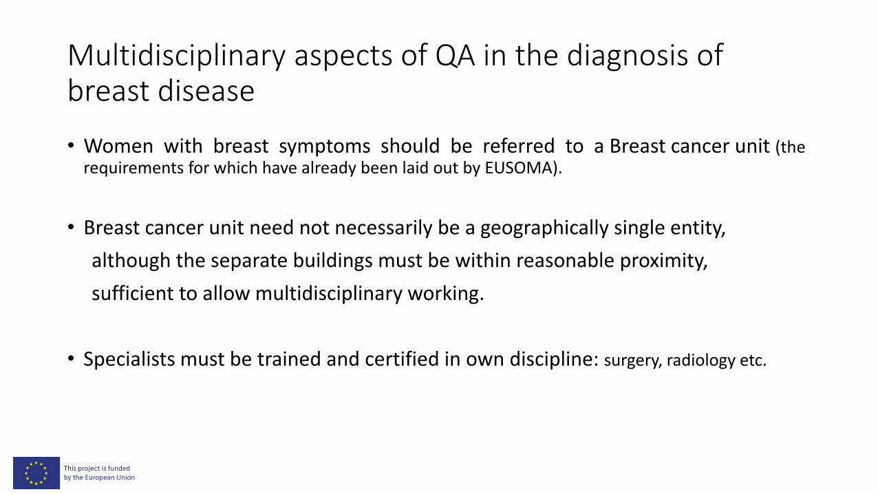
Multidisciplinary aspects of QA in the diagnosis of breast disease
• Women with breast symptoms should be referred to a Breast cancer unit (the requirements for which have already been laid out by EUSOMA).
• Breast cancer unit need not necessarily be a geographically single entity,
although the separate buildings must be within reasonable proximity,
sufficient to allow multidisciplinary working.
• Specialists must be trained and certified in own discipline: surgery, radiology etc.
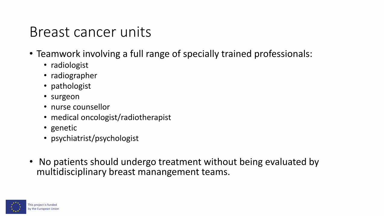
Breast cancer units
• Teamwork involving a full range of specially trained professionals:• radiologist• radiographer • pathologist• surgeon • nurse counsellor • medical oncologist/radiotherapist• genetic• psychiatrist/psychologist
• No patients should undergo treatment without being evaluated by multidisciplinary breast manangement teams.

Multidisciplinary aspects of QA in the diagnosis of breast disease• Screening is predominantly a radiological procedure with particular emphasis placed on
the optimal balance of sensitivity and specificity.
• The radiologist has the role of prime responsibility in screening.
• In symptomatic activity the clinician has the role of prime responsibility.
• The role of imaging, interpretation and cytological/histological sampling procedures is crucial in the cancer diagnostics.
• Triple assessment, i.e. clinical examination, imaging, and cytological / histologicalsampling is still regarded as the gold standard.

Epidemiology group
• Quality assurance:• Coverage• Responce rate• BIRADS clasification• Time between exam and reporting
• Ensuring quality:• Communication with GP• Quality of promotional activity
• Obstacles• IT – upgrading needed, lack of buget• Data base for invitation – updating of data• Commnunication with GP and RTG units - ?• Not enough appointments for mammography – Lack o resources, investment urgently need• Lack of human and equipment resouces – PP should became priority in practice
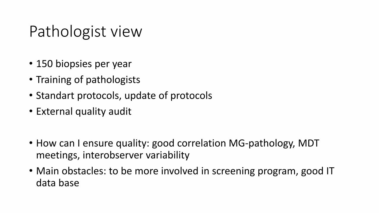
Pathologist view
• 150 biopsies per year
• Training of pathologists
• Standart protocols, update of protocols
• External quality audit
• How can I ensure quality: good correlation MG-pathology, MDT meetings, interobserver variability
• Main obstacles: to be more involved in screening program, good IT data base

• 2 pathologists per unit
• At least 150 biopsies per year
• Standart procedures:
• Implementation: comunication among MDT members, working groupfor coordination

Top Related