


Languages
Pages
Legal


Proteomic and virus-induced gene silencing (VIGS) analyses reveal thatGossypol, Brassinosteroids and Jasmonic acid contribute to the resistance ofcotton to Verticillium dahliae
Authors:
Wei Gao, Lu Long, Longfu Zhu*, Li Xu, Wenhui Gao, Longqing Sun, Linlin Liu,and Xianlong Zhang*
* Corresponding author: Xianlong Zhang, or Longfu Zhu, National Key Laboratory
of Crop Genetic Improvement, Huazhong Agricultural University, Wuhan, Hubei
430070, China
Telephone: +86-27-8728-0510
Fax: +86-27-8728-0196
E-mail: [email protected], or [email protected]
Address: National Key Laboratory of Crop Genetic Improvement, Huazhong
Agricultural University, Wuhan, Hubei 430070, China
Running title: Proteomic of cotton defense response to Verticillium dahliae
1
MCP Papers in Press. Published on September 9, 2013 as Manuscript M113.031013
Copyright 2013 by The American Society for Biochemistry and Molecular Biology, Inc.

Abbreviations:
2-DE, two-dimensional gel electrophoresis; qPCR, quantitative real time PCR; At,
Arabidopsis thaliana; Gh, Gossypium hirsutum; Gb, Gossypium barbadense; hpi,
hours past inoculation; ssp, sample spot protein; NCBI, National Center for
Biotechnology information; VIGS, virus-induced gene silencing; ROS, reactive
oxygen species; JA, jasmonic acid; BRs, brassinosteroids; BL, brassinolide; SA,
salicylic acid; DI, disease index
2

Summary
Verticillium wilt causes massive annual losses of cotton yield, but the mechanism of
cotton resistance to Verticillium dahliae is complex and poorly understood. In this
study, a comparative proteomic analysis was performed in resistant cotton (Gossypium
barbadense cv. '7124') upon infection with V. dahliae. A total of 188 differentially
expressed proteins were identified by mass spectrometry (MALDI-TOF/TOF)
analysis and could be classified into 17 biological processes based on Gene Ontology
annotation. Most of these proteins were implicated in stimulus response, cellular
processes and metabolic processes. Based on the proteomic analysis, several genes
involved in secondary metabolism, reactive oxygen burst and phytohormone signaling
pathways were identified for further physiological and molecular analysis. The roles
of the corresponding genes were further characterized by employing virus-induced
gene silencing (VIGS). Based on the results, we suggest that the production of
gossypol is sufficient to affect the cotton resistance to V. dahliae. Silencing of
GbCAD1, a key enzyme involving in gossypol biosynthesis, compromised cotton
resistance to V. dahliae. Reactive oxygen species and salicylic acid (SA) signaling
may be also implicated as regulators in cotton responsive to V. dahliae according to
the analysis of GbSSI2, an important regulator in the crosstalk between SA and
jasmonic acid (JA) signal pathways. Moreover, brassinosteroids (BRs) and JA
signaling may play essential roles in the cotton disease resistance to V. dahliae. The
BR signaling was activated in cotton upon inoculation with V. dahliae and the disease
resistance of cotton was enhanced after exogenous application of brassinolide (BL).
Meanwhile, JA signaling was also activated in cotton after inoculation with V. dahliae
and BL application. These data provide highlights in the molecular basis of cotton
resistance to V. dahliae.
Introduction
3

Cotton (Gossypium spp.) is one of the most important economic crops globally.
However, the yield of cotton is restricted by many unfavorable environmental
conditions including biotic and abiotic stresses. Among these stresses, Verticillium
wilt, a soil-borne vascular disease caused by Verticillium dahliae, is a devastating
disease of cotton worldwide (1), reducing the quality and yield of the fiber; up to 30%
yield reductions can occur during a severe outbreak of the disease (2). Few
germplasms have been found with resistance to V. dahliae in upland cotton
(Gossypium hirsutum), which contributes approximately 95% of the total cotton yield
(3), and the complex genetics of resistance to V. dahliae in upland cotton cultivars has
prevented the efficient breeding of disease-resistant cotton (4-6). Moreover, the
fungus can survive in soil for many years even in the absence of hosts (2). These
issues make it difficult to develop an effective and practical management plan for the
control of V. dahliae in cotton production.
Plants have evolved a complete, multilayered immune system that includes
constitutive and inducible defenses to counteract colonization by pathogens (7).
Several endogenous signal molecules, such as salicylic acid (SA), ethylene (ET) and
jasmonic acid (JA), are synthesized and activate distinct defense pathways involved in
complex defense signaling networks (8). Among these molecules, JA usually acts
with ethylene to induce resistance against necrotrophic pathogens, whereas
SA-mediated defense responses are effective against hemi-biotrophs and biotrophs
and are critical for systemic acquired resistance (SAR) (9). In addition, defense
signaling pathways mediated by SA and JA frequently act antagonistically to mediate
defense against specific types of pathogens (10-12). For example, SA accumulation
and SA-derived signaling are induced by virulent Pseudomonas syringae infection,
which enhances susceptibility to Alternaria brassicicola by inhibiting JA-mediated
defense responses in Arabidopsis (11). Nevertheless, the phytotoxin coronatine, a
structural analog of JA produced by Pseudomonas syringae, can suppress SA-derived
responses in the host (10). Other endogenous signal molecules, such reactive oxygen
species (ROS), auxin and brassinosteroids (BRs), can also influence defense signaling
4

and resistance (13-17). Plants have evolved regulatory defense mechanisms to adapt
efficiently to changes in their complex environment during pathogen invasion.
Crosstalk among signal molecules provides plants with a powerful capacity to finely
regulate the immune response. The molecular mechanisms involved in plant immunity
are complex.
During the past two decades, extensive studies have enriched our understanding of
the molecular mechanism of cotton resistance to V. dahliae. Production of
phytoalexins, including terpenoids and phenylpropanoid substances, is induced
quickly in cotton upon infection by V. dahliae (18, 19). Gossypol is one of the most
important sesquiterpene phytoalexin, which exists specifically in cotton and plays a
crucial role in the defense against the invasion of pathogens and insects (20). Though
many phytoalexin-related genes have been shown to be important in mediating cotton
defense, the molecular mechanism is unknown (21, 22). As sequencing technology
develops, a number of genes related to disease resistance (e.g., aerobic metabolism
enzymes, pathogen-related proteins, ethylene biosynthesis and response genes, etc.)
have been identified from resistant cotton cultivars (Gossypium barbadense cv. '7124'
or 'Pima 90') through suppression subtractive hybridization (SSH) (23, 24). Using
RNA-Seq-dependent transcriptional analysis, a subset of genes participating in lignin
metabolism was demonstrated to be very important in the resistance of cotton to V.
dahliae (19). Additionally, defense- and stress-related proteins, such as
pathogenesis-related proteins and proteins likely to be involved in the oxidative burst,
sugars, ethylene signaling and isoprenoid synthesis, have recently been suggested to
be involved in cotton response to V. dahliae (25, 26). Most of the candidate genes
involving in disease resistance are isolated from transcriptomic analysis, while only
few genes have been functionally characterized (27, 28). Ve1 is the only R gene
isolated using map-based cloning from tomato (Solanum lycopersicum) and has been
shown to provide race-specific resistance to race 1 strains of V. dahliae and V.
albo-atrum in tomato and Arabidopsis (17, 29). Although several genes homologous
5

to Ve1 have been cloned from cotton (30, 31), it is unclear whether Ve1-mediated
resistance signaling also exists in cotton.
In this study, we performed a comparative proteomic analysis on Mock and V.
dahliae inoculated roots of G. barbadense cv. '7124', which shows high resistance to V.
dahliae, at different time points. A number of differentially expressed proteins were
identified. Furthermore, three classes of genes, involved in gossypol metabolism, BR
signaling, and JA signaling were characterized using virus-induced gene silencing
(VIGS). Our data suggest that gossypol, BRs and JA act as important factors in
contributing resistance of cotton to V. dahliae. For the first time, proteomics was
combined with VIGS to discover and validate defense-related genes in cotton. Our
research provides new insights into the molecular basis of cotton defense against V.
dahliae.
Experimental Procedures
Plant growth and pathogen inoculation
Seeds of G. barbadense cv. '7124' (resistant) and G. hirsutum cv. 'YZ-1' (susceptible)
were grown in a controlled environment chamber under a 14 h light/10 h dark cycle at
28 °C for 2 weeks.
The defoliating isolate 'V991' of V. dahliae was grown on a potato-dextrose agar
medium for 4 d; the fungus was then incubated in Czapek’s medium (NaNO3, 0.3%
w/v; MgSO4, 0.1% w/v; KH2PO4, 0.1% w/v; FeSO4, 0.0002% w/v; KCl, 0.1% w/v;
sucrose, 3% w/v; pH 6.0) at 25 °C for 5 d. The concentration of spores was adjusted
to approximately 106 conidia per mL with deionized water for inoculation. The cotton
seedlings were removed from the soil and dip-infected with the liquid containing V.
dahliae spores. The seedlings were incubated at 25 °C under a 14 h/10 h light/dark
photoperiod, and the roots were harvested at 1, 6, 12, 24, 48 and 72 h after inoculation.
Seedlings treated with sterile distilled water in the same manner were used as a Mock
6

treatment. Roots were stored at -80 °C until protein extraction was performed.
Protein extraction and 2D electrophoresis
For 2D-PAGE, proteins from cotton roots were prepared according to Yao and Pan (32,
33) with minor modifications. Frozen root tissues were ground in liquid nitrogen to a
fine powder and incubated in extraction buffer (-20 °C precooled acetone, 12% w/v
trichloroacetic acid, and 0.07% w/v DTT). After washing twice with cold acetone
containing 0.07% (w/v) DTT, the vacuum-dried powder was suspended in extraction
buffer [30% sucrose, 50 mM Tris-HCl (pH 8.0), 2% SDS, 2 mM PMSF, 0.07% DTT,
and an equal volume of Tris-saturated phenol (pH 8.0)]. The phenol phase was then
precipitated with 5 volumes of 0.1 M ammonium acetate in methanol at -20 °C. The
collected protein pellets were washed twice with 80% methanol and 80% acetone.
After air-drying, the pellets were dissolved in lysis buffer (8 M urea, 2 M thiourea, 2%
CHAPS, 1% DTT, and 2% v/v IPG buffer, pH 4-7). Protein concentration was
determined using a 2-D quant kit (Bio-Rad, USA).
The first dimensional gel separation was performed according to the manufacturer’s
protocol with modifications (Bio-Rad, USA). Total proteins (1.0 mg) were diluted in
450 μL rehydration buffer (8 M urea, 2 M thiourea, 2% CHAPS, 1% DTT, and 2% v/v
IPG buffer, pH 4-7) and loaded on immobilized pH gradient (IPG) strips (24 cm, pH
4-7 nonlinear) (Bio-Rad, USA). The IPG strips were rehydrated for 12 h at room
temperature and focused at gradient steps of 50 V for 1 h, 500 V for 1 h, 1000 V for 1
h, and 10,000 V for 4 h, with a final step of 10,000 V toward a total of 90 kVh. Before
second dimension analysis, IPG strips were incubated in equilibration buffer (50 mM
Tris-HCl, pH 8.8; 6 M urea; 30% glycerol; and 2% SDS) containing 1% DTT and
then in equilibration buffer containing 2% (w/v) iodoacetamide. For the second
dimension, IPG strips were fixed on 12.5% acrylamide gels. Then, 2D-PAGE was
performed using the Ettan DALT six electrophoresis unit (GE Healthcare, USA) at 5
W per gel for 1 h and then 15 W per gel for 6 h until the bromophenol blue dye front
reached the bottom of the gels.
7

Gels were stained with Coomassie Brilliant Blue solution (0.2% Coomassie
Brilliant Blue G250, 20% methanol, 10% phosphoric acid, and 10% ammonium
sulfate). The 2D gels were scanned using a GS-800 Calibrated Densitometer (Bio-Rad,
USA). Protein spots were detected using PDQuest software (Bio-Rad, USA). After
volumetric quantification and matching, differences in protein content between Mock
and inoculated samples were analyzed using a Student t-test and calculated as the fold
ratio. Data was from three biological replicates; a threshold of P ≤0.05 and fold
change of ≥2 or ≤0.5 was used to identify significantly differentially expressed protein
spots.
MALDI-TOF/TOF analysis
Differentially expressed proteins were excised from the gels and destained with 25
mM NH4HCO3 in 50% ACN until the CBB disappeared. After being washed twice
with 100% ACN for 10 min, proteins were digested in-gel using trypsin (Promega,
Madison, USA) overnight at 37 °C. The gel pieces were extracted once with
extraction buffer (67% ACN and 5% TFA). After being dried completely, the samples
were resuspended in 0.1% TFA and then mixed in a 1:1 ratio with a matrix consisting
of a saturated solution of CHCA in 50% ACN containing 0.1% TFA. Samples were
then spotted onto a freshly cleaned target plate. After air-drying, the crystallized spots
were analyzed using an ABI 4800 MALDI-TOF/TOF Plus mass spectrometer
(Applied Biosystems, Foster City, USA). Both the MS and MS/MS data were
integrated and performed using GPS Explorer V3.6 software (Applied Biosystems).
Proteins were successfully identified with a 95% or higher confidence interval using
the MASCOT V2.3 search engine (Matrix Science, London, UK) and searching in the
Gossypium EST database (release data 20120128; 2476590 sequences; 555009942
residues). The other search parameters were the enzyme trypsin; one missed cleavage
site; partial modifications of cysteine carbamido methylation and methionine
oxidization; no fixed modifications; peptide tolerance of 100 ppm; and fragment mass
tolerance of 0.3 Da.
8

RNA isolation, reverse transcription-PCR (RT-PCR) and quantitative real-time
PCR (qPCR)
Total RNA was isolated from cotton as previously described (34). First strand cDNA
was synthesized from 5 μg of total RNA using the Superscript first-strand synthesis
system (Invitrogen, Foster City, CA, USA). For RT-PCR, an aliquot of the reverse
transcription product was used as the template. The ubiquitin7 gene (DQ116441) of
cotton was used as an internal control. Primers were designed as shown in Table S1.
qPCR was performed according to the guidelines of the Minimum Information for
Publication of Quantitative Real Time PCR Experiments (35). Diluted cDNA was
used for qPCR with SYBR green using ABI 7500 Real Time PCR system (Applied
Biosystems, USA). The expression analysis data are presented as the means ± SD
from three biologically independent experiments. All primers for qPCR were designed
using Primer Express 5.0 software (Applied Biosystems, USA) and are shown in
Table S2.
Virus-induced gene silencing (VIGS) in cotton followed by pathogen inoculation
The TRV vectors and Agrobacterium tumefaciens for VIGS were prepared according
to Fradin et al. (17). Inserts to generate TRV:GbCAD1, TRV:Gb14-3-3c,
TRV:Gb14-3-3d, TRV:GbSSI2 and positive control TRV:GbCLA1 (cloroplastos
alterados 1) were amplified from the cDNA of G. barbadense cv. '7124'. Primer pairs
to generate TRV vectors are shown in Table S3. PCR fragments were digested with
BamHI and KpnI and then ligated into the TRV:00 plasmid (36). The constructs were
transformed into A. tumefaciens GV3101 by electroporation. TRV vectors were
agro-infiltrated as described (31) into the cotyledons of 10-day-old seedlings of G.
barbadense cv. '7124' or G. hirsutum cv. 'YZ-1'. The seedlings were then grown at
25 °C with a 16 h/8 h light/dark photoperiod cycle in a controlled environment
chamber. As shown in Fig. S1, the leaf bleaching phenotype was observed 2 weeks
after infiltration in TRV:GbCLA1 plants. Therefore, inoculation with V. dahliae isolate
'V991' was also performed 2 weeks after infiltration. The rate of diseased plants and
9

disease index were scored with at least 16 plants per treatment and repeated at least
for three times. Plant disease index (DI) is calculated as the following formula:DI = ∑( ×the number of seedlings at level n)
4×the number of total seedlings×100, n denotes disease level, cotton seedlings
were divided into 5 levels based on their disease severity after V. dahliae inoculation
(level 0, 1, 2, 3, 4) according to Xu et al. (37), DI reflects the disease infection status
of a population, not an individual plant; higher DI means more serious infection.
Treatments with methyl jasmonate (MeJA) and brassinolide (BL)
For treatments with MeJA and BL, the concentrations were 200 μM and 5 μg/pot,
respectively. Cotton seedlings were cultured in a pot (four or five plants per pot) in a
greenhouse and three-leaf stage plants were treated with MeJA or BL by soil drench
application.
Measurement of total SA and JA levels
The extraction and measurement of the endogenous SA and JA of cotton seedlings
were performed as described (38). Three replicates of each frozen sample
(approximately 100 mg for each replicate) were ground to a fine powder in liquid
nitrogen and mixed with 750 μL cold extraction buffer (methanol:water:acetic acid,
80:19:1, v/v/v). After shaking for 16 h at 4 °C in the dark, the supernatants were
collected and then filtered using a syringe-facilitated 13-mm diameter nylon filter
with a pore size of 0.22 μm (Nylon 66; Jin Teng Experiment Equipment Co., Ltd, Tian
jin, China). Filtrates were dried using nitrogen gas at room temperature and then
dissolved in 200 μL methanol. An aliquot of dissolved sample was further diluted 100
times using methanol for the quantification of SA because cotton contains high levels
of SA. Supernatants were analyzed on an HPLC-MS/MS (1200L LC-MS system,
Varian).
DAB staining and H2O2 measurement
For H2O2 detection, leaves were incubated in 1 mg/mL pH 3.8 DAB-HCl
10

(Sigma-Aldrich, USA) in the dark for 8 h. The leaves were then cleared by boiling in
alcoholic lactophenol (95% ethanol:lactophenol, 2:1 v/v) for 20 min. The reddish
color of the leaves as evidence of H2O2 was visualized by light microscopy. Hydrogen
peroxide content was measured using an H2O2 Quantitative Assay Kit (TIANGEN,
China).
Trypan blue staining
Trypan blue staining was performed as described by Choi and Hwang (39). Leaves
were stained by boiling in lactophenol-trypan blue (10 mL lactic acid, 10 mL glycerol,
10 g phenol, and 10 mg trypan blue dissolved in 10 mL distilled water) followed by
destaining with chloral hydrate (2.5 g/mL).
Gossypol visualization and measurement
Gossypol was visualized by photography within 5 min after dropping a saturated
solution of antimony trichloride (SbCl3) in 40% perchloric acid (HClO4) on fresh
free-hand sections (approximately 100 mm thick) from the same sites of Mock and
infected cotton roots. Images were captured using an Olympus light microscope
equipped with an Olympus U-PMTVC adapter and a Leica DC300F camera, and
processed using Leica IM50 Image Manager V1.20 software.
Sesquiterpene aldehydes (gossypol equivalents) were extracted and quantitated using
the phloroglucinol/HCl method (22).
Statistical analysis
For all generated data, at least three biological replicates were performed. The data are
presented as the mean ± SD. Statistical significance was determined using two-tailed
unpaired Student’s t-tests, and P values <0.05 were considered statistically significant.
11

Results
Two cotton cultivars, G. barbadense cv. '7124' (resistant) and G. hirsutum cv. 'YZ-1'
(susceptible) were inoculated with a highly aggressive defoliating fungal pathogen V.
dahliae strain 'V991' (Fig. 1A). Approximately two weeks later, susceptible plants
exhibited visual symptoms of cotyledon wilting, leaf chlorosis, and severe stunting,
while resistant plants only showed slight stunting compared with the control.
Therefore, the potential presence of a special mechanism and related defense response
to V. dahliae in '7124' were investigated using comparative proteomics.
Proteomics analysis and identification of cotton defense-related proteins
Proteins were isolated from the roots of '7124' during 'V991' inoculation and screened
using 2-DE gels. Approximately 1500 protein spots were detected on each 2-DE gel
of Mock and infected cotton roots at 1, 6, 12, 24, 48, and 72 hpi. Differences in
protein content between Mock and inoculated samples were analyzed using a Student
t-test and calculated as the fold ratio. Data was from three biological replicates; a
threshold of P ≤0.05 and fold change of ≥2 or ≤0.5 was used to identify significantly
differentially expressed protein spots. A total of 274 proteins with significant
expression changes after V. dahliae inoculation compared with Mock were
successfully identified using MALDI-TOF/TOF (Fig. 1B-D). These differently
expressed proteins were mostly observed at 1, 24 and 48 hpi (22.63%, 27.01% and
24.09%, respectively); 130 proteins were up-regulated while 144 proteins were
down-regulated (Table 1).
Among these 274 proteins, several were detected at multiple time points. Thus, 188
unique proteins with significant expression changes during V. dahliae infection were
obtained after removing redundant proteins. Detailed information about these proteins
is shown in Table S4. All these proteins were divided into three groups according to
their expression patterns (up-regulated, down-regulated and other patterns of
regulation; Fig. 1B-D); 47% of the 188 proteins were down-regulated during infection
12

(Fig. S2A). To globally understand the potential function of these 188 proteins, GO
functional classification analysis was performed to calculate the functional category
distribution based on level 2 biological processes (Fig. S2B). As a result, all identified
proteins could be classified into 17 biological processes, including metabolism,
cellular process, response to stimulus, biogenesis, immune system, etc. In addition to
the response to stimulus-related proteins, more proteins involved in metabolic and
cellular processes were found to be down-regulated, which may indicate that the
normal development of cotton was retarded by the infection of the fungus.
A set of disease-related genes involved in the complex biological processes of
cotton defense against V. dahliae was identified, including oxidative burst-, auxin
response-, secondary metabolism- and pathogenesis-related genes. The expression
level of several proteins participating in the regulation of redox homeostasis,
including peroxidase (GbPOD, ssp318), ascorbate peroxidase (GbAPX2, ssp4017),
monodehydroascorbate reductase (GbMDAR6, ssp9503) and malate dehydrogenase
(GbMDH, ssp4212), were found to be clearly influenced by the inoculation with V.
dahliae (Fig. 2A). Meanwhile, a class of genes potentially involved in disease
resistance was a group of the auxin signaling-related genes, such as the WD40
repeat-like superfamily protein (GbWD40, ssp1434), amino peptidase P1 (GbAPP1,
ssp3827), auxin-induced protein pcnt115 (GbAKR, ssp7212) and pyruvate
dehydrogenase E1a-like subunit (GbIAR4, ssp8314). These proteins are involved in
the accumulation, polar transport or homeostasis of auxin (40-42), and their
expression levels changed dramatically in cotton after inoculation with V. dahliae (Fig.
2B). Furthermore, secondary metabolism-related proteins, such as related to lignin
metabolism, were found to be activated during the inoculation. Two important
proteins participating in lignin metabolism, phenylcoumaran benzylic ether
reductase-like protein (GbPRR1, ssp4252) and caffeoyl-CoAO-methyltransferase
(GbCCoAOMT, ssp8129) (19), were increased significantly upon infection with the
fungus (Fig. 2C). Several other proteins involved in signal transduction, such as
mitogen-activated protein kinase (GbMAPK4, ssp6425) and protein phosphatase 2c
13

(GbPP2C, ssp2123), were also clearly induced during the interaction between cotton
and V. dahliae (Fig. 2C).
Gossypol is involved in the resistance of cotton to V. dahliae
Among these identified proteins, the protein GbCAD1 (ssp2124), related to the
biosynthesis of the terpenoid phytoalexin gossypol (18), was clearly up-regulated in
the roots after inoculation with V. dahliae (Fig. 3A). To investigate and verify the
participation of gossypol in response to V. dahliae, the expression of genes involved in
gossypol biosynthesis was determined by RT-PCR. As shown in Fig. 3B, the
expression of GbCAD1 was enhanced after inoculation with V. dahliae compared with
mock-treated samples, which was consistent with the expression pattern at the protein
level. Furthermore, two other key genes (farnesyl diphosphate synthase, GbFPS;
cytochrome P450 mono-oxygenase, GbCYP706B1) were also up-regulated, whereas
GbdHG-6-OMT, a negative regulator of gossypol biosynthesis, was down-regulated,
indicating that inoculation with V. dahliae could activate the biosynthesis of the
terpenoid phytoalexin gossypol in cotton.
VIGS was employed to better understand the function of GbCAD1 in the cotton
defense response. Two weeks after Agrobacterium infiltration in '7124', the transcripts
of GbCAD1 were significantly reduced in TRV:GbCAD1 plants compared with
TRV:00 plants, as shown by RT-PCR analysis (Fig. 3C), indicating that GbCAD1 was
effectively silenced in cotton. Upon inoculation with 'V991', more wilting and
etiolated leaves were observed in TRV:GbCAD1 plants compared with the control
(Fig. 3C). To investigate whether the gossypol production was altered after GbCAD1
was silenced, the phloroglucinol/HCl method was used to measure the content of
gossypol equivalents (22). Gossypol equivalents in both roots and leaves of
TRV:GbCAD1 plants was significantly lower compared with the control 12 d after
VIGS infiltration (Fig. 3D). Statistical analysis of data for the rate of diseased plants
and the disease index showed that deficiency of GbCAD1 compromises resistance to
V. dahliae (Fig. 3E).
14

Because GbCAD1 has been shown to be an important enzyme in gossypol
biosynthesis, the changes in gossypol production in cotton after pathogen inoculation
were evaluated. Plants were harvested 5 d after inoculation and stained with
SbCl3-HClO4, and the results show the presence of gossypol in vascular bundles more
frequently in inoculated plants compared with uninfected plants, and the production of
gossypol equivalents in infected seedlings was enhanced compared with uninfected
plants (Fig. 3F, G). These results indicate that gossypol production was significantly
increased upon V. dahliae infection, and inhibiting the biosynthesis of gossypol
impairs cotton resistance to V. dahliae.
BR signaling positively regulates the resistance of cotton to V. dahliae
Four Gb14-3-3 proteins, the homologous genes of which in Arabidopsis were reported
to interact with the BIN2-phosphorylated targets in the BZR1 protein (43), were all
down-regulated significantly in the protein expression profile (Fig. 4A). The
transcripts of four Gb14-3-3 protein-encoding genes were confirmed using RT-PCR
analysis. The transcriptional levels of Gb14-3-3c (ssp127) and Gb14-3-3d (ssp4002)
were suppressed in cotton after inoculation with V. dahliae, which is consistent with
the protein analysis (Fig. 4B). While the transcripts of Gb14-3-3a (ssp12) and
Gb14-3-3b (ssp123) were not significantly changed and there were no detectable
effect on disease resistance of cotton when silencing these two genes with VIGS (data
not shown). Therefore, Gb14-3-3c (ssp127) and Gb14-3-3d (ssp4002) were chosen for
further study.
Seedlings of susceptible cultivar 'YZ-1' infiltrated with Agrobacterium carrying
TRV: Gb14-3-3c and TRV:Gb14-3-3d were prepared to investigate the roles of these
two Gb14-3-3 proteins in cotton. The results of the RT-PCR analysis showed that the
transcripts of Gb14-3-3c and Gb14-3-3d were successfully suppressed in seedlings 2
weeks after VIGS infiltration (Fig. 4C). The rate of diseased plants and the disease
index were calculated, and the results show that silencing of Gb14-3-3c and
Gb14-3-3d improves resistance to the pathogen based on the reduced wilting and
15

etiolated leaves observed in TRV:Gb14-3-3c and TRV:Gb14-3-3d seedlings (Fig. 4D).
This suggests that the 14-3-3 proteins function as negative regulators of resistance to
V. dahliae in cotton.
Based on previous studies, the 14-3-3 genes described above are involved in the
negative regulation of the brassinosteroid (BR) signaling pathway in plants (43-45). It
is possible that BR signaling is involved in regulating the resistance of cotton to V.
dahliae. To verify this hypothesis, the expression patterns of three genes involved in
the BR signaling pathway were analyzed in roots after inoculation with 'V991' (Fig.
5A). Brassinosteroid insensitive 1 (BRI1), a BR receptor gene, and Brassinazole
resistant 1 (BZR1), a positive BR response factor, were up-regulated, while
Brassinosteroid insensitive 2 (BIN2), a negative response receptor of BR, was
down-regulated in roots after inoculation with V. dahliae.
To characterize the role of BR in the cotton plant response to V. dahliae, exogenous
BL (brassinolide, a biologically active BR; 5 μg/pot) was applied to seedlings of
'YZ-1' 24 h before inoculation with 'V991'. Seedlings treated with exogenous BL
exhibited significantly more resistance to V. dahliae than mock-treated plants (Fig. 5B,
C). Meanwhile, the expression of Gb14-3-3c and Gb14-3-3d was significantly
inhibited after the seedlings were treated with BL (Fig. 5D). This demonstrates a role
for BR as a positive regulator of cotton resistance to V. dahliae.
SA and JA have been reported to play important roles in plant immunity, so we also
analyzed expression of SA and JA signaling pathway-related genes in cotton roots
after treatment of BL. Surprisingly, most of genes involved in JA signaling were
up-regulated in cotton plants after treatment of BL (Fig. 5D), but there were no
obvious changes in the transcripts of SA signaling pathway-related genes (data not
shown).
GbSSI2 influences the cotton resistance by altering SA- and JA-mediated defensesignaling
16
A novel protein identified as stearoylacyl-carrier protein desaturase (ssp4333,
gi|48813830), which shares 78% amino acid sequence identity with SSI2
(AT2G43710) in Arabidopsis, was significantly induced by V. dahliae and named
GbSSI2 (Fig. 6A). The transcript of this gene also accumulated within 1 hour after
inoculation with V. dahliae, as shown by RT-PCR analysis (Fig. 6B). To further
investigate the function of GbSSI2 in cotton, VIGS was performed to silence the
expression of GbSSI2 in the resistant cultivar '7124'. The RT-PCR analysis showed
that GbSSI2 transcripts were significantly reduced in cotton after infiltration with
Agrobacterium containing TRV:GbSSI2 vector (Fig. 5C). Furthermore, spontaneous
lesions in stems and yellow leaf veins were found in GbSSI2-silenced plants (Fig. 6C).
These spontaneous lesions expanded during plant growth and severely blocked cotton
development, which is similar to the phenotype of ssi2 in Arabidopsis.
Spontaneous lesions in the Arabidopsis mutant ssi2 are caused by intense
hypersensitive cell death because of the accumulation of reactive oxygen species
(ROS) (46). Biochemical quantitative determination of the H2O2 content of
GbSSI2-silenced cotton revealed levels significantly higher than those in control
plants (Fig. 6D). Supporting this, accumulation of H2O2 as visualized by DAB
staining confirmed the higher levels of H2O2 in the silenced plants (Fig. 6D).
Hypersensitive cell death is generally considered a major result of plant disease
resistance mediated by SA signaling (47). The endogenous SA content was measured
and found to be significantly increased in GhSSI2-silenced plants compared with
control plants (Fig. 6E). Meanwhile, expression of GbNPR1, GbPR1 and GbPR5,
three important genes involved in the SA signaling pathway, was analyzed using
qPCR, and results show that the transcripts of all three genes were up-regulated
significantly after silencing of GbSSI2 in cotton (Fig. 6E).
Leaves from GbSSI2-silenced cotton were used for fungal inoculation with 'V991'.
Mycelia were observed 3 d after dropping spore suspension (106 conidia per mL) onto
the leaf surface. Silencing of GbSSI2 greatly increased the mycelia growth of V.
17

dahliae and accelerated cell death at the infected location, as indicated by trypan blue
staining (Fig. 6F). After inoculation with V. dahliae, statistical analysis showed that
hyphal cover area and trypan blue staining area of TRV:SSI2 plant leaves were
significantly larger than those of TRV:00 plant leaves (Fig. 6G). These results show an
essential role of GbSSI2 participating in cotton resistance to V. dahliae and
demonstrated that the accumulation of H2O2 and the activation of the SA signaling
pathway led to enhanced infection of cotton by V. dahliae.
Defense signaling pathways mediated by SA and JA frequently act antagonistically
to mediate defense against specific types of pathogens (10-12). To explore whether
activation of the SA pathway in TRV:GbSSI2 plants influences JA signal transduction,
the expression of genes participating in the biosynthesis of JA (GbLOX1, GbAOS, and
GbOPR3) and its signal transduction (GbJAZ1, GbJAZ6, GbMYC2 and GbPR4) was
analyzed. qPCR results show that the transcripts of genes involved in both JA
biosynthesis and JA signal transduction were inhibited in TRV:GbSSI2 plants (Fig.
7A). Moreover, the content of JA was significantly decreased in TRV:GbSSI2 plants
compared with the control (Fig. 7B), which was in accordance with the expression
patterns of related genes (Fig. 7A). All the mentions above indicate that GbSSI2 may
influence the cotton resistance by altering SA- and JA-mediated defense signaling.
Discussion
Verticillium wilt is a serious disease that significantly affects the yield and quality of
cotton. Understanding the molecular mechanism of cotton-V. dahliae interactions is a
key aim to support for disease resistance breeding in cotton. As the development of
sequencing technologies progresses, diverse differentially expressed genes following
cotton inoculation with V. dahliae have been identified at the transcriptional level (19,
24), while the functional roles of few candidate genes have been confirmed. Despite
the relative simplicity and high throughput, the transcriptional data is complex, and so
difficult to draw meaningful conclusions, and does not readily provide new functional
18

insights. For example, it has been shown that the transcriptional level does not
necessarily correlate with the protein level (48). Here, a complete analysis of protein
expression using 2-DE and MALDI-TOF/TOF, combined with expression analysis
and functional studies using VIGS and infection phenotyping, offers a useful
approach to reveal the molecular basis of cotton defense against V. dahliae.
Utilizing the resistant cultivar '7124' from G. barbadense, we identified 188
differentially expressed proteins through a proteomics approach from roots inoculated
with V. dahliae. Among these proteins, those that were down-regulated by the fungus
were the majority of differentially expressed proteins, which may imply that
metabolism is repressed to in turn repress the plant immune network. This idea is
consistent with our previous results obtained using transcriptional analysis by
RNA-seq (19). According to the results of GO functional classification, the three
largest biological processes, including 'cellular process', 'metabolism' and 'stimulus
response', which together represent the principal fundamental cell functional
processes, were involved in the response of cotton to V. dahliae. This suggests that the
host undergoes major physiological changes in response to the plant-pathogen
interaction.
Secondary metabolites become especially abundant in plant cells when plants are
threatened by diverse pathogens in nature (19, 24, 25). Generally, the production of
secondary metabolites, such as phytoalexins and lignin, can inhibit the growth of
pathogens directly or provide a physical barrier against the invading plant pathogens.
Our previous studies have demonstrated the importance of lignin involvement in the
phenylpropanoid pathway; lignin was found to contribute to cotton disease resistance
(19). In this study, GbCCoAOMT, a protein involved in lignin biosynthesis, was also
identified and was significantly up-regulated at the protein level upon infection of V.
dahliae.
Gossypol, the major type of sesquiterpenoid, is a cotton-specific secondary
metabolite that has been shown to be associated with disease resistance (18, 20, 21,
19

49-51). The involvement of gossypol metabolites in the interaction between cotton
and V. dahliae was further confirmed in the current study. GbCAD1, an important
enzyme involved in gossypol biosynthesis (18, 51), was identified through our
proteomics analysis. Gossypol content was dramatically increased by fungal infection,
and silencing of GbCAD1 by VIGS enhanced cotton susceptibility to the pathogen.
Moreover, it was speculated that downregulation of dHG-6-OMT could maintain a
relatively high terpenoid content and significantly increase the resistance of G.
hirsutum to fungi and pests (51). In our study, GbdHG-6-OMT (ssp1320), a gene
negatively regulating resistance, was suppressed in cotton roots during V. dahliae
inoculation. Other genes involved in gossypol biosynthesis, including GbFPS and
GbCYP706B1, were up-regulated upon infection of V. dahliae, as shown by RT-PCR
analysis and so leading to enhanced resistance. These results suggest that the
accumulation of gossypol is an effective strategy for the response of cotton to V.
dahliae.
Recent advances in plant immunity research have provided new insights into the
underlying defense signaling network by diverse small-molecule hormones, such as
auxin, BR, JA, and SA (8, 16). These signaling pathways cross-communicate in an
antagonistic or synergistic manner, providing the plant with a powerful capacity to
finely regulate its immune response (16). However, evidence is accumulating that
pathogens can manipulate hormone-regulated signaling pathways to evade host
immune responses (16). We isolated a number of proteins involved in the
phytohormone signaling network with diverse expression patterns during V. dahliae
infection, which implies that complicated cross-talk of phytohormone signaling
pathways is involved in the incompatible interaction between cotton and V. dahliae.
Over the past decade, extensive research efforts have demonstrated that BRs can act
as regulatory factors in biotic stress responses in plants (52-56). BZR1 and BZR2 are
two key transcription factors that mediate BR signals by regulating downstream gene
expression. In addition, 14-3-3 proteins have been demonstrated to be negative
20

regulators of BR signaling by regulating the subcellular localization and activity of
both BZR1 and BZR2 (44). Four Gb14-3-3 proteins involved in BR signaling were
identified in our proteomic analysis to have decreased in abundance in cotton after
inoculation with V. dahliae. Our RT-PCR data also showed that the genes positively
regulating BR signaling were induced in cotton after inoculation with V. dahliae.
Silencing of Gb14-3-3c and Gb14-3-3d in cotton through VIGS enhanced the
resistance of cotton to V. dahliae. Meanwhile, exogenous BL treatment has a similar
effect on cotton disease resistance to V. dahliae. Moreover, SERK3/BAK1 is a
coreceptor that physically associates with BRI1 for BR-dependent signaling and has
been shown to be required for Ve1-mediated resistance in tomato and Arabidopsis (17,
29). The above results demonstrate that BRs and the BR signaling pathway positively
regulate the resistance of cotton during V. dahliae infection. This study therefore
provides new information about the function of BR in cotton disease resistance.
In plants, ROS play a dual role as both toxic byproduct of normal cell metabolism
and regulatory molecules in stress perception and signal transduction (57). The
expression levels of several proteins that participate in the regulation of redox
homeostasis, GbPOD, GbAPX2, GbMDAR6, and GbMDH, were found to be
influenced, and most were down-regulated, in cotton upon infection with V. dahliae.
At the same time, SSI2, a stearoyl-ACP-desaturase, was induced during the
interaction between cotton and V. dahliae. Silencing of GbSSI2 in cotton led to the
accumulation of H2O2 and impaired the resistance of cotton to V. dahliae. These
results imply that ROS in cotton may be negatively correlated with disease resistance
to V. dahliae. However, ROS with other signaling molecules are complex in plant
cells (14, 47), and the exact role of ROS involved in cotton defense against V. dahliae
invasion remains to be discovered.
Two phytohormones SA and JA are known to play major roles in regulating plant
defense response against various pathogens. SA participates in the activation of the
defense response against biotrophic and hemi-biotrophic pathogens, while JA/ET
mediates plant defenses against necrotrophic pathogens (16, 58). Previous studies
21

have also shown that SA is a potent suppressor of JA-mediated defenses against
necrotrophs (8, 16). A balance between glycerol-3-phosphate (G3P) and oleic acid
levels mediated by SSI2 is critical for the regulation of SA- and JA-mediated defense
signaling in the plant (59). Silencing of GbSSI2 in cotton through VIGS
simultaneously up-regulates SA synthesis and SA-mediated responses and inhibits
JA-inducible defenses, resulting in increased susceptibility to V. dahliae, which is
similar to the results from a mutation or silencing of SSI2 in Arabidopsis or rice to
pathogens (60-62). When cotton plants were invaded by V. dahliae, protein levels of
GbSSI2 increased significantly, as shown by proteomic analysis. Therefore, we
speculated that the JA signaling pathway may be activated in this process. Our
speculation was verified by the upregulation of transcripts of JA signaling
pathway-related genes in the roots of '7124' inoculation by V. dahliae (Fig. 7C).
GaWRKY1, which participates in the regulation of gossypol biosynthesis in cotton,
may also be induced by JA (22). Arabidopsis MYC2, a positive regulator in the JA
signaling pathway, is involved in the induction of sesquiterpene synthase genes (63).
Our results also confirm this, as GbFPS, GbCYP706B1, GbCAD1 and WRKY1 were
significantly induced in Gossypium barbadense cv. '7124' treated with MeJA (Fig. S3).
All these result indicate that gossypol, the sesquiterpene specifically synthesized in
cotton, is induced and regulated by JA. Recently, BAK1 was shown to regulate the
accumulation of jasmonic acid, while BAK1-silenced plants showed attenuated JA and
JA-isoleucine bursts (52, 64), and JA application to the BAK1-silenced plants restored
the induction of defensive trypsin proteinase inhibitors to higher levels. Furthermore,
Ve1-mediated resistance could be compromised in the bak1-4, coi1-16 and jar1-1
mutants in Arabidopsis (29). We also monitored the expression pattern of JA
synthesis- and signaling pathway-related genes in cotton roots treated with BL. All
these genes are induced by BL, indicating JA signaling pathway is active in cotton
plants treated with BL (Fig. 5D). These data demonstrate that cross-talk between BR
and JA signaling may exist in cotton and may positively contribute to the disease
resistance of cotton to V. dahliae. However further research is required to define the
22

precise underlying molecular mechanisms.
In summary, comparative proteomics was employed in our study to identify disease
response proteins and to understand the mechanism of cotton resistance to the fungal
pathogen V. dahliae. Combined with VIGS, we revealed that gossypol, BRs and JA
act as important factors in the resistance of cotton to V. dahliae. These data provide
important information to highlight the molecular processes of disease resistance in
cotton and facilitate further studies in cotton breeding for disease resistance.
Acknowledgements
We thank Dr. Bart P. H. J. Thomma (Laboratory of Phytopathology, Wageningen
University, The Netherlands) for providing the VIGS vectors, Dongqin Li (National
Key Laboratory of Crop Genetic Improvement, China) for technical advice in
measuring hormone content and Liebo Shu for technical assistance in MS analysis
(Shanghai Boyuan Biotechnology, China). This work was supported financially by the
National High-tech program (2013AA102601-4) and the National Natural Science
Foundation of China (31271772).
References
1. Sal'kova, E. G., and Guseva, N. N. (1965) The role of pectolytic enzymes of the verticillium
dahliae fungus in the development of cotton wilt. Dokl. Akad. Nauk SSSR 163, 515-522
2. Cai, Y. F., He, X. H., Mo, J. C., Sun, Q., Yang, J. P., and Liu, J. G. (2009) Molecular research and
genetic engineering of resistance to Verticillium wilt in cotton: A review. Afr. J. Biotechnol. 8,
7363-7372
3. Zhang, J., Sanogo, S., Flynn, R., Baral, J., Bajaj, S., Hughs, S. E., and Percy, R. (2012)
Germplasm evaluation and transfer of Verticillium wilt resistance from Pima (Gossypium barbadense)
to Upland cotton (G. hirsutum). Euphytica 187, 147-160
4. Aguado, A., Santos, B. D. L., Blanco, C., and Romero, F. (2008) Study of gene effects for cotton
23

yield and Verticillium wilt tolerance in cotton plant (Gossypium hirsutum L.). Field Crops Res. 107,
78-86
5. Jiang, F., Zhao, J., Zhou, L., Guo, W., and Zhang, T. (2009) Molecular mapping of Verticillium
wilt resistance QTL clustered on chromosomes D7 and D9 in upland cotton. Sci. China C Life Sci. 52,
872-884
6. Zhang, J. F., Lu, Y., Adragna, H., and Hughs, E. (2005) Genetic Improvement of New Mexico
Acala Cotton Germplasm and Their Genetic Diversity. Crop Sci. 45, 2363
7. Jones, J. D. G., and Dangl, J. L. (2006) The plant immune system. Nature 444, 323-329
8. Bari, R., and Jones, J. D. (2009) Role of plant hormones in plant defence responses. Plant Mol.
Biol. 69, 473-488
9. Dempsey, D. A., and Klessig, D. F. (2012) SOS - too many signals for systemic acquired
resistance? Trends Plant Sci. 17, 538-545
10. Brooks, D. M., Bender, C. L., and Kunkel, B. N. (2005) The Pseudomonas syringae phytotoxin
coronatine promotes virulence by overcoming salicylic acid-dependent defences in Arabidopsis
thaliana. Mol. Plant Pathol. 6, 629-639
11. Spoel, S. H., Johnson, J. S., and Dong, X. (2007) Regulation of tradeoffs between plant defenses
against pathogens with different lifestyles. Proc. Natl. Acad. Sci. U. S. A. 104, 18842-18847
12. Spoel, S. H., Koornneef, A., Claessens, S. M., Korzelius, J. P., Van Pelt, J. A., Mueller, M. J.,
Buchala, A. J., Metraux, J. P., Brown, R., Kazan, K., Van Loon, L. C., Dong, X., and Pieterse, C. M.
(2003) NPR1 modulates cross-talk between salicylate- and jasmonate-dependent defense pathways
through a novel function in the cytosol. Plant Cell 15, 760-770
13. Grant, M. R., and Jones, J. D. (2009) Hormone (dis)harmony moulds plant health and disease.
Science 324, 750-752
14. Li, A., Zhang, R., Pan, L., Tang, L., Zhao, G., Zhu, M., Chu, J., Sun, X., Wei, B., Zhang, X., Jia, J.,
and Mao, L. (2011) Transcriptome analysis of H2O2-treated wheat seedlings reveals a
H2O2-responsive fatty acid desaturase gene participating in powdery mildew resistance. PLoS ONE 6,
e28810
15. Lamb, C., and Dixon, R. A. (1997) The oxidative burst in plant disease resistance. Annu. Rev.
Plant Physiol. Plant Mol. Biol. 48, 251-275
16. Pieterse, C. M., Leon-Reyes, A., Van der Ent, S., and Van Wees, S. C. (2009) Networking by
small-molecule hormones in plant immunity. Nat. Chem. Biol. 5, 308-316
17. Fradin, E. F., Zhang, Z., Juarez Ayala, J. C., Castroverde, C. D., Nazar, R. N., Robb, J., Liu, C. M.,
and Thomma, B. P. (2009) Genetic dissection of Verticillium wilt resistance mediated by tomato Ve1.
Plant Physiol. 150, 320-332
18. Liu, C. J., Heinstein, P., and Chen, X. Y. (1999) Expression pattern of genes encoding farnesyl
diphosphate synthase and sesquiterpene cyclase in cotton suspension-cultured cells treated with fungal
elicitors. Mol. Plant. Microbe Interact. 12, 1095-1104
19. Xu, L., Zhu, L., Tu, L., Liu, L., Yuan, D., Jin, L., Long, L., and Zhang, X. (2011) Lignin
metabolism has a central role in the resistance of cotton to the wilt fungus Verticillium dahliae as
revealed by RNA-Seq-dependent transcriptional analysis and histochemistry. J. Exp. Bot. 62,
5607-5621
20. Luo, P., Wang, Y. H., Wang, G. D., Essenberg, M., and Chen, X. Y. (2001) Molecular cloning and
functional identification of (+)-delta-cadinene-8-hydroxylase, a cytochrome P450 mono-oxygenase
(CYP706B1) of cotton sesquiterpene biosynthesis. Plant J. 28, 95-104
24

21. Townsend, B. J., Poole, A., Blake, C. J., and Llewellyn, D. J. (2005) Antisense suppression of a
(+)-delta-cadinene synthase gene in cotton prevents the induction of this defense response gene during
bacterial blight infection but not its constitutive expression. Plant Physiol. 138, 516-528
22. Xu, Y. H., Wang, J. W., Wang, S., Wang, J. Y., and Chen, X. Y. (2004) Characterization of
GaWRKY1, a cotton transcription factor that regulates the sesquiterpene synthase gene
(+)-delta-cadinene synthase-A. Plant Physiol. 135, 507-515
23. Zuo, K., Wang, J., Wu, W., Chai, Y., Sun, X., and Tang, K. (2005) Identification and
characterization of differentially expressed ESTs of Gossypium barbadense infected by Verticillium
dahliae with suppression subtractive hybridization. Mol. Biol. 39, 191-199
24. Xu, L., Zhu, L. F., Tu, L. L., Guo, X. P., Long, L., Sun, L. Q., Gao, W., and Zhang, X. L. (2011)
Differential gene expression in cotton defence response to Verticillium dahliae by SSH. J Phytopathol
159, 606-615
25. Wang, F. X., Ma, Y. P., Yang, C. L., Zhao, P. M., Yao, Y., Jian, G. L., Luo, Y. M., and Xia, G. X.
(2011) Proteomic analysis of the sea-island cotton roots infected by wilt pathogen Verticillium dahliae.
Proteomics 11, 4296-4309
26. Zhao, F. a., Fang, W., Xie, D., Zhao, Y., Tang, Z., Li, W., Nie, L., and Lv, S. (2012) Proteomic
identification of differentially expressed proteins in Gossypium thurberi inoculated with cotton
Verticillium dahliae. Plant Sci. (Amsterdam, Neth.) 185–186, 176-184
27. Munis, M. F., Tu, L., Deng, F., Tan, J., Xu, L., Xu, S., Long, L., and Zhang, X. (2010) A
thaumatin-like protein gene involved in cotton fiber secondary cell wall development enhances
resistance against Verticillium dahliae and other stresses in transgenic tobacco. Biochem. Biophys. Res.
Commun. 393, 38-44
28. Shi, J., An, H. L., Zhang, L., Gao, Z., and Guo, X. Q. (2010) GhMPK7, a novel multiple
stress-responsive cotton group C MAPK gene, has a role in broad spectrum disease resistance and plant
development. Plant Mol. Biol. 74, 1-17
29. Fradin, E. F., Abd-El-Haliem, A., Masini, L., van den Berg, G. C. M., Joosten, M. H. A. J., and
Thomma, B. P. H. J. (2011) Interfamily transfer of tomato Ve1 mediates Verticillium resistance in
Arabidopsis. Plant Physiol. 156, 2255-2265
30. Zhang, B., Yang, Y., Chen, T., Yu, W., Liu, T., Li, H., Fan, X., Ren, Y., Shen, D., Liu, L., Dou, D.,
and Chang, Y. (2012) Island cotton Gbve1 gene encoding a receptor-like protein confers resistance to
both defoliating and non-defoliating isolates of Verticillium dahliae. PLoS ONE 7, e51091
31. Gao, X., Wheeler, T., Li, Z., Kenerley, C. M., He, P., and Shan, L. (2011) Silencing GhNDR1 and
GhMKK2 compromises cotton resistance to Verticillium wilt. Plant J. 66, 293-305
32. Yao, Y., Yang, Y. W., and Liu, J. Y. (2006) An efficient protein preparation for proteomic analysis
of developing cotton fibers by 2-DE. Electrophoresis 27, 4559-4569
33. Pang, C. Y., Wang, H., Pang, Y., Xu, C., Jiao, Y., Qin, Y. M., Western, T. L., Yu, S. X., and Zhu, Y.
X. (2010) Comparative proteomics indicates that biosynthesis of pectic precursors is important for
cotton fiber and Arabidopsis root hair elongation. Mol. Cell. Proteomics 9, 2019-2033
34. Zhu, L., Tu, L., Zeng, F., Liu, D., and Zhang, X. (2005) An improved simple protocol for isolation
of high quality RNA from Gossypium spp. suitable for cDNA library construction. Acta Agronomic
Sinica 31, 1657-1659
35. Bustin, S. A., Benes, V., Garson, J. A., Hellemans, J., Huggett, J., Kubista, M., Mueller, R., Nolan,
T., Pfaffl, M. W., Shipley, G. L., Vandesompele, J., and Wittwer, C. T. (2009) The MIQE guidelines:
minimum information for publication of quantitative real-time PCR experiments. Clin. Chem. 55,
25

611-622
36. Liu, Y., Schiff, M., Marathe, R., and Dinesh-Kumar, S. P. (2002) Tobacco Rar1, EDS1 and
NPR1/NIM1 like genes are required for N-mediated resistance to tobacco mosaic virus. Plant J. 30,
415-429
37. Xu, F., Yang, L., Zhang, J., Guo, X., Zhang, X., and Li, G. (2012) Prevalence of the defoliating
pathotype of Verticillium dahliae on cotton in central china and virulence on selected cotton cultivars. J
Phytopathol 160, 369-376
38. Bowling, S. A., Guo, A., Cao, H., Gordon, A. S., Klessig, D. F., and Dong, X. (1994) A mutation
in Arabidopsis that leads to constitutive expression of systemic acquired resistance. Plant Cell 6,
1845-1857
39. Choi du, S., and Hwang, B. K. (2011) Proteomics and functional analyses of pepper abscisic
acid-responsive 1 (ABR1), which is involved in cell death and defense signaling. Plant Cell 23,
823-842
40. Parry, G., Ward, S., Cernac, A., Dharmasiri, S., and Estelle, M. (2006) The Arabidopsis suppressor
of auxin resistance proteins are nucleoporins with an important role in hormone signaling and
development. Plant Cell 18, 1590-1603
41. Sieburth, L. E., Muday, G. K., King, E. J., Benton, G., Kim, S., Metcalf, K. E., Meyers, L.,
Seamen, E., and Van Norman, J. M. (2006) SCARFACE encodes an ARF-GAP that is required for
normal auxin efflux and vein patterning in Arabidopsis. Plant Cell 18, 1396-1411
42. Quint, M., Barkawi, L. S., Fan, K. T., Cohen, J. D., and Gray, W. M. (2009) Arabidopsis IAR4
modulates auxin response by regulating auxin homeostasis. Plant Physiol. 150, 748-758
43. de Vries, S. C. (2007) 14-3-3 proteins in plant brassinosteroid signaling. Dev. Cell 13, 162-164
44. Gampala, S. S., Kim, T. W., He, J. X., Tang, W., Deng, Z., Bai, M. Y., Guan, S., Lalonde, S., Sun,
Y., Gendron, J. M., Chen, H., Shibagaki, N., Ferl, R. J., Ehrhardt, D., Chong, K., Burlingame, A. L.,
and Wang, Z. Y. (2007) An essential role for 14-3-3 proteins in brassinosteroid signal transduction in
Arabidopsis. Dev. Cell 13, 177-189
45. Wang, H., Yang, C., Zhang, C., Wang, N., Lu, D., Wang, J., Zhang, S., Wang, Z. X., Ma, H., and
Wang, X. (2011) Dual role of BKI1 and 14-3-3 s in brassinosteroid signaling to link receptor with
transcription factors. Dev. Cell 21, 825-834
46. Kachroo, P., Kachroo, A., Lapchyk, L., Hildebrand, D., and Klessig, D. F. (2003) Restoration of
defective cross talk in ssi2 mutants: role of salicylic acid, jasmonic acid, and fatty acids in
SSI2-mediated signaling. Mol. Plant. Microbe Interact. 16, 1022-1029
47. Govrin, E. M., and Levine, A. (2000) The hypersensitive response facilitates plant infection by the
necrotrophic pathogen Botrytis cinerea. Curr. Biol. 10, 751-757
48. Gygi, S. P., Rochon, Y., Franza, B. R., and Aebersold, R. (1999) Correlation between protein and
mRNA abundance in yeast. Mol. Cell. Biol. 19, 1720-1730
49. Liu, J., Benedict, C. R., Stipanovic, R. D., and Bell, A. A. (1999) Purification and characterization
of S-adenosyl-L-methionine: desoxyhemigossypol-6-O-methyltransferase from cotton plants. An
enzyme capable of methylating the defense terpenoids of cotton. Plant Physiol. 121, 1017-1024
50. Tan, X. P., Liang, W. Q., Liu, C. J., Luo, P., Heinstein, P., and Chen, X. Y. (2000) Expression
pattern of (+)-delta-cadinene synthase genes and biosynthesis of sesquiterpene aldehydes in plants of
Gossypium arboreum L. Planta 210, 644-651
51. Liu, J., Benedict, C. R., Stipanovic, R. D., Magill, C. W., and Bell, A. A. (2002) Cloning and
expression of desoxyhemigossypol-6-O-methyltransferase from cotton (Gossypium barbadense). J.
26

Agric. Food Chem. 50, 3165-3172
52. Choudhary, S. P., Yu, J. Q., Yamaguchi-Shinozaki, K., Shinozaki, K., and Tran, L. S. (2012)
Benefits of brassinosteroid crosstalk. Trends Plant Sci. 17, 594-605
53. Krishna, P. (2003) Brassinosteroid-Mediated Stress Responses. J. Plant Growth Regul. 22,
289-297
54. Nakashita, H., Yasuda, M., Nitta, T., Asami, T., Fujioka, S., Arai, Y., Sekimata, K., Takatsuto, S.,
Yamaguchi, I., and Yoshida, S. (2003) Brassinosteroid functions in a broad range of disease resistance
in tobacco and rice. Plant J. 33, 887-898
55. Vriet, C., Russinova, E., and Reuzeau, C. (2012) Boosting crop yields with plant steroids. Plant
Cell 24, 842-857
56. Wang, H., Nagegowda, D. A., Rawat, R., Bouvier-Nave, P., Guo, D., Bach, T. J., and Chye, M. L.
(2012) Overexpression of Brassica juncea wild-type and mutant HMG-CoA synthase 1 in Arabidopsis
up-regulates genes in sterol biosynthesis and enhances sterol production and stress tolerance. Plant
Biotechnol. J. 10, 31-42
57. Gadjev, I., Vanderauwera, S., Gechev, T. S., Laloi, C., Minkov, I. N., Shulaev, V., Apel, K., Inze,
D., Mittler, R., and Van Breusegem, F. (2006) Transcriptomic footprints disclose specificity of reactive
oxygen species signaling in Arabidopsis. Plant Physiol. 141, 436-445
58. Bernoux, M., Ellis, J. G., and Dodds, P. N. (2011) New insights in plant immunity signaling
activation. Curr. Opin. Plant Biol. 14, 512-518
59. Kachroo, A., Venugopal, S. C., Lapchyk, L., Falcone, D., Hildebrand, D., and Kachroo, P. (2004)
Oleic acid levels regulated by glycerolipid metabolism modulate defense gene expression in
Arabidopsis. Proc. Natl. Acad. Sci. U. S. A. 101, 5152-5157
60. Gao, Q. M., Venugopal, S., Navarre, D., and Kachroo, A. (2011) Low oleic acid-derived
repression of jasmonic acid-inducible defense responses requires the WRKY50 and WRKY51 proteins.
Plant Physiol. 155, 464-476
61. Kachroo, A., Lapchyk, L., Fukushige, H., Hildebrand, D., Klessig, D., and Kachroo, P. (2003)
Plastidial fatty acid signaling modulates salicylic acid- and jasmonic acid-mediated defense pathways
in the Arabidopsis ssi2 mutant. Plant Cell 15, 2952-2965
62. Jiang, C. J., Shimono, M., Maeda, S., Inoue, H., Mori, M., Hasegawa, M., Sugano, S., and
Takatsuji, H. (2009) Suppression of the rice fatty-acid desaturase gene OsSSI2 enhances resistance to
blast and leaf blight diseases in rice. Mol. Plant. Microbe Interact. 22, 820-829
63. Hong, G. J., Xue, X. Y., Mao, Y. B., Wang, L. J., and Chen, X. Y. (2012) Arabidopsis MYC2
interacts with DELLA proteins in regulating sesquiterpene synthase gene expression. Plant Cell 24,
2635-2648
64. Yang, D. H., Hettenhausen, C., Baldwin, I. T., and Wu, J. (2011) BAK1 regulates the
accumulation of jasmonic acid and the levels of trypsin proteinase inhibitors in Nicotiana attenuata's
responses to herbivory. J. Exp. Bot. 62, 641-652
Figure legends:
Fig. 1. Disease symptoms on two kinds of cotton cultivars and representative
27

2-DE maps of total differential expression proteins isolated from the resistant
cotton cultivar. (A) Infection of cotton seedlings with V. dahliae. After seeding for
two weeks, the seedlings of G. barbadense cv. '7124' (resistant) and G. hirsutum cv.
'YZ-1' (susceptible) were dip-infected with the liquid containing V. dahliae spores and
treated with sterile distilled water as a Mock treatment. At least 20 plants were used
for V. dahliae inoculation per experiment and the experiment was repeated for three
times at the same condition. The Mock and infected roots of '7124' harvested at 1, 6,
12, 24, 48, and 72 hpi were used for protein and RNA extraction. Photos were taken
14 d post inoculation. (B) Protein spots of downregulation, (C) upregulation and (D)
other regulatory patterns are indicated in the representative 2-DE maps. The
differently expressed proteins were labeled in the images.
Fig. 2. Representative protein spots shown on 2-DE maps and quantification of
the signal intensities. (A) Oxidative burst-related proteins. GbPOD, peroxidase;
GbAPX2, ascorbate peroxidase; GbMDAR6, monodehydro ascorbate reductase;
GbMDH, malate dehydrogenase. (B) Auxin signaling-related proteins. GbWD40,
WD40 repeat-like superfamily protein; GbAPP1, amino peptidase P1; GbAKR,
auxin-induced protein pcnt115; GbIAR4, pyruvate dehydrogenase E1a-like subunit.
(C) Other disease-related proteins. GbPRR1, phenylcoumaran benzylic ether
reductase-like protein; GbCCoAOMT, caffeoyl-CoAO-methyltransferase; GbMAPK4,
mitogen-activated protein kinase; GbPP2C, protein phosphatase 2c. Proteins with
differential expression were shown in 2-DE maps and the signal intensities obtained
from three independent 2-DE gels. Error bars represent the standard deviation of three
biological replicates; asterisks indicate statistically significant differences, as
determined by the Student t-test (*P < 0.05; **P < 0.01).
Fig. 3. Gossypol is involved in the resistance of cotton to V. dahliae. (A) Shown are
representative protein spot (GbCAD1) upon inoculation with V. dahliae and
quantification of the signal intensities obtained from three independent 2-DE maps.
Error bars represent the standard deviation of three biological replicates; asterisks
28

indicate statistically significant differences, as determined by the Student t-test (**P <
0.01). (B) RT-PCR analysis of gossypol biosynthesis-related genes at the
transcriptional level in G. barbadense cv. '7124' upon inoculation with V. dahliae.
PCR was performed by 28 cycles of amplification for GbUB7 and 29 cycles for other
genes. GbFPS, farnesyl diphosphate synthase; GbCYP706B1, cytochrome P450
mono-oxygenase. (C) Disease symptoms induced on empty vector control (TRV:00) or
GbCAD1-silenced (TRV:GbCAD1) cotton plants after inoculation with V. dahliae
strain 'V991'. Ten-day-old G. barbadense cv. '7124' seedlings were hand-infiltrated
with Agrobacterium carrying individual genes in the VIGS vector. Two weeks after
infiltration, the seedlings were dip-inoculated with V. dahliae. Photos were taken at 12
d after inoculation. (D) Accumulation of sesquiterpene aldehydes (gossypol
equivalents) of control (TRV:00) and GbCAD1-silenced (TRV:GbCAD1) cotton plants.
The roots and leaves of control and GbCAD1-silenced cotton plants were collected for
sesquiterpene aldehydes extraction and quantitation 12 d after VIGS-infiltration. Error
bars represent the standard deviation of three replicates (n=12); asterisks indicate
statistically significant differences, as determined by the Student t-test (**P < 0.01).
(E) Rate of diseased plants and disease index measurement of control (TRV:00) and
GbCAD1-silenced (TRV:GbCAD1) cotton plants after inoculation with conidial
suspension of 'V991'. The rate of diseased plants and disease index were measured at
4 d after the plants beginning to present disease symptoms. Error bars represent the
standard deviation of three biological replicates (n≥16); asterisks indicate statistically
significant differences, as determined by the Student t-test (**P < 0.01). (F) Detection
of gossypol accumulation in cotton roots inoculated with 'V991'. Radial sections of
uninfected cotton roots display less orange-staining gossypol than infected cotton
roots. Scale bars: 400 μm and 200μm, respectively. (G) Accumulation of
sesquiterpene aldehydes (gossypol equivalents) of G. barbadense cv. '7124' inoculated
with 'V991'. After inoculation with 'V991' for 5 d, the Mock and infected roots of
'7124' were harvested for sesquiterpene aldehydes extraction and quantitation. Error
bars represent the standard deviation of three replicates (n=12); asterisks indicate
statistically significant differences, as determined by the Student t-test (**P < 0.01).
29

Fig. 4. Silencing of Gb14-3-3 enhances cotton resistance to V. dahliae. (A) Shown
are representative protein spots (Gb14-3-3c, Gb14-3-3d) upon inoculation with V.
dahliae and quantification of the signal intensities obtained from three independent
2-DE maps. Error bars represent the standard deviation of three biological replicates;
asterisks indicate statistically significant differences, as determined by the Student
t-test (*P < 0.05; **P < 0.01). (B) RT-PCR analysis of the expression pattern of
Gb14-3-3c and Gb14-3-3d at the transcriptional level in G. barbadense cv. '7124'
upon inoculation with V. dahliae. PCR was performed by 28 cycles of amplification
for GbUB7 and 30 cycles for other genes. (C) Disease symptoms induced on TRV:00,
TRV:Gb14-3-3c or TRV:Gb14-3-3d cotton after inoculation with V. dahliae strain
'V991'. Ten-day-old G. hirsutum cv. 'YZ-1' seedlings were hand-infiltrated with
Agrobacterium carrying individual genes in the VIGS vector. Two weeks after
infiltration, the seedlings were dip-inoculated with V. dahliae. Photos were taken at 9
d after inoculation. (D) Rate of diseased plants and disease index measurement of
TRV:00, TRV:Gb14-3-3c and TRV:Gb14-3-3d cotton plants after inoculation with
conidial suspension of 'V991' by root dipping method. The rate of diseased plants and
disease index were measured at 4 d after the plants beginning to present disease
symptoms. Error bars represent the standard deviation of three biological replicates
(n≥16); asterisks indicate statistically significant differences, as determined by the
Student t-test (**P < 0.01).
Fig. 5. BL enhances disease resistance caused by V. dahliae in cotton. (A) RT-PCR
analysis of BR signaling pathway-related genes at the transcriptional level in G.
barbadense cv. '7124' upon inoculation with V. dahliae. PCR was performed by 28
cycles of amplification for GbUB7 and 29 cycles for other genes. BZR1, Brassinazole
resistant 1; BRI1, Brassinosteroid insensitive 1; BIN2, Brassinosteroid insensitive 2.
(B) Effect of 5 μg/pot BL application on G. hirsutum cv. 'YZ-1' seedlings inoculated
with V. dahliae. Each experiment contains 16 plants and the experiment repeats for
three times. Photograph of representative disease symptoms taken 9 d after
inoculation. (C) Rate of diseased plants and disease index of water- and BL-treated
30

cotton plants. Plants were treated with BL by soil drenching 24 h prior to challenge
inoculation with V. dahliae. The rate of diseased plants and disease index were
measured at 4 d after the plants beginning to present disease symptoms. Error bars
represent the standard deviation of three biological replicates (n≥16); asterisks
indicate statistically significant differences, as determined by the Student t-test (**P <
0.01). (D) qPCR analysis of the Gb14-3-3c and Gb14-3-3d transcripts in control and
BL treatments. JA signaling-related genes were detected at the transcriptional levels
by qPCR. Error bars represent the standard deviation for three independent
experiments, and three technical replicates were analyzed; asterisks indicate
statistically significant differences, as determined by the Student t-test (*P < 0.05;
**P < 0.01).
Fig. 6. Activation of ROS and SA enhances the susceptibility of GbSSI2-silenced
cotton to V. dahliae. (A) Shown are representative protein spot (GbSSI2) upon
inoculation with V. dahliae and quantification of the signal intensities obtained from
three independent 2-DE maps. Error bars represent the standard deviation of three
biological replicates; asterisks indicate statistically significant differences, as
determined by the Student t-test (*P < 0.05; **P < 0.01). (B) RT-PCR analysis of the
expression pattern of GbSSI2 at the transcriptional level in G. barbadense cv. '7124'
upon inoculation with V. dahliae. PCR was performed by 28 cycles of amplification
for GbUB7 and 30 cycles for GbSSI2. (C) Spontaneous lesion formation on the stems
and leaves of TRV:GbSSI2 plants. The cotyledons of 10-day-old seedlings of G.
barbadense cv. '7124' were hand-infiltrated with Agrobacterium carrying either
TRV:GbSSI2 or VIGS-vector control (TRV:00). Spontaneous lesion was present on the
stems and leaves of GbSSI2-silenced plants 12 d after infiltration. RT-PCR analysis
indicated that the transcripts of GbSSI2 were reduced 12 d after infiltration. (D) DAB
staining and measurement of H2O2 accumulation in TRV:00 and TRV:GbSSI2 leaves.
Error bars represent the standard deviation of three replicates (n=12); asterisks
indicate statistically significant differences, as determined by the Student t-test (**P <
0.01). (E) Detection of SA signaling pathway-related genes and alteration of SA levels
31

in TRV:00 and TRV:GbSSI2 plants. Error bars of qRT analysis represent the standard
deviation for three independent experiments, and three technical replicates were
analyzed; Error bars of SA levels represent the standard deviation of three biological
replicates (n=10); asterisks indicate statistically significant differences, as determined
by the Student t-test (**P < 0.01). (F) Disease symptoms induced on the leaves of
TRV:00 and TRV:GbSSI2 cotton plants 3 d after inoculation with spore suspension of
V. dahliae (106 conidia per mL). Mycelia growth on leaves inoculated with 'V991' ([F],
upper panel). Trypan blue staining of 'V991'-infected leaves ([F], lower panel). Scale
bars: 1 mm. (G) Hyphae cover area and trypan blue staining area of 'V991'-infected
leaves. Error bars represent the standard deviation of three replicates (n=12); asterisks
indicate statistically significant differences, as determined by the Student t-test (**P <
0.01).
Fig. 7. The JA signaling pathway is suppressed in GbSSI2-silenced cotton and
activated in cotton after inoculation with V. dahliae. (A) Detection of JA signaling
pathway-related genes in TRV:00 and TRV:GbSSI2 plants by qPCR. Error bars
represent the standard deviation for three independent experiments, and three
technical replicates were analyzed; asterisks indicate statistically significant
differences, as determined by the Student t-test (**P < 0.01). (B) Detection of JA
levels in TRV:00 and TRV:GbSSI2 plants. Error bars represent the standard deviation
of three biological replicates (n=10); asterisks indicate statistically significant
differences, as determined by the Student t-test (**P < 0.01). (C) RT-PCR analysis of
JA signaling pathway-related genes in G. barbadense cv. '7124' upon inoculation with
V. dahliae. PCR was performed by 28 cycles of amplification for GbUB7 and 29
cycles for other genes.
32

Table 1. Number of differentially expressed proteins at different times after inoculation
Time after inoculation (h) Total1h 6h 12h 24h 48h 72h
up 26 24 12 42 22 4 130
% up 20.00% 18.46% 9.23% 32.31% 16.92% 3.08%
down 36 5 9 32 44 18 144
% down 25.00% 3.47% 6.25% 22.22% 30.56% 12.50%
total 62 29 21 74 66 22 274
% total 22.63% 10.58% 7.66% 27.01% 24.09% 8.03%
33

Figures
Fig. 1
Fig. 2
34

Fig. 3
Fig. 4
35

Fig. 5
36

Fig. 6
37
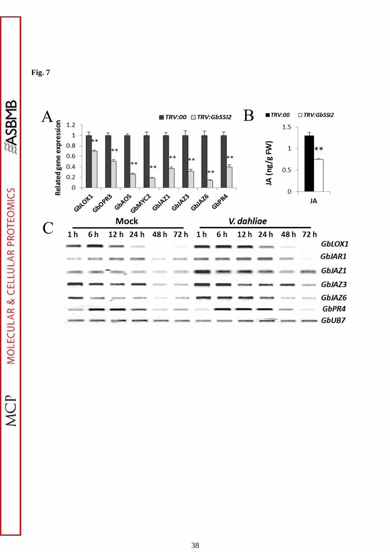
Fig. 7
38
Top Related