


Languages
Pages
Legal


ORIGINAL CONTRIBUTION
Protective effects of garlic extract on cardiac function, heart ratevariability, and cardiac mitochondria in obese insulin-resistantrats
Luerat Supakul • Hiranya Pintana •
Nattayaporn Apaijai • Siriporn Chattipakorn •
Krekwit Shinlapawittayatorn • Nipon Chattipakorn
Received: 17 July 2013 / Accepted: 3 October 2013
� Springer-Verlag Berlin Heidelberg 2013
Abstract
Purpose Garlic has been shown to exhibit antioxidant
effects and cardioprotective properties. However, the
effects of garlic extract on the heart in insulin resistance
induced by long-term high-fat-diet consumption are not
well defined. Therefore, we sought to determine the effects
of garlic extract in the obese insulin-resistant rats.
Methods Male Wistar rats (180–200 g) were divided into
two groups: normal-diet or high-fat-diet (n = 24/group)
fed for 12 weeks. Rats in each groups were divided into
three subgroups (n = 8 each): vehicle or garlic extract (250
or 500 mg/kg/day, respectively) treated for 28 days. At the
end of the treatment, the metabolic parameters, heart rate
variability (HRV), cardiac function, and cardiac mito-
chondrial function were determined.
Results Rats that received a high-fat-diet for 12 weeks had
increased body weight, visceral fat, plasma insulin levels, total
cholesterol, oxidative stress levels, depressed HRV, and cardiac
mitochondrial dysfunction. Garlic extract at both concentrations
significantly decreased the plasma insulin, total cholesterol,
homeostasis model assessment index, and oxidative stress lev-
els. Furthermore, garlic extract at both doses restored the HRV,
cardiac function, and cardiac mitochondrial function.
Conclusion We concluded that garlic extract at both
concentrations exerted cardioprotective effects against
cardiac dysfunction and mitochondrial dysfunction in
obese insulin-resistant rats.
Keywords Garlic extract � High-fat-diet � Insulin
resistance � Cardiac function �Mitochondrial function
Abbreviations
BCA Bicinchoninic acid
DCFDA Dichlorohydrofluorescein diacetate
ECG Electrocardiogram
EDP End diastolic pressure
EGTA Ethylene glycol bis (2-amino ethylether)-N,N,
N,N-tetraacetic acid
ESP End systolic pressure
HOMA Homeostasis model assessment
HPLC High-performance liquid chromatography
HR Heart rate
HRV Heart rate variability
JC-1 5,50,6,60-Tetrachloro-1,10,3,30-tetraethylbenzim-
idazolcarbocyanine iodide
MDA Malondialdehyde
DWm Mitochondrial membrane potential changes
ROS Reactive oxygen species
SV Stroke volume
TBA Thiobarbituric acid
Introduction
Long-term consumption of high-fat-diet is one of the
causes of obesity and has been shown to lead to insulin
L. Supakul � H. Pintana � N. Apaijai � S. Chattipakorn �K. Shinlapawittayatorn � N. Chattipakorn (&)
Cardiac Electrophysiology Research and Training Center,
Faculty of Medicine, Chiang Mai University, Chiang Mai 50200,
Thailand
e-mail: [email protected]
L. Supakul � H. Pintana � N. Apaijai � K. Shinlapawittayatorn �N. Chattipakorn
Cardiac Electrophysiology Unit, Department of Physiology,
Faculty of Medicine, Chiang Mai University, Chiang Mai,
Thailand
S. Chattipakorn
Department of Oral Biology and Diagnostic Science, Faculty
of Dentistry, Chiang Mai University, Chiang Mai, Thailand
123
Eur J Nutr
DOI 10.1007/s00394-013-0595-6

resistance [1]. Insulin resistance is part of the metabolic
syndrome and can be characterized by hyperinsulinemia
with euglycemia [2, 3]. A previous study demonstrated that
insulin resistance can lead to oxidative stress as indicated
by increased reactive oxygen species (ROS) and malondi-
aldehyde (MDA) levels [4]. In addition, insulin resistance
has been shown to affect the heart by altering both the
mechanical function and cardiac autonomic balance. In the
past decades, heart rate variability (HRV) has been used to
indicate cardiac autonomic tone balance. Depressed HRV
has been shown to be associated with a poor prognosis in
post-myocardial infarction and heart failure patients [5].
Previous studies also demonstrated that obese insulin-
resistant rats induced by a high-fat-diet consumption
developed left ventricular dysfunction as well as depressed
HRV [2]. Moreover, cardiac mitochondrial dysfunction
was also observed in these obese insulin-resistant rats,
which could be responsible for decreased left ventricular
function.
Garlic (Allium sativum) has been used as a spice or
medicinal herb for many centuries [6]. It contains several
sulfur compounds including allylmethy, diallyl, dimethyl-
monotohexasulfide, diallydisulfide, allylmethyltrisulfide,
diallytrisulfide, vinyldithiins and ajoenes [7]. However, it
has been shown that the most biologically active com-
pounds contained within garlic are allicin [8] and several
sulfur-based compounds [9]. Garlic has been shown to
exert beneficial effects such as antibacterial [10] and
antifungal properties [11]. Interestingly, various studies
have reported that garlic also exhibits cardio protective
properties such as prevention of hyperlipidemia [12],
antithrombotic [13], and antiarrhythmic effects [14]. In
addition, garlic has been shown to increase the antioxidant
level in rats with cardio toxicity induced by doxorubicin
[15]. Despite these beneficial effects of garlic extract, its
cardioprotective effects in insulin resistance induced by
long-term high-fat-diet consumption are not well defined.
In the present study, we determined the effects of garlic
extract on the heart in obese insulin-resistant rats induced
by long-term high-fat-diet consumption. We hypothesized
that garlic extract administration could improve insulin
resistance and attenuate cardiac dysfunction and cardiac
mitochondrial dysfunction, which are impaired by long-
term high-fat-diet consumption.
Materials and methods
Animals and diet
All experiments were approved by the Institutional Animal
Care and Use Committees of the Faculty of Medicine,
Chiang Mai University, Chiang Mai, Thailand. Male
Wistar rats weighing 180–200 g were obtained from the
National Animal Center, Salaya Campus, Mahidol Uni-
versity, Thailand. Rats were kept at room temperature, 12-h
light/dark cycle and water ad libitum. Rats were randomly
divided into two groups to receive either a normal-diet or a
high-fat-diet (n = 24/group) for 12 weeks. In the normal-
diet group, rats were fed with standard laboratory chow
containing 19.77 % energy from fat. In high-fat-diet group,
rats were fed with diet containing 59.28 % energy from fat.
Then, rats in each group were divided into three subgroups
to receive one of the following treatments: vehicle (normal
saline), garlic extract 250, or 500 mg/kg/day (n = 8/
group). All rats received these treatments by intragastric
gavage for 28 days. The garlic extract solution was pre-
pared from commercially available garlic extract powder
capsules (Immunitop, Bangkok, Thailand) dissolved in
deionized water (2 ml/kg of body weight). Each capsule
contains 370 mg of garlic extract, which has 3,500 lg of
allicin, or approximately 1 % allicin in garlic extract per
capsule. Garlic extract solution was administrated once
daily via gavage feeding. Food intake was recorded every
day, and body weight was recorded weekly. Lead II elec-
trocardiogram (ECG) was recorded for HRV determina-
tion. Both ECG and blood samples were collected at
baseline, 4, 8, 12 weeks and at the end of treatment. After
28 days of treatment, rats were anesthetized and cardiac
function was determined by using the pressure–volume
catheter (Scisense, Ontario, Canada) [16]. At the end of
each study, the heart was removed and divided into two
parts for determining cardiac mitochondrial function and
cardiac MDA levels.
Plasma glucose, cholesterol, and insulin level
determination
Plasma glucose and total cholesterol levels were measured
by colorimetric assay using a commercial kit (Biotech,
Bankok, Thailand) [3]. Plasma insulin levels were mea-
sured by a sandwich ELISA kit (Linco Research, St.
Charles, MO) [3]. Insulin resistance was assessed by using
the Homeostasis Model Assessment (HOMA) which is a
mathematical model describing the degree of insulin
resistance. A higher HOMA index indicates a higher
degree of insulin resistance [3].
HRV analysis
Heart rate variability was used to determine cardiac sym-
pathovagal balance by using Power Lab (AD Instrument,
Sydney, Australia) and Chart 5.0 program [17]. Lead II
ECG was recorded for HRV analysis. In the present study,
frequency domain method was used to represent the HRV
and were determined using a MATLAB program [2, 18].
Eur J Nutr
123
High-frequency (HF) component generally represents car-
diac parasympathetic activity, whereas low-frequency (LF)
component usually represents cardiac sympathetic and
parasympathetic activity. LF/HF ratio was used to indicate
cardiac sympathovagal balance [19].
Cardiac and plasma MDA level determination
Cardiac and plasma MDA levels were determined by using
high-performance liquid chromatography (HPLC) base
assay [16]. Cardiac tissues were homogenized in phosphate
buffer pH 2.8. Plasma and cardiac tissues were mixed with
H3PO4 and thiobarbituric acid (TBA) to create TBA-
reactive substances. The plasma and cardiac TBA-reactive
substance concentrations were determined directly from a
standard curve and reported as MDA equivalent concen-
trations [20].
Cardiac function measurement
Rats were anesthetized via intramuscular injection using a
combination of zoletil (50 mg/kg, Virbac, Laboratories,
Carros, France) and xylazine (0.15 mg/kg, Laboratory
Carlier, SA, Barcelona, Spain). A ventral midline incision
at the neck was performed for tracheostomy. The rats were
ventilated with room air. The right carotid artery was
cannulated with a pressure–volume catheter (Scisense,
Ontario, Canada) for measuring left ventricular pressure
and volume for 20 min [2, 21, 22]. Cardiac function
parameters including heart rate, end systolic and end dia-
stolic pressure, maximum and minimum dP/dt, and stroke
volume were determined using the analytical software
program (Labscribe, Dover, NH) [2, 21, 22].
Cardiac mitochondrial isolation and mitochondrial
function determination
Cardiac mitochondrial isolation was performed as reported
previously [23]. The ventricles were removed and
homogenized in ice-cold buffer containing sucrose
(300 mmol/l), TES sodium salt (5 mM), and ethylene
glycol bis (2-amino ethylether)-N,N,N,N-tetraacetic acid
(EGTA) (0.2 mM), pH 7.2 (4 �C). The tissue was finely
minced and homogenized by using a homogenizer. After-
ward, the homogenate was centrifuged at 8009g for 5 min.
The supernatant was collected and centrifuged at
8,8009g for 5 min. The mitochondrial pellet was sus-
pended in ice-cold buffer and centrifuged once more at
8,8009g for 5 min. Protein concentration was determined
via the bicinchoninic acid (BCA) assay [23]. In this
study, cardiac mitochondrial function was determined
by measuring the mitochondrial ROS production, mito-
chondrial membrane potential changes, and mitochondrial
swelling. The morphology of cardiac mitochondria was
also determined using the transmission electron micro-
scope [2, 21, 22].
Determination of cardiac mitochondrial ROS
production
ROS was measured with the dye dichlorohydrofluorescein
diacetate (DCFDA) [24]. Isolated cardiac mitochondria
was incubated with two lM DCFDA at 25 �C for 20 min.
DCFDA fluorescence is excited at the wavelength of
485 nm, and the emission is detected at the wavelength of
530 nm. ROS concentration was determined using a fluo-
rescent microplate reader.
Determination of cardiac mitochondrial membrane
potential changes
Cardiac mitochondrial membrane potential changes (DWm)
were measured using the dye 5,50,6,60-tetrachloro-1,10,3,30-tetraethylbenzimidazolcarbocyanine iodide (JC-1). Isolated
cardiac mitochondria were stained with JC-1 (5 lM) at
37 �C for 30 min [25]. Mitochondrial membrane potential
was determined as fluorescence intensity and was measured
by a fluorescent microplate reader. JC-1 monomer (green)
fluorescence was excited at the wavelength of 485 nm and
the emission detected at the wavelength of 530 nm. JC-1
aggregate form (red) fluorescence was excited at the
wavelength of 485 nm and the emission detected at the
wavelength of 590 nm. The change in mitochondrial
membrane potential was calculated as the ratio of red to
green fluorescence intensity. The depolarization of cardiac
mitochondrial membrane potential was indicated by a
decreased red/green fluorescence intensity ratio [2, 23, 26].
Determination of cardiac mitochondrial swelling
Isolated cardiac mitochondria (0.4 mg/ml) was incubated
in 1.5 ml of respiration buffer (containing 100 mM KCl,
50 mM sucrose, 10 mM HEPES, 5 mM KH2PO4, pH 7.4 at
37 �C) [23]. Cardiac mitochondrial swelling was detected
using the spectrophotometer at a wavelength of 540 nm. A
decrease in the absorbance of the mitochondrial suspension
indicated mitochondrial swelling [23]. Transmission elec-
tron microscope was used to visualize the mitochondrial
morphology [3, 21].
Statistical analysis
All data were expressed as mean ± SE. One way ANOVA
followed by LSD post hoc test was used to determine the
difference between groups. P value \0.05 was considered
statistically significant.
Eur J Nutr
123

Results
Effects of high-fat-diet consumption on metabolic
parameters in rats
At the baseline, metabolic parameters were not signifi-
cantly different between the normal-diet group and the
high-fat-diet group (Table 1). After 12 weeks of high-fat-
diet consumption, body weight, plasma cholesterol, vis-
ceral fat, and plasma and cardiac MDA levels were sig-
nificantly increased, compared with the normal-diet-fed
rats. High-fat-diet-fed rats exhibited a significant increase
in plasma insulin level. Moreover, high-fat-diet-fed rats
also exhibited a significant increase in the HOMA index,
which is an indicator of insulin resistance (Table 1).
Effects of garlic extract on metabolic parameters
in obese insulin-resistant rats
In normal-diet rats treated with garlic extract at the dose of
250 and 500 mg/kg/day, the metabolic parameters in these
groups were not different from those in normal-diet rats
treated with vehicle (Table 2). In high-fat-diet-fed rats,
garlic extract at the dose of 250 and 500 mg/kg/day sig-
nificantly reduced plasma cholesterol, plasma insulin,
HOMA index, visceral fat, and plasma and cardiac MDA
levels, without altering plasma glucose, body weight, and
food intake (Table 2). The metabolic parameters in high-
fat-diet-fed rats treated with garlic extract of both doses
were not different.
Effects of garlic extract on HRV in obese insulin-
resistant rats
At the baseline, the LF/HF ratio was not different between
normal-diet- and high-fat-diet-fed rats. In high-fat-diet rats,
an increased in LF/HF ratio was observed at week eight
and reached the maximal level at week 12 (Fig. 1), indi-
cating depressed HRV. After the treatment of garlic extract
for 28 days at the dose of 250 and 500 mg/kg/day, the LF/
HF ratio was significantly decreased, compared with high-
fat-diet rats treated with vehicle (Fig. 2). There was no
Table 1 Metabolic parameters of normal-diet- and high-fat-diet-fed rats at baseline and at week 12
Metabolic parameters Baseline Week 12
ND HF ND HF
Body weight (g) 189 ± 4 190 ± 4 424 ± 9* 545 ± 18*,#
Food intake (g) 24 ± 0.31 25 ± 0.39 23 ± 1 23.92 ± 1
Plasma insulin (ng/ml) 1.59 ± 0.52 1.57 ± 0.42 1.93 ± 0.05 3.66 ± 0.95*,#
Plasma glucose (mg/dl) 137 ± 6 140 ± 13 147 ± 7 149 ± 5
HOMA index 14.16 ± 4.66 17.87 ± 3.16 16.68 ± 3.16 28.35 ± 2.16*,#
Plasma total cholesterol (mg/dl) 65 ± 13 63 ± 14 77 ± 2 105 ± 6*,#
Plasma MDA (lmol/ml) 2.08 ± 0.11 1.97 ± 0.16 2.17 ± 0.23 5.28 ± 1*,#
ND normal-diet, HF high-fat-diet
* P \ 0.05 versus Baseline, # P \ 0.05 versus ND week 12
Table 2 Effects of garlic extracts dose 250 and 500 mg/kg on metabolic parameters of normal- and high-fat-diet-fed rats
Metabolic parameters NDV NDG250 NDG500 HFV HFG250 HFG500
Body weight (g) 461 ± 11 467 ± 17 476 ± 16 567 ± 17* 571 ± 16* 574 ± 27*
Food intake (g) 22 ± 0.72 21 ± 0.79 21 ± 0.70 23 ± 0.59 23 ± 0.72 23 ± 0.35
Visceral fat (g) 27 ± 5 20 ± 2 22 ± 1 63 ± 4* 50 ± 3*,# 51 ± 3*,#
Plasma insulin (ng/ml) 1.97 ± 0.30 1.31 ± 0.63 1.56 ± 0.29 3.84 ± 0.39* 1.56 ± 0.1# 2.26 ± 0.41#
Plasma glucose (mg/dl) 155 ± 7 150 ± 13 142 ± 9 161 ± 12 149 ± 9 143 ± 14
HOMA index 16.19 ± 0.36 11.78 ± 3.44 13.79 ± 2.87 25.64 ± 1.52* 13.52 ± 1.29# 17.64 ± 3.13#
Plasma total cholesterol (mg/dl) 83 ± 7 76 ± 10 76 ± 4 161 ± 9* 84 ± 6# 79 ± 9#
Plasma MDA (lmol/ml) 1.96 ± 0.09 1.88 ± 0.02 1.87 ± 0.11 6.91 ± 0.04* 1.76 ± 0.04*,# 1.85 ± 0.06#
Cardiac MDA (lmol/mg protein) 5.94 ± 2.14 5.29 ± 0.82 5.54 ± 0.89 11.67 ± 2.53* 5.57 ± 1.16# 5.03 ± 0.62#
NDV normal-diet ? vehicle, NDG250 normal-diet ? garlic extract at dose 250 mg/kg, NDG500 normal-diet ? garlic extract at dose 500 mg/
kg, HFV high-fat-diet ? vehicle, HFG250 high-fat-diet ? garlic extract at dose 250 mg/kg, HFG500 high-fat-diet ? garlic extract at dose
500 mg/kg
* P \ 0.05 versus NDV, # P \ 0.05 versus HFV
Eur J Nutr
123

difference in the LF/HF ratio in high-fat-diet rats treated
with garlic extract of both doses. Furthermore, the LF/HF
ratio in the HF groups treated with garlic extract of both
doses was not different from the NDV group.
Effects of garlic extract on cardiac function in insulin-
resistant rats
In the normal-diet groups, heart rate (HR), end systolic
pressure (ESP), end diastolic pressure (EDP), -dP/dt, ?dP/
dt, and stroke volume (SV) were not different among
rats treated with vehicle, 250-mg/kg garlic extract, and
500-mg/kg garlic extract. In high-fat-diet rats, HR, EDP,
and -dP/dt were significantly increased, whereas the ESP,
?dP/dt, and SV were decreased, compared with rats on a
normal-diet. Interestingly, in high-fat-diet groups treated
with garlic extract at doses of 250 and 500 mg/kg/day, HR,
EDP, and -dP/dt were significantly decreased, whereas the
ESP, ?dP/dt, and SV were significantly increased, com-
pared with high-fat-diet rats (Table 3). There was no dif-
ference in the cardiac function parameters in the high-fat-
diet rats treated with garlic extract of both doses.
Effects of garlic extract on cardiac mitochondrial
function
Cardiac mitochondrial ROS production
In the normal-diet group treated with garlic extract at doses
of 250 and 500 mg/kg/day, the ROS level was not different
from that in the normal-diet group treated with vehicle
(Fig. 3). In the high-fat-diet group treated with vehicle,
ROS production was significantly increased compared with
the normal-diet group. In high-fat rats treated with both
garlic extract doses, the ROS level was significantly
decreased compared with the high-fat-diet group treated
with vehicle (Fig. 3). There was no difference in the ROS
production in the high-fat-diet rats treated with garlic
extract of both doses.
Cardiac mitochondrial membrane potential changes
(DWm)
In the normal-diet rats treated with garlic extract at doses
of 250 and 500 mg/kg/day, DWm were not different com-
pared to normal-diet rats treated with vehicle (Fig. 4). In
the high-fat-diet group treated with vehicle, DWm were
significantly decreased when compared with the normal-
diet group, indicating mitochondrial depolarization. In both
garlic-extract-treated groups, DWm were significantly
increased when compared with high-fat-diet group treated
with vehicle and was not different from the normal-diet
group (Fig. 4). There was no difference in the DWm in the
high-fat-diet rats treated with garlic extract of both doses.
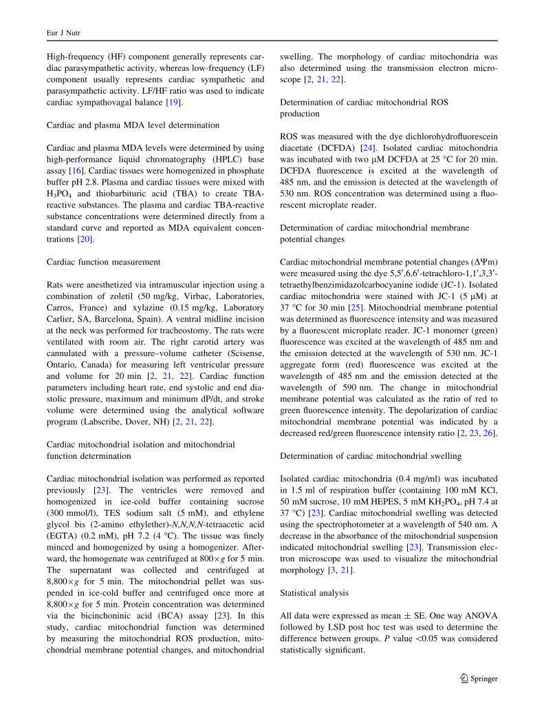
Cardiac mitochondrial swelling
In the normal-diet group treated with garlic extract at doses
of 250 and 500 mg/kg/day, the absorbance was not dif-
ferent when compared with normal-diet-fed rats (Fig. 5). In
high-fat-diet group treated with vehicle, the absorbance
was significantly decreased when compared with normal-
diet group, indicating cardiac mitochondrial swelling.
However, in garlic-extract-treated groups, the absorbance
was restored back to a normal level (Fig. 5). There was no
difference in the absorbance in the high-fat-diet rats treated
Fig. 1 LF/HF ratio in normal-diet- and high-fat-diet-fed rats. LF/HF
ratio was increased at week eight and 12 of high-fat-diet consumption,
indicating impaired cardiac sympathovagal balance. LF/HF low-
frequency/high-frequency ratio, W4 week four of high-fat-diet con-
sumption, W8 week eight of high-fat-diet consumption, W12 week 12
of high-fat-diet consumption, ND normal-diet group, HF high-fat-diet
group. *P \ 0.05 versus baseline
Fig. 2 LF/HF ratio in normal-diet and high-fat-diet rats treated with
garlic extract at the doses of 250 and 500 mg/kg/day. Garlic extract at
both doses could reduce LF/HF ratio back to the normal level. LF/HF
low-frequency/high-frequency ratio, ND normal-diet group, HF
high-fat-diet group, NDV normal-diet ? vehicle, NDG250 normal-
diet ? garlic extract at the dose of 250 mg/kg, NDG500 normal-
diet ? garlic extract at the dose of 500 mg/kg, HFV high-
fat-diet ? vehicle, HFG250 high-fat-diet ? garlic extract at the dose
of 250 mg/kg, HFG500 high-fat-diet ? garlic extract at the dose of
500 mg/kg. *P \ 0.05 versus NDV, #P \ 0.05 versus HFV
Eur J Nutr
123

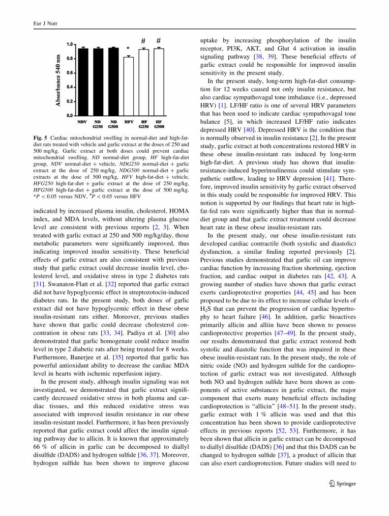
with garlic extract of both doses. Representative pictures of
cardiac mitochondria from an electron microscope are
shown in Fig. 6. Electron microscopic images revealed that
high-fat-diet caused unfolded cristae compared to normal
cardiac mitochondria, with marked swelling in the high-fat-
diet treated with vehicle group (Fig. 6d). Garlic extract at
both concentrations restored cardiac mitochondrial swell-
ing caused by high-fat-diet-induced insulin resistance
(Fig. 6e, f).
Discussion
The major findings of this study are as follows. In obese
insulin-resistant rats induced by long-term high-fat-diet
consumption, garlic extract could (1) improve metabolic
parameters and oxidative stress, (2) restore cardiac sym-
pathovagal tone balance, (3) prevent cardiac systolic and
diastolic dysfunction, and (4) restore cardiac mitochondrial
dysfunction. Previous studies demonstrated that long-term
high-fat-diet-fed rats for 12 weeks could cause insulin
resistance [1, 2, 26], by reducing the interaction between
insulin and insulin receptor substrate-1 (IRS-1), leading to
decreased insulin sensitivity and insulin resistance [27]. It
has been shown that insulin resistance can cause oxidative
stress by increasing ROS production leading to cardio-
vascular disease [28]. Since insulin resistance is strongly
associated with oxidative stress, reducing oxidative stress
under this condition has been shown to improve insulin
sensitivity as well as cardiac function [29, 30]. In the
present study, our findings that rats fed with high-fat-diet
for 12 weeks have developed insulin resistance as
Table 3 Effects of garlic extracts on cardiac function and hemodynamic parameters of normal-diet- and high-fat-diet-fed rats
Cardiac function NDV NDG250 NDG500 HFV HFG250 HFG500
HR(bpm) 324 ± 24 311.05 ± 40 333 ± 39 419 ± 3* 307 ± 27# 315 ± 19#
ESP(mmHg) 132 ± 8 145 ± 15 148 ± 12 92 ± 14* 134 ± 9# 141 ± 9#
EDP(mmHg) 16 ± 2 16 ± 0.02 15 ± 1.19 40 ± 3.12* 16 ± 0.28# 16 ± 0.23#
?dP/dt(mmHg/sec) 8,828 ± 43 9,830 ± 130 9,336 ± 351 5,410 ± 218* 9,894 ± 66# 9,612 ± 85#
-dP/dt(mmHg/sec) -5,960 ± 274 -5,030 ± 691 -5,240 ± 367 -3,809 ± 224* -5,074 ± 683# -5,334 ± 658#
SV(ll/g) 1.06 ± 0.04 1.03 ± 0.11 1.08 ± 0.07 0.74 ± 0.05* 0.94 ± 0.05# 0.99 ± 0.05#
NDV normal-diet ? vehicle, NDG250 normal-diet ? garlic extract at dose 250 mg/kg, NDG500 normal-diet ? garlic extract at dose 500 mg/
kg, HFV high-fat-diet ? vehicle, HFG250 high-fat-diet ? garlic extract at dose 250 mg/kg, HFG500 high-fat-diet ? garlic extract at dose
500 mg/kg
* P \ 0.05 versus NDV, # P \ 0.05 versus HFV
Fig. 3 Cardiac mitochondrial ROS production in normal-diet and
high-fat-diet rats treated with vehicle and garlic extract at the doses of
250 and 500 mg/kg. Garlic extract at both doses could reduce ROS
production in high-fat-diet-fed rats back to normal level. ROS reactive
oxygen species, ND normal-diet group, HF high-fat-diet group, NDV
normal-diet ? vehicle, NDG250 normal-diet ? garlic extract at the
dose of 250 mg/kg, NDG500 normal-diet ? garlic extract at the dose
of 500 mg/kg, HFV high-fat-diet ? vehicle, HFG250 high-fat-
diet ? Garlic extract at the dose of 250 mg/kg, HFG500 high-fat-
diet ? garlic extract at the dose of 500 mg/kg. *P \ 0.05 versus
NDV, #P \ 0.05 versus HFV
Fig. 4 Cardiac mitochondrial membrane potential changes in nor-
mal-diet and high-fat-diet rats treated with vehicle and garlic extract
at the doses of 250 and 500 mg/kg. Garlic extract at both doses
completely prevented cardiac mitochondria membrane depolarization.
ND normal-diet group, HF high-fat-diet group, NDV normal-
diet ? vehicle, NDG250 normal-diet ? garlic extract at the dose of
250 mg/kg, NDG500 normal-diet ? garlic extract at the dose of
500 mg/kg, HFV high-fat-diet ? vehicle, HFG250 high-fat-
diet ? garlic extract at the dose of 250 mg/kg, HFG500 high-fat-
diet ? garlic extract at the dose of 500 mg/kg. *P \ 0.05 versus
NDV, #P \ 0.05 versus HFV
Eur J Nutr
123
indicated by increased plasma insulin, cholesterol, HOMA
index, and MDA levels, without altering plasma glucose
level are consistent with previous reports [2, 3]. When
treated with garlic extract at 250 and 500 mg/kg/day, those
metabolic parameters were significantly improved, thus
indicating improved insulin sensitivity. These beneficial
effects of garlic extract are also consistent with previous
study that garlic extract could decrease insulin level, cho-
lesterol level, and oxidative stress in type 2 diabetes rats
[31]. Swanaton-Flatt et al. [32] reported that garlic extract
did not have hypoglycemic effect in streptozotocin-induced
diabetes rats. In the present study, both doses of garlic
extract did not have hypoglycemic effect in these obese
insulin-resistant rats either. Moreover, previous studies
have shown that garlic could decrease cholesterol con-
centration in obese rats [33, 34]. Padiya et al. [30] also
demonstrated that garlic homogenate could reduce insulin
level in type 2 diabetic rats after being treated for 8 weeks.
Furthermore, Banerjee et al. [35] reported that garlic has
powerful antioxidant ability to decrease the cardiac MDA
level in hearts with ischemic reperfusion injury.
In the present study, although insulin signaling was not
investigated, we demonstrated that garlic extract signifi-
cantly decreased oxidative stress in both plasma and car-
diac tissues, and this reduced oxidative stress was
associated with improved insulin resistance in our obese
insulin-resistant model. Furthermore, it has been previously
reported that garlic extract could affect the insulin signal-
ing pathway due to allicin. It is known that approximately
66 % of allicin in garlic can be decomposed to diallyl
disulfide (DADS) and hydrogen sulfide [36, 37]. Moreover,
hydrogen sulfide has been shown to improve glucose
uptake by increasing phosphorylation of the insulin
receptor, PI3K, AKT, and Glut 4 activation in insulin
signaling pathway [38, 39]. These beneficial effects of
garlic extract could be responsible for improved insulin
sensitivity in the present study.
In the present study, long-term high-fat-diet consump-
tion for 12 weeks caused not only insulin resistance, but
also cardiac sympathovagal tone imbalance (i.e., depressed
HRV) [1]. LF/HF ratio is one of several HRV parameters
that has been used to indicate cardiac sympathovagal tone
balance [5], in which increased LF/HF ratio indicates
depressed HRV [40]. Depressed HRV is the condition that
is normally observed in insulin resistance [2]. In the present
study, garlic extract at both concentrations restored HRV in
these obese insulin-resistant rats induced by long-term
high-fat-diet. A previous study has shown that insulin-
resistance-induced hyperinsulinemia could stimulate sym-
pathetic outflow, leading to HRV depression [41]. There-
fore, improved insulin sensitivity by garlic extract observed
in this study could be responsible for improved HRV. This
notion is supported by our findings that heart rate in high-
fat-fed rats were significantly higher than that in normal-
diet group and that garlic extract treatment could decrease
heart rate in these obese insulin-resistant rats.
In the present study, our obese insulin-resistant rats
developed cardiac contractile (both systolic and diastolic)
dysfunction, a similar finding reported previously [2].
Previous studies demonstrated that garlic oil can improve
cardiac function by increasing fraction shortening, ejection
fraction, and cardiac output in diabetes rats [42, 43]. A
growing number of studies have shown that garlic extract
exerts cardioprotective properties [44, 45] and has been
proposed to be due to its effect to increase cellular levels of
H2S that can prevent the progression of cardiac hypertro-
phy to heart failure [46]. In addition, garlic bioactives
primarily allicin and alliin have been shown to possess
cardioprotective properties [47–49]. In the present study,
our results demonstrated that garlic extract restored both
systolic and diastolic function that was impaired in these
obese insulin-resistant rats. In the present study, the role of
nitric oxide (NO) and hydrogen sulfide for the cardiopro-
tection of garlic extract was not investigated. Although
both NO and hydrogen sulfide have been shown as com-
ponents of active substances in garlic extract, the major
component that exerts many beneficial effects including
cardioprotection is ‘‘allicin’’ [48–51]. In the present study,
garlic extract with 1 % allicin was used and that this
concentration has been shown to provide cardioprotective
effects in previous reports [52, 53]. Furthermore, it has
been shown that allicin in garlic extract can be decomposed
to diallyl disulfide (DADS) [36] and that this DADS can be
changed to hydrogen sulfide [37], a product of allicin that
can also exert cardioprotection. Future studies will need to
Fig. 5 Cardiac mitochondrial swelling in normal-diet and high-fat-
diet rats treated with vehicle and garlic extract at the doses of 250 and
500 mg/kg. Garlic extract at both doses could prevent cardiac
mitochondrial swelling. ND normal-diet group, HF high-fat-diet
group, NDV normal-diet ? vehicle, NDG250 normal-diet ? garlic
extract at the dose of 250 mg/kg, NDG500 normal-diet ? garlic
extracts at the dose of 500 mg/kg, HFV high-fat-diet ? vehicle,
HFG250 high-fat-diet ? garlic extract at the dose of 250 mg/kg,
HFG500 high-fat-diet ? garlic extract at the dose of 500 mg/kg.
*P \ 0.05 versus NDV, #P \ 0.05 versus HFV
Eur J Nutr
123
explore possible role of NO and hydrogen sulfide on car-
dioprotection in this obese insulin-resistant model.
The heart is an organ that consumes much energy for
contraction and relaxation [54]. Cardiac mitochondria are
crucial organelles which generate energy for cardiomyo-
cytes [55]. In this study, we found that high-fat-diet con-
sumption for 12 weeks led to cardiac mitochondrial
dysfunction as indicated by increased mitochondrial ROS
Fig. 6 Representative pictures of cardiac ultrastructural morphology
by transmission electron microscopy (original magnification,
915,000). Normal cardiac mitochondria from the heart of the
normal-diet-fed rat is shown with apparent folded cristae (a). Cardiac
mitochondria from the heart of normal-diet-fed rat treated with garlic
250 mg/kg (b) and 500 mg/kg (c) also demonstrated apparent folded
cristae similar to that seen in (a). Cardiac mitochondrial swelling was
observed in the high-fat-diet-fed rat as indicated by unfolded cristae,
compared to that in the normal-diet group (d). Garlic extract at
250 mg/kg (e) and 500 mg/kg (f) preserved cardiac mitochondrial
morphology in the heart of obese insulin-resistant rats, as indicated by
clear folded cristae in cardiac mitochondria. ND normal-diet, G250
garlic extract at the dose of 250 mg/kg, G500 garlic extracts at the
dose of 500 mg/kg, HF high-fat-diet
Eur J Nutr
123

production, mitochondrial membrane potential depolariza-
tion, and mitochondrial swelling. We found that garlic
extract treatment restored cardiac mitochondrial function
by decreased mitochondrial ROS production and prevented
mitochondrial membrane depolarization and mitochondrial
swelling. The improved cardiac mitochondrial function by
garlic extract treatment in these obese insulin-resistant rats
could also be responsible for improved cardiac contractile
function found in this study.
In conclusion, long-term high-fat-diet consumption for
12 weeks could lead to insulin resistance, HRV depression,
cardiac contractile dysfunction, and cardiac mitochondrial
dysfunction. Treatment with garlic extract could attenuate
insulin resistance, HRV depression, cardiac dysfunc-
tion, and cardiac mitochondrial dysfunction in obese
insulin-resistant rats induced by long-term high-fat-diet
consumption.
Acknowledgments This work is supported by the Thailand
Research Fund Senior Scholar Grant RTA 5580006 (NC), BRG
5480003 (SC), and MRG5580125 (KS).
Conflict of interest The authors declare that they have no conflict
of interest.
References
1. Pratchayasakul W, Kerdphoo S, Petsophonsakul P, Pongchaide-
cha A, Chattipakorn N, Chattipakorn SC (2011) Effects of high-
fat diet on insulin receptor function in rat hippocampus and the
level of neuronal corticosterone. Life Sci 88:619–627
2. Apaijai N, Pintana H, Chattipakorn SC, Chattipakorn N (2012)
Cardioprotective effects of metformin and vildagliptin in adult
rats with insulin resistance induced by a high-fat diet. Endocri-
nology 153:3878–3885
3. Pipatpiboon N, Pratchayasakul W, Chattipakorn N, Chattipakorn
SC (2012) PPARgamma agonist improves neuronal insulin
receptor function in hippocampus and brain mitochondria func-
tion in rats with insulin resistance induced by long term high-fat
diets. Endocrinology 153:329–338
4. Houstis N, Rosen ED, Lander ES (2006) Reactive oxygen species
have a causal role in multiple forms of insulin resistance. Nature
440:944–948
5. Chattipakorn N, Incharoen T, Kanlop N, Chattipakorn S (2007)
Heart rate variability in myocardial infarction and heart failure.
Int J Cardiol 120:289–296
6. Sungnoon R, Chattipakorn N (2005) Anti-arrhythmic effects of
herbal medicine. Indian Heart J 57:109–113
7. Gupta N, Porter TD (2001) Garlic and garlic-derived compounds
inhibit human squalene monooxygenase. J Nutr 131:1662–1667
8. Ali M, Al-Qattan KK, Al-Enezi F, Khanafer RM, Mustafa T
(2000) Effect of allicin from garlic powder on serum lipids and
blood pressure in rats fed with a high cholesterol diet. Prosta-
glandins Leukot Essent Fatty Acids 62:253–259
9. Weber ND, Andersen DO, North JA, Murray BK, Lawson LD,
Hughes BG (1992) In vitro virucidal effects of Allium sativum
(garlic) extract and compounds. Planta Med 58:417–423
10. Harris JC, Cottrell SL, Plummer S, Lloyd D (2001) Antimicrobial
properties of Allium sativum (garlic). Appl Microbiol Biotechnol
57:282–286
11. Yoshida S, Kasuga S, Hayashi N, Ushiroguchi T, Matsuura H,
Nakagawa S (1987) Antifungal activity of ajoene derived from
garlic. Appl Environ Microbiol 53:615–617
12. Pedraza-Chaverri J, Medina-Campos ON, Granados-Silvestre
MA, Maldonado PD, Olivares-Corichi IM, Hernandez-Pando R
(2000) Garlic ameliorates hyperlipidemia in chronic aminonu-
cleoside nephrosis. Mol Cell Biochem 211:69–77
13. Ali M, Thomson M, Alnaqeeb MA, Al-Hassan JM, Khater SH,
Gomes SA (1990) Antithrombotic activity of garlic: its inhibition
of the synthesis of thromboxane-B2 during infusion of arachi-
donic acid and collagen in rabbits. Prostaglandins Leukot Essent
Fatty Acids 41:95–99
14. Isensee H, Rietz B, Jacob R (1993) Cardioprotective actions of
garlic (Allium sativum). Arzneimittelforschung 43:94–98
15. Alkreathy H, Damanhouri ZA, Ahmed N, Slevin M, Ali SS,
Osman AM (2010) Aged garlic extract protects against doxoru-
bicin-induced cardiotoxicity in rats. Food Chem Toxicol
48:951–956
16. Thephinlap C, Phisalaphong C, Lailerd N, Chattipakorn N,
Winichagoon P, Vadolus J, Fucharoen S, Porter JB, Srichaira-
tanakool S (2011) Reversal of cardiac iron loading and dys-
function in thalassemic mice by curcuminoids. Med Chem
7:62–69
17. Pongchaidecha A, Lailerd N, Boonprasert W, Chattipakorn N
(2009) Effects of curcuminoid supplement on cardiac autonomic
status in high-fat-induced obese rats. Nutrition 25:870–878
18. Apaijai N, Pintana H, Chattipakorn SC, Chattipakorn N (2013)
Effects of vildagliptin vs. sitagliptin, on cardiac function, heart
rate variability, and mitochondrial function in obese insulin
resistant rats. Br J Pharmacol
19. Ohuchi H, Suzuki H, Yasuda K, Arakaki Y, Echigo S, Kamiya T
(2000) Heart rate recovery after exercise and cardiac autonomic
nervous activity in children. Pediatr Res 47:329–335
20. Mateos R, Lecumberri E, Ramos S, Goya L, Bravo L (2005)
Determination of malondialdehyde (MDA) by high-performance
liquid chromatography in serum and liver as a biomarker for
oxidative stress. Application to a rat model for hypercholester-
olemia and evaluation of the effect of diets rich in phenolic
antioxidants from fruits. J Chromatogr B Analyt Technol Biomed
Life Sci 827:76–82
21. Palee S, Weerateerangkul P, Surinkeaw S, Chattipakorn S,
Chattipakorn N (2011) Effect of rosiglitazone on cardiac elec-
trophysiology, infarct size and mitochondrial function in ischae-
mia and reperfusion of swine and rat heart. Exp Physiol
96:778–789
22. Surinkaew S, Kumphune S, Chattipakorn S, Chattipakorn N
(2013) Inhibition of p38 MAPK during ischemia, but not reper-
fusion, effectively attenuates fatal arrhythmia in ischemia/reper-
fusion heart. J Cardiovasc Pharmacol 61:133–141
23. Thummasorn S, Kumfu S, Chattipakorn S, Chattipakorn N (2011)
Granulocyte-colony stimulating factor attenuates mitochondrial
dysfunction induced by oxidative stress in cardiac mitochondria.
Mitochondrion 11:457–466
24. Novalija E, Kevin LG, Eells JT, Henry MM, Stowe DF (2003)
Anesthetic preconditioning improves adenosine triphosphate
synthesis and reduces reactive oxygen species formation in
mitochondria after ischemia by a redox dependent mechanism.
Anesthesiology 98:1155–1163
25. Tong V, Teng XW, Chang TK, Abbott FS (2005) Valproic acid
II: effects on oxidative stress, mitochondrial membrane potential,
and cytotoxicity in glutathione-depleted rat hepatocytes. Toxicol
Sci 86:436–443
26. Pintana H, Apaijai N, Pratchayasakul W, Chattipakorn N, Chat-
tipakorn SC (2012) Effects of metformin on learning and memory
behaviors and brain mitochondrial functions in high fat diet
induced insulin resistant rats. Life Sci 91:409–414
Eur J Nutr
123
27. Griffin ME, Marcucci MJ, Cline GW, Bell K, Barucci N, Lee D,
Goodyear LJ, Kraegen EW, White MF, Shulman GI (1999) Free
fatty acid-induced insulin resistance is associated with activation
of protein kinase C theta and alterations in the insulin signaling
cascade. Diabetes 48:1270–1274
28. Ceconi C, Boraso A, Cargnoni A, Ferrari R (2003) Oxidative
stress in cardiovascular disease: myth or fact? Arch Biochem
Biophys 420:217–221
29. Hfaiedh N, Murat JC, Elfeki A (2011) Compared ability of garlic
(Allium sativum) extract or alpha-tocopherol ? magnesium
association to reduce metabolic disorders and oxidative stress in
diabetic rats. Phytother Res 25:821–827
30. Padiya R, Khatua TN, Bagul PK, Kuncha M, Banerjee SK (2011)
Garlic improves insulin sensitivity and associated metabolic
syndromes in fructose fed rats. Nutr Metab (Lond) 8:53
31. Saravanan G, Prakash J (2004) Effect of garlic (Allium sativum)
on lipid peroxidation in experimental myocardial infarction in
rats. J Ethnopharmacol 94:155–158
32. Swanston-Flatt SK, Day C, Bailey CJ, Flatt PR (1990) Traditional
plant treatments for diabetes. Studies in normal and streptozoto-
cin diabetic mice. Diabetologia 33:462–464
33. Warshafsky S, Kamer RS, Sivak SL (1993) Effect of garlic on
total serum cholesterol. A meta-analysis. Ann Intern Med
119:599–605
34. Yeh YY, Liu L (2001) Cholesterol-lowering effect of garlic
extracts and organosulfur compounds: human and animal studies.
J Nutr 131:989S–993S
35. Banerjee SK, Dinda AK, Manchanda SC, Maulik SK (2002)
Chronic garlic administration protects rat heart against oxidative
stress induced by ischemic reperfusion injury. BMC Pharmacol 2:16
36. Amagase H (2006) Clarifying the real bioactive constituents of
garlic. J Nutr 136:716S–725S
37. Benavides GA, Squadrito GL, Mills RW, Patel HD, Isbell TS,
Patel RP, Darley-Usmar VM, Doeller JE, Kraus DW (2007)
Hydrogen sulfide mediates the vasoactivity of garlic. Proc Natl
Acad Sci U S A 104:17977–17982
38. Manna P, Jain SK (2011) Hydrogen sulfide and L-cysteine
increase phosphatidylinositol 3,4,5-trisphosphate (PIP3) and
glucose utilization by inhibiting phosphatase and tensin homolog
(PTEN) protein and activating phosphoinositide 3-kinase
(PI3 K)/serine/threonine protein kinase (AKT)/protein kinase
Czeta/lambda (PKCzeta/lambda) in 3T3l1 adipocytes. J Biol
Chem 286:39848–39859
39. Xue R, Hao DD, Sun JP, Li WW, Zhao MM, Li XH, Chen Y, Zhu
JH, Ding YJ, Liu J, Zhu YC (2013) Hydrogen sulfide treatment
promotes glucose uptake by increasing insulin receptor sensitivity
and ameliorates kidney lesions in type 2 diabetes. Antioxid
Redox Signal 19:5–23
40. Incharoen T, Thephinlap C, Srichairatanakool S, Chattipakorn S,
Winichagoon P, Fucharoen S, Vadolas J, Chattipakorn N (2007)
Heart rate variability in beta-thalassemic mice. Int J Cardiol
121:203–204
41. Emdin M, Gastaldelli A, Muscelli E, Macerata A, Natali A,
Camastra S, Ferrannini E (2001) Hyperinsulinemia and
autonomic nervous system dysfunction in obesity: effects of
weight loss. Circulation 103:513–519
42. Alkreathy H, Damanhouri ZA, Ahmed N, Slevin M, Ali SS,
Osman AM (2010) Aged garlic extract protects against doxoru-
bicin-induced cardiotoxicity in rats. Food Chem Toxicol
48:951–956
43. Chang SH, Liu CJ, Kuo CH, Chen H, Lin WY, Teng KY, Chang
SW, Tsai CH, Tsai FJ, Huang CY, Tzang BS, Kuo WW (2011)
Garlic Oil Alleviates MAPKs- and IL-6-mediated Diabetes-
related Cardiac Hypertrophy in STZ-induced DM Rats. Evid
Based Complement Alternat Med 2011:950150
44. Rahman K, Lowe GM (2006) Garlic and cardiovascular disease:
a critical review. J Nutr 136:736S–740S
45. Ried K, Frank OR, Stocks NP, Fakler P, Sullivan T (2008) Effect
of garlic on blood pressure: a systematic review and meta-ana-
lysis. BMC Cardiovasc Disord 8:13
46. Givvimani S, Munjal C, Gargoum R, Sen U, Tyagi N, Vacek JC,
Tyagi SC (2011) Hydrogen sulfide mitigates transition from
compensatory hypertrophy to heart failure. J Appl Physiol
110:1093–1100
47. Asdaq SM, Inamdar MN (2010) Potential of garlic and its active
constituent, S-allyl cysteine, as antihypertensive and cardiopro-
tective in presence of captopril. Phytomedicine 17:1016–1026
48. Liu C, Cao F, Tang QZ, Yan L, Dong YG, Zhu LH, Wang L,
Bian ZY, Li H (2010) Allicin protects against cardiac hypertro-
phy and fibrosis via attenuating reactive oxygen species-depen-
dent signaling pathways. J Nutr Biochem 21:1238–1250
49. Sun X, Ku DD (2006) Allicin in garlic protects against coronary
endothelial dysfunction and right heart hypertrophy in pulmonary
hypertensive rats. Am J Physiol Heart Circ Physiol 291:H2431–
H2438
50. Li XH, Li CY, Xiang ZG, Hu JJ, Lu JM, Tian RB, Jia W (2012)
Allicin ameliorates cardiac hypertrophy and fibrosis through
enhancing of Nrf2 antioxidant signaling pathways. Cardiovasc
Drugs Ther 26:457–465
51. Liu Y, Qi H, Wang Y, Wu M, Cao Y, Huang W, Li L, Ji Z, Sun
H (2012) Allicin protects against myocardial apoptosis and
fibrosis in streptozotocin-induced diabetic rats. Phytomedicine
19:693–698
52. Cho SJ, Rhee DK, Pyo S (2006) Allicin, a major component of
garlic, inhibits apoptosis of macrophage in a depleted nutritional
state. Nutrition 22:1177–1184
53. Zhang Y, Yao HP, Huang FF, Wu W, Gao Y, Chen ZB, Liang
ZY, Liang TB (2008) Allicin, a major component of garlic,
inhibits apoptosis in vital organs in rats with trauma/hemorrhagic
shock. Crit Care Med 36:3226–3232
54. Matsuoka S, Sarai N, Jo H, Noma A (2004) Simulation of ATP
metabolism in cardiac excitation-contraction coupling. Prog
Biophys Mol Biol 85:279–299
55. Lehman JJ, Barger PM, Kovacs A, Saffitz JE, Medeiros DM,
Kelly DP (2000) Peroxisome proliferator-activated receptor
gamma coactivator-1 promotes cardiac mitochondrial biogenesis.
J Clin Invest 106:847–856
Eur J Nutr
123
Top Related