
![RESEARCH Open Access Female sex hormones mediate the ...€¦ · asthma [29], ameliorating lung inflammation and decreasing lung remodeling in murine asthma model [30]. Taking these](https://static.fdocuments.in/doc/165x107/60e8a293f8b4061fc9701e9f/research-open-access-female-sex-hormones-mediate-the-asthma-29-ameliorating.jpg)


Languages
Pages
Legal

JPET #215152
1
Title page
Prevention of Bleomycin-Induced Lung Inflammation and Fibrosis in Mice by Naproxen and JNJ7777120 Treatment Arianna Carolina Rosa, Alessandro Pini, Laura Lucarini, Cecilia Lanzi, Eleonora Veglia, Robin L. Thurmond, Holger Stark and Emanuela Masini
Departments of Drug Science and Technology, University of Turin, Turin, Italy (A.C.R., E.V.); NEUROFARBA, Section of Pharmacology (L.L., C.L., E.M.); Experimental and Clinical Medicine (A.P.), University of Florence, 50139 Florence, Italy; Janssen Research & Development, L.L.C., San Diego, CA, USA (R.L.T) and Heinrich-Heine Düsseldorf University, Institute of Medicinal Chemistry, D40225 Düsseldorf, Germany (H.S.)
This article has not been copyedited and formatted. The final version may differ from this version.JPET Fast Forward. Published on September 2, 2014 as DOI: 10.1124/jpet.114.215152
at ASPE
T Journals on D
ecember 11, 2021
jpet.aspetjournals.orgD
ownloaded from

JPET #215152
2
Running Title page Running title: Effect of JNJ7777120 and naproxen on lung fibrosis in mice Corresponding author: Emanuela Masini, MD Department of NEUROFARBA, Section of Pharmacology, University of Florence, Viale G. Pieraccini n.6, 50139 Florence, Italy Phone ++39 055 4271233 Email: [email protected]
Text pages 26
Tables 0
Figures 7
References 39
Word number
Abstract 232
Introduction 603
Discussion 1104
NONSTANDARD ABBREVATIONS: COX, cyclooxygenase; H4R, histamine H4 receptor; MDA, Malonyldialdehyde, MPO; myeloperoxidase; NSAID, non-steroidal anti-inflammatory drug; 8-OHdG, 8-hydroxy-2'-deoxyguanosine; PAO, pressure at the airway opening; PAS, periodic acid-Schiff; ROS, reactive oxygen species; TBARS, thiobarbituric acid-reactive substance; JNJ7777120, 1-[(5-chloro-1H-indol-2-yl)carbonyl]-4-methylpiperazine; (TGF)-β, Transforming Growth Factor; PGE2, Prostaglandin E2; IL10, Interleukin-10
Section category: Inflammation, Immunopharmacology, and Asthma
This article has not been copyedited and formatted. The final version may differ from this version.JPET Fast Forward. Published on September 2, 2014 as DOI: 10.1124/jpet.114.215152
at ASPE
T Journals on D
ecember 11, 2021
jpet.aspetjournals.orgD
ownloaded from

JPET #215152
3
ABSTRACT
Pulmonary fibrosis, a progressive and lethal lung disease characterized by inflammation and
accumulation of extracellular matrix components, is one of major therapeutic challenge, where new
therapeutic strategies are warranted. COX inhibitors have been previously utilized to reduce
inflammation. Histamine H4 receptor (H4R), largely expressed in haematopoietic cells, has been
identified as a novel target for inflammatory and immune disorders. The aim of this study was to
evaluate the effect of JNJ7777120, a selective H4R antagonist, and naproxen, a well-known NSAID,
and their combination in a murine model of bleomycin-induced fibrosis. Bleomycin (0.05 IU) was
instilled intra-tracheally to C57BL/6 mice, which were then treated by micro-osmotic pump with
vehicle, JNJ7777120 (40 mg/kg/b.wt.), naproxen (21 mg/kg/b.wt.) or a combination of both.
Airway resistance to inflation, an index of lung stiffness, was assessed and lung specimens were
processed for inflammation, oxidative stress and fibrosis markers. Both the drugs alone were able to
reduce the airway resistance to inflation induced by bleomycin and the inflammatory response by
decreasing the COX-2 and MPO expression and activity and TBARS and 8-OHdG production.
Lung fibrosis was inhibited as demonstrated by the reduction of tissue levels of TGF-β, collagen
deposition, relative goblet cell number and smooth muscle layer thickness. Our results demonstrate
that both JNJ7777120 and naproxen exert an anti-inflammatory and anti-fibrotic effect increased by
their combination, which could be an effective therapeutic strategy in the treatment of pulmonary
fibrosis.
This article has not been copyedited and formatted. The final version may differ from this version.JPET Fast Forward. Published on September 2, 2014 as DOI: 10.1124/jpet.114.215152
at ASPE
T Journals on D
ecember 11, 2021
jpet.aspetjournals.orgD
ownloaded from

JPET #215152
4
Introduction
Pulmonary fibrosis is a disease causing considerable morbidity and mortality and it is one of
the major therapeutic challenges (Hauber and Blaukovitsch, 2010; Raghu et al., 2011).
The hallmark of pulmonary fibrosis is a patho-physiological response of the lungs to chronic injury
and inflammation that manifests as abnormal and excessive deposition of collagen and other
extracellular matrix components. Accumulation of vascular exudates and inflammatory cells within
the injured alveolar space lead to epithelial cell injury. These exudates enhance the proliferation of
resident fibroblasts and their trans-differentiation into myofibroblasts (activated collagen-secreting
fibroblasts) as well as the transformation of epithelial cells, a typical feature of all fibrotic diseases
(Kisseleva and Brenner, 2008). The myofibroblasts, organized into agglomerations of cells known
as fibroblastic foci, produce an excessive tissue matrix, especially collagen, and the fibrosis
becomes established. (du Bois, 2010). This results in progressive airway stiffening and thickening
of the air-blood membrane, which make breathing difficult and eventually leads to respiratory
failure.
Compelling evidences suggest that inflammatory cells influence fibrosis by releasing pro-
fibrotic mediators (Stramer et al., 2007). However, there is no effective therapy available that can
favourably influence the course of the disease (Hauber and Blaukovitsch, 2010; Raghu et al., 2011).
The use of glucocorticoids or immunosuppressive medications has been the conventional
pharmacological approach, although current reviews suggest that there is no therapeutic benefit
with these drugs in comparison with their significant side effects (Carter, 2011). In this context,
non-steroidal antinflammatory drugs (NSAIDs), inhibiting the biosynthesis of prostanoids, have
been proposed as a possible therapy in pulmonary fibrosis. Naproxen, a well-known classical
NSAID, was found to be effective in reducing lung inflammation and preventing collagen
accumulation in the model of bleomycin-induced lung fibrosis (Masini et al., 2005; Pini et al.,
2012). However, the clinical relevance of NSAIDs is questioned by their ineffectiveness in
improving pulmonary function or survival in patients with idiopathic pulmonary fibrosis (Davies et
This article has not been copyedited and formatted. The final version may differ from this version.JPET Fast Forward. Published on September 2, 2014 as DOI: 10.1124/jpet.114.215152
at ASPE
T Journals on D
ecember 11, 2021
jpet.aspetjournals.orgD
ownloaded from
JPET #215152
5
al., 2003; Richeldi et al., 2003). Our data suggest that prostaglandin biosynthesis inhibition could
have favorable effects during the overt inflammatory phase of the disease and not when the fibrosis
is already established (Pini et al., 2012). Therefore, options oriented towards new therapeutic
targets and combined therapies which could override the limits of the existing anti-inflammatory
drugs are urgently required. In particular, it could be possible to design combination-based
approaches targeting events that are down-stream in the disease cascade compared with those that
are up-stream, as fibroblast activation is crucial to the disease progression (Sivakumar et al., 2012).
In vitro studies have shown that histamine is able to stimulate foreskin fibroblast
proliferation, collagen synthesis (Garbuzenko et al., 2002) and conjunctival fibroblast migration
(Leonardi et al., 1999). The observation that histamine H4 receptor (H4R) is present in the bronchial
epithelial, smooth muscle and micro-vascular endothelial cells of the lung (Gantner et al., 2002),
suggests a possible involvement of this receptor in different airway diseases. More recently,
Kohyama et al., (2010) demonstrated, in human foetal lung fibroblasts, that JNJ7777120, a selective
H4R antagonist, prevents fibronectin-induced lung fibroblast migration, thus suggesting that H4R
could represent an attractive target for the development of new drugs for lung fibrosis treatment
(Kohyama et al., 2010). In addition, H4R antagonists were found to reduce collagen deposition and
Goblet cell hyperplasia in a model of allergic asthma (Cowden et al., 2010). The aim of the present
study was to validate the hypothesis that a combination-based strategy with an H4R antagonist and a
NSAID could be effective in pulmonary fibrosis. For this purpose we have evaluated the relative
effect of the H4R selective antagonist JNJ7777120, naproxen and their combination in the in vivo
mouse model of bleomycin-induced lung fibrosis.
This article has not been copyedited and formatted. The final version may differ from this version.JPET Fast Forward. Published on September 2, 2014 as DOI: 10.1124/jpet.114.215152
at ASPE
T Journals on D
ecember 11, 2021
jpet.aspetjournals.orgD
ownloaded from

JPET #215152
6
Methods
Animals. Fifty male C57BL/6 mice, about 2-months old and weighing 25-30 g were used for the
experiments. They were purchased from a commercial dealer (Harlan, Udine, Italy) and housed in a
controlled environment for 3 days at 22°C with a 12-h light/12-h dark cycle before use. During the
experimental time the animals were maintained in the same conditions as reported above and
provided with standard chow and water at libitum. The experimental protocols were designed in
compliance with the Italian and the European Community regulations on animal experimentation
for scientific purposes (D.M. 116192; O.J. of E.C. L358/1 12/18/1986) and in agreement with the
Good Laboratory Practice. The protocols were approved by the Ethical Committee of the University
of Florence, Italy. Experiments were carried out at the Centre for Laboratory Animal Housing and
Experimentation (CeSAL), University of Florence, Italy.
Surgery and Treatments. The mice were anesthetized with zolazepam/tiletamine (Zoletil, Virbac
Srl, Milan, Italy; 50 μg/g in 100 μl saline, i.p.); 40 were treated with bleomycin (0.05 IU in 100 μl
saline) and other 10 with 100 μl saline (referred to as non-fibrotic negative controls), both delivered
by tracheal injection. The bleomycin-treated mice were randomly assessed to receive, by
subcutaneusly implanted micro-osmotic pump (ALZET Osmotic Pumps, Cupertino, CA, USA),
vehicle alone (referred to as fibrotic positive controls), the 1-[(5-chloro-1H-indol-2-yl)carbonyl]-4-
methylpiperazine, JNJ7777120 (Hill et al., 1997) kindly provided by Johnson & Johnson
Pharmaceutical and Development, San Diego, CA, USA (total dose 40 mg/kg b.wt.), the 2-(6-
methoxynaphthalen-2-yl)propanoic acid, known as naproxen, (total dose 21 mg/kg b.wt.) or a
combination of both for 15 days after surgery. The micro-osmotic pump allows the release of 0.11
µl/h of drug, for a daily dosage of 1.056 mg/kg for JNJ7777120 and of 0.55 mg/kg for naproxen.
The dose of JNJ7777120 administered with the micro-osmotic pump was selected as the lower
dose, which exerted an effect in a sub-chronic model of asthma (Cowden et al., 2010). Naproxen
dose (0.55 mg/kg/die) was selected according to our previous results obtained in the same animal
This article has not been copyedited and formatted. The final version may differ from this version.JPET Fast Forward. Published on September 2, 2014 as DOI: 10.1124/jpet.114.215152
at ASPE
T Journals on D
ecember 11, 2021
jpet.aspetjournals.orgD
ownloaded from
JPET #215152
7
model of fibrosis (Pini et al., 2012) where 1mg/Kg already showed the maximal effect; in the
present study we investigated the efficacy of the combination of the two compounds in order to
reduce the toxicity of naproxen (ED50 3.7 mg/kg).
Functional Assay of Fibrosis. At day 14 after surgery, the mice were subjected to measurement of
airway resistance to inflation, a functional parameter related to fibrosis-induced lung stiffness, using
a constant volume mechanical ventilation method (Masini et al., 2005). Briefly, upon anesthesia,
the mice were operated to insert a 22-gauge cannula (Venflon 2, 0.8 mm diameter) into the trachea
and then ventilated with a small-animal respirator (Ugo Basile, Bologna, Italy), adjusted to deliver a
tidal volume of 0.8 ml at a rate of 20 strokes/min. Changes in lung resistance to inflation (pressure
at the airway opening, PAO) were registered by a high sensitivity pressure transducer (P75 type
379, HSE, Germany) connected to a polygraph (Harvard, UK; settings: gain 1, chart speed 25
mm/s). Inflation pressure was measured for at least 3 min. In each mouse, PAO measurements
(expressed as mm on the chart) were carried out on at least 40 consecutive tracings of respiratory
strokes after breathing stabilization and then averaged.
Lung Tissue Sampling. After the functional assay, the animals were killed with lethal dose of
anesthetic drugs and the whole left lungs were excised and fixed by immersion in 4% formaldehyde
in PBS for histological analysis. The right lungs were weighed, quickly frozen and stored at -80° C.
When needed for the biochemical assays, these samples were thawed at 4° C, homogenized on ice
in 50 mM Tris-HCl buffer containing 180 mM KCl and 10 mM EDTA, final pH 7.4, and then
centrifuged at 10,000 g, 4° C, for 30 min, unless otherwise reported. The supernatants and the
pellets were collected and used for separate assays, as detailed below.
Histology and Computer-Aided Densitometry of Lung Collagen. Histological sections, 6 μm
thick, were cut from paraffin-embedded lung samples and stained with hematoxylin and eosin for
This article has not been copyedited and formatted. The final version may differ from this version.JPET Fast Forward. Published on September 2, 2014 as DOI: 10.1124/jpet.114.215152
at ASPE
T Journals on D
ecember 11, 2021
jpet.aspetjournals.orgD
ownloaded from

JPET #215152
8
routine observation, periodic acid-Schiff (PAS) or modified Azan method (Pini et al., 2010) for the
evaluation of goblet cells and collagen deposition. Staining was performed in a single session, to
minimize the artifactual differences in collagen staining. For each mouse, 20 photomicrographs of
peri-bronchial connective tissue were randomly taken using a digital camera connected to a light
microscope with a 40x objective (test area of each micrograph: 38,700 μm2). Measurements of
optical density (OD) of the aniline blue-stained collagen fibers were carried out using ImageJ 1.33
image analysis program (http://rsb.info.nih.gov/ij), upon appropriate threshold selection to exclude
aerial air spaces and bronchial/alveolar epithelium, as previously described (Pini et al., 2010). For
morphometry of smooth muscle layer thickness and bronchial goblet cell numbers, both key
markers of airway remodeling, lung tissue sections were stained with hematoxylin and eosin or with
PAS staining for mucins, respectively. Digital photomicrographs of medium- and small-sized
bronchi were randomly taken. Measurements of the thickness of the bronchial smooth muscle layer
were carried out on the digitized images using the above-mentioned software. PAS-stained goblet
cells and total bronchial epithelial cells were counted on bronchial cross-section profiles, and the
percentage of goblet cells was calculated. For all these parameters, values are means ± S.E.M. of
individual mice (twenty images each) from the different experimental groups.
Determination of Transforming Growth Factor (TGF)-β Levels. The levels of TGF-β, the major
pro-fibrotic cytokine involved in fibroblast activation (Wynn, 2008), were measured on aliquots
(100 μl) of lung homogenate supernatants using the Flow Cytomix assay (Bender Medsystems
GmbH, Vienna, Austria), following the protocol provided by the manufacturer. Briefly, a
suspension of anti-TGF-β-coated beads was incubated with the samples (and a TGF-β standard
curve) and then with biotin-conjugated secondary antibodies and streptavidin-phycoerythrin.
Fluorescence was read with a cytofluorimeter (Epics XL, Beckman Coulter, Milan, Italy). Values
are expressed as pg/mg proteins, the latter determined with the Bradford method (Bradford, 1976)
over an albumin standard curve.
This article has not been copyedited and formatted. The final version may differ from this version.JPET Fast Forward. Published on September 2, 2014 as DOI: 10.1124/jpet.114.215152
at ASPE
T Journals on D
ecember 11, 2021
jpet.aspetjournals.orgD
ownloaded from
JPET #215152
9
Determination of Myeloperoxidase (MPO) Activity. This tissue indicator of leukocyte
recruitment was determined as described by the literature (Mullane et al., 1985). Briefly, frozen
lung tissue samples of about 50-70 mg were homogenized in a solution containing 0.5%
hexadodecyltrimethyl-ammonium bromide dissolved in 10 mmol/L potassium phosphate buffer, pH
7 and then centrifuged for 30 min at 20,000 g at 4° C. An aliquot of the supernatant was then
allowed to react with a solution of tetra-methyl-benzidine (1.6 mmol/L) and 0.1 mmol/L H2O2. The
rate of change in absorbance was measured spectrophotometrically at 650 nm. Myeloperoxidase
activity was defined as the quantity of enzyme degrading 1 µmol of peroxide per min at 37°C and
was expressed in mU/mg of proteins, determined with the Bradford method (Bradford, 1976) over
an albumin standard curve
Determination of Prostaglandin E2 (PGE2) and Interleukin (IL)10 levels. The levels of PGE2,
the major cyclooxygenase product generated by activated inflammatory cells, and the levels of IL10,
a well-known anti-inflammatory cytokine, were measured on aliquots (100 μl) of lung homogenate
supernatants using a commercial ELISA BiotrakTM kits (Amesham Biosciences, U.K.) following the
protocol provided by the manufacturer. The values are expressed as ng/mg of proteins, the latter
determined with the Bradford method (Bradford, 1976) over an albumin standard curve.
Determination of Oxidative Stress Parameters. Malonyldialdehyde (MDA) is an end-product of
peroxidation of cell membrane lipids caused by oxygen-derived free radicals and is considered a
reliable marker of inflammatory tissue damage. It was determined as thiobarbituric acid-reactive
substance (TBARS) levels as described previously (Ohkawa et al., 1979). Approximately 100 mg of
lung tissue were homogenized with 1 ml of 50 mmol/L Tris-HCl buffer containing 180 mmol/L
KCl and 10 mmol/L EDTA, final pH 7.4. 0.5 ml of 2-thiobarbituric acid (1% w/v) in 0.05 mol/L
NaOH and 0.5 ml of HCl (25% w/v in water) were added to 0.5 ml of sample. The mixture was
This article has not been copyedited and formatted. The final version may differ from this version.JPET Fast Forward. Published on September 2, 2014 as DOI: 10.1124/jpet.114.215152
at ASPE
T Journals on D
ecember 11, 2021
jpet.aspetjournals.orgD
ownloaded from

JPET #215152
10
placed in test tubes, sealed with screw caps, and heated in boiling water for 10 min. After cooling,
the chromogen was extracted in 3 ml of 1-butanol, and the organic phase was separated by
centrifugation at 2,000 x g for 10 min. The absorbance of the organic phase was read
spectrophotometrically at 532 nm wave length. The values are expressed as nmol of TBARS (MDA
equivalents)/mg of protein, using a standard curve of 1,1,3,3-tetramethoxypropane.
As indicator of oxidative DNA damage the levels of 8-hydroxy-2'-deoxyguanosine (8-OHdG) were
determined as previously described (Lodovici et al., 2000). Briefly, lung samples were
homogenized in 1ml of 10 mM PBS, pH 7.4, sonicated on ice for 1 min, added with 1 ml of 10
mmol/L Tris-HCl buffer, pH 8, containing 10 mmol/L EDTA, 10 mmol/L NaCl, and 0.5% SDS,
incubated for 1 h at 37°C with 20 μg/ml RNase 1 (Sigma-Aldrich) and overnight at 37°C under
argon in the presence of 100 μg/ml proteinase K (Sigma-Aldrich). The mixture was extracted with
chloroform/isoamyl alcohol (10/2 v/v). DNA was precipitated from the aqueous phase with 0.2
volumes of 10 mmol/L ammonium acetate, solubilized in 200 μl of 20 mmol/L acetate buffer, pH
5.3, and denatured at 90°C for 3 min. The extract was then supplemented with 10 IU of P1 nuclease
(Sigma-Aldrich) in 10 μl and incubated for 1 h at 37°C with 5 IU of alkaline phosphatase (Sigma-
Aldrich) in 0.4 mol/L phosphate buffer, pH 8.8. All of the procedures were performed in the dark
under argon. The mixture was filtered by an Amicon Micropure-EZ filter (Millipore Corporation,
Billerica, MA), and 50 μl of each sample was used for 8-OHdG determination using a Bioxytech
enzyme immunoassay kit (Oxis, Portland, OR), following the instructions provided by the
manufacturer. The values are expressed as ng 8-OHdG/mg of proteins, the latter determined with
the Bradford method (Bradford, 1976) over an albumin standard curve.
Determination of Smad3 Level expression. Tissue samples were homogenized on ice and lysed as
previously reported (Sassoli et al., 2012). One mg of total protein extract was pre-cleared by Protein
G (Sigma-Aldrich, St Louis, MO, USA) for 1 h at 4°C. After centrifugation, the supernatants were
collected and incubated overnight at 4°C with 4 μg of goat polyclonal anti-Smad4 antibody (Santa
This article has not been copyedited and formatted. The final version may differ from this version.JPET Fast Forward. Published on September 2, 2014 as DOI: 10.1124/jpet.114.215152
at ASPE
T Journals on D
ecember 11, 2021
jpet.aspetjournals.orgD
ownloaded from

JPET #215152
11
Cruz Biotechnologies, Santa Cruz, CA, USA). The immunocomplexes were recovered using
Protein G, subjected to electrophoresis, blotted with rabbit polyclonal anti-Smad3 (1:1000 in T-
TBS; Cell-Signaling Technology, Danvers, MA, USA) and then re-probed with anti-Smad4
antibody (1:1000 in T-TBS).
Statistical Analysis. For each assay, data were reported as mean values (± S.E.M) of individual
average measures of the different animals per group. Significance of differences among the groups
was assessed by one-way ANOVA followed by Newman-Keuls post-hoc test for multiple
comparisons, using Graph Pad Prism 4.03 statistical software (GraphPad Software, INC., San
Diego, CA, USA).
This article has not been copyedited and formatted. The final version may differ from this version.JPET Fast Forward. Published on September 2, 2014 as DOI: 10.1124/jpet.114.215152
at ASPE
T Journals on D
ecember 11, 2021
jpet.aspetjournals.orgD
ownloaded from
JPET #215152
12
Results
Functional Assay of Fibrosis. Intratracheal bleomycin caused a statistically significant increase in
airway stiffness as judged by the significant elevation of PAO (Fig.1) in the fibrotic positive
controls compared with the non-fibrotic negative ones (+2.06 ± 0.25 mm; P<0.001). Both naproxen
and JNJ7777120 given alone caused a significant reduction of bleomycin-induced airway stiffness.
The efficacy of JNJ7777120 seemed slightly higher than that of equimolar naproxene (-1.30 ± 0.21
mm and -1.07 ± 0.22 mm for JNJ7777120 and naproxen, respectively), although the differences did
not reach statistical significance. Similar results were obtained when the two drugs were co-
administrated.
Morphological and morphometrical analyses. Intratracheal bleomycin administration was found
to cause lung inflammation and fibrosis. By computer-aided densitometry on Azan-stained sections
(Fig. 2), which allows the determination of the OD of collagen fibers, the lungs of the fibrotic
positive controls demonstrated an increase in collagen fibers, that was significantly reduced by both
JNJ7777120 and naproxen given alone (P<0.01, and P<0.05, respectively). When the two drugs
were given together a trend towards an increased effect was observed. The extent of the
inflammatory infiltrate, which was composed mainly of macrophages, lymphocytes and neutrophils,
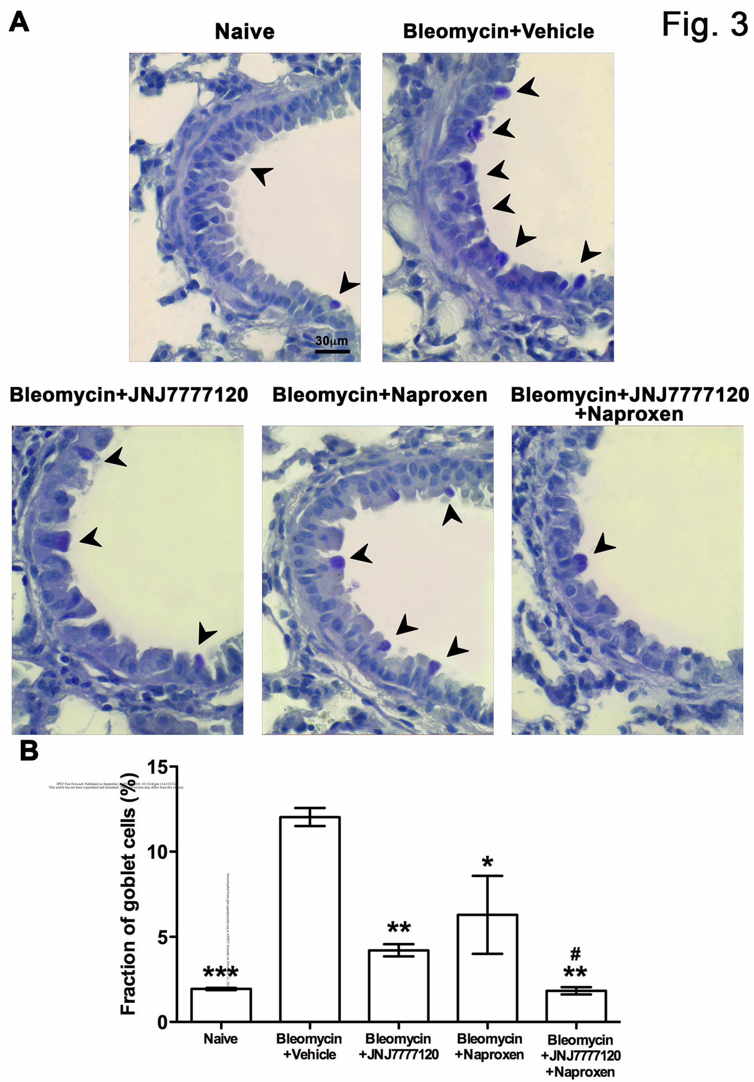
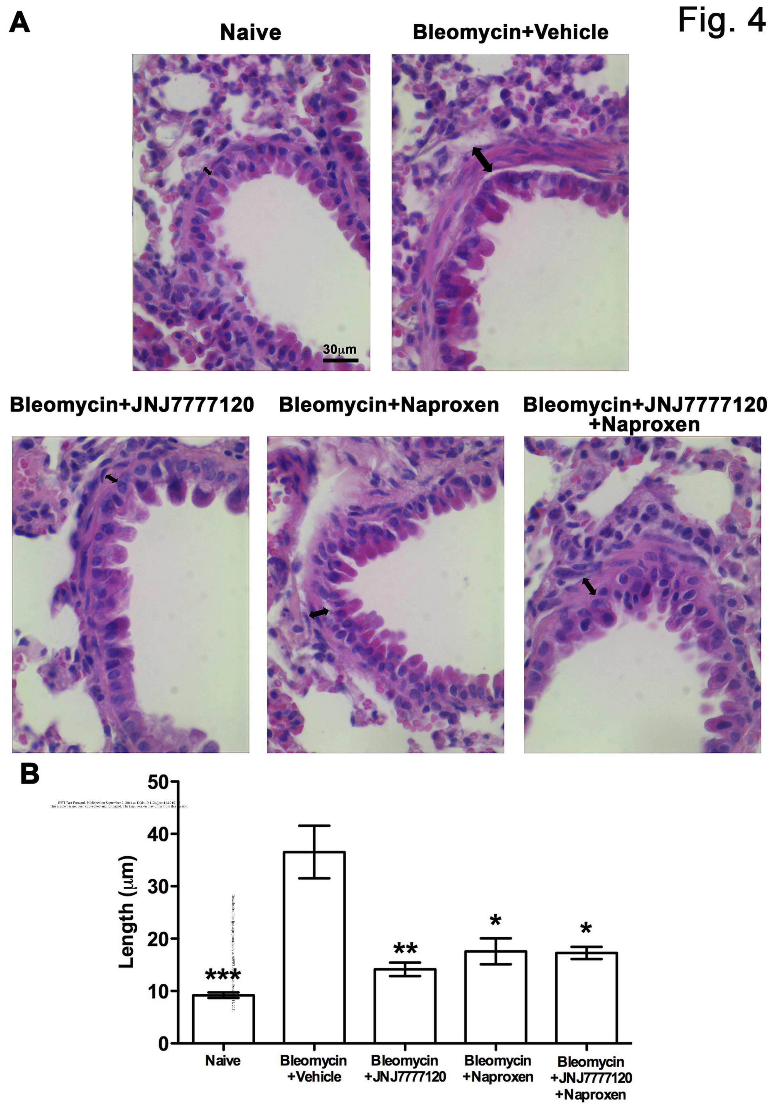
was reduced by all the treatments. We then evaluated bronchial remodelling by measuring the
relative number of goblet cells (Fig. 3) and the thickness of the smooth muscle (Fig. 4), key
histological parameters of inflammation-induced adverse bronchial remodelling (Bai and Knight,
2005). As expected, both these parameters were significantly increased after intratracheal
bleomycin treatment (+10.09 ± 1.30 %, P<0.001 for goblet cells number and +27.38 ± 3.35 μm,
P<0.001 for thickness of the smooth muscle). JNJ7777120 and naproxen both alone or in
combination were able to significantly reduce the percentage of PAS-positive goblet cells over total
bronchial epithelial cells (-7.83 ± 1.21 %, P<0.05 and -5.74 ± 1.38 %, P<0.05, respectively), as well
as the thickness of the airway smooth muscle layer (-22.40 ± 3.03 µm, P<0.01 and -18.97 ± 3.50
This article has not been copyedited and formatted. The final version may differ from this version.JPET Fast Forward. Published on September 2, 2014 as DOI: 10.1124/jpet.114.215152
at ASPE
T Journals on D
ecember 11, 2021
jpet.aspetjournals.orgD
ownloaded from
JPET #215152
13
μm, P<0.05, respectively). Notably, the combination of the two drugs showed a statistically
significant reduction of the fraction of goblet cells compared to the treatment with naproxen alone (-
4.46 ± 1.38 %; P<0.05).
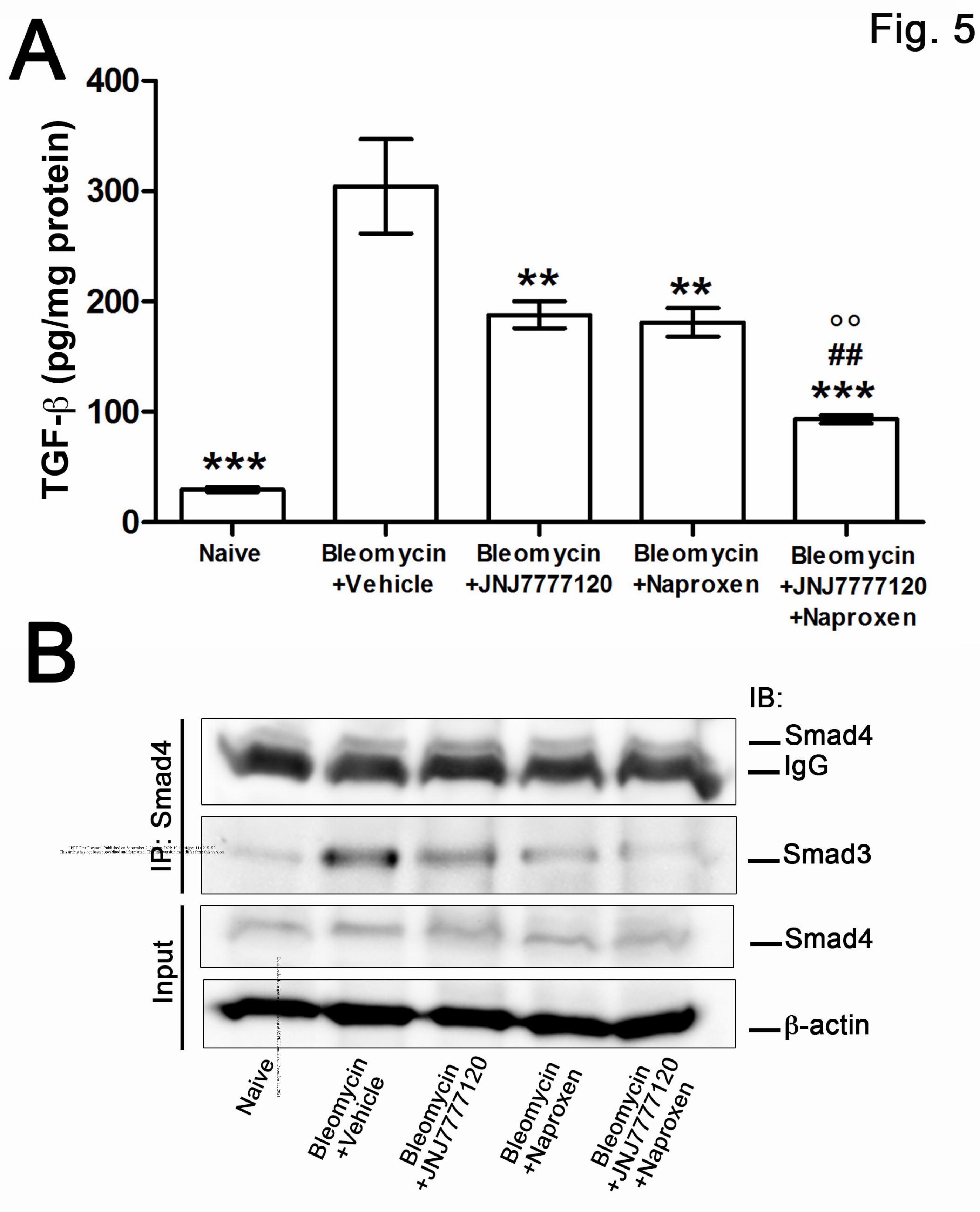
Determination of inflammation and fibrosis parameters. Assay of TGF-β (Fig. 5A), a major
pro-fibrotic cytokine, showed that this molecule significantly increased in the fibrotic positive
controls (+274.9 ± 13.68 pg/mg protein; P<0.001). JNJ7777120 or naproxen treatment caused a
statistically significant and comparable reduction of TGF-β (-116.2 ± 6.98 pg/mg protein and -123.0
± 6.84 pg/mg protein, respectively; P<0.01 for both). Notably, the co-administration of both drugs
was more effective than JNJ7777120 or naproxen alone (-93.83 ± 6.68 pg/mg protein vs.
JNJ7777120 and -87.12 ± 5.60 pg/mg protein vs. naproxen; P<0.01). As the Smad3/4 complex is
necessary for activation of TGF-β signaling (Chen et al., 2005), we investigated the effect of the
pharmacological treatment on the complex formation by Western blotting analysis performed on the
immunoprecipitated Smad4 protein. As shown in Fig. 5B, just above the heavy chain of IgG, we
identify a 61 kDa protein band consistent with Smad4. Notably, when we blotted with the anti-
Smad3 antibody, we observed a profound up-regulation in the positive fibrotic control that was
prevented by JNJ7777120 and naproxen given alone. Intriguingly, as shown for TGF-β levels, the
co-administration of the two drugs was more effective.
Determination of MPO (Fig. 6A), an index of leukocyte accumulation into the inflamed lung tissue,
showed that this parameter was significantly increased in the fibrotic positive controls compared
with non-fibrotic negative ones. Administration of JNJ7777120 or naproxen caused a statistically
significant reduction of MPO induced by bleomycin (-9.53 ± 0.28, -9.39 ± 0.027 mU/mg protein for
JNJ7777120 and naproxen, respectively; P<0.001). When the two drugs were co-administrated an
increased effect was observed (-11.27 ± 0.34; P<0.05).
Determination of PGE2 (Fig. 6B), the major cyclooxygenase product generated by activated
inflammatory cells, fibroblasts included, showed that this mediator was markedly increased in the
This article has not been copyedited and formatted. The final version may differ from this version.JPET Fast Forward. Published on September 2, 2014 as DOI: 10.1124/jpet.114.215152
at ASPE
T Journals on D
ecember 11, 2021
jpet.aspetjournals.orgD
ownloaded from

JPET #215152
14
fibrotic positive controls when compared with the non-fibrotic negative controls (+52.08 ± 2.69
pg/mg protein; P<0.001). JNJ7777120 or naproxen given alone caused a statistically significant
reduction of PGE2 level, with naproxen, as expected, more effective than JNJ7777120 (-16.20 ±
2.36 pg/mg protein for JNJ7777120 and -35.30 ± 2.36 pg/mg protein for naproxen; P<0.01).
However, the combination of the two drugs was more effective in reducing PGE2 production in
comparison to the single drugs (-28.26 ± 2.23 pg/mg protein vs JNJ7777120, P<0.01; and -9.16 ±
2.23 pg/mg protein vs naproxen P<0.001). To confirm the anti-inflammatory effects of the two
studied drugs we evaluated IL10 production, a regulatory cytokine (Fig. 6C). As expected,
bleomycin-treated animals showed a significant reduction in IL10 levels (-17.05 ± 1.51, P<0.001)
whereas the administration of both JNJ7777120 or naproxen alone or in combination caused a
statistically significant increased of IL10 levels (P<0.01). When the two drugs were co-
administrated a potentiated effect was observed.
Evaluation of oxidative stress parameters. Measurement of TBARS (Fig. 7A), a reliable marker
of oxidative tissue injury being the end products of cell membrane lipid peroxidation by ROS, and
of 8-OHdG (Fig. 7B), an indicator of oxidative DNA damage, showed that they were markedly
increased in fibrotic positive controls (+41.33 ± 11.15 nmol/mg protein and +51.25 ± 2.77 ng/mg
protein, respectively; P<0.001) compared with non-fibrotic negative control; these parameters were
significantly reduced by JNJ7777120 (-18.17 ± 5.66 nmol/mg protein, -33.46 ± 2.65 ng/mg protein,
respectively; P<0.01) or naproxen (-23.50 ± 7.32 nmol/mg protein, -33.48 ± 2.65 ng/mg protein,
respectively; P<0.01). Notably, the combination of JNJ7777120 and naproxen was more effective
in reducing MDA levels than JNJ7777120 (-14.08 ± 4.8 nmol/mg protein; P<0.05) or naproxen (-
8.74 ± 2.98 nmol/mg protein; P<0.01) treatment alone. A slight but not significant effect was also
observed also in 8-OHdG production.
This article has not been copyedited and formatted. The final version may differ from this version.JPET Fast Forward. Published on September 2, 2014 as DOI: 10.1124/jpet.114.215152
at ASPE
T Journals on D
ecember 11, 2021
jpet.aspetjournals.orgD
ownloaded from
JPET #215152
15
Discussion
The data presented in this work demonstrate the anti-inflammatory and anti-fibrotic
properties of naproxen and JNJ7777120 in a mouse model of lung fibrosis. We selected the model
of bleomycin, intra-tracheally delivered, because it is the best characterized murine model in use
today for lung fibrosis. Intratracheal delivery of bleomicyn to rodents results in a direct damage
initially to alveolar epithelial cells, followed by the development of neutrophilic and lymphocytic
pan-alveolitis within the first week and the development of fibrosis by day 14, with maximal
responses generally around days 21–28 (Moore and Hogaboam, 2008). Chronic inflammatory
conditions in the lungs lead to permanent structural changes and remodeling of the airway walls,
whose fibrosis is a major constituent. Nowadays, there are no approved drugs that counteract the
pathological mechanism apart for some potential treatments targeting TGF-β1 pathway, e.g.
pirfenidone (Paz and Shoenfeld, 2010). Our experiments were performed in C57BL/6 mice, which
are reported to be more susceptible to bleomycin-induced fibrosis than other strains, such as Balb/c
mice (Schrier et al., 1983; Harrison and Lazo, 1987). Both drugs given alone or in combination
were administered after the onset of bleomycin-induced lung injury, by using a subcutaneously
implanted micro-osmotic pump. This system allows sustained and long-term drug delivery, thus
overcoming the pharmacokinetic limits of JNJ7777120, which has a maximal oral bioavailability of
about 30% and a terminal half-life in mice of 1 h (Thurmond et al., 2004).
In our experimental model JNJ7777120, a selective receptor antagonist for H4R has been
compared with equimolar doses of naproxen. Compound JNJ7777120 has shown strong anti-
inflammatory properties, significantly decreasing the histological and biochemical inflammatory
parameters, such as the number of infiltrating leukocytes, PGE2 and IL10 levels. When looking at
the reduction of PGE2 levels, which solely depend on COX inhibition, naproxen had a higher
efficacy than JNJ7777120 (p<0.001). On the other hand, when considering the other parameters,
such as inhibition of leukocyte infiltration in the lung tissue and oxidative stress markers,
JNJ7777120 was a little more effective than naproxen. When the two drugs were given together we
This article has not been copyedited and formatted. The final version may differ from this version.JPET Fast Forward. Published on September 2, 2014 as DOI: 10.1124/jpet.114.215152
at ASPE
T Journals on D
ecember 11, 2021
jpet.aspetjournals.orgD
ownloaded from

JPET #215152
16
observed an additional effect conceivably determined by the complementary mechanisms acting on
different pro-inflammatory targets. However, it can be speculated that anti-TGF-β activity of
JNJ7777120 may indirectly affects eicosanoid pathways, by reducing TGF-β/Smad signaling which
is known to induce COX-2. Moreover, the hypothesis of a link between PGE2 and TGF-β, already
suggested (Alfranca et al., 2008), is herein supported; indeed, also naproxen down-regulates TGF-β
levels and Smad3/4 complex formation. COX-1 and COX-2 inhibitors, such as indomethacin,
diclofenac, meloxican and naproxen were reported to reduce lung collagen accumulation,
inflammation and oxidative stress in bleomicyn-induced lung fibrosis model (Thrall et al., 1979;
Chandler and Young, 1989; Arafa et al., 2007; Pini et al., 2012), a model that highlights the
inflammatory component of the pathology. Indeed high levels of PGE2 were reported in our positive
fibrotic controls. However, evidence exist that PGE2 has anti-inflammatory effects and inhibits
collagen production (Saltzman et al., 1982) and that PGE2 levels in patients with pulmonary fibrosis
are significantly decreased, suggesting an ambiguous role of PGE2 in lung fibroblast homeostasis. It
is possible that prostaglandin inhibition can have a positive effect on the onset of lung fibrosis
during the initial phase of inflammation and a detrimental effect during the fibrogenic events.
On the other side, the available data strongly support the H4R as a novel target for the
pharmacological modulation of immune and inflammatory disorders (Masini et al., 2013). Indeed,
in inflammatory lung disorders, histamine acts as a mediator of both acute and chronic phases. The
H4R are present in low amounts in the lung, where its expression in bronchial epithelial and smooth
muscle cells and micro-vascular endothelial cells (Gantner et al., 2002) can differently contribute to
airway diseases. H4R mediates redistribution and recruitment of mast cells in mucosal epithelium
after allergen exposure (Thurmond et al., 2004), mediates the synergistic action of histamine and
CXCL12, a chemokine involved in airway allergic disorders (Godot et al., 2007) and mediates the
recruitment and response of Treg cells (Morgan et al., 2007).
The relevant effects of JNJ7777120 could be explained through the marked decrease on
leukocyte infiltration in this in vivo model of pulmonary fibrosis, thus adding further evidence to the
This article has not been copyedited and formatted. The final version may differ from this version.JPET Fast Forward. Published on September 2, 2014 as DOI: 10.1124/jpet.114.215152
at ASPE
T Journals on D
ecember 11, 2021
jpet.aspetjournals.orgD
ownloaded from

JPET #215152
17
role of this receptor in controlling leukocyte trafficking and pro-inflammatory responses (Zampeli
and Tiligada, 2009). These data suggest that JNJ7777120 may exert not only a favorable effect
during the overt inflammatory phase of the disease, as previously reported with naproxen (Pini et
al., 2012), but also during fibroblast proliferation. Indeed, previous data demonstrated a role for
histamine and H4R in fibroblast activation (Cowden et al., 2010), which is a pivotal event in the
pathological process related to the disease progression (Garbuzenko et al., 2002; Kohyama et al.,
2010). Here we report the effects of JNJ7777120 on the lung TGF-β pathway, collagen deposition
and goblet cell hyperplasia. In keeping with the demonstration that histamine modulates TGF-
β/Smad signaling in conjunctival fibroblasts (Leonardi et al., 2010), our data confirm that the pro-
fibrotic effect of histamine is regulated by the activation of H4R. Taken together these results
clearly indicate the anti-fibrotic effect of the H4R antagonist.
On this background, our study points at the association between the two drugs as a promising
therapeutic approach for pulmonary fibrosis. When naproxen and JNJ7777120 were co-
administrated, additive positive effects were measured on lung TGF-β signalling modulation,
PGE2, leukocytes and goblet cell infiltration, confirming the effects of these drugs on the
inflammatory phase. A single time point (24 days) was planned for this study. At this time-point a
maximal fibrotic response was observed and JNJ7777120 and naproxen, given in combination,
exerted a maximal effect on most of the studied parameters.
Our data suggest that Smad3/4 complex formation plays a major role in determining the
pharmacological efficacy of the combination strategy herein proposed. Indeed, we propose that
JNJ7777120, reducing TGF-β and Smad3/4 complex formation, contributes to PGE2 reduction,
which is further affected by naproxen, inhibiting COX-2. Therefore, by abolishing the PGE2-
mediated positive feed-back on TGF-β/Smad signaling this strategy results more effective than the
each drug alone.
This article has not been copyedited and formatted. The final version may differ from this version.JPET Fast Forward. Published on September 2, 2014 as DOI: 10.1124/jpet.114.215152
at ASPE
T Journals on D
ecember 11, 2021
jpet.aspetjournals.orgD
ownloaded from

JPET #215152
18
In conclusion, the results of the present study, supporting the hypothesis that H4R antagonism exerts
anti-inflammatory and anti-fibrotic effects in the model of bleomycin-induced lung fibrosis, indicate
the therapeutic potential of the combination of H4R antagonists and NSAIDs. Although
JNJ7777120 itself is emerging as a promising therapeutic agent in lung inflammation and fibrosis,
the association with naproxen can have a distinct advantage over the single drug for the potentiating
effect on the inhibition of inflammatory and pro-fibrotic parameters. Moreover, this strategy could
override the safety limitations of the existing anti-inflammatory drugs in the treatment of pulmonary
fibrosis.
This article has not been copyedited and formatted. The final version may differ from this version.JPET Fast Forward. Published on September 2, 2014 as DOI: 10.1124/jpet.114.215152
at ASPE
T Journals on D
ecember 11, 2021
jpet.aspetjournals.orgD
ownloaded from

JPET #215152
19
Authorship contributions
Participated in research design: A.C.R., H.S., R.L.T., E.M.
Conducted experiments: A.C.R., A.P., L.L., E.V.,C.L.
Performed data analysis : A.P.
Wrote or contributed to the writing of the manuscript: A.C.R., E.V., E.M.
This article has not been copyedited and formatted. The final version may differ from this version.JPET Fast Forward. Published on September 2, 2014 as DOI: 10.1124/jpet.114.215152
at ASPE
T Journals on D
ecember 11, 2021
jpet.aspetjournals.orgD
ownloaded from

JPET #215152
20
References
Alfranca A, Lopez-Oliva JM, Genis L, Lopez-Maderuelo D, Mirones I, Salvado D, Quesada AJ,
Arroyo AG and Redondo JM (2008) PGE2 induces angiogenesis via MT1-MMP-mediated
activation of the TGFbeta/Alk5 signaling pathway. Blood 112:1120-1128.
Arafa HM, Abdel-Wahab MH, El-Shafeey MF, Badary OA and Hamada FM (2007) Anti-fibrotic
effect of meloxicam in a murine lung fibrosis model. Eur J Pharmacol 564:181-189.
Bai TR and Knight DA (2005) Structural changes in the airways in asthma: observations and
consequences. Clin Sci (Lond) 108:463-477.
Bradford MM (1976) A rapid and sensitive method for the quantitation of microgram quantities of
protein utilizing the principle of protein-dye binding. Anal Biochem 72:248-254.
Carter NJ (2011) Pirfenidone: in idiopathic pulmonary fibrosis. Drugs 71:1721-1732.
Chandler DB and Young K (1989) The effect of diclofenac acid (Voltaren) on bleomycin-induced
pulmonary fibrosis in hamsters. Prostaglandins Leukot Essent Fatty Acids 38:9-14.
Chen HB, Rud JG, Lin K and Xu L (2005) Nuclear targeting of transforming growth factor-beta-
activated Smad complexes. J Biol Chem 280:21329-21336.
Cowden JM, Riley JP, Ma JY, Thurmond RL and Dunford PJ (2010) Histamine H4 receptor
antagonism diminishes existing airway inflammation and dysfunction via modulation of Th2
cytokines. Respir Res 11:86.
Davies HR, Richeldi L and Walters EH (2003) Immunomodulatory agents for idiopathic pulmonary
fibrosis. Cochrane Database Syst Rev:CD003134.
du Bois RM (2010) Strategies for treating idiopathic pulmonary fibrosis. Nat Rev Drug Discov
9:129-140.
Gantner F, Sakai K, Tusche MW, Cruikshank WW, Center DM and Bacon KB (2002) Histamine
h(4) and h(2) receptors control histamine-induced interleukin-16 release from human
CD8(+) T cells. J Pharmacol Exp Ther 303:300-307.
This article has not been copyedited and formatted. The final version may differ from this version.JPET Fast Forward. Published on September 2, 2014 as DOI: 10.1124/jpet.114.215152
at ASPE
T Journals on D
ecember 11, 2021
jpet.aspetjournals.orgD
ownloaded from

JPET #215152
21
Garbuzenko E, Nagler A, Pickholtz D, Gillery P, Reich R, Maquart FX and Levi-Schaffer F (2002)
Human mast cells stimulate fibroblast proliferation, collagen synthesis and lattice
contraction: a direct role for mast cells in skin fibrosis. Clin Exp Allergy 32:237-246.
Godot V, Arock M, Garcia G, Capel F, Flys C, Dy M, Emilie D and Humbert M (2007) H4
histamine receptor mediates optimal migration of mast cell precursors to CXCL12. J Allergy
Clin Immunol 120:827-834.
Harrison JH, Jr. and Lazo JS (1987) High dose continuous infusion of bleomycin in mice: a new
model for drug-induced pulmonary fibrosis. J Pharmacol Exp Ther 243:1185-1194.
Hauber HP and Blaukovitsch M (2010) Current and future treatment options in idiopathic
pulmonary fibrosis. Inflamm Allergy Drug Targets 9:158-172.
Kohyama T, Yamauchi Y, Takizawa H, Kamitani S, Kawasaki S and Nagase T (2010) Histamine
stimulates human lung fibroblast migration. Mol Cell Biochem 337:77-81.
Leonardi A, Di Stefano A, Motterle L, Zavan B, Abatangelo G and Brun P (2010) Transforming
growth factor-beta/Smad - signalling pathway and conjunctival remodelling in vernal
keratoconjunctivitis. Clin Exp Allergy 41:52-60.
Leonardi A, Radice M, Fregona IA, Plebani M, Abatangelo G and Secchi AG (1999) Histamine
effects on conjunctival fibroblasts from patients with vernal conjunctivitis. Exp Eye Res
68:739-746.
Lodovici M, Casalini C, Cariaggi R, Michelucci L and Dolara P (2000) Levels of 8-
hydroxydeoxyguanosine as a marker of DNA damage in human leukocytes. Free Radic Biol
Med 28:13-17.
Masini E, Bani D, Vannacci A, Pierpaoli S, Mannaioni PF, Comhair SA, Xu W, Muscoli C,
Erzurum SC and Salvemini D (2005) Reduction of antigen-induced respiratory
abnormalities and airway inflammation in sensitized guinea pigs by a superoxide dismutase
mimetic. Free Radic Biol Med 39:520-531.
This article has not been copyedited and formatted. The final version may differ from this version.JPET Fast Forward. Published on September 2, 2014 as DOI: 10.1124/jpet.114.215152
at ASPE
T Journals on D
ecember 11, 2021
jpet.aspetjournals.orgD
ownloaded from

JPET #215152
22
Masini E, Lucarini L, Sydbom A, Dahlén B and Dahlén S-E (2013) Histamine in asthmatic and
fibrotic disorders, in Histamine H4 receptor: A novel drug target (Stark H ed) pp 145-171,
Versita, London.
Moore BB and Hogaboam CM (2008) Murine models of pulmonary fibrosis. Am J Physiol Lung
Cell Mol Physiol 294:L152-160.
Morgan RK, McAllister B, Cross L, Green DS, Kornfeld H, Center DM and Cruikshank WW
(2007) Histamine 4 receptor activation induces recruitment of FoxP3+ T cells and inhibits
allergic asthma in a murine model. J Immunol 178:8081-8089.
Mullane KM, Kraemer R and Smith B (1985) Myeloperoxidase activity as a quantitative assessment
of neutrophil infiltration into ischemic myocardium. J Pharmacol Methods 14:157-167.
Ohkawa H, Ohishi N and Yagi K (1979) Assay for lipid peroxides in animal tissues by
thiobarbituric acid reaction. Anal Biochem 95:351-358.
Paz Z and Shoenfeld Y (2010) Antifibrosis: to reverse the irreversible. Clin Rev Allergy Immunol
38:276-286.
Pini A, Shemesh R, Samuel CS, Bathgate RA, Zauberman A, Hermesh C, Wool A, Bani D and
Rotman G (2010) Prevention of bleomycin-induced pulmonary fibrosis by a novel
antifibrotic peptide with relaxin-like activity. J Pharmacol Exp Ther 335:589-599.
Pini A, Viappiani S, Bolla M, Masini E and Bani D (2012) Prevention of bleomycin-induced lung
fibrosis in mice by a novel approach of parallel inhibition of cyclooxygenase and nitric-
oxide donation using NCX 466, a prototype cyclooxygenase inhibitor and nitric-oxide
donor. J Pharmacol Exp Ther 341:493-499.
Raghu G, Collard HR, Egan JJ, Martinez FJ, Behr J, Brown KK, Colby TV, Cordier JF, Flaherty
KR, Lasky JA, Lynch DA, Ryu JH, Swigris JJ, Wells AU, Ancochea J, Bouros D, Carvalho
C, Costabel U, Ebina M, Hansell DM, Johkoh T, Kim DS, King TE, Jr., Kondoh Y, Myers
J, Muller NL, Nicholson AG, Richeldi L, Selman M, Dudden RF, Griss BS, Protzko SL and
Schunemann HJ (2011) An official ATS/ERS/JRS/ALAT statement: idiopathic pulmonary
This article has not been copyedited and formatted. The final version may differ from this version.JPET Fast Forward. Published on September 2, 2014 as DOI: 10.1124/jpet.114.215152
at ASPE
T Journals on D
ecember 11, 2021
jpet.aspetjournals.orgD
ownloaded from
JPET #215152
23
fibrosis: evidence-based guidelines for diagnosis and management. Am J Respir Crit Care
Med 183:788-824.
Richeldi L, Davies HR, Ferrara G and Franco F (2003) Corticosteroids for idiopathic pulmonary
fibrosis. Cochrane Database Syst Rev:CD002880.
Saltzman LE, Moss J, Berg RA, Hom B and Crystal RG (1982) Modulation of collagen production
by fibroblasts. Effects of chronic exposure to agonists that increase intracellular cyclic
AMP. Biochem J 204:25-30.
Sassoli C, Pini A, Chellini F, Mazzanti B, Nistri S, Nosi D, Saccardi R, Quercioli F, Zecchi-
Orlandini S and Formigli L (2012) Bone marrow mesenchymal stromal cells stimulate
skeletal myoblast proliferation through the paracrine release of VEGF. PLoS One 7:e37512.
Schrier DJ, Kunkel RG and Phan SH (1983) The role of strain variation in murine bleomycin-
induced pulmonary fibrosis. Am Rev Respir Dis 127:63-66.
Sivakumar P, Ntolios P, Jenkins G and Laurent G (2012) Into the matrix: targeting fibroblasts in
pulmonary fibrosis. Curr Opin Pulm Med 18:462-469.
Stramer BM, Mori R and Martin P (2007) The inflammation-fibrosis link? A Jekyll and Hyde role
for blood cells during wound repair. J Invest Dermatol 127:1009-1017.
Thrall RS, McCormick JR, Jack RM, McReynolds RA and Ward PA (1979) Bleomycin-induced
pulmonary fibrosis in the rat: inhibition by indomethacin. Am J Pathol 95:117-130.
Thurmond RL, Desai PJ, Dunford PJ, Fung-Leung WP, Hofstra CL, Jiang W, Nguyen S, Riley JP,
Sun S, Williams KN, Edwards JP and Karlsson L (2004) A potent and selective histamine
H4 receptor antagonist with anti-inflammatory properties. J Pharmacol Exp Ther 309:404-
413.
Wynn TA (2008) Cellular and molecular mechanisms of fibrosis. J Pathol 214:199-210.
Zampeli E and Tiligada E (2009) The role of histamine H4 receptor in immune and inflammatory
disorders. Br J Pharmacol 157:24-33.
This article has not been copyedited and formatted. The final version may differ from this version.JPET Fast Forward. Published on September 2, 2014 as DOI: 10.1124/jpet.114.215152
at ASPE
T Journals on D
ecember 11, 2021
jpet.aspetjournals.orgD
ownloaded from

JPET #215152
24
Footnotes
This work was supported by COST Action BM0806 (A.C.R., A.P.; H.S. and E.M.) and from Ente
Cassa di Risparmio di Firenze, Florence, Italy (E.M.). Part of this study was presented at the 40th
EHRS meeting, 11-14th May 2011 Sochi, Russia and published in abstract form in Inflamm. Res.
Sep 2011; 60(2): S335-S335.
This article has not been copyedited and formatted. The final version may differ from this version.JPET Fast Forward. Published on September 2, 2014 as DOI: 10.1124/jpet.114.215152
at ASPE
T Journals on D
ecember 11, 2021
jpet.aspetjournals.orgD
ownloaded from

JPET #215152
25
Legends for Figures
Fig. 1. Spirometric evaluation. Bar graph and statistical analysis of differences of pressure at the
airway opening (PAO) values (means± S.E.M) between the different experimental groups (one way
ANOVA; n=10 animals per group). **P<0.01 and ***P<0.001 vs. bleomycin + vehicle.
Fig. 2. Evaluation of lung fibrosis. Panel A: representative micrographs of Azan-stained lung tissue
sections from mice of the different experimental groups. Collagen fibers are deep blue stained. The
lung from fibrotic control treated with vehicle showed marked fibrosis in the peri-bronchial stroma,
which was absent in the lung from non-fibrotic negative control and reduced by both JNJ7777120
and naproxen either alone or in combination. Panel B: bar graph showing the optical density (OD,
means± S.E.M.) of Azan-stained collagen fibers of the different experimental groups (one way
ANOVA, n=10 mice per group). *P<0.05, **P<0.01 and ***P<0.001 vs. bleomycin + vehicle.
Fig. 3 Goblet cell hyperplasia. Panel A: Representative micrographs of PAS-stained sections.
Arrows indicate the goblet cells. Panel B: bar graph showing the fraction of goblet cells (% means ±
S.E.M.) in the different experimental groups (one way ANOVA, n=10 animals per group). *P<0.05,
**P<0.01 and ***P<0.001 vs. bleomycin + vehicle; # P<0.05 vs. bleomycin + naproxen.
Fig. 4. Evaluation of the muscular remodeling. The smooth muscle thickness was assessed by
computer-aided morphometry on hematoxylin and eosin-stained lung sections. Panel A:
representative micrographs of the sections. The thickness of the smooth muscle layer is indicated by
double arrows. Panel B: bar graph showing the length (μm) of the muscular fiber (means ± S.E.M.)
in the different experimental groups (one way ANOVA, n=10 animal per group). *P<0.05,
**P<0.01vs. bleomycin+vehicle.
This article has not been copyedited and formatted. The final version may differ from this version.JPET Fast Forward. Published on September 2, 2014 as DOI: 10.1124/jpet.114.215152
at ASPE
T Journals on D
ecember 11, 2021
jpet.aspetjournals.orgD
ownloaded from

JPET #215152
26
Fig. 5. Evaluation of fibrotic key mediators. Panel A: Bar graph showing the lung tissue levels of
the pro-fibrotic cytokine TGF-β (means ± S.E.M.) of the different experimental groups (one way
ANOVA, n=10 animals per group). Panel B: Smad4 and Smad3 expression level in the noted
experimental conditions, assayed by Western Blotting analysis performed on the
immunoprecipitated Smad4 protein. Note that the profound up-regulation of Smad3 expression
level in the positive fibrotic control was prevented by the treatment with both JNJ7777120 or
naproxen alone. The co-administration of the two drugs was more effective. **P<0.01 and
***P<0.001 vs. bleomycin + vehicle; °°P<0.01 vs. bleomycin+JNJ7777120; ##P<0.01 vs.
bleomycin + naproxen.
Fig. 6. Evaluation inflammation parameters. Panel A: bar graph showing the tissue level (means ±
S.E.M.) of the enzyme MPO, evaluated as the quantity of enzyme degrading 1 μmol of
peroxide/min at 37 °C, of the different experimental groups. Panel B: bar graph showing the tissue
levels of the PGE2 (means ± S.E.M.) of the different experimental groups. Panel C. Bar graph
showing the tissue levels of the anti-inflammatory cytokine IL10 (means ± S.E.M.) of the different
experimental groups (one way ANOVA, n=10 animals per group). **P<0.01 and ***P<0.001 vs.
bleomycin + vehicle; °P<0.05 and °°P<0.01 vs. bleomycin+JNJ7777120; # P<0.05 and ###P<0.001
vs. bleomycin + naproxen.
Fig. 7. Evaluation of oxidative stress parameters. Panel A: bar graph showing the lung tissue of
TBARS (means ± S.E.M.) in the different experimental groups. Panel B: bar graph showing the
levels of 8-OHdG (means ± S.E.M.) in the different experimental groups (one way ANOVA, n=10
animals/group). °P<0.05 vs. bleomycin + JNJ7777120; ##P<0.01 vs. bleomycin+naproxen;
**P<0.01 and ***P<0.001 vs. bleomycin + vehicle.
This article has not been copyedited and formatted. The final version may differ from this version.JPET Fast Forward. Published on September 2, 2014 as DOI: 10.1124/jpet.114.215152
at ASPE
T Journals on D
ecember 11, 2021
jpet.aspetjournals.orgD
ownloaded from

This article has not been copyedited and formatted. The final version may differ from this version.JPET Fast Forward. Published on September 2, 2014 as DOI: 10.1124/jpet.114.215152
at ASPE
T Journals on D
ecember 11, 2021
jpet.aspetjournals.orgD
ownloaded from

This article has not been copyedited and formatted. The final version may differ from this version.JPET Fast Forward. Published on September 2, 2014 as DOI: 10.1124/jpet.114.215152
at ASPE
T Journals on D
ecember 11, 2021
jpet.aspetjournals.orgD
ownloaded from
This article has not been copyedited and formatted. The final version may differ from this version.JPET Fast Forward. Published on September 2, 2014 as DOI: 10.1124/jpet.114.215152
at ASPE
T Journals on D
ecember 11, 2021
jpet.aspetjournals.orgD
ownloaded from
This article has not been copyedited and formatted. The final version may differ from this version.JPET Fast Forward. Published on September 2, 2014 as DOI: 10.1124/jpet.114.215152
at ASPE
T Journals on D
ecember 11, 2021
jpet.aspetjournals.orgD
ownloaded from
This article has not been copyedited and formatted. The final version may differ from this version.JPET Fast Forward. Published on September 2, 2014 as DOI: 10.1124/jpet.114.215152
at ASPE
T Journals on D
ecember 11, 2021
jpet.aspetjournals.orgD
ownloaded from

This article has not been copyedited and formatted. The final version may differ from this version.JPET Fast Forward. Published on September 2, 2014 as DOI: 10.1124/jpet.114.215152
at ASPE
T Journals on D
ecember 11, 2021
jpet.aspetjournals.orgD
ownloaded from

This article has not been copyedited and formatted. The final version may differ from this version.JPET Fast Forward. Published on September 2, 2014 as DOI: 10.1124/jpet.114.215152
at ASPE
T Journals on D
ecember 11, 2021
jpet.aspetjournals.orgD
ownloaded from
Top Related