


Languages
Pages
Legal

Polystyrene (nano)composites with possible antibacterial effect MERINSKA D., DUJKOVA Z.
Department of polymer engineering
Tomas Bata University in Zlin
Nám.TGM 275, Zlín 76272
CZECH REPUBLIC
[email protected] http://web.utb.cz
Abstract: - The possibility of the preparation of PS (nano)composites with antibacterial properties was studied.
Mentioned polymer matrix was mixed with several types of (nano)fillers with the suppose of their antibacterial effect
in order to evaluate their influence on material properties of prepared samples with the regard of simultaneous
antibacterial effect. The XRD and TEM for nanoclays and hardness, tensile stress, permeability for oxygen and
nitrogen and antibacterial effect for all of them were evaluated. Generally, properties of PS were not significantly
influenced by any of added filler.
Key-Words: polystyrene, composite, nanocomposite, antibacterial effect, food packaging
1 Introduction Nanofiller/polymer nanocomposites have been studied
extensively during the last two decades, because they
have been expected to obtain improved properties in
the comparison with the neat polymer matrix and
commonly used fillers [1-6]. One of the used polymer
matrices for the preparation is polystyrene (PS). It is a
commonly used polymer with various applications,
e.g. insulation, packaging, in household and in
automotive industry. Unfortunately, PS also exhibits
some disadvantageous properties, i.e. relatively high
flammability. This is the reason why polystyrene
composites and later nanocomposites have been
studied because of the possibility of PS properties
improvement [7-11].
Lately PS in several forms has been used also in food
packaging industry. It is very well known the using of
PS for caps for coffee or bowls for hot food where the
good thermal isolation of expanded PS. Generally, in
the food packaging the hygienic quality of packed
food is important. Commonly this request is reached
by the addition of antibacterial agents. There are a few
types of these agents and the most used is silver in
different forms.
Polymer silver nanocomposites are advanced
functional materials composed of silver nanoparticles
dispersed inside the polymeric matrix [12-15] and/or
coated by polymer, thus forming a core-shell structure
[16-19].
As a result, the produced material combines the
suitable properties of both partners. Among the
numerous nanoparticles that have been used as
polymer functionalizing agent, silver nanoparticles
represent the most sought-after (nano)material. This is
mainly due to their unique catalytic [20-22], electric
[23–24], optical [25–26], and, particularly,
antimicrobial properties [27-31], which are well
established and extensively investigated mainly in
colloidal systems.
This work was focused on the comparison of several
additive types in order to evaluate their possible
antibacterial effect in PS matrix with the keeping of
mechanical properties and permeability with the aim to
find new applications for PS as food packaging
materials.
2 Experimental
2.1 Materials
Commercial polystyrene KRASTEN® 174 from
SYNTHOS Kralupy a.s., Czech Republic was used as a
polymer matrix. Unmodified clay CloisiteNa+
(Southern Clay Chemistry, US), as layered nanofiller.
Two different types of silver particles were used – Ag
powder, size of particles lower than 100 nm, and Ag
acetate, both from Aldridge. In addition, commercial
antibacterial compound Irgaguard B 5000 from BASF
was used. All fillers were added in 1wt %, resp. 3 wt%.
2.2 Preparation of composites and specimens Composites were prepared by mixing and compounding
in a Brabender kneader. Temperature was 170 °C and
the speed of the kneading parts was 30 rpm. Time of
mixing was 10 min.
The XRD measurement was performed on the
specimens prepared by pressing in a hot press at for 3
min at 210 °C followed by cutting of a circle shape.
The thickness was 1 mm.
Mathematical Methods and Techniques in Engineering and Environmental Science
ISBN: 978-1-61804-046-6 377
In order to prepare specimens for the mechanical
property evaluation the pellets were molded according
to ISO 294–1 at the equipment BATTENFELD 500
CD+, melt temperature 220 °C, form temperature 45
°C.
At the same conditions the samples for the
permeability evaluation were prepared, with the
thickness of 50 µm.
2.3 Instrumentation The level of exfoliation in the prepared samples
after compounding process was measured by XRD
patterns.
Compounded samples were analyzed by XRD powder
diffractometer (INEL) equipped with the curved
position sensitive detector CPS 120 (120° 2θ),
reflection mode with a germanium monochromator
(Cuα1 radiation). Samples were placed into holder and
exposed for 2000s.
For the transmission electron microscopy (JEM
200CX), the specimens were cut using Leica cryo-
ultramicrotome at sample temperature -100°C and
knife temperature -50°C to obtain ultra-thin sections
with the thickness approximately 50 nm and an
acceleration voltage of 100 kV was used.
Mechanical properties were measured according to
ISO 527-2/1A/X on the Zwick 145665. All presented
data are the arithmetic average taken from 10
measurements.
The permeability was measured for oxygen and
nitrogen, with the bodies cut from thin film. The
pressure was 2.10
5 Pa, at the temperature of 35°C.
Last evaluated property was the antibacterial effect
of added additives.
Agar media:
Mueller-Hinton agar (peptone 17.5 g/l, beef extract
4.0 g/l, starch 1.5 g/l, agar 20 g/l and distilled water)
was used for testing of antibacterial properties and
Sabouraud agar (casein peptone 5 g/l, meat peptone 5
g/l, glucose 40 g/l, agar 15 g/l and distilled water) was
used for testing of antifungal properties.
Procedure:
Petri plates with Mueller-Hinton agar were inoculated
by cell suspension of Staphylococcus aureus CCM
3953 or Escherichia coli CCM 3954 (50 µl were
spread onto each plate) and prepared plastic samples 2
x 2 cm were put on the surface. After 5 hours the
samples were removed and all the plates were
incubated for 48 hours at 37°C. After that, bacterial
growth or inhibition of growth was noted and
compared to blank test, in which plastic samples
without antimicrobial agent were used.
3 Problem Solution
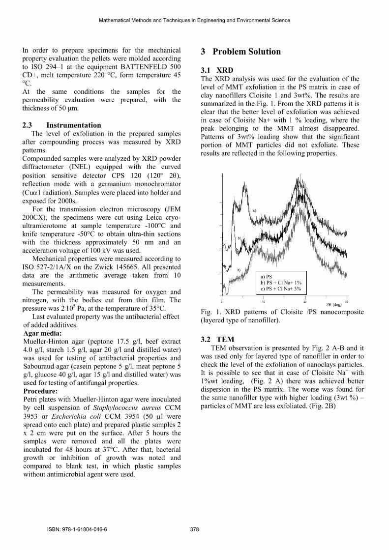
3.1 XRD The XRD analysis was used for the evaluation of the
level of MMT exfoliation in the PS matrix in case of
clay nanofillers Cloisite 1 and 3wt%. The results are
summarized in the Fig. 1. From the XRD patterns it is
clear that the better level of exfoliation was achieved
in case of Cloisite Na+ with 1 % loading, where the
peak belonging to the MMT almost disappeared.
Patterns of 3wt% loading show that the significant
portion of MMT particles did not exfoliate. These
results are reflected in the following properties.
Fig. 1. XRD patterns of Cloisite /PS nanocomposite
(layered type of nanofiller).
3.2 TEM
TEM observation is presented by Fig. 2 A-B and it
was used only for layered type of nanofiller in order to
check the level of the exfoliation of nanoclays particles.
It is possible to see that in case of Cloisite Na+ with
1%wt loading, (Fig. 2 A) there was achieved better
dispersion in the PS matrix. The worse was found for
the same nanofiller type with higher loading (3wt %) –
particles of MMT are less exfoliated. (Fig. 2B)
a) PS b) PS + Cl Na+ 1%
c) PS + Cl Na+ 3%
Mathematical Methods and Techniques in Engineering and Environmental Science
ISBN: 978-1-61804-046-6 378

A) B)
Fig. 2. TEM pictures: A) Cloisite Na+ 1%, B) Cloisite
Na+ 3%
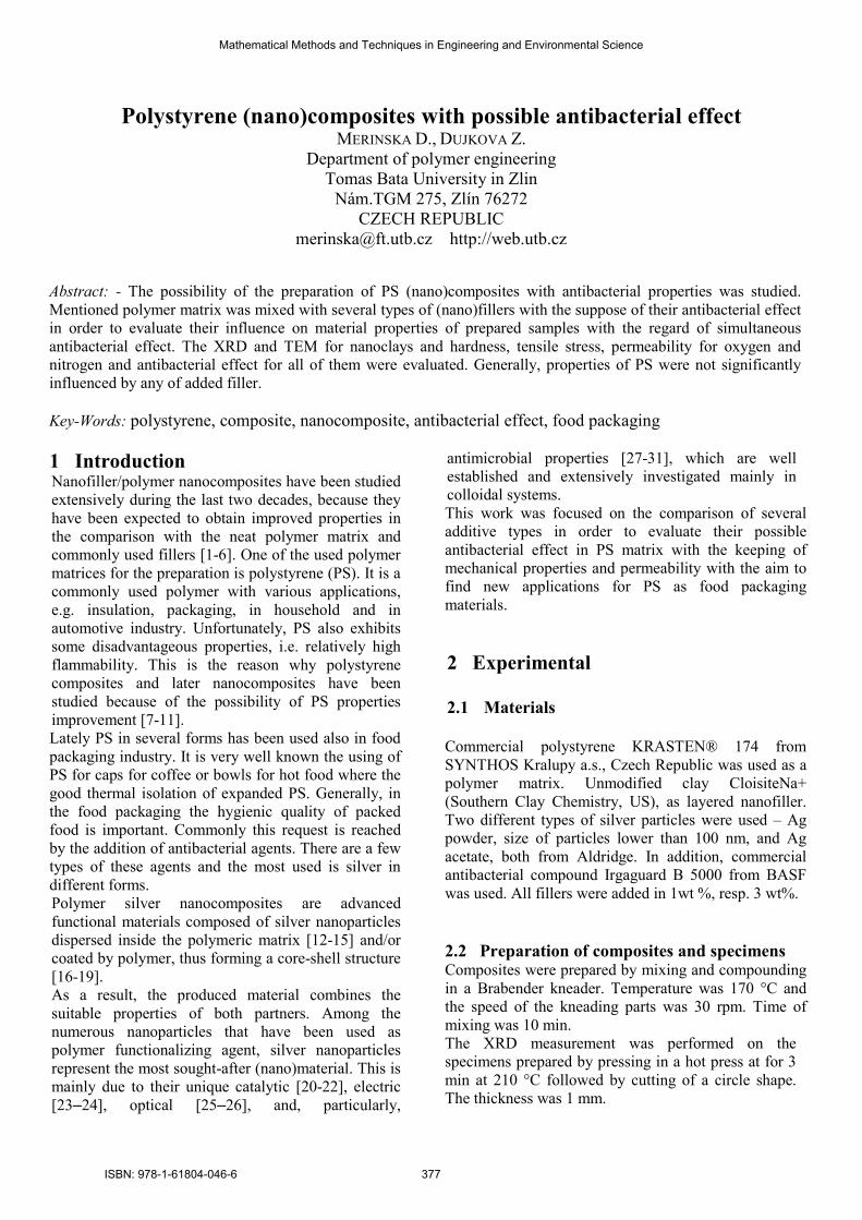
3.3 Hardness The influence of nanofillers on the hardness was the next
observed fact. From the graph in Fig. 2 it is possible to
deduce that the hardness of prepared samples stayed
almost the same, it was observed only very slight
increasing. Thus, added nanofillers do not influence the
hardness.
0
10
20
30
40
50
60
70
80
90
neat
PS
3% IR
G
1%IR
G
3% C
l Na+
1% C
l Na+
3% A
g
1% A
g
3% A
g Ac
1% A
g Ac
samples
Sh
ore
A
1% 3%
Fig. 2. Hardness
3.4 Mechanical properties In the evaluation of mechanical properties, the tensile
strength, modulus and impact strength were measured.
Here the result of tensile strength is presented.
0
5
10
15
20
25
30
neat P
S
3% IR
G
1%IR
G
3% C
l Na+
1% C
l Na+
3% A
g
1% A
g
3% A
g Ac
1% A
g Ac
samples
Te
ns
ile
str
en
gh
t (M
Pa
)
1% 3%
Fig. 3. Mechanical properties – tensile strength
The evaluation of tensile strength brought a little bit
different data. In almost all cases it can be seen that the
value of the tensile strength is lower in the comparison
with the neat PS matrix. The decrease is higher for
samples with 3 wt % loading, which can be caused by
worse dispersion of MMT or Ag agglomerates.
Generally, the decrease is not significant; it is only about
5%. To sum up, the tensile strength is not influenced
significantly.
3.5 Permeability
Barrier properties of prepared samples were
evaluated because the possible using of this type of
material in packaging. The most common gases -
oxygen and nitrogen - were observed. The results of
measurement are summarized in the graph in Fig. 4.
0,00E+00
1,00E-16
2,00E-16
3,00E-16
4,00E-16
5,00E-16
6,00E-16
7,00E-16
8,00E-16
9,00E-16
neat PS
IRG
Cl Na+ Ag
Ag Ac
samples
Perm
. co
ef.
(m
ol/s.m
.Pa)
O2 N2
Fig. 4. Permeability coefficient for O2 and N2
The result is very similar to the previous
measurements. The permeability coefficient is not
significantly different for neat and filled PS matrix
for both evaluated gases. Also here the barrier
property is not negatively influenced by used
nanofillers.
3.6 Antibacterial effect
As it was mentioned earlier, the aim of this work
was to compare several additive types in order to
evaluate their possible antibacterial effect in PS matrix
with the keeping of mechanical properties and
permeability. The mostly researched types of bacteria are
two the most widened - Escherichia coli or
Staphylococcus aureus (see Fig 5).
100 nm 100 nm
Mathematical Methods and Techniques in Engineering and Environmental Science
ISBN: 978-1-61804-046-6 379
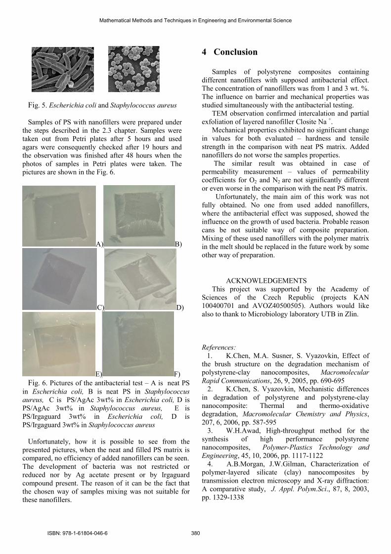
Fig. 5. Escherichia coli and Staphylococcus aureus
Samples of PS with nanofillers were prepared under
the steps described in the 2.3 chapter. Samples were
taken out from Petri plates after 5 hours and used
agars were consequently checked after 19 hours and
the observation was finished after 48 hours when the
photos of samples in Petri plates were taken. The
pictures are shown in the Fig. 6.
A) B)
C) D)
E) F)
Fig. 6. Pictures of the antibacterial test – A is neat PS
in Escherichia coli, B is neat PS in Staphylococcus
aureus, C is PS/AgAc 3wt% in Escherichia coli, D is
PS/AgAc 3wt% in Staphylococcus aureus, E is
PS/Irgaguard 3wt% in Escherichia coli, D is
PS/Irgaguard 3wt% in Staphylococcus aureus
Unfortunately, how it is possible to see from the
presented pictures, when the neat and filled PS matrix is
compared, no efficiency of added nanofillers can be seen.
The development of bacteria was not restricted or
reduced nor by Ag acetate present or by Irgaguard
compound present. The reason of it can be the fact that
the chosen way of samples mixing was not suitable for
these nanofillers.
4 Conclusion
Samples of polystyrene composites containing
different nanofillers with supposed antibacterial effect.
The concentration of nanofillers was from 1 and 3 wt. %.
The influence on barrier and mechanical properties was
studied simultaneously with the antibacterial testing.
TEM observation confirmed intercalation and partial
exfoliation of layered nanofiller Closite Na +.
Mechanical properties exhibited no significant change
in values for both evaluated – hardness and tensile
strength in the comparison with neat PS matrix. Added
nanofillers do not worse the samples properties.
The similar result was obtained in case of
permeability measurement – values of permeability
coefficients for O2 and N2 are not significantly different
or even worse in the comparison with the neat PS matrix.
Unfortunately, the main aim of this work was not
fully obtained. No one from used added nanofillers,
where the antibacterial effect was supposed, showed the
influence on the growth of used bacteria. Probable reason
cans be not suitable way of composite preparation.
Mixing of these used nanofillers with the polymer matrix
in the melt should be replaced in the future work by some
other way of preparation.
ACKNOWLEDGEMENTS
This project was supported by the Academy of
Sciences of the Czech Republic (projects KAN
100400701 and AVOZ40500505). Authors would like
also to thank to Microbiology laboratory UTB in Zlin.
References:
1. K.Chen, M.A. Susner, S. Vyazovkin, Effect of
the brush structure on the degradation mechanism of
polystyrene-clay nanocomposites, Macromolecular
Rapid Communications, 26, 9, 2005, pp. 690-695
2. K.Chen, S. Vyazovkin, Mechanistic differences
in degradation of polystyrene and polystyrene-clay
nanocomposite: Thermal and thermo-oxidative
degradation, Macromolecular Chemistry and Physics,
207, 6, 2006, pp. 587-595
3. W.H.Awad, High-throughput method for the
synthesis of high performance polystyrene
nanocomposites, Polymer-Plastics Technology and
Engineering, 45, 10, 2006, pp. 1117-1122
4. A.B.Morgan, J.W.Gilman, Characterization of
polymer-layered silicate (clay) nanocomposites by
transmission electron microscopy and X-ray diffraction:
A comparative study, J. Appl. Polym.Sci., 87, 8, 2003,
pp. 1329-1338
Mathematical Methods and Techniques in Engineering and Environmental Science
ISBN: 978-1-61804-046-6 380

5. J.M. Yeh, C.P. Chin, Structure and properties of
poly(o-methoxyaniline)-clay nanocomposite materials,
J. Appl. Pol. Sci., 88, 4, 2003pp. 1072-1080
6. S.J.Ahmadi, Y.D. Huang, W. Li, Synthetic routes,
properties and future applications of polymer-layered
silicate nanocomposites, J. Mat. Sci., 39, 6, 2004, pp.
1919-1925
7. S.Su, D.D. Jiang, C. A. Wilkie, Novel
polymerically-modified clays permit the preparation of
intercalated and exfoliated nanocomposites of styrene
and its copolymers by melt blending, Pol. Degr, Stab.,
83, 2, 2004, pp. 333-346
8. K.Chen, C.A. Wilkie, S. Vyazovkin,
Nanoconfinement revealed in degradation and relaxation
studies of two structurally different polystyrene-clay
systems, J.Phys. Chem., 111, 2007, pp. 12685-12692
9. G. Chigwada, D.Y. Wang, C.A. Wilkie, Pol.
Degr. Stab., 91, 4, 848-855, (2006).
10. G. Chigwada, D.Y. Wang,D.D. Jiang, C.A.
Wilkie, Styrenic nanocomposites prepared using a novel
biphenyl-containing modified clay, Pol. Degr. Stab., 91,
4, 2006, 755-762
11. S.Nazarenko, P. Meneghetti, P. Julmon, Gas
barrier of polystyrene montmorillonite clay
nanocomposites: Effect of mineral layer aggregation, J.
Pol. Sci., Part B: Pol. Phys., 45, 2007, pp. 1733-1753
12. G. F. Prozorova, S. A Korzhova, T. V Kon'kova.
et al., Specific features of formation of silver
nanoparticles in the polymermatrix, Doklady Chem., 437,
2011, pp. 47-49
13. Ch. Triebel, S. Vasylyev, C. Damm et
al.Polyurethane/silver-nanocomposites with enhanced
silver ion release using multifunctional invertible
polyesters
J Mat Chem, 21, 12, 2011, pp. 4377-4383
14. M. A Kudryashov, A. I. Mashin, A. S. Tyurin et
al., Morphology of a silver/polyacrylonitrile
nanocomposite
Tech Phys, 56, 1, 2011, pp. 92-96
15. P. Dallas, V. K. Sharma, R. Zboril, Silver
polymeric nanocomposites as advanced antimicrobial
agents: Classification, synthetic paths, applications, and
perspectives
Adv in Colloid and Interface Sci, 166, 1-2, 2011, pp. 119-
135
16. R. Gunawidjaja, T. Myint, H. Eilers, Synthesis
of silver/SiO(2)/Eu:Lu(2)O(3) core-shell nanoparticles
and their polymer nanocomposites, Powder Technology,
2, 2011, pp. 157-166
17. R P Jose; A Urrutia; J Goicoechea; et al. An
antibacterial coating based on a polymer/sol-gel hybrid
matrix loaded with silver nanoparticles, *anoscale Res
Lett, 6, 2011, Article Number: 305
18. S. Pyne, P. Sarkar, S. Basu et al.Synthesis and
photo physical properties of Au @ Ag (core @ shell)
nanoparticles disperse in polyvinylalcohol matrix,
J.*anoparticle Res, 13,4, 2010, pp. 1759-1767
19. S.K. Tripathy, J.-N. Jo, H.-M. Song et al.,
Fabrication and optical study of Ag@SnO(2) core-shell
structure nanoparticle thin films, App. Phys. A-Mat. Sci
& Proc., 104, 2, 2010, pp. 601-607
20. A. Roucoux, J. Schulz, H. Patin, Reduced
transition metal colloids: A novel family of reusable
catalysts? Chem Rev, 102, 10, 2002, pp. 3757-3778
0 21. N. Severin, S. Kirstein, S.M. Sokolov, J.P. Rabe,
Rapid Trench Channeling of Graphenes with Catalytic
Silver Nanoparticles, *ano Lett , 9, 1, 2009, pp.457
22. A.M. Signori, K.D.O. Santos, R. Eising, B.L.
Albuquerque et al., Formation of Catalytic Silver
Nanoparticles Supported on Branched Polyethyleneimine
Derivatives, Langmuir, 26, 22, 2010,pp. 17772-17779
23. M. E. Leyva, F. G. Garcia, A. A. Alencar de
Queiroz et al.,Electrical properties of the DGEBA/PANI-
Ag composites, J Mater Sci-Mater in Electronics, 22, 4,
2011, pp. 376-383
24. M. K. Abyaneh, S. Jafarkhani, S. K.Kulkarni,
Electrical transport behaviour of silver-PMMA
nanocomposite films at low temperature, J Experimental
*anoscience, 6, 2, 2011, pp. 159-173
25. C. K. Rowan, I. Paci, Optical Properties of
Ag/Polyvinylidene Fluoride Nanocomposites: A
Theoretical Study, J Phys Chem C, 115, 16, 2011, pp.
8316-8324
26. V.Chaudhary, A. K. Thakur, A. K. Bhowmick,
Improved optical and electrical response in metal-
polymer nanocomposites for photovoltaic applications
J Mater Sci, 46, 2011, pp. 6096-6105
27. M. Y. Mamaghani, M. Pishvaei, B. Kaffashi,
Synthesis of latex based antibacterial acrylate
polymer/nanosilver via in situ miniemulsion
polymerization, Macromol Res, 19, 3, 2011, pp. 243-
249
28. Q. Shi, N. Vitchuli, J. Nowak et al., One-step
synthesis of silver nanoparticle-filled nylon 6 nanofibers
and their antibacterial properties
J Mat Chem, 21, 28, 2011, pp. 10330-10335
29. V. Melinte, T. Buruiana, I. D. Moraru et al.,
Silver-Polymer Composite Materials with Antibacterial
Properties, Digest Journal of *anomaterials and
Biostructures, 6, 1, 2011, pp. 213-223
30. S. Lischer, E. Koerner, D. J. Balazs et al.,
Antibacterial burst-release from minimal Ag-containing
plasma polymer coatings, Journal of the Royal Society
Interface, 60, 2011, pp. 1019-1030
31. H. Stara; Z. Stary, H. Muenstedt, Silver
Nanoparticles in Blends of Polyethylene and a
Superabsorbent Polymer: Morphology and Silver Ion
Release, Macromol Mater Eng, 296, 5, 2011, pp. 423-
427
Mathematical Methods and Techniques in Engineering and Environmental Science
ISBN: 978-1-61804-046-6 381
Top Related