


Languages
Pages
Legal

PHT 313Lab (1)
Staphylococci
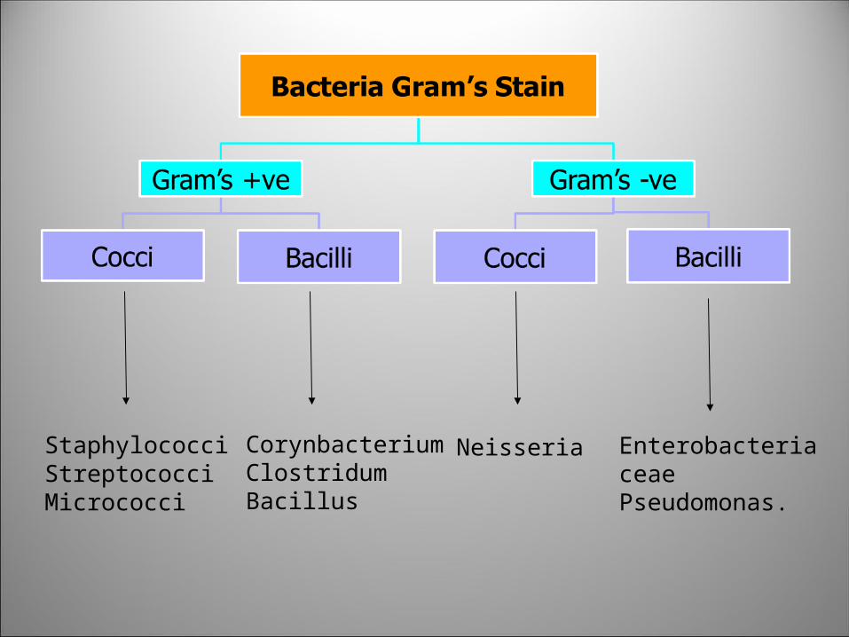
StaphylococciStreptococciMicrococci
NeisseriaCorynbacteriumClostridumBacillus
Enterobacteriaceae Pseudomonas.
Staphylococci
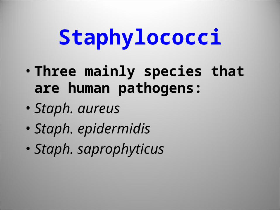
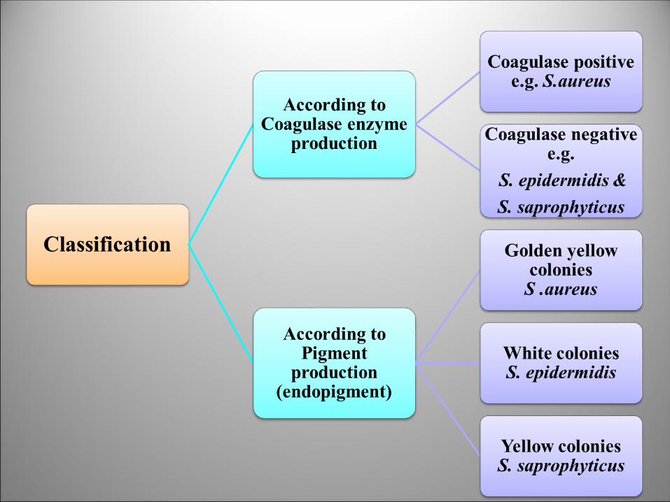
• Three mainly species that are human pathogens:
• Staph. aureus
• Staph. epidermidis
• Staph. saprophyticus
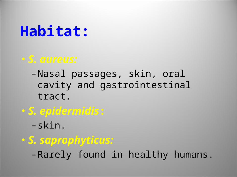
Habitat:
• S. aureus:– Nasal passages, skin, oral cavity and
gastrointestinal tract.
• S. epidermidis:– skin.
• S. saprophyticus: – Rarely found in healthy humans.
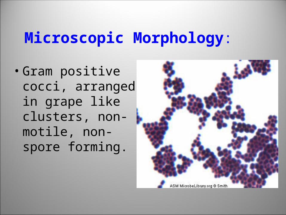
Microscopic Morphology:
• Gram positive cocci, arranged in grape like clusters, non-motile, non-spore forming.
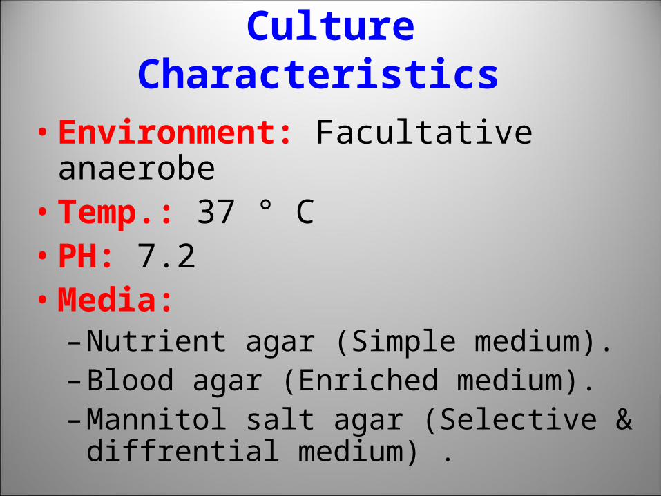
Culture Characteristics
• Environment: Facultative anaerobe• Temp.: 37 ° C• PH: 7.2• Media:
– Nutrient agar (Simple medium).– Blood agar (Enriched medium).– Mannitol salt agar (Selective & diffrential
medium) .
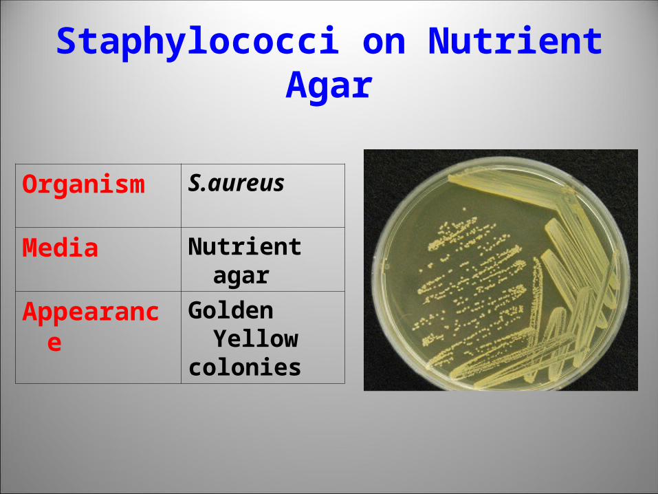
Staphylococci on Nutrient Agar
Organism S.aureus
Media Nutrient agar
Appearance Golden Yellow
colonies
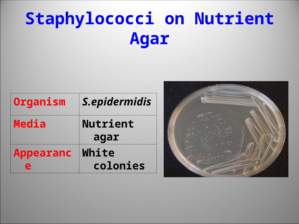
Staphylococci on Nutrient Agar
Organism S.epidermidis
Media Nutrient agar
Appearance White colonies
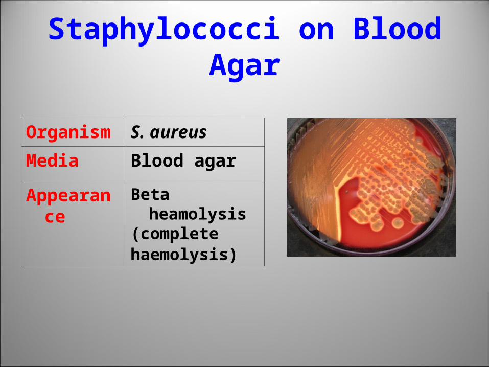
Staphylococci on Blood Agar
Organism S. aureus
Media Blood agar
Appearance Beta heamolysis(completehaemolysis)
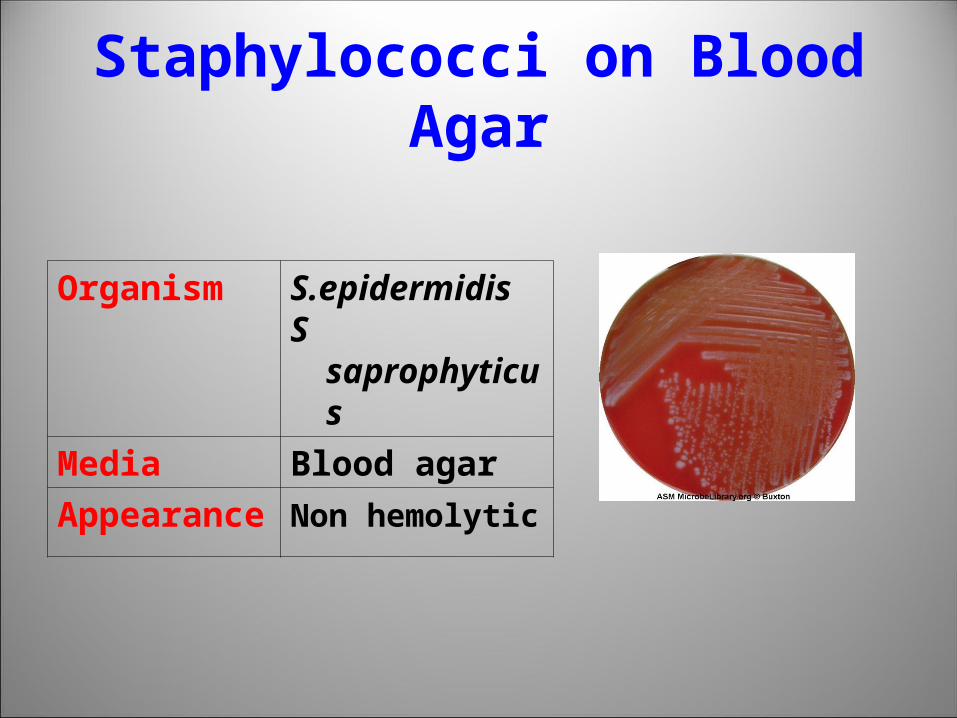
Staphylococci on Blood Agar
Organism S.epidermidisS saprophyticus
Media Blood agar
Appearance Non hemolytic
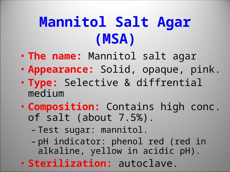
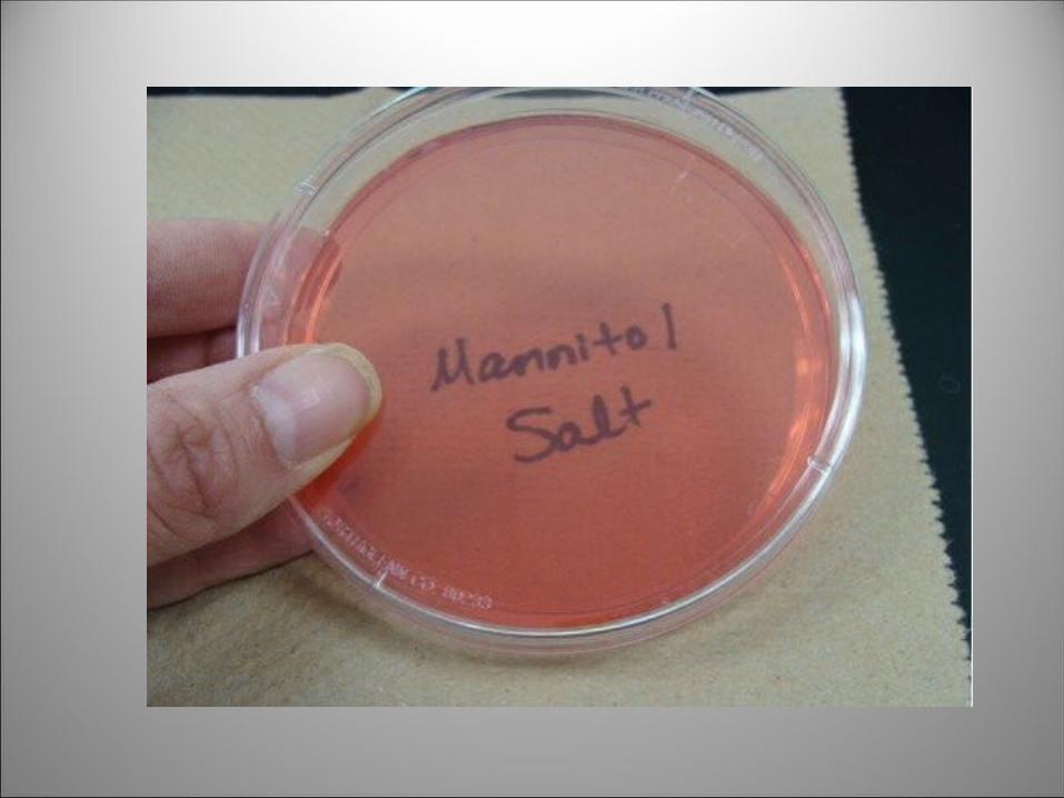
Mannitol Salt Agar (MSA)
• The name: Mannitol salt agar • Appearance: Solid, opaque, pink.• Type: Selective & diffrential medium• Composition: Contains high conc. of salt
(about 7.5%).– Test sugar: mannitol.– pH indicator: phenol red (red in alkaline, yellow in
acidic pH).
• Sterilization: autoclave.
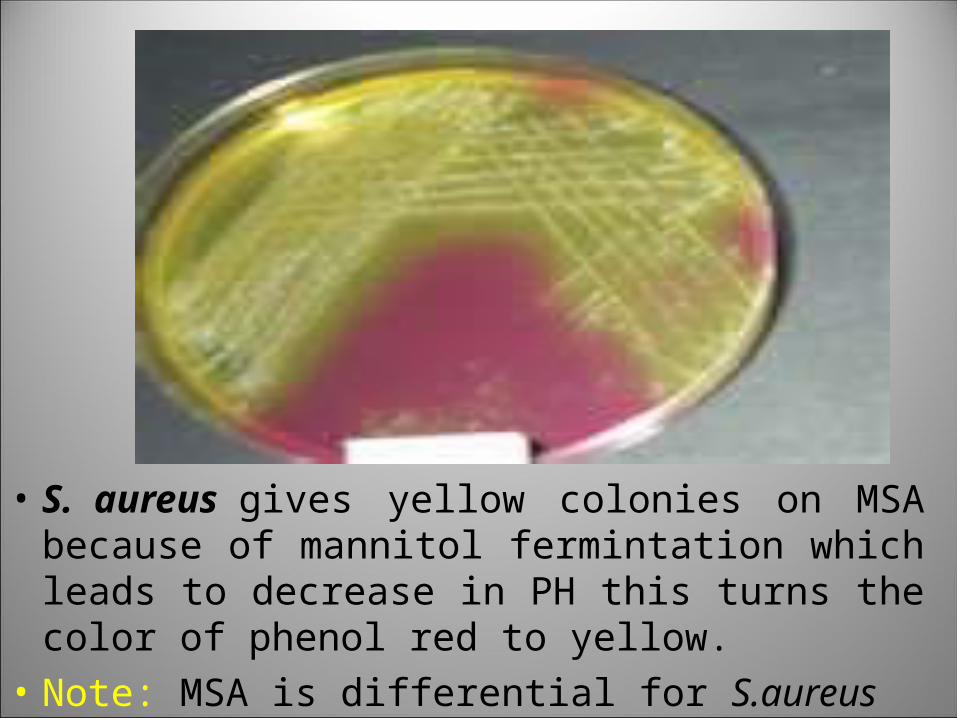
• S. aureus gives yellow colonies on MSA because of mannitol fermintation which leads to decrease in PH this turns the color of phenol red to yellow.
• Note: MSA is differential for S.aureus
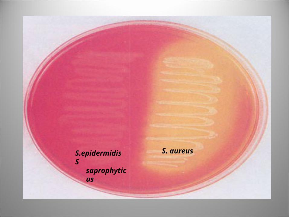
S. aureus S.epidermidisS saprophyticus
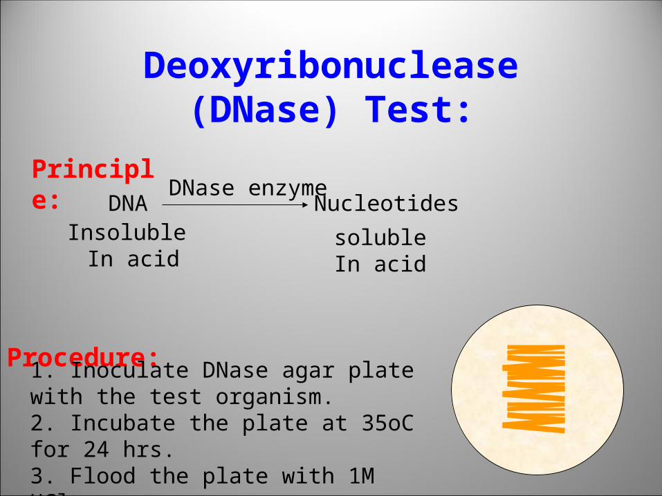
Deoxyribonuclease (DNase) Test: Principle:
DNADNase enzyme
NucleotidesInsoluble
In acidsoluble In acid
Procedure:
1. Inoculate DNase agar plate with the test organism. 2. Incubate the plate at 35oC for 24 hrs.3. Flood the plate with 1M HCl.
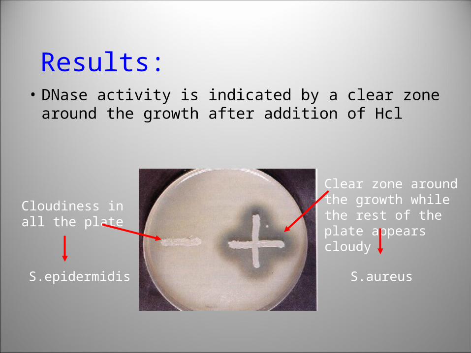
Results:• DNase activity is indicated by a clear zone
around the growth after addition of Hcl
Clear zone around the growth while the rest of the plate appears cloudy
Cloudiness in all the plate
S.epidermidis S.aureus
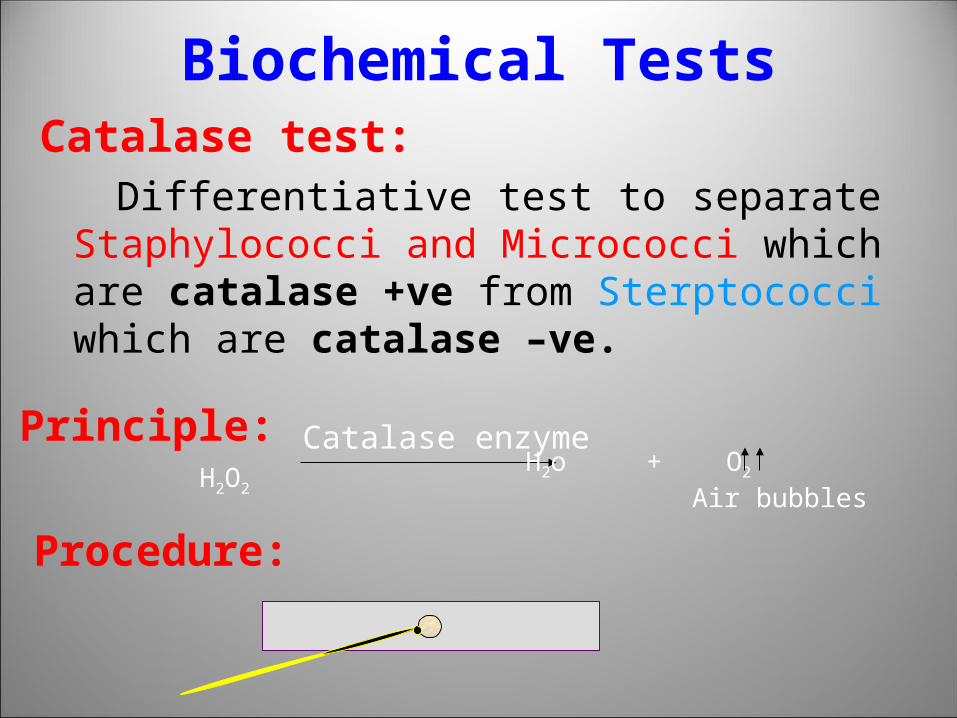
Biochemical TestsCatalase test: Differentiative test to separate Staphylococci
and Micrococci which are catalase +ve from Sterptococci which are catalase –ve.
H2O2
Catalase enzymeH2o + O2
Air bubbles
Principle:
Procedure:
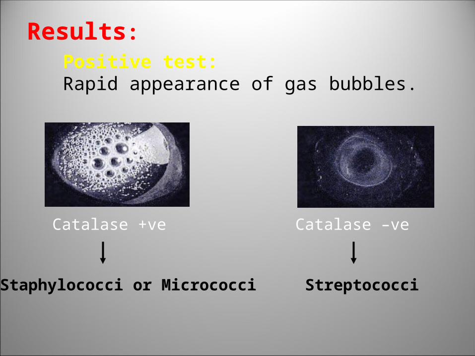
Results:Positive test: Rapid appearance of gas bubbles.
Staphylococci or Micrococci
Catalase –ve
Streptococci
Catalase +ve
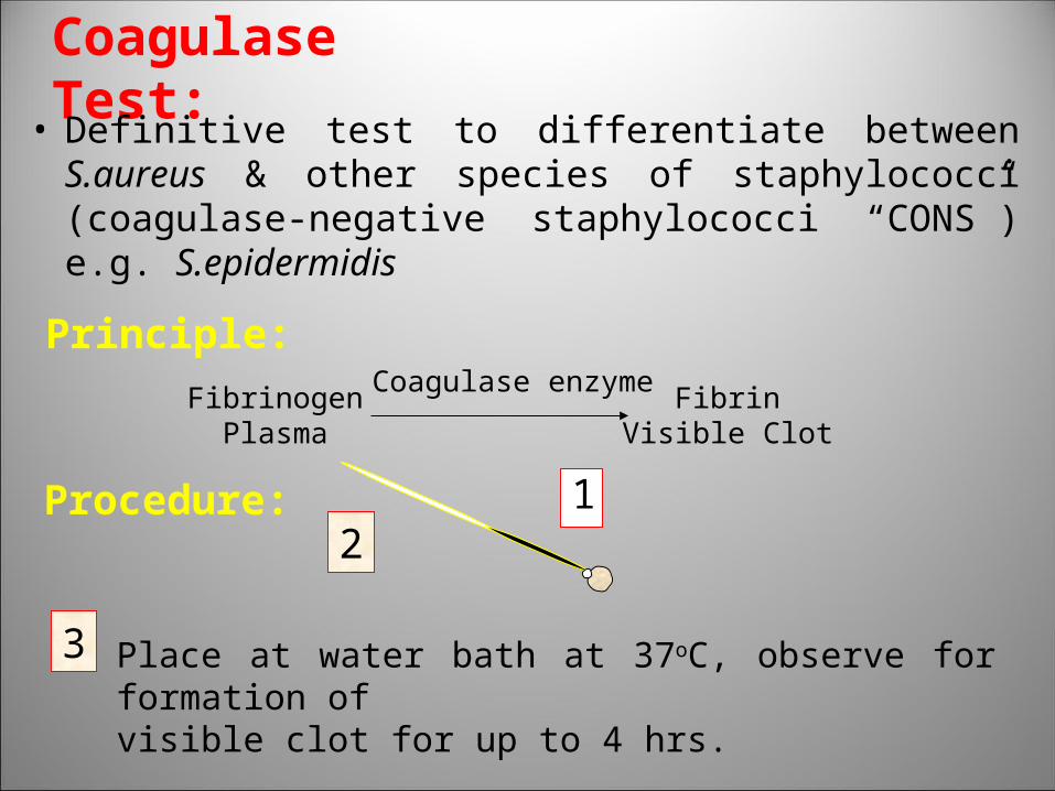
Coagulase Test:• Definitive test to differentiate between S.aureus &
other species of staphylococci (coagulase-negative staphylococci “CONS”) e.g. S.epidermidis
Principle:
FibrinogenPlasma
Coagulase enzymeFibrin
Visible Clot
Procedure:2
3 Place at water bath at 37oC, observe for formation of visible clot for up to 4 hrs.
1
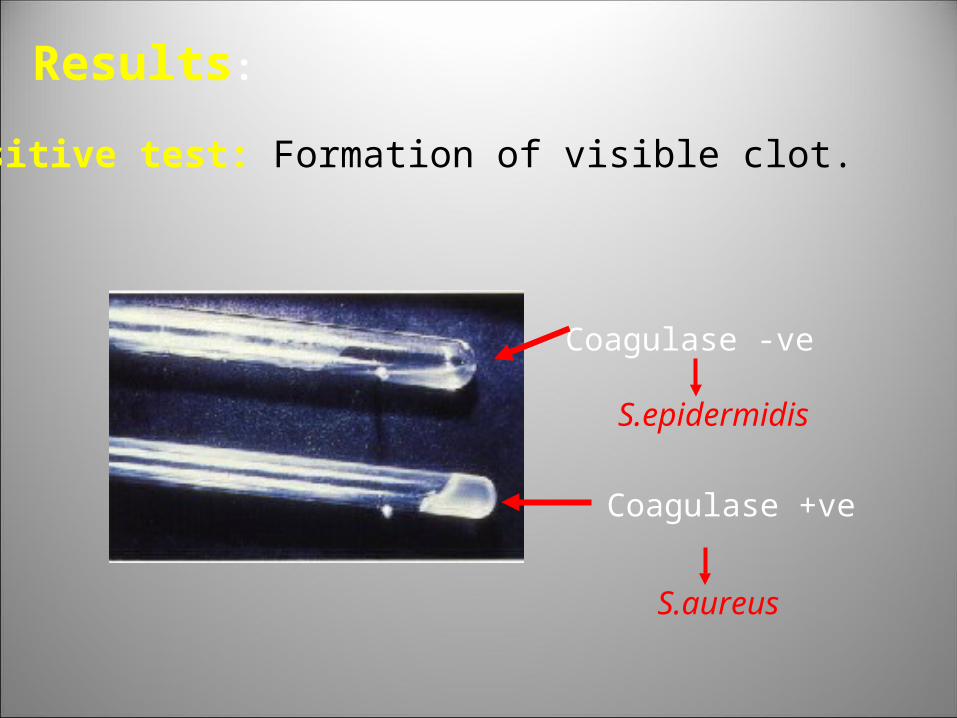
Results:
Positive test: Formation of visible clot.
Coagulase +ve
S.aureus
Coagulase -ve
S.epidermidis
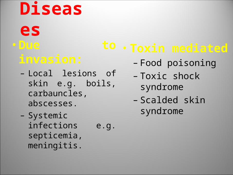
Diseases• Due to invasion:
– Local lesions of skin e.g. boils, carbauncles, abscesses.
– Systemic infections e.g. septicemia, meningitis.
• Toxin mediated– Food poisoning– Toxic shock syndrome– Scalded skin syndrome
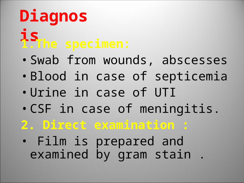
Diagnosis 1.The specimen:• Swab from wounds, abscesses• Blood in case of septicemia • Urine in case of UTI• CSF in case of meningitis.2. Direct examination :• Film is prepared and examined by gram
stain .
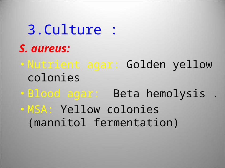
3.Culture : S. aureus:
• Nutrient agar: Golden yellow colonies
• Blood agar: Beta hemolysis .
• MSA: Yellow colonies (mannitol fermentation)
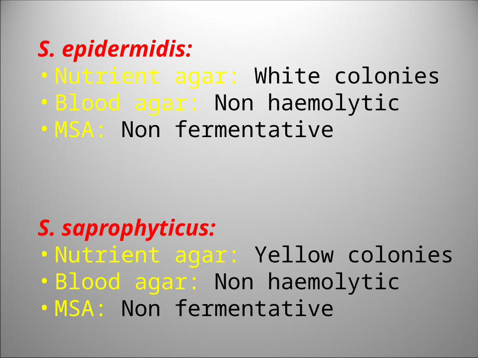
S. epidermidis:• Nutrient agar: White colonies• Blood agar: Non haemolytic• MSA: Non fermentative
S. saprophyticus:• Nutrient agar: Yellow colonies• Blood agar: Non haemolytic• MSA: Non fermentative
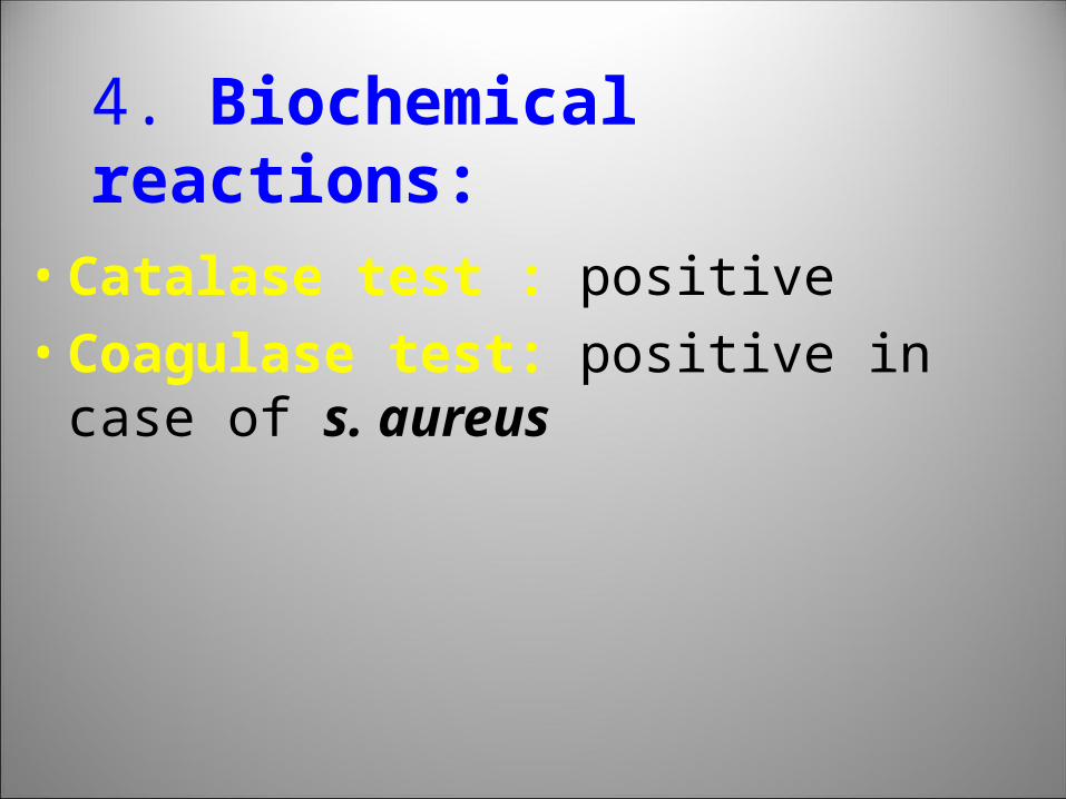
4. Biochemical reactions:
• Catalase test : positive
• Coagulase test: positive in case of s. aureus

Top Related