


Languages
Pages
Legal


p53 gene mutations in human tumors
Lung (897) 56% Adrenal (31) 23% Colon (960) 50% Breast (1536) 22% Esophagus (279) 45% Endometrium (224) 22% Ovary (386) 44% Mesothelioma (23) 22% Pancreas (170) 44% Renal (102) 19% Skin (220) 44% Thyroid (299) 13% Gastric (314) 41% He matological (1916) 12% He ad & neck (524) 37% Carcinoid (13) 11% Bladder (308) 34% Melanoma (70) 9% Sarc oma (339) 31% Parathyroid (13) 8% Prostate (87) 30% Cevix (350) 7% He patoce llular (716) 29% Neuroblastoma (212) 1% Brain (456) 25% others (155) 0%
Tumor type (n) p53 mutation Tumor ty pe ( n)
p53 mutation
All tumors: 37%
Greenblatt et al. (1995) Cancer Res. 54:4855
50%

p53 and Ink4a are the two most frequently mutated genes in human tumors
LocusChromosome location type of alterations
estimated frequency of alterations
p53
INK4a
17p13
9p21
nucleotide substitution
homozygous deletion nucleotide substitution small deletion/insertion promoter methylation
~ 50%
~ 40%

p53(low)
p53(high)
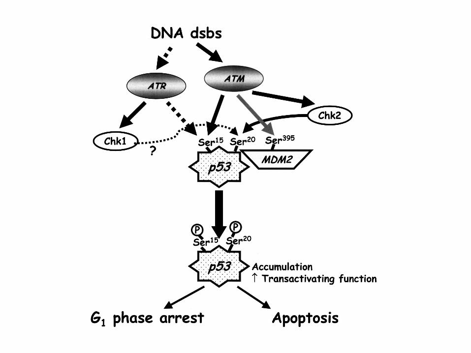
Cell cycle arrest Apoptosis
Cellular Stresses(e.g. DNA damage)
The Basic Paradigm of p53 Function

The Discovery of p53
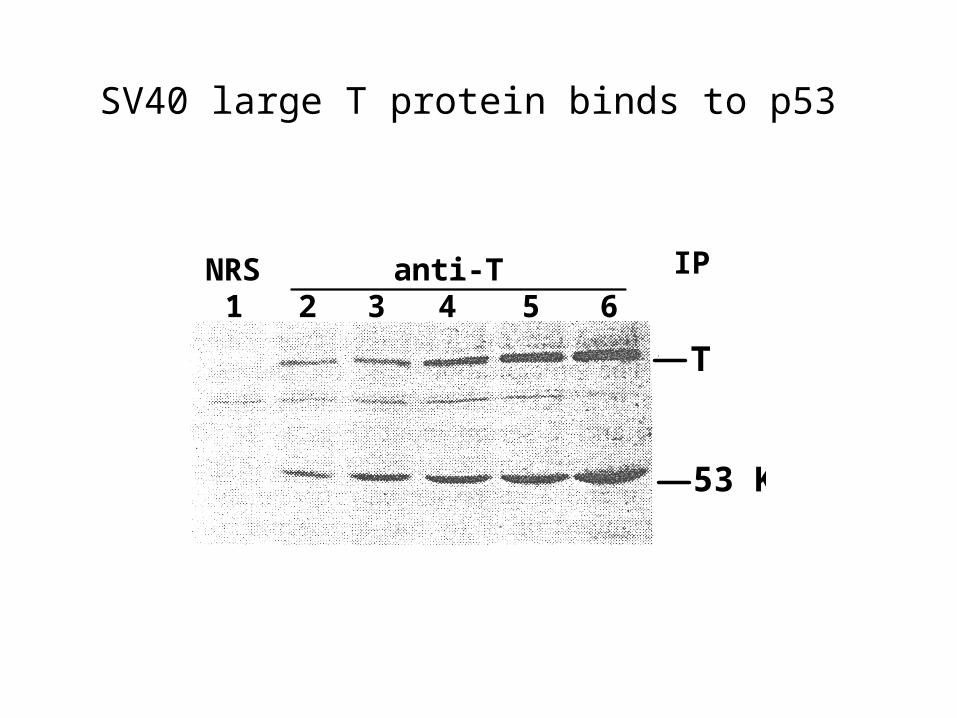
SV40 large T protein binds to p53
T
53 K
61 2 3 4 5
IPNRS anti-T

Li Fraumeni Syndrome
An inherited neoplastic disease with autosomal dominant trait
Characterised by multiple primary neoplasms in children and young adults
A predominance of soft tissue sarcomas (e.g. breast)
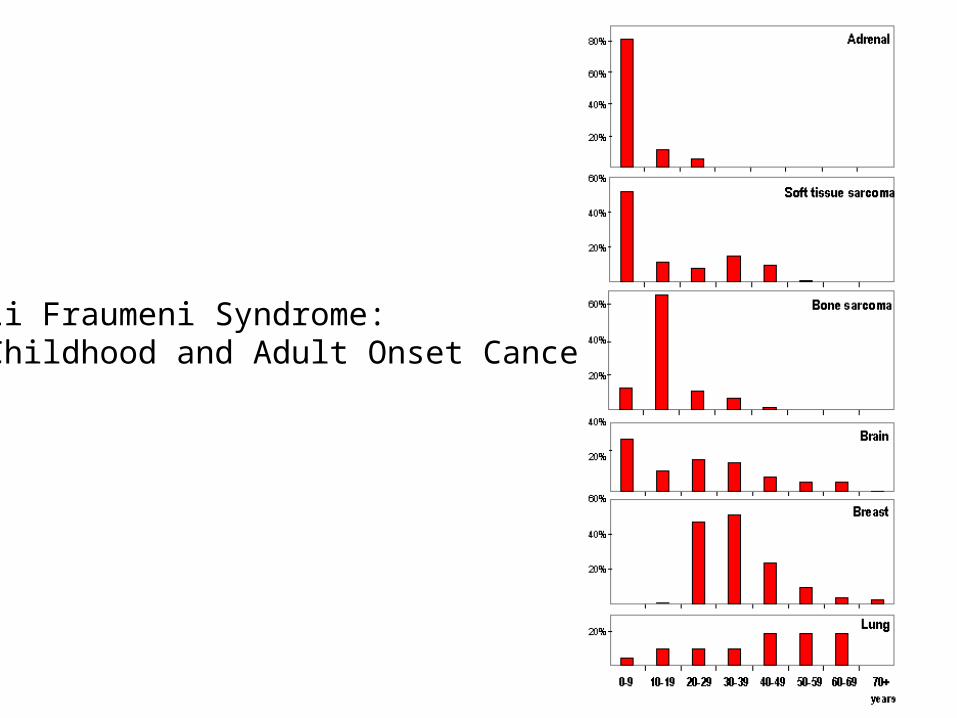
Li Fraumeni Syndrome:Childhood and Adult Onset Cancers
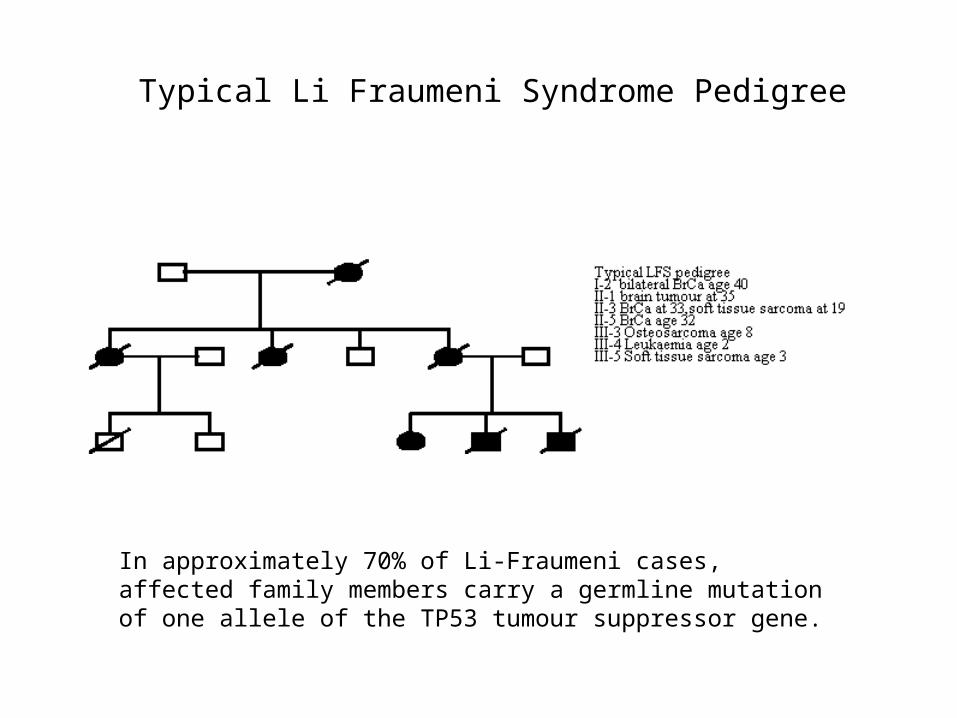
Typical Li Fraumeni Syndrome Pedigree
In approximately 70% of Li-Fraumeni cases, affected family members carry a germline mutation of one allele of the TP53 tumour suppressor gene.

Inactivation of p53 in Friend murine erythroleukemia

p53 Mutant Mice Develop Cancer

p53 Mutations in Human Tumors are Found with HighFrequency In the DNA Binding Domain
In 143 families reported:point mutations (85%)deletions (9%)splice mutations (3.5%) insertions (2%)

Ribbon Model Space Filling Model
p53 Binds DNA
The most common mutation changes arginine 248, colored red here. Notice how it snakes into the minor groove of the DNA (shown in blue and green), forming a strong stabilizing interaction. When mutated to another amino acid, this interaction is lost. Other key sites of mutation are shown in pink, including arginine residues 175, 249, 273 and 282, and glycine 245.

p53 Binds DNA as a Tetramer

p53 Functions in Cell Cycle Checkpoints

p53 Mutant Cells Lack Cell Cycle Checkpoint Function


p53 activates p21 (WAF1)
p53
WAF1
0 4 6 8 16 hr
El-Deiry et al. (1993) Cell 75:817


The Structure of RAD9
BRCT domains required for oligomerization in response to DNA damageBRCT domains are found in BRCA1Rad9 and BRCA1 both may act as scaffoldsFHA domains bind specific phosphopeptides
Rad53=Chk2Toh and Lowndes, Biochem Soc Trans. 2003. 31:242-6.
1309
SCD=S/T Cluster Domain

The Function of RAD9
(Rad53=chk2)(Rad9 similar to BRCA1)
(Mec1=ATM)
Toh and Lowndes, Biochem Soc Trans. 2003. 31:242-6.
Top Related