


Languages
Pages
Legal

• Describe the cardiac anatomy for patients with Hypoplastic Left Heart Syndrome (HLHS)
• Compare staged surgical repairs for HLHS
• Identify goals of surgical interventions for staged repairs for HLHS
• Identify components of family education during prenatal counseling, postoperative care, as well as discharge for patients with HLHS
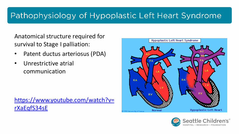
Anatomical structure required for survival to Stage I palliation:
• Patent ductus arteriosus (PDA)
• Unrestrictive atrial communication
https://www.youtube.com/watch?v=rXaEqfS34sE
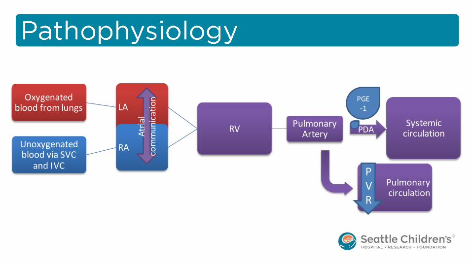
Systemic circulation
Pulmonary Artery
RV
LA Oxygenated
blood from lungs
RA Unoxygenated blood via SVC
and IVC
Pulmonary circulation
Atr
ial
com
mun
icat
ion
PDA
PGE-1
PVR
• Right Ventricle (RV) cardiac output (CO) is increased by 2-5 times its normal
– Shock
– Necrotizing enterocolitis (NEC)
– Poor perfusion to the brain and coronary arteries
• Prenatal diagnosis is usually made at 18-20 weeks gestation
• Family will meet with Prenatal Cardiology, at which time they will be provided with an overwhelming amount of information
• Anatomy of HLHS
• Staged palliations
• Common procedures
• Impact on other organs
• Likelihood of transplant
• Lifelong chronic disease
Family is counselled on 3 options: 1. Intervention 2. Palliative 3. Termination (In the state of
Washington a pregnancy can be terminated up to 24 weeks, if the pregnancy is further along, then this option is not provided)
* In the past 2 years, among 24 HLHS patients seen by SCH team, 7 chose termination and 1-2 chose comfort care, thus ~30% of families chose no intervention
• Ultimate goal is to separate the pulmonary and systemic circulations
• This is performed in 3 staged surgeries referred to “palliations”
• They are called palliations because they provide circulation that is compatible with life, however their hearts cannot actually be “repaired”
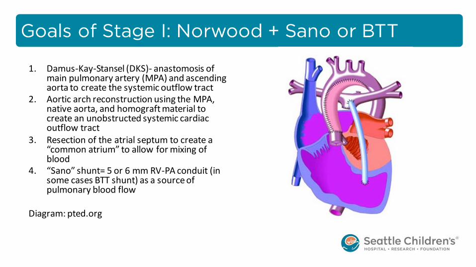
1. Damus-Kay-Stansel (DKS)- anastomosis of main pulmonary artery (MPA) and ascending aorta to create the systemic outflow tract
2. Aortic arch reconstruction using the MPA, native aorta, and homograft material to create an unobstructed systemic cardiac outflow tract
3. Resection of the atrial septum to create a “common atrium” to allow for mixing of blood
4. “Sano” shunt= 5 or 6 mm RV-PA conduit (in some cases BTT shunt) as a source of pulmonary blood flow
Diagram: pted.org
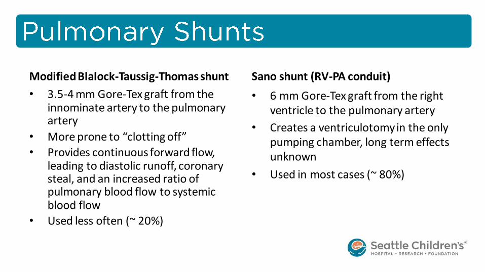
Modified Blalock-Taussig-Thomas shunt
• 3.5-4 mm Gore-Tex graft from the innominate artery to the pulmonary artery
• More prone to “clotting off”
• Provides continuous forward flow, leading to diastolic runoff, coronary steal, and an increased ratio of pulmonary blood flow to systemic blood flow
• Used less often (~ 20%)
Sano shunt (RV-PA conduit)
• 6 mm Gore-Tex graft from the right ventricle to the pulmonary artery
• Creates a ventriculotomy in the only pumping chamber, long term effects unknown
• Used in most cases (~ 80%)
https://www.youtube.com/watch?v=-87kq98l1kk
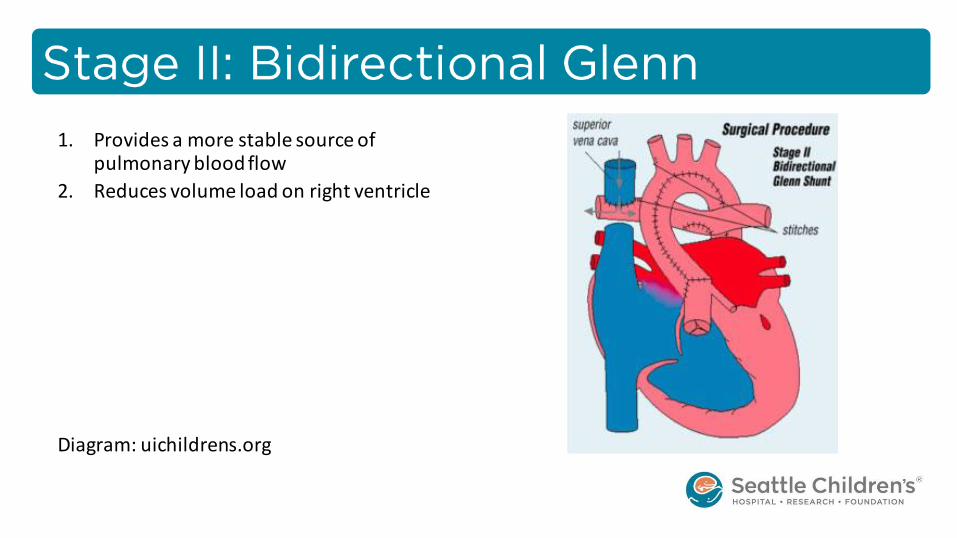
1. Provides a more stable source of pulmonary blood flow
2. Reduces volume load on right ventricle
Diagram: uichildrens.org
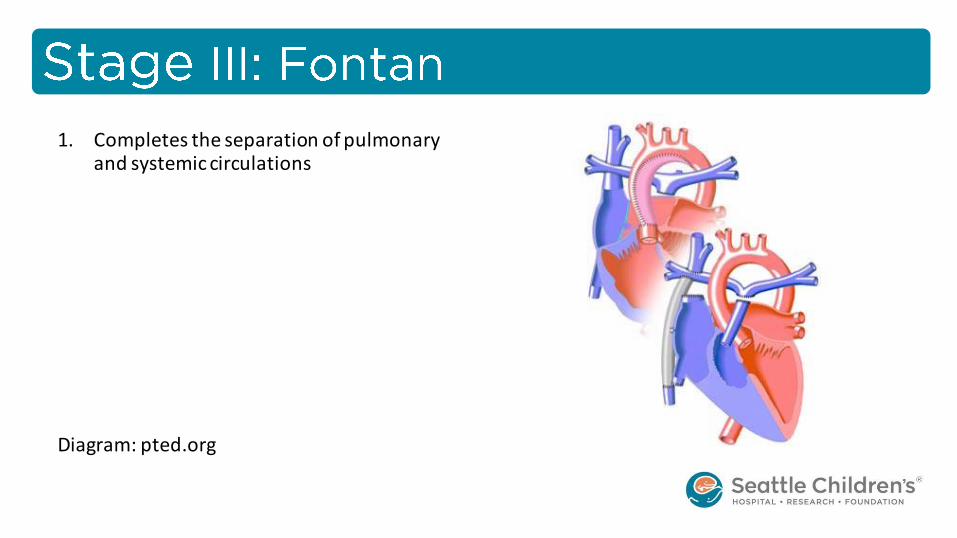
1. Completes the separation of pulmonary and systemic circulations
Diagram: pted.org
• Usually return to ICU with open sternum, delayed sternal closure expected on POD 2-4 in most cases
• Typically intubated between 3-7 days, though sometimes longer
• Expected length of stay in ICU is about 1-2 weeks
• Usually another 2+ weeks on the surgical floor
• Must stay local through Stage 2 palliation for close follow up with Single Ventricle Team
• Arrhythmias
• Infection
• Vocal cord dysfunction/paralysis
• Necrotizing enterocololitis
• Renal injury
• Low cardiac output stateECMOdeath
• Close monitoring of oxygen saturations (should ideally be 75-85%)
• Feeding advancement (including working with OT) while being closely monitored for NEC
• Managing diuretic therapy and often times pulmonary overcirculation
• Monitoring for potential aortic arch obstruction (daily upper/lower BPs)
• SINGLE VENTRICLE TEACHING
• Work of breathing
• Oxygen saturation (goal of 75-85%)
• Perfusion
• Upper AND lower blood pressure (to evaluate for potential coarctation)
• Feeding tolerance/advancement (more to come in upcoming slide)
• Weight gain
• Parents knowledge/comfort/level of involvement
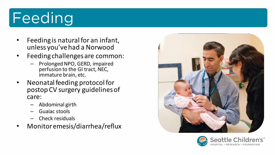
• Feeding is natural for an infant, unless you’ve had a Norwood
• Feeding challenges are common: – Prolonged NPO, GERD, impaired
perfusion to the GI tract, NEC, immature brain, etc.
• Neonatal feeding protocol for postop CV surgery guidelines of care: – Abdominal girth – Guaiac stools – Check residuals
• Monitor emesis/diarrhea/reflux
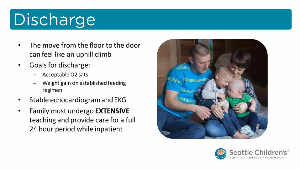
• The move from the floor to the door can feel like an uphill climb
• Goals for discharge: – Acceptable O2 sats
– Weight gain on established feeding regimen
• Stable echocardiogram and EKG
• Family must undergo EXTENSIVE teaching and provide care for a full 24 hour period while inpatient
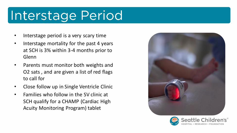
• Interstage period is a very scary time
• Interstage mortality for the past 4 years at SCH is 3% within 3-4 months prior to Glenn
• Parents must monitor both weights and O2 sats , and are given a list of red flags to call for
• Close follow up in Single Ventricle Clinic
• Families who follow in the SV clinic at SCH qualify for a CHAMP (Cardiac High Acuity Monitoring Program) tablet
• Hypoplastic left heart syndrome is a devastating diagnosis for parents
• At minimum these children will undergo 3 major surgeries, in addition to several other procedures
• Interstage mortality, though decreasing, remains a major consideration in the care of these fragile patients
• Many will require a heart transplant at some point in their lives
• With or without the need for transplantation, the diagnosis of HLHS carries with it a lifetime of chronic illness
Pediatric Heart Surgery: A Ready Reference for Professionals- fifth edition, 2012
https://www.chop.edu
Top Related