


Languages
Pages
Legal


G. COLONNA M.D.,
G. Lorusso M.D., S. Santoro M.D.
Eye Department “S. Maria degli Angeli” Hospital
Putignano (Bari) – ITALY
Head of Dpt. S. Santoro MD.
New topographic custom ablation
procedure for treating irregular
astigmatism post keratoplasty with high
frequency (1 KHz) excimer laser.
ESCRS – Berlin 2008

On what information do we base
our CA planning in virgin eyes?
Ablation planning has traditionally been based on information that reflect visual function
– M.R., Autorefractometry, WF aberrometry, Placido based topography
– Data dependent on patients fixation
– Information (maps) is referenced to the rotational position of the fixating eye
Such concept seems to be logical and has been highly successful in treatments of spherocylindrical errors and even High Order Aberrations in virgin eyes
– As the goal of the surgery is improvement of a well functioning intact visual system
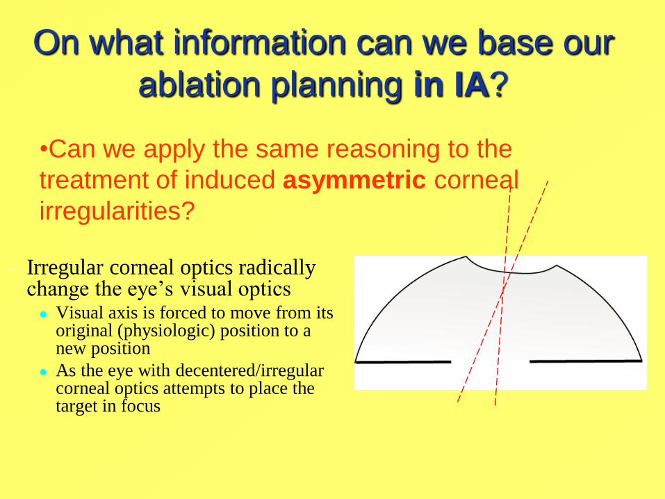
On what information can we base our
ablation planning in IA?
– Irregular corneal optics radically change the eye’s visual optics Visual axis is forced to move from its
original (physiologic) position to a new position
As the eye with decentered/irregular corneal optics attempts to place the target in focus
•Can we apply the same reasoning to the
treatment of induced asymmetric corneal
irregularities?

– Eye with Irregular corneal optics assumes a new rotational position so that x, y position of the corneal intercept of visual axis and its tilt change
An ablation plan, that uses topography or WF information
referenced to the visual axis, would attempt to optimize
the corneal optics on the basis of a pathological
rotational position

What kind of information is
used by current technologies?
Information bound to visual axis / line of sight:
– - Wavefront guided
- Topography guided - based on placido ring
information (”corneal wavefront”)
Information reflecting global 3D corneal
morphology independent on the visual axis
Topography guided - based on elevation
data (acquired by triangulation)
“ C I P T A”

What consequences the use of the
two concepts has in treatments of
IA?
The most important issues in treatments of
eyes with irregular astigmatism are:
– Corneal tissue sparing = crucial in previously
treated cases or where a lamella has been placed
– Smooth transition towards the untreated cornea (=
Biologic tolerance = Better chance for a permanent
effect)

Purpose
To evaluate the efficacy and safety of topographic
guided ablation using a new scheimpflug image based
topographer. All eyes show an irregular corneal
astigmatism post keratoplastic surgery not correctable
with spectacles. Objective was to regularize the anterior
cornea surface to eliminate the cylindrical component of
manifest refraction due to the cornea.
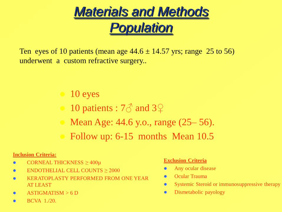
Materials and Methods
Population
Ten eyes of 10 patients (mean age 44.6 ± 14.57 yrs; range 25 to 56)
underwent a custom refractive surgery..
10 eyes
10 patients : 7♂ and 3♀
Mean Age: 44.6 y.o., range (25– 56).
Follow up: 6-15 months Mean 10.5
Inclusion Criteria:
CORNEAL THICKNESS ≥ 400µ
ENDOTHELIAL CELL COUNTS ≥ 2000
KERATOPLASTY PERFORMED FROM ONE YEAR
AT LEAST
ASTIGMATISM > 6 D
BCVA 1./20.
Exclusion Criteria
Any ocular disease
Ocular Trauma
Systemic Steroid or immunosuppressive therapy
Dismetabolic payology

Materials and Methods
The iVis Platform
All patients were treated in transepithelial procedure with the italian excimer
laser IRES 1,000 Hz (iVIS Technologies, Taranto, Italy). The ablation profile,
calculated by CIPTA software (iVIS), was based upon Topography (Precisio,
iVIS). The transepithelial excimer laser ablation was planned to leave a regular
and smooth anterior surface of the cornea. All treatments were planned
controlling the transition zone curvature over the transplanted cornea to reduce
the possibility of any regression process related to the epithelium
Frequency = 1000 Hz
Gaussian flying spot = 0.65mm
Constant frequency on surface
area
Setup / Calibration = totally
automated
C.I.P.T.A.

Pre bcva+1=-7(10) 20/20 Post ucva 20/20
Pre bcva=-4(50) 20/20 Post ucva 20/25
Post ucva 20/20Pre bcva=-8(80) 20/32

Results Six Months follow up
All eyes were re-epithelializated within 7 days. At a minimum of six months
follow-up, the best spectacle corrected visual acuity improved from the pre-op
mean values of 20/32 to 20/20 post-operatorely, the uncorrected visual acuity
improved from the pre-op mean values of 20/125 to 20/32 post-operatorely and
the mean corneal astigmatism was reduced from a mean of 8.0D ± 2.73 to
1.75D ± 0.25. The topographic pattern improved showing a regular cornea
surface in all eyes. No adverse events have been reported neither during the
surgery and the followup period.

Conclusion
Eye Department “S. Maria degli Angeli” HospitalPutignano (Bari) – ITALY
Head of Dpt. S. Santoro MD.
Topography guided customized ablation combined with
an ultrafast excimer laser with transepithelial
procedure is a safe and effective technique for treating
irregular astigmatism post keratoplasty, improving the
patient’s VA and his quality of vision.
Top Related