


Languages
Pages
Legal


Republic of Iraq
Ministry of Higher Education
and Scientific Research
University of Baghdad
College of Education for Pure Science
(Ibn Al-Haitham)/ Department of Biology
New Immunological Technique for
Diagnosis of Candida albicans Infection
A Thesis
Submitted to the Council of the College of Education for Pure
Science (Ibn Al-Haitham), University of Baghdad in Partial
Fulfillment of the Requirements for the Degree of Master in Science of Biology /Immunology
By
Russul Arkan Hassan
B.Sc. Biology, College of Education for Pure Science (Ibn Al-
Haitham) / University of Baghdad (2016)
Supervised By
Assistant Professor
Dr. Hazima Mossa AL-Abassi
August /2019 A.D. Thul Huja /1440 A.H.

﷽ وعنده مفاتيح الغيب ل يعلمها إلا هو ويعلم ﴿
ما في البر والبحر وما تسقط من ورقة إلا
ول يعلمها ول حباة في ظلمات الرض
﴾رطب ول يابس إلا في كتاب مبين
(59)أية -سورة االنعام


To the big heart in universe and the
smile of my life my mother and father.
To those my heart always remembers
them, to my brother and my sisters.
To the two person who guides my way in
science and knowledge Dr. Hazima
mossa Al-Abassi and Dr. Ali Abdul
Hussain Mahdi
RUSSUL
Dedication

Acknowledgment
Before and after everything I must thank my God
(Allah) for providing me the strength to finish this study.
All thanks are to his Almighty, who gave me the right
direction and courage to perform this research work.
I would like to express my thanks very much to the
Head of the Department of Biology/ College of
Education for Pure Sciences (Ibn Al-Haitham) for
providing this occasion to opportunity my research
work. Iwould also like to thank all members of this
department for giving me their valuable advice.
I would like to express my deep thanks and sincere
gratitude to my supervisor Dr. Hazima M. K. Al-Abassi
for her valuable scientific guidance, understanding,
encouragement that made it possible for me to
accomplish this study. I would like to express my deep
thanks and sincere gratitude Dr. Ali Abdul Hussain
Mahdi who guided me with his valuable advice and
support which helped us in completing this research.
Also, I would like to express grateful to my Family for
their constant encouragement, patience, continuous
care and support all of them helped me greatly during
the period of this study.
RUSSUL
I
Abstract
In this study, included produce a new technique for diagnosis of
Candida albicans (skin, mouth, and vagina) infection. This study
included 100 samples of the patient (immunocompromised women with
average age (55-57) year and babies with average age (1-18 month),
patients infected with various (fungi, bacteria) infection, in addition to 20
samples for an apparently healthy individual (a control). Samples were
collected from AL- Dowaly Private Hospital in Baghdad during the
period January 2019 to June 2019. All infections were diagnosed by
consultant medical staff at the hospital by using several tests including
(germ tube, culture, Vitek and API20C). Cases enrolled in this study were
as follow:
Thirty- Samples from patients with C. albicans infection (skin
scales).
Fifteen-Samples from patients with Aspergillus spp. infection (skin
scales).
Fifteen-Samples from patients with Tricophactone spp. (infection
(skin scales).
{Skin scales were isolated from fingers foot (intertriginous)}
Ten-Samples from patients with C. albicans infection (skin swabs).
Ten-Samples from patient's bacteria with Staphylococcus aureus
infection (skin swabs).
Ten-Samples from patients (1-18 month) infected with C. albicans
(mouth swabs).
Ten-Samples from patients with C. albicans infection (vagina
swabs).

II
The Samples were examined using the new technique compared to the
routine methods of diagnosis, we found the new technique gave a positive
result for samples infected with C. albicans, while the rest of the samples
non - C. albicans infection (fungal, bacteria) gave a negative result.
It was shown that the Vitek test was more sensitivity (100%) and
specific (100%) for diagnosis of C. albicans than other methods (API20C
sensitivity (100%) and specific (97%), Colonial morphology ''culture''
sensitivity (96%) and specific (93%) and germ tube test sensitivity (98%)
and specific (95%). The results of the new technique test in this study for
the diagnosis of C. albicans was similar to the result of Vitek test in
sensitivity (100%) and specificity (100%), the benefits of this new
technique are to provide information with high accuracy, economical (low-
cost) and reduced time consumption. for example, physicians can use
through the attending visit of the patient without having to be sent to the
laboratory.

List of contents
III
List of contents
Title Page No.
Abstract I
List of content III
List of table VI
List of figure VII
List of abbreviations VIII
CHAPTER ONE
1-1 Introduction 1
1.2 Aims of the study 3
CHAPTER TWO
2.1 Antigen –Antibody Interaction 4
2.2 Factors Affecting Antigen Antibody Reactions 5
2.3. Immunological diagnostic Techniques 6
2.3.1. Fluorescent Immunoassay 6
2.3.2. Flow Cytometry 7
2.3.3. Radioimmunoassay 7
2.3.4. Enzyme Linked Immuno-Sorbent Assay, or ELISA 8
2.4. Candida Spp 9
2.5. Candida albicans 10
2.6. Candidiasis 12
2.7. Classification of candidiasis infection 12

List of contents
IV
2.7. 1. Superficial infection 12
2.7.1.1. Oral candidiasis 12
2.7.1.2. Cutaneous candidiasis 14
2.7.1.3. Vaginal candidiasis 15
2.7.2 Systemic infections 16
2.8. Pathogenicity and Virulence of C. albicans 16
2.9. Immune responses to Candida spp. 17
2.10. Methods Laboratory diagnosis of C. albicans 20
2.11.1. Direct examination 20
2.11.2. Culture 20
2.11.3. Germ Tube Test 20
2.11.4. API Yeast Identification System 22
2.11.5. Vitek Yeast Identification System [ specific Biochemical
reaction]
22
2.11.6. PCR (Polymerase Chain Reaction) 22
2.12. Diagnosis of Disseminated Candidiasis 23
CHAPTER THREE
3. Materials and Methods 24
3.1 Subject 24
3.2. Equipments. 25
3.3. Methods 28
3.3.1. Laboratory diagnosis of C. albicans 28
3.3.1.1. Cultuer 28

List of contents
V
3.3.1.2. Germ tube test 28
3.3.1.3. Vitek Yeast Identification System 29
3.3.1.4. API 20C Yeast Identification System 29
3.3.2. Laboratory diagnosis of the other samples 30
3.4. Preparation new kit to diagnose Ab-C. albicans 30
3.4.1. Procedure 30
3.6. Diagnosis of C. albicans infection by New Kit 33
CHAPTER FOURE
4.1. Isolation and Diagnosis of C. albicans 34
4.2. Sensitivity and Specificity for Diagnosis of C. albicans 36
4.3. Experiments results in which a new technique was used to
diagnose samples
39
CHPTER FIVE
5.1 Discussion 44
Conclusions and Recommandinations
Conclusions 48
Recommandinations 49
References 50
Abstract in Arabic أ

List of Tables
VI
List of tables
No. Table Titer Page
3-1 The general Equipments utilized in this study. 25
3-2 The Materials utilized in this study 25
3-3 The devices which used in the study 26
3-4 The kit and material 26
3-5 Isobornyl acrylate Basic information 32
3-6 Isobornyl acrylate Chemical Propertie 32
4-1 Diagnosis C. albicans and other (fungi, bacteria)
infection by routine methods and new technique
35
4-2 The reliable of the test by using sensitivity and
specificity
36
4-3 Sensitivity and Specificity for Diagnosis of C. albicans
by routine methods and new technique
37

List of figure
VII
List of figure
N0. Figure Titer Page No.
2-1 Colony of C. albicans was growth on soubourod agar 11
2-2 Oral candidiasis [thrush] 13
2-3 Cutaneous Candidasis 15
2-4 Germ tube formation of C. albicans 21
3-1 Isobornyl acrylate structure 30
4-1 Sensitivity (%) and specificity (%)for Diagnosis of C.
albicans by routine methods and new technique
38
4-2 The difference between sample of skin scale infected with
C. albicans and a non-infected sample of skin scales (as
control) under UV. Light
39
4-3 The difference between sample of skin scale infected with
C. albicans and sample of skin scale infected with
Aspergillus spp. under UV. Light
40
4-4 The difference between sample of skin scale infected with
C. albicans and sample of skin scale infected with
Tricophacton spp. under UV. Light.
41
4-5 The difference between sample of skin swab infected with
C. albicans and sample of skin swab infected with
Staphylococcus aureus under UV. Light.
42
4-6 The mouth-swab infected with C. albicans 43
4-7 The vagina-swab infected with C. albicans 43
List of figure
VIII
List of abbreviations
C. albicans Candida albicans
Ag-Ab interaction Antigen-Antibody interaction
IgG Immunoglobulin Gamma
FITC fluorescein isothiocyanate
FC-assay Flow cytometry assay
RIA Radioimmunoassay
ELISA Enzyme Linked Immuno-Sorbent assay
VBC Vitek biochemical card
IL-17 Interleukin -17
IL-4 Interleukin -4
IFN-γ Interferon Gamma
UV-Light Ultraviolet-Light
UV-Stain Ultraviolet-Stain
DC Disseminated Candidiasis
PCR Polymerase Chain Reaction

Chapter one INTRODUCTION

Chapter one Introduction
1
1.1 Introduction
Immunological techniques are a wide varieties of methods and
specialized experimental protocols devised by immunologists for inducing,
measuring, and characterizing immune responses (Van Emon.,2016). They
allow the immunologists to alter the immune system through cellular,
molecular and genetic manipulation. It's used usually to diagnosis human
diseases. Laboratory tests vary widely in clinical immunology; some are
essential for diagnosis while others are useful in sub classifying disorders.
Some are of research interest only, but may add to our immunological
armamentarium in the future. In this regard, it is important to understand
that these tests do vary in their sensitivity and specificity. The sensitivity
of a test is defined as the number of diseased individuals that are positive
for the test compared with those who are negative. These techniques have
developed and used in the medical and biotechnology fields. And the
immunological techniques used in the diagnosis, which relied on the
principle of antigen reaction with antibodies (e.g. ELISA, Immune
Fluorescent and Radioimmunoassay) (Carpenter.,1975; Schultz.,2009;
Zabriskie.,2009& Dunbbar.,2012).
Among species of genus Candida, C. albicans is the pathogen most
frequently isolated from the human body, including the oral cavity and
gastrointestinal and able of causing life-threatening opportunistic fungal
infections. C. albicans is considered opportunistic pathogen can cause
harm under abnormal conditions (Vilela et al., 2015; Strijbis et al., 2014).
The included ratio infection of candidiasis in Iraq from (2017 - 2018) was
(15-14.8) / 105 aura of the population respectively (Minster of health).
Chapter one Introduction
2
When the immune system is suppressed, this yeast can multiply rapidly,
penetrate the intestinal lining and move into the blood stream, Yeast
population is controlled by probiotic or beneficial bacteria (Mohamed et
al., 2010).
The immune mechanisms of defence against fungal infections are
numerous, and range from protective mechanisms that were present early
in evolution (innate immunity) to sophisticated adaptive mechanisms that
are induced specifically during infection and disease (Abood,
2014).
Innate immunity is the first line of unspecific host defense against
pathogens carried out by macrophages, neutrophils, and dendritic cells
(Kiyoura and Tamai, 2015). In adaptive immunity both CD4+ (T helper
cells) and CD8+ (cytotoxic T cells) have been reported a play role in
antifungal immunity, the nature of the T cell response is established by the
cytokine of the T cells during encounter activation: IL-12/IFN𝛾 for Th1
cells, IL-4 for Th2 cells (Naglik, 2014 and Sadeq.,2017).

Chapter one Introduction
3
1.2. Aims of the study
The laboratory diagnosis of candidiasis depends on the infection
caused by it. Prompt and accurate identification of Candida species is very
essential for effective therapeutic outcome. Conventional methods for the
diagnosis of candidiasis are less sensitive and time consuming, (e.g.
culture, germ tube, Vitek and API20C). So this study aims to produce a kit
with more sensitivity, specificity, accuracy and safety than routine method
to diagnose C. albicans to achieve this, the following approach were
adopted:
1- Prepare a new kit for diagnosis of Candida albicans using (specific
antibody conjugate with UV-stain).
2- Detect the immune complex by using harmless UV-light.
3- Design of protocol to diagnose other microorganisms depended on
this principle study (more specific and sensitive than classical test).

Chapter two LITERATURE REVIEW
Chapter two Literature review
4
2.1. Antigen –antibody Interaction
Antibody – Antigen interactions are much like other receptor-ligand
interactions, physicochemical forces are involved in the interaction
between an antibody and an antigen that are similar to those between an
enzyme and its substrate (or competitive inhibitor) or between a receptor,
such as an insulin receptor, and a ligand, such as insulin. These forces
derive from four sources: (1) Ionic bond (2) hydrogen bonds (3) van der
Waals forces, and (4) hydrophobic interactions. Many experimental
approaches have been used to define the structure of the antibody – binding
site for antigen (Zabriskie.,2009; Helbert., 2016).
The first correct description of the antigen-antibody reaction was
given by Richard J. Goldberg at the University of Wisconsin in 1952
(Goldberg.,1952). It came to be known as "Goldberg's theory" (of antigen-
antibody reaction) (Spiers., 1958).
Many serologic techniques are used to detect the interaction of
antigens with antibodies. These methods are suitable for the detection and
quantitation of antibodies to infectious agents, as well as microbial and
non-microbial antigens. Antigen-Antibody reactions can be classified into
two categories:
1- Precipitation reactions
Antibody and soluble antigen interacting in aqueous solution form a
lattice that eventually develops into a visible precipitate. Antibodies that
aggregate soluble antigens are called precipitins. Although formation of
the soluble Ag-Ab complex occurs within minutes, formation of the visible
precipitate occurs more slowly and often takes a day or two to reach
completion. Formation of an Ag-Ab lattice depends on the valency of both
Chapter two Literature review
5
the antibody and antigen. (Wild., 2001). In the clinical laboratory several
applications of the precipitation reaction are used. These methods include:
1- Immunodiffusion: These are of two types: single and double
immunodiffusion.
2- Electroimmunodiffusion (Stevens& Miller.,2016).
2- Agglutination reactions
The interaction between antibody and a particulate antigen results in
visible clumping called agglutination. Antibodies that produce such
reactions are called agglutinins. Agglutination reactions are similar in
principle to precipitation reactions; they depend on the crosslinking of
polyvalent antigens (Stites et al.,1997; Stevens& Miller.,2016 and
Shoemark et al., 2017).
2.2. Factors affecting antigen antibody reactions
Many factors affect the interaction between antigen and antibody
(Stevens & Miller., 2016); these include:
1- Specificity: The ability of a particular antibody to combine with one
antigen instead of another is referred to as specificity.
2- Temperature: The optimum temperature needed to reach
equilibrium in an antibody-antigen reaction differs for different
antibodies. IgM antibodies are cold reacting with thermal range 4-
22°C, and IgG antibodies are warm reacting, with an optimum
temperature of reaction at 37°C.
3- pH: The optimum pH for all reactions has 7.0 is used for routine
laboratory testing.
4- Ionic strength: The concentration of salt in the reaction medium has
an effect on antibody uptake by the membrane bound erythrocyte
antigens. Sodium and chloride ions in solution have inhibition effect.

Chapter two Literature review
6
5- Concentration: Under normal condition the concentration of antigen
and antibody should be optimal but some time this thing fail to be
happen in which excess antibody or antigen concentration will result
in false reaction, some times known as zonal reaction. When the
concentration of antigen is excess it is known as post zone reaction;
excess antibody is referred as prozone reaction. This phenomenon
can by overcome by serial dilution until optimum amount of antigen
and antibody will present (Lisova et al.,2014).
6- Cross reactivity: When some of the determinants of an antigen are
shared by similar antigenic determinants on the surface apparently
unrelated molecules, a proportion of the antibodies directed against
one kind of antigen will also react with the other kind of antigen. This
is called cross reactivity (Wrammert et al.,2011).
2.3. Immunological diagnostic techniques
It's used usually to diagnosis human disease, these techniques have
developed and used in the medical and biotechnology fields (Schultz.,
2009), some type of immunological techniques:
2.3.1. Fluorescent immunoassay
Immunofluorescence use antibodies to which fluorescent
compounds (fluorochromes) have been covalently attached. One of the
fluorescent compound widely used by immunologists is fluorescein
isothiocyanate (FITC), which couples to free amino groups on proteins.
FITC emits a greenish light when exposed to ultraviolet(UV) light.
Fluorescence microscopes equipped with UV sources are used to examine
samples that have been exposed to fluorescent antibodies. This test is used
widely to detect antigens in cells or tissue sections (Helbert.,2016;
Shoemark et al., 2017). There are three types of Immunofluorescence:

Chapter two Literature review
7
1- Direct immunofluorescence: This technique is used to detect
antigen in clinical samples using specific fluorochrome labeled
antibody.
2- Indirect immunofluorescence: Is uses two antibodies; the
unlabeled first (primary) antibody specifically binds the target
molecule, and the secondary antibody, which carries the
fluorophore, recognizes the primary antibody and binds to it (Odell
et al.,2013).
3- Micro immunofluorescence: This is a serological technique used
to detect antibodies in patient serum. (Owen & Punt et al.,2013; Poot
et al.,2016).
2.3.2. Flow cytometry
Flow cytometry is a technique used to enumerate cells that express
an antigen. The cells are stained with antibody specific for the cell-surface
antigen. The antibody is coupled to specific fluorescent reagents, such as
FITC (several other different colored fluors are available), and is then
passed through the flow cytometer. The number of stained cells can be
counted, such as the number of CD4+T cells (Helbert., 2016).
2.3.3. Radioimmunoassay
In radioimmunoassay, radioisotopes can be used to measure the
concentration of antigen or antibody in serum sample. If antibody
concentration is measured radioactive labeled antibody competes with
patient unlabeled antibody for binding sites on a known amount of antigen.
The main advantage of the radioimmunoassay method is the extreme
sensitivity and ability to detect trace amounts of antigen or antibody. In
addition, a large number of tests can be performed in a relatively short time
Chapter two Literature review
8
period. The disadvantage is the hazards and instability of isotopes
(Turgeon., 2013).
However, with the proliferation of RIAs came increasing levels of
worry about the amount of radioactivity generated by research and clinical
laboratories and the associated risks to the technical staff and to the
environment.
2.3.4. Enzyme linked immuno-sorbent assay, or ELISA
The Enzyme Linked Immuno-Sorbent Assay (ELISA) is a commonly
used analytical biochemistry assay, first described by Engvall and
Perlmann in 1972. This assay was used to measure antigen or antibody
presence and concentration. ELISA is a very sensitive and simple test, it
has been used as a diagnostic tool in medicine, plant pathology, and
biotechnology, as well as a quality control check in various industries.
In the simplest form of an ELISA, antigens from the sample are
attached to a well surface. Then, the antibody is applied over the surface
so it can bind to the antigen. This antibody is linked to an enzyme, and in
the final step, a substance containing the enzyme's substrate is added. The
subsequent reaction produces a detectable signal, most commonly a color
change ( Cheng., 2010; Helbert.,2016). There are four types of enzyme
linked immuno-sorbent assay, or ELISA:
1- Direct ELISA
Antigens are detected by antibodies directly linked to the enzyme
(Spence and Zachary.,2018).
2- Indirect ELISA
In indirect ELISA, both a primary antibody and a secondary antibody
are used. In this case, the primary antibody is not labeled with an enzyme.
Instead, the secondary antibody is labeled with an enzyme.

Chapter two Literature review
9
The primary antibody binds to the antigen Installer to the plate, and then
the enzyme-labeled secondary antibody binds to the primary antibody.
Finally, the enzyme linked to the secondary antibody reacts with its
substrate to produce a visible signal that can be measured (Al-
Lammi,2009; Schmidt et al.,2012).
3- Sandwich ELISA
In sandwich ELISA, however, it is the antibody that is immobilized
to the plate, and this antibody is called capture antibody. In addition to
capture antibody, sandwich ELISA also involves the use of detection
antibodies, which generally include the unlabeled primary detection
antibody and the enzyme-labeled secondary detection antibody. Antigens
were added into the wells and antibodies are then added directly linked to
the enzyme (Kragstrup et al.,2013).
4- Competitive ELISA
Also known as inhibition ELISA or competitive immunoassay, this
assay measures the concentration of an antigen by detection of signal
interference. The sample antigen competes with a reference antigen for
binding to a specific amount of labeled antibody. The labeled antigen and
the sample antigen (unlabeled) compete for binding to the primary
antibody. The lower the amount of antigen in the sample, the stronger the
signal due to more labeled antigen in the well (Charbonnet et al.,2014).
2.4. Candida spp.
The term Candida comes from the Latin word 'candid' a which
means white, the spores of candida are harmless, polymorphic fungus that
becomes invasive and pathogenic pseudohyphae when there is a defect in
the balance of flora the host (Sharif.,2012 and Abood., 2014).

Chapter two Literature review
10
About 14 Candida species have been implicated in human infections,
with C. albicans being the most prevalent among the yeast isolates
(Meurman, et al., 2007). The most frequently isolated species is C.
albicans, but Candida tropicalis, Candida glabrata, Candida krusei, and
Candida parapsilosisare also emerging as important etiologic agents of
Candida infection (Coleman et al.,2010 and Mikko et al.,2014).
Many fungal pathogens of humans such as C. albicans is capable of
growing as unicellular budding yeast cells or as filamentous hyphae or
Pseudohyphae it is called fungal polymorphic, which normally grow in
filamentous forms outside the human body, but transform to yeast forms in
human tissues (Vazquez and Sobel.,2003; Brown et al., 2012; Marttila et
al., 2013).
2.5. Candida albicans
C. albicans lives in 80% of the human population without causing
harmful effects, although the overgrowth of the fungus results in
candidiasis [candidosis]. C. albicans is the most common and well-studied
of the disease-causing Candida spp, that naturally colonizes in skin, genital
and intestinal mucosa. Under normal circumstances, the fungus does not
cause disease, but the absence of appropriate immune recognition and
response mechanisms can lead to the inability to control C. albicans
colonization and invasion. Candidiasis is often observed in
immunocompromised individuals such as (HIV-infected patients (AIDS),
Cancer Chemotherapy, Organ or Bone marrow transplantation]. In
addition, hospital-acquired infections by C. albicans have become a cause
of major health concerns (Vargas et al.,2005; David.,2010; Agha et
al.,2011 and Ajah.,2016).
Chapter two Literature review
11
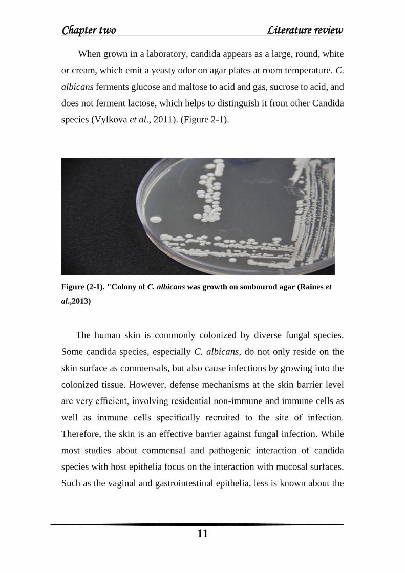
When grown in a laboratory, candida appears as a large, round, white
or cream, which emit a yeasty odor on agar plates at room temperature. C.
albicans ferments glucose and maltose to acid and gas, sucrose to acid, and
does not ferment lactose, which helps to distinguish it from other Candida
species (Vylkova et al., 2011). (Figure 2-1).
Figure (2-1). "Colony of C. albicans was growth on soubourod agar (Raines et
al.,2013)
The human skin is commonly colonized by diverse fungal species.
Some candida species, especially C. albicans, do not only reside on the
skin surface as commensals, but also cause infections by growing into the
colonized tissue. However, defense mechanisms at the skin barrier level
are very efficient, involving residential non-immune and immune cells as
well as immune cells specifically recruited to the site of infection.
Therefore, the skin is an effective barrier against fungal infection. While
most studies about commensal and pathogenic interaction of candida
species with host epithelia focus on the interaction with mucosal surfaces.
Such as the vaginal and gastrointestinal epithelia, less is known about the

Chapter two Literature review
12
mechanisms underlying Candida interaction with the skin. (Kashem &
Kaplan.,2016and Kühbacher et al., 2017).
2.6. Candidiasis
Candidiasis was one of the fungal infections caused by a type of
yeasts that belong to the genus Candida. (Fenn et al.,1999;
Noble&Johnson.,2007; Brown et al.,2012). candidiasis is also technically
known as candidosis, moniliasis, and oidiomycosis. Candidiasis symptoms
vary depending on the area of the body that is infected (Marttila et
al.,2013). Candidiasis encompasses infections that range from superficial,
such as oral thrush and vaginitis, to systemic and potentially life-
threatening diseases. Candida infections of the latter category are also
referred to as candidemia and are usually confined to severely
immunocompromised persons, such as cancer, transplant, and AIDS
patients, as well as nontrauma emergency surgery patients
(Noble&Johnson.,2007; Marttila et al.,2013).
2.7. Classification of Candidiasis infection
Candidiasis may be divided into the following types: (Marttila et
al.,2013)
2.7.1. Superficial Infection
2.7.1.1. Oral Candidiasis
The mucous membranes lining the mouth are infected by oral
candidiasis also known as oral thrush (Melo et al.,2004). The mouth is the
main pathway through which most pathogenic bacteria enter Therefore, the
study of oral health contributes to the reduction of multiple bacterial
infections within the body (Todar.,2002). Of the most resulting infections

Chapter two Literature review
13
common of candida albicans yeast, they appear as a simple tumor or
ulceration in any part of the oral cavity and then develop into white spots
that may combine to form a membrane containing large numbers of Pseudo
hyphae and playstyle spores as well as endothelial cells (Noltte.,1982).
This type of injury appears in newborns mainly because they acquire yeast
when passing through the mother-birth canal (Emmons et al.,1977; Kwon-
Chung and Bennett.,1992), as the bacteria passes from the mother to the
child through saliva directly. During kissing, talking or sneezing on the
face of the child or using contaminated substances such as spoons and
toothbrushes (Steven.,1996). The infection in adults is chronic and
characterized by the formation of the membrane thicker with the presence
of heartburn and dehydration in the affected part and more frequent in
people with pulmonary tuberculosis and leukemia or HIV infection (AIDS)
(Meurman et al.,2007; Mahdi.,2015). (Figure 2-2).
Figure (2-2) Oral candidiasis [thrush] (Coleman et al.,2010)

Chapter two Literature review
14
2.7.1.2. Cutaneous candidiasis
The infection in adults is chronic and characterized by the fact
that the membrane, which is formed thicker with the presence of heartburn
and dryness in the affected part and the incidence of infection in people
suffering from tuberculosis, Acute leukemia or HIV / AIDS. The infection
occurs in the outer part of the body (skin, hair, and nails) by the Candida
albicans because of its possession of the two types of enzymes are
proteinase and keratinize. The injury includes an area of Infra-axillaries,
Infra-mummeries and Inter-triginous, where these places are characterized
by warmth and humidity (Figure 2-3). The Included ratio infection of
Cutaneous Candidiasis was 85% in Iraq (Minster of health). The infection
begins in the form of vesicles, and then turns into specific spots that are
dark red and wet accompanied by itching and movement and tend the outer
layer to be scaly and fast division. Also, may be accompanied by a
secondary infection with bacteria Staphylococcus aureus (Notle.,1982;
Negi et al.,1984).
The infection of the nail known as chronic Paronychia which occurs as
a result of the infection of candidiasis of the subcutaneous plate causing
inflammation of the pterygium with redness and painful swelling and the
collection of inflammatory secretions. In case of people who use water for
a long time and people with diabetes (Vazquez and Soble.,1995;
Hay.,2018), infected nails and become a brown color.

Chapter two Literature review
15
Figure)2-3( Cutaneous Candidasis
2.7.1.3. Vaginal candidiasis
C. albicans yeast is a natural flora in vaginal secretions in the case of
a balance between the bacteria and this yeast, but when there is an
imbalance, this yeast grows and becomes more increasing and this
indicates the occurrence infected with this yeast (Murray et al.,2000).
Vaginal candidiasis is a common fungal infection that occurs under
predisposing conditions such as, diabetes, antibiotic therapy, and
contraceptive. This disease is a common disorder among women. (Watson
and Calabretto,2007). It is caused by the overgrowth of Candida species in
the vagina and is characterized by vaginal secretion white or yellow thick
textures accompanied by heartburn at the site of injury, the appearance of
secretions varies from one case to another depending on the severity of the
infection (Fidel,2004; Sobel, 2007; (Achkar and Fries, 2010 and Abood.,
2014).

Chapter two Literature review
16
2.7.2 Systemic Infections
This infection included different system such as: -
1- Urinary tract candidiasis
2- Respiratory tract candidiasis
3- Alimentary tract infection
4- Candida of central nervous system
5- Candidal meningitis
6- Endocarditis
7- Disseminated candidiasis [e.g. "hepatosplenic candidiasis", which
sometimes follows neutropenia; or "candidemia", a form of
septicemia] (Vylkova et al.,2011 and Antinori et al.,2016).
2.8. Pathogenicity and virulence of C. albicans
Pathogenicity is a qualitative characteristic of the micro-organism by
which it can induce disease (pathogen) or not (non-pathogen). Virulence is
a quantitative trait that indicates the amount of damage induced by the
pathogen on the host. The most virulent pathogen causes the most serious
damage (disease) to the host. Both pathogenicity and virulence are
controlled by pathogenicity and virulence genes. The dimorphism, germ
tube formation and hemolysis are indicators of pathogenicity of C.
albicans. C. albicans possess a group of virulence factors that include
surface molecules aids in adhesion to cell surface of the host, acidic
protease enzymes and ability to transform to the filamentous form inside
the body. The later factor enables the fungus to escape from the
bloodstream and penetration of epithelial tissues then the growth in the
internal tissues. It aids also in the resistance of phagocytes where it ruptures
the cell and continue growing (Hidalgo and Vazquez,2005; Shirtliff et
al.,2009).

Chapter two Literature review
17
The ability of C. albicans to switch morphology between yeast and
hyphal form is crucial to its ability to adhere surfaces and colonize tissue
(Saville et al.,2003). The ability of the pathogenic strains to transform from
white cells to opaque cells is associated with changes in the size, shape,
adhesion, hyphae formation, sensitivity to drugs and neutrophils and
pathogenicity. The white cells are more virulent in systemic infections
while the opaque is more successful in skin infections. (Williams, et
al.,2000; Munro et al.,2005; Forche et al.,2008 and Hassan et al.,2014).
2.9. Immune responses to Candida spp.
The emergence of novel pathogenic fungi and the lack of fungal
vaccines have focused on an acute interest in illustrating immune defense
mechanisms against fungi. And these mechanisms of immune defense
occur by two pathway innate and adaptive immune responses (Devine and
Marsh, 2009; LeibundGut-Landmann et al., 2012).
Innate immunity was the first line of unspecific host defense against
pathogens. First step for the immune responses interacts with epithelial
cells that play a critical role in protecting the body against invasion, the C.
albicans can recognized by many receptors including Toll-like receptors
(TLRs), C-type lectin receptors (CLRs), mannose receptor (MR) that can
induce pro-inflammatory cytokine, chemokine (Kiyoura and Tamai, 2015).
Beyond the critical role of phagocytosis in host defense, phagocytosis was
one of the first processes of innate immunity, carried out by macrophages,
neutrophils, and dendritic cells to engulf C. albicans (Underhill, 2005;
Wellington et al., 2009). The C. albicans cell wall components (mannan,
glucan and chitin) considered as immunostimulatory for activate
phagocytosis (Kiyoura and Tamai, 2015). Mannan, which known as the
serotypes of distinguish Candida spp. located on the outer surface of the
C. albicans cell wall. The structure of C. albicans mannan involves in its

Chapter two Literature review
18
recognition through macrophages (McKenzie et al., 2010). Heat-killed C.
albicans raised level of surface expression to 1, 3-glucan, that commonly
available within mannan in the pathogen cell wall (Jouault et al., 2006;
Wellington et al., 2009). Phagocytosis of glucans by macrophages can
improve killing pathogen through production of reactive oxygen species
(ROS), which considered as crucial chemical reactive to get rid of
pathogen, while a live C. albicans inhibition ROS production in process of
phagocytes (Wellington et al., 2009; Kankkunen et al., 2010).
Subsequently, producing many antimicrobial peptides that secreted and
synthesized by different cells involving epithelial cells, neutrophils and
immune cells such as LL-37, β Defensin and Histatins. LL-37 was
chemotactic produce by neutrophils and monocytes. Which have
advantage in suppress C. albicans adhesion on plastic surfaces
(LópezGarcía et al., 2005; Tsai et al., 2011;). The β Defensin family,
produced by epithelial cells have antimicrobial activity against gram
negative and gram positive bacteria, enveloped viruses and fungi in vitro.
Histatins were proteins that secreted by human parotid and submandibular,
sublingual saliva in humans, that have fungicidal activity against C.
albicans and also may have role in causing small membrane defects
(Kiyoura and Tamai, 2015).
Adaptive immune cells, such as T cells and B cells, express genetically
rearranged surface receptors that are exquisitely specific for a particular
antigen. Innate and adaptive immune responses are inextricably
intertwined, and a successful adaptive immune response is a coordinated
effort requiring activation of tissue-resident cells, antigen-presenting cells,
and antigen-specific T and B cells. recovery of tissue homeostasis involves
a balance of TH1- and TH17-type responses and Treg cells. For example,
activation of CLRs in vivo leads to the development of TH1- and TH17-

Chapter two Literature review
19
type CD4+ T cells. The TH1 response provides protective immunity
against fungi through enhancing the functions of phagocytic cells through
the production of IFN-γ and promotion of B-cell production of opsonizing
antifungal antibodies. TH17 responses are also critical because they
activate tissue cells, such as epithelial cells and fibroblasts, resulting in the
production of chemokines that recruit phagocytes to the site of the immune
response, through the production of (IL-17 family of cytokines particularly
IL-17A and, IL-22) (Verma et al.,2015; Jolink et al.,2017 and Bartemes &
Kita.,2018).
Th1 and Th17 cells are the principal T helper subsets that contribute
to protective immunity to several pathogenic fungi. (Kagami et al,2010;
Saijo et al,2010). Apart from conventional Th1/Th2 responses, Th17 cells
have recently been described as an important Th cell subtype conferring
protection against extracellular bacterial and fungal infections (Curtis and
Way, 2009). IL-17A, the major cytokine secreted by Th17 cells, possesses
multiple proinflammatory functions, such as recruiting neutrophils (Brown
,2011), activating neutrophil/macrophage phagocytosis activity, and
inducing b-defensin release (Kao et al,2008). Therefore, IL-17 is regarded
as an important component in host defense against C. albicans infection.
(Acosta-Rodriguez et al,2007; Tomalka et al,2011).
For a long time, it was assumed that cell-mediated immunity (CMI)
was remarkable, but humoral immunity had no or little role in immunity.
Whatever, it was admitted now that CMI importance mechanism in
defence, but that specific kinds of antibody response were protective.
Usually, Th 1-type CMI wanted for riddance of a fungal infection, while
Th2 immunity commonly leads to susceptibility to infection (Blanco and
Garcia, 2008).

Chapter two Literature review
20
2.10. Methods Laboratory diagnosis of C. albicans
2.10.1. Direct examination
Direct microscopic examination is a rapid method for diagnosis
of candidiasis. It requires less expertise (Deorukhkar & Saini.,2014). The
swab (sample) with KOH (10%) solution is examined and fixed on the
slide. The solution analyzes the epithelial cells accompanying the sample,
leaving the yeast cells, allowing to see the false filaments and the oval
shape of the yeast. For Candida, this method is commonly used in
laboratories for its speed of giving the result, but is inaccurate (Kerawala
and Newlands.,2010).
2.10.2 Culture
The sample is taken from the affected area by candidiasis. And the
planning survey is carried out on a container planting plate on the
appropriate medium for its growth (Sabroad Dextrose Acar, Blood acar
medium, Candida Chrome acar medium). And incubated in the incubator
at a temperature of 37°C for 48 hours. And through the color, shape and
growth method of the colonies can identify the cause of the injury zone.
This method is less useful than direct examination using a microscope
because it takes a long time and is very sensitive. The sample can be
contaminated making it useless in diagnosis (Purkait., 2011).
2.10.3 Germ tube test
In diagnostic mycology the basic work up for yeast identification
starts with a germ tube test. Germ tube formation was first reported by
Reynolds and Braude and hence the germ tube test is also known as
Reynolds-Braude Phenomenon (Deorukhkar et al., 2012).

Chapter two Literature review
21
This is a rapid method for identifying C. albicans and C. dubliniensis
by its ability to produce short, slender, tube like structures which is called
the germ tubes when it is incubated in serum at 37°C.
Distinguish between species belonging to the genus Candida, If C.
albicans isolates produce the germ tube when incubated with the human
serum at a temperature of 37°C for three hours and is important as a
diagnostic character to distinguish it from other types of Candida (Milne.,
1996), as Campbell et al.,1998 mentioned that C. dubliniensis and has the
ability to produce the germ tube.
In this test the observer must be able to differentiate between the germ
tube and the pseudohyphae. The elongated daughter cells from the mother
cell without constriction at their origin are referred to as germ tubes
whereas constriction at the origin of the mother cells is called the
pseudohyphae (Kim et al., 2002). A criterion for germ tube positivity is
observation of minimum five germ tube in the entire wet mount
preparation. Negative results are confirmed by examining at least 10 high
power fields for the presence of germ tubes (Deorukhkar et al., 2012 c).
(Figure 2-4).
Figure (2-4). Germ tube formation of C. albicans (Bedini et al.,2006)

Chapter two Literature review
22
2.10.4. Analytical profile index (API) Yeast Identification System
The API Candida system consists of a single-use disposable plastic
strip with 10 wells to perform 12 colorimetric biochemical tests: five sugar
assimilation tests (for glucose, galactose, sucrose, trehalose, and raffinose)
and seven enzymatic tests (for b-maltosidase, a-amylase, b-xylosidase, b-
glucuronidase, urea hydrolysis, N-acetylb-glucosaminidase, and b-
galactosidase). Inoculation of the wells was performed by adding a yeast
suspension to the dehydrated substrates. The results were read after
incubation for 18 to 24 hat 35°C.A four-digi tnumerical profile was
generated for each isolate depending upon the reactions it produced.
Identifications were made by referring to the list of numerical profiles and
a computer program provided by the manufacturer. (Colin et al.,1999;
Hazen and Howell., 2003).
2.10.6. Vitek Yeast identification system [ specific biochemical
reaction]
2.10.5. Polymerase chain reaction (PCR)
One of the methods used for accurate diagnosis is a DNA-based
method known as Polymerase Chain Reaction (PCR) (White et al., 2006).
They are widely used if an expansion by a small fraction of DNA that is
estimated by micrograms and by decoupling the DNA bar and then re-link
with new tapes and this process is done by controlling the temperature
(Mothershed and Whitney.,2006) and it passes by three stages:
1- Denaturation .
2- Annealing.
3- Elongation.
Chapter two Literature review
23
The basis of PCR work is to rely on a thermal cycle that uses
thermodynamics from DNA reactions by rotating the heating and cooling
of PCR samples after the temperature is determined (Sharkey et al.,1994).
This technique was depended in the clinical diagnosis of Candida, in
addition to other pathogenic fungi(Kato.,2001). And it is one of the most
effective techniques in the diagnosis of yeast and mold, which is better than
the traditional methods as well as accompanying many of the obstacles in
the diagnosis of isolates within the same type and in addition to that, it
takes a long time (weeks or more) for the emergence of the result ( Thomas
., 2003) in addition to the similarity of the behavior of yeast in many of the
parietal characteristics, and on the agricultural circles and the difficulty of
distinguishing them even using the differential chromar Acar (Mancini and
Ossi ., 2005 and Al-khafaji.,2017).
2.11. Diagnosis of disseminated candidiasis
As compared to superficial and mucocutaneous candidiasis, the clinical
presentations of Disseminated Candidiasis (DC) are non-specific and
complicated. Therefore, the rapid and precise laboratory diagnosis of DC
is crucial, not only for species identification, but also for timely institution
of appropriate antifungal treatment (Ellepola and Morrison, 2005; Pappas
et al., 2009). The laboratory diagnosis of DC involves the use of
immunodiagnostic and other non-cultural methods in addition to culture.
The varied nature of clinical forms the specimen collection difficultin DC.
In most cases, invasive procedures are necessary for the specimen
collection (Segal & Elad., 2005 and Mikko et al., 2014).

Chapter three MATERIALS AND METHODS

Chapter three Materials and methods
24
3. Materials and Methods
3.1. Subjects
The case study included 100 samples of the patient
(immunocompromised women with average age (55-57) year and babies
with average age (1-18 month), patients infected with various (fungi,
bacteria) infection, in addition to 20 samples for an apparently healthy
individual (a control). Patients were attended to AL- Dowaly Private
Hospital in Baghdad during the period January 2019 to June 2019. All
infections were diagnosed by consultant medical staff at the hospital by
using several tests including (germ tube, culture, Vitek and API20C).
Cases enrolled in this investigation were as follow:
Thirty - Samples from patients with C. albicans infection (skin scales).
Fifteen -Samples from patients with Aspergillus spp. infection (skin
scales).
Fifteen -Samples from patients with Tricophactone (infection (skin
scales).
{Skin scales were isolated from fingers (intertriginous)}
Ten -Samples from patients with C. albicans infection (skin swabs).
Ten -Samples from patient's infected with Staphylococcus aureus (skin
swabs).
Ten -Samples from babies (1-18) month infected with C. albicans
(mouth swabs).
Ten -Samples from patients with C. albicans infection (vagina swabs).
The Samples were examined using the new technique and compared
with the routine methods of diagnosis.

Chapter three Materials and methods
25
3.2. Equipments
Instruments and equipment used in this study are listed in the tables
below:
Table (3-1) general Equipments utilized in this study.
Country Company Equipment
China Surgiphath Cover slip
U.S.A Whatman Filter paper
China Surgipath Forceps
China Surgipath Glass microscope slide
China boiBact Gas burner
China Surgipath Loop
China Bioche-max Micro- pipette
China Surgipath Petri dishes
China Scalped blade
China Swab culture
USA corporation Test tube
China Medeco 1ml and,200ml ,1ml Tips-Pipette
UK Cardinal Health Wooden applicator stick
Table (3-2) Materials utilized in this study
Country Company Materials
USA Baxter Normal saline
UK Biomax Potassium hydroxide solution
UK Biomax Sabouraud agar
Germany Bio-Rad SDS (sodium dodecyl sulfate)

Chapter three Materials and methods
26
Table (3-3) devices which used in the study
Country Company Devises
Korea Fine Tech Hood Lab
Germany MEMERT Incubator
Italy Optika Microscope
Turkey Beko Refrigerator
Taiwan Major Ccience UV-Light System
Table (3-4) Kit and material
Country Company Kit and Material
China Elabscience Antibody Specific
C. albicain
California LibreTexts UV-Stain

Chapter three Materials and methods
27
Protocol
100 Samples(infection) / 20 Samples (control)
Type of Samples: Skin scales, Skin swabs, Mouth and vaginal
swabs.
Samples were collected from AL- Dowaly Private Hospital in
Baghdad during the period January 2019 to May 2019.
Diagnosis of samples by
consultant medical staff
using routine methods
Diagnosis of samples by a
new technique
Germ tube
Culture
Vitek
API20C
Kit for specific antibodies
for C. albicans+ UV-stain
The samples were
diagnosed by UV –light
(harmless)

Chapter three Materials and methods
28
3.3. Methods
3.3.1. Laboratory diagnosis of C. albicans
3.3.1.1. Culture:
Sabouraud's dextrose agar (65 g) was suspended in 1L distilled
water. And well until a uniform suspension was obtained. After heated with
frequent agitation and boiled then sterilized at 121°C for 15 min, then after
48, and 72 hours of incubation.
3.3.1.2. Germ tube test
Principle assay:
The Germ Tube Test is a screening procedure used to differentiate
C. albicans from other yeast.
Procedure assay:
1. 0.5 ml [11 drops] of sheep serum was add in a 10 x 75 mm tube.
2. A light suspension of the suspect yeast colonies was made by touching
1-2 large colonies or 3-4 smaller colonies with a sterile wooden applicator
stick and then the sheep serum was inoculated with the applicator stick.
Note: Too large inoculum will inhibit germ tube formation.
3. The tube was incubated for 2-3 hours in a 35 - 37°C incubator.
Warning: Do not over-incubate the tube. Candida tropicalis may produce
pseudo-germ tubes after 3 hours of incubation.
4. A drop of the suspension was placed on a slide using a Pasteur pipette
and coverslip.

Chapter three Materials and methods
29
5. The wet mount was examined microscopically for production of germ
tubes (long tube-like projections extending out from the yeast cells).
(Mahdi.,2015).
3.3.1.3. Vitek yeast identification system (automatic system).
3.3.1.4. API 20C Yeast identification system {Done according to
company biomerieux}
Procedure assay:
1- The basal medium was melted in the ampoules by placing them in
an autoclave for 2 minutes or in a boiling water
2- The ampoules were placed in a water bath at 48 to 50 C, and were
allowed to cool.
3- An incubation tray was preped. A squeeze bottle was used to
dispense 20 ml of water into the tray, and then the strip was placed into
the incubation tray.
4- The ampoules according were opened to the manufacturer's
instructions, and The molten medium were inoculated with an applicator
stick that has touched one or two colonies (>2 mm diameter), adjusted to
a density just below 1+ on a Wickerham card.
5- The strip was inoculated (20 cupules; approximately 0.2 ml each) by
using a Pateur pipette and following the manufacturer's directions, and
then the lid was placed on the tray.
6- The trays were incubated at 28 to 30 C for 72 hours. record the results
were read and recorded after 48 hours (David.,2010).

Chapter three Materials and methods
30
3.3.2. Laboratory diagnosis of the other samples such as
(Aspergillus spp , Tricophacton and Staphylococcus aureus bacteria) was
diagnosed by consultant medical staff at the Dowaly Private Hospital in
Baghdad ,and using the methods for special diagnosing these samples such
as (Culture ,and other methods).
3.4. Preparation new kit to diagnose Ab-C. albicans
3.4.1. Procedure
I) Preparation of stain (Manufactured stain)
1. 200ml of distilled water added to 100ml from concentrated liquid
stain (100%).
2. The mixture has been shaken for one min.
3. Figure (3-1) shows the composition of the UV-stain. Tables (3-5),
(3-6) showing basic information and chemical properties of stain (López-
García.,2007).
Figure (3-1) Isobornyl acrylate structure

Chapter three Materials and methods
31
II) Preparation of conjugated reagent
1. 100ml of stain solution added to 50 ml from (Antibody-C.
albicans).
Note: The concentration of the antibody is (10%).
2. Shake the mixture for one min.
3. The mixture incubated 37°C for (24 hours) was the optimization
time to incubate the mixture and happened association between the stain
and antibody- C. albicans. Because (5,10,15,20) hours are not appropriate
for a link to occur (Mahdi.,2015).
4. Then kept at 4ₒC until to use.
III) preparation procedures to diagnosis the samples
1. Infected samples (skin scales, skin swab, mouth and vagina swabs)
were taken by a doctor.
2. Samples were placed on the slide and fixed by exposure to heat
(benzene burner).
3. Add 2 drops (0.02ml) from conjugated reagent then left it for (1
minute) was the best time to incubate the sample and give the result.
Because more than 1 minute gives false results due to the strength of the
dye concentration that makes all the components of the sample to be
pigmented.
4. Finally washed by Distilled water.
5. Then the samples are examined by UV-light (+ve result was
glowing, -ve result was non-glowing.

Chapter three Materials and methods
32
Table (3-5) Isobornyl acrylate basic information
Product Name: Isobornyl acrylate
Chemical Abstract
Service (CAS) 5888-33-5
Molecular formula C13H20O2
Molecular weight 208.3
EINECS: 227-561-6
Hazard Moderately toxic by ingestion. Low toxicity by ski
n contact and mild eye irritant.
Table (3-6) Isobornyl acrylate chemical propertie
Melting Point <-35°C
Boiling Point 119-121 °C15 mm Hg(lit.)
Density 0.986 g/mL at 25 °C(lit.)
Refractive Index n20/D 1.476(lit.)
Fahrenheit Point 207 °F
Water Solubility Difficult to mix in water.
Sensitive Light Sensitive
CAS DataBase Reference 5888-33-5(CAS DataBase Reference)

Chapter three Materials and methods
33
3.5. Diagnosis of C. albicans infection by New technique
Experiments were performed on the samples to check the sensitivity
and specificity of the new technique
1- The first experiment was carried out on infected skin scales from
women with C. albicans and non-infected skin scales (Control). These
samples have been treated in mentioned steps previously in the procedure.
2- The second experiment was conducted on infected skin scales from
women with C. albicans and Aspergillus spp. These samples have been
treated in mentioned steps previously in the procedure.
3- The third experiment was conducted on infected skin scales from
women with C. albicans and Tricophacton spp. These samples have been
treated in mentioned steps previously in the procedure.
4- The fourth experiment was carried out on infected skin-swabs with C.
albicans and bacteria Staphlococcus auries (swabs) from women. These
samples have been treated in mentioned steps previously in the procedure.
5- The fifth experiment was carried out on the infected mouth-swabs with
C. albicans from babies. These samples have been treated in Mentioned
steps previously in the procedure.
6-The sixth experiment was carried out on the infected vagina-swabs with
C. albicans. These samples have been treated in mentioned steps
previously in the procedure.

Chapter four RESULT

Chapter four Results
34
4.1. Isolation and diagnosis of C. albicans
Table (4-1) showed the comparison among some types of tests used to
diagnose C. albicans and other fungi, bacterial infections. It have been
shown that the Vitek test was used to diagnose (+ve 60) samples of C.
albicans (skin scales, skin swabs, mouth and vaginal swabs) versus zero
isolations for a non C. albicans infection, while API20C test was used to
diagnose (+ve 60) samples of C. albicans versus (+ve 1) isolations for a
non C. albicans infection ,as well as to culture test ( diagnosed (+ve 58)
isolations of C. albicans versus (+ve 3) isolations for a non C. albicans
infection) and Germ tube test ( diagnosed (+ve 59) isolations of C. albicans
versus (+ve 2) isolations for a non C. albicans infection). As for the new
technique which diagnose (60)+ve isolate of C. albicans versus to zero for
a non- C. albicans.

Chapter four Results
35
Table (4-1) diagnosis C. albicans and other (fungi, bacteria) Infection
by routine methods and new technique
Types of
samples( fungi,
bacteria)
No.
samples
New
technique
Routine techniques
C. albicans
infection
60
+ve 60
Culture Germ
Tube
Test
Vitek API20C
+ve 58 +ve 59 +ve 60 +ve 60
-ve 0 -ve 2 -ve 1 -ve 0 -ve 0
Other (Fungi &
Bacteria) infection
40
-ve 40
-ve 37
-ve 38
-ve 40
-ve 39
+ve 0
+ve 3
+ve 2
+ve 0
+ve 1
(+ve) Number of samples infected with C. albicans.
(-ve) Number of non - infected samples with C. albicans.

Chapter four Results
36
4.2. Sensitivity and specificity for diagnosis of C. albicans
Results in table (4-2) showed (True Positive, False Negative, True
Negative and False Postive) of samples per all tests to diagnosis of C.
albicans.
(TP) The number of samples infected with C. albicans and have a
positive result.
(FN) The number of samples non-infected by C. albicans and have
a negative result.
(TN) The number of samples non-infected by C. albicans and have
a negative result (other fungi and bacteria).
(FP) The number of samples non- infected with C. albicans and
have a positive result.
bleTa (4-2)The reliable of the test by using sensitivity and specificity
Tests for Diagnostic of C.
albicans
(TP) (FN) (TN) (FP)
Colonial morpholy (Culture) 60 2 40 3
Germ tube test 60 1 40 2
Vitek 60 0 40 0
API20C 60 0 40 1
New technique 60 0 40 0
Table (4-3) and Figure (4-1) showed the comparing between some types
of tests used in C. albicans diagnosis. It was shown that the Vitek test was
more sensitivity (100%) and specificity (100%) for diagnosis of C.
albicans than other methods (API20C sensitivity (100%) and specificity

Chapter four Results
37
(97%), Colonial morphology ''culture'' sensitivity (96%) and specificity
(93%) and germ tube test sensitivity (98%) and specificity (95%).
The results of New Technique assay for C. albicans was similar to the
results of Vitek test in sensitivity and specificity (100%), but it was
different regarding all these tests due to its dependent on antibodies
(against C. albicans), in preparation the specialized kit to diagnose of C.
albicans Consists of (Antibody conjugated with UV-Stain) ready for
interaction with C. albicans antigens. The sensitivity and specificity of the
new technique was recognized by the two laws:
- Sensitivity= TP/(TP+FN)
- Specificity= TN/(TN+FP)
Table (4-3) sensitivity and specificity for diagnosis of C. albicans by
Routine methods and new technique.
Tests for Diagnostic of
C. albicans
Sensitivity
TP/(TP+FN)
Specificity
TN/(TN+FP)
Colonial Morpholy (Culture) 96% 93%
Germ Tube Test 98% 95%
Vitek 100% 100%
API20C 100% 97%
New Technique 100% 100%

Chapter four Results
38
Figure (4-1) sensitivity (%) and specificity (%)for diagnosis of C.
albicans by routine methods and new technique
96
98
100 100 100
93
95
100
97
100
COLONIAL MORPHOLOGY
(CULTURE)
GERM TUBE TEST VITEK API20C NEW TECHNIQUE
Sensitivity and Specificity
Sensitivity Specificity

Chapter four Results
39
4.3. Experiments results in which a new technique was used
to diagnose samples
1- The first experiment results
As shown in figure (4-2), the result was (+ve) for a glowing sample
of skin scale infected with C. albicans as compared with the (-ve) result for
a non-glowing sample (non-infected) of skin scale (as control/negative).
Figure (4-2) shows the difference between sample of skin scale
infected with C. albicans and a non-infected sample of skin scales (as
control/negative) under UV. Light.

Chapter four Results
40
2- The second experiment results
As shown in figure (4-3), the result was (+ve) for a glowing sample
of skin scale infected with C. albicans as compared with the (-ve) result for
a non-glowing sample of skin scale infected with Aspergillus spp.
Figure (4-3) shows the difference between sample of skin scale infected
with C. albicans and sample of skin scale infected with Aspergillus spp.
under UV. Light.
Chapter four Results
41
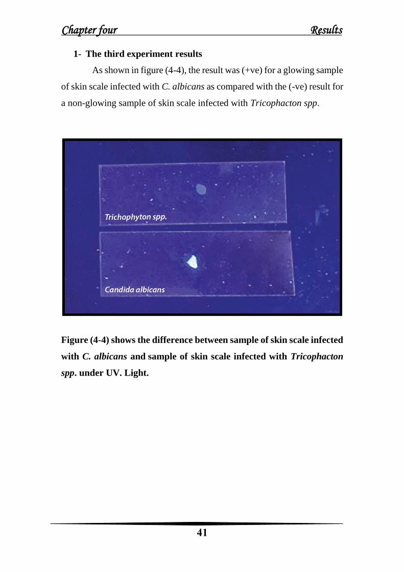
1- The third experiment results
As shown in figure (4-4), the result was (+ve) for a glowing sample
of skin scale infected with C. albicans as compared with the (-ve) result for
a non-glowing sample of skin scale infected with Tricophacton spp.
Figure (4-4) shows the difference between sample of skin scale infected
with C. albicans and sample of skin scale infected with Tricophacton
spp. under UV. Light.

Chapter four Results
42
4- The Fourth experiment results
As shown in figure (4-5), the result was (+ve) for a glowing sample of
skin swab infected with C. albicans as compared with the (-ve) result for a
non-glowing sample of skin swab infected with Staphylococcus aureus.
Figure (4-5) shows the difference between sample of skin swab infected
with C. albicans and sample of skin swab infected with Staphylococcus
aureus under UV. Light.
Chapter four Results
43
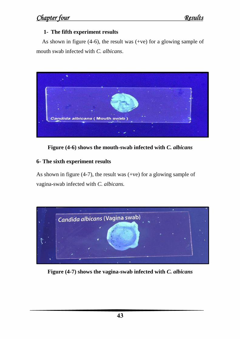
1- The fifth experiment results
As shown in figure (4-6), the result was (+ve) for a glowing sample of
mouth swab infected with C. albicans.
Figure (4-6) shows the mouth-swab infected with C. albicans
6- The sixth experiment results
As shown in figure (4-7), the result was (+ve) for a glowing sample of
vagina-swab infected with C. albicans.
Figure (4-7) shows the vagina-swab infected with C. albicans

Chapter five DISCUSSION
Chapter five Discussion
44
1.5 Discussion
The case study included 100 samples of the patient
(immunocompromised women with average age (55-57) year and babies
with average age (1-18 month), patients infected with various (fungi,
bacteria) infection, in addition to 20 samples for an apparently healthy
individual (a control). Patients were attended to AL- Dowaly Private
Hospital in Baghdad during the period January 2019 to June 2019. All
infections were diagnosed by consultant medical staff at the hospital by
using several tests including (germ tube, culture, Vitek and API20C).
Results of new technique was as follow: in figure (4-2) the result was
(+ve) for a glowing sample of skin scale infected with C. albicans as
compared with the (-ve) result for a non-glowing sample (non-infected) of
skin scale (as control). As shown figures (4-3)and (4-4) the result was
(+ve) for a glowing sample of skin scale infected with C. albicans as
compared with the (-ve) result for a non-glowing samples of skin scales
infected with Aspergillus spp and Tricophactone spp., while shown in
figures (4-5),(4-6)and (4-7) the result was (+ve) for a glowing sample of
skin swab and (mouth, vagina) swabs infected with C. albicans as
compared with the (-ve) result for a non-glowing sample of skin swab
infected with Staphylococcus aureus.
The comparison between the new technique with the current routine
tests was performed to demonstrate the feature of the new technique, and
the comparison are as follow:
Table (4-3) figure (4-1) shown that the new technique is more
sensitivity and specificity (100%) Compared to culture Which was
sensitive to (96%) and specialized (93%), because of the culture method

Chapter five Discussion
45
characterizes by several mistakes, this method is very sensitive, the sample
can be contaminated making it useless in diagnosis. It also can identify the
cause of the injury zone through phenotypic diagnosis (color, shape and
growth method of the colonies in agar medium). Candida spp. colonies
appear on medium within 24 to 72 hours. Some species may require more
than 3 days to appear on culture medium, these results agree with studies
(Segal and Elad.,2005 and Purkait., 2011), while the new technique does
not take a long time it need one minute to get a result and there is no
contamination.
Table (4-3) figure (4-1) shown that the new technique is more sensitive
and specialized (100%) compared with the germ tube test which has a
sensitivity (98%) and specificity (95%). This is a rapid method for
identifying C. albicans and C. dubliniensis by its ability to produce short,
slender, tube like structures called germ tubes when it is incubated in serum
at 37°C for 2 hours. Due to the time required to prepare human serum and
safety problems concerned with its use, many clinical microbiological
laboratories have started using non-human serum media for testing germ
tube production. These include egg white, saliva, tissue culture medium,
sheep serum, and various media. It's need to be accurate in the time and
temperature for example, incubating period for more than 3 hours may
produce pseudo-germ tubes. The observer must be able to differentiate
between the germ tube and the pseudohyphae, any observer must be
experienced in diagnosis. These results agree with studies (Milne., 1996;
Kim et al., 2002 and Deorukhkar et al., 2012), while the new technique
dissent need any factors of the above.

Chapter five Discussion
46
Table (4-3) figure (4-1) show that the new technique is more sensitive
and specificity (100%) compared with the API20C test to the sensitivity
(100%) and specificity (97%), due to the convergence of results ratios the
API20C test has less subjective errors in the interpretation of results, it is a
costly commercial system, they have several advantages like rapid
identification, require no or less supplemental tests. These results agree
with (Deorukhkar and Saini., 2014), while the new technique is low cost
commercially.
Table (4-3) figure (4-1) shows that the new technique of diagnosing
C. albicans is similar to the Vitek test with sensitivity and specificity
(100%) This is because the Vitek system is widely used for rapid
identification and susceptibility testing and sensitivity testing for many
microorganisms (e.g. Yeasts, bacteria, viruses, and parasites) these studies
agree with ( Mondelli et al., 2012), it's a fast and accurate diagnostic
technique in all routine laboratories. The study which perform by
(Ligozziet.,2002) that proved This technology has evolved so that it can
also, diagnose the specific antibodies for each pathogenic microorganism
by diagnostic card. These results agree with (Graf et al., 2000; David., 2010
and Deorukhkar &Saini,.2014; Q. Badr and Abaas., 2017). The new
technique is used to measure the antigens' presence of C. albicans and is
done by adding conjugated similar to the conjugated of ELIZA, but the
difference is the addition of a stain and its binding to the Fc-antibody part
instead of the enzyme, this association is caused by the hydrophobic effect
Because nonpolar molecules are clustered together, away from water
molecules, Large molecules can contain non-polar regions, they tend to be
near to each other and cause a change in the shape of the molecule so that
it becomes near to each other and away from water molecules (Jordan and

Chapter five Discussion
47
Gibb.,2015). This is why Strong physical bonds were formed between the
stain, which is a non-polar molecule with a hydrophobic surface with the
antibody security acid (hydrophobic). The immune-complex exposed to
UV-Light as a source. The positive sample glowed while the negative
sample doesn't glow. The present diagnostic kit is considering a good tool
for a diagnosis of C. albicans infection in all infected areas (skin, mouth,
and vagina).
Conclusions and recommendation
Conclusions Recommendation .
48
Conclusions
1- The new technique was more sensitivity (100%) and
specificity (100%)compared to the routine method.
2- The present diagnostic kit is considering a good tool to
diagnosis of C. albicans infection in all infected area (skin,
mouth and vagina).
3- It does not take a long time Where the duration of its work to
give the result to be one minute, and their cost is low.
4- Local Kit for diagnosis of C. albicans was successfully
prepared and produce in this study.
Conclusions Recommendation .
49
Recommendation
1- Development of the new technique to diagnose other samples,
not skin only, for example blood, sputum and other samples
infected with C. albicans.
2- Preparation of a kit to diagnose the rest of fungi and bacteria.
3- The new technique can be used by the physicians without
having to be sent to the laboratory.
4- To determine the precise accuracy, we recommend to apply
this diagnostic kit on infected animals by inoculation infection
in mouth and skin or blood then observe the animals to get
more safety and accuracy.

References

References .
50
References
• Abood, M.S. (2014). Immunological and molecular study of Candida
spp causing vulvovaginal candidiasis and the role of Lactic acid bacteria
as probiotic in vivo and in vitro. PhD thesis, collage of science for
woman, Baghdad University. IRAQ.
• Agha, M.A; Agha, S.A; Sharafat, S.; Zafar, M.N; Khanani, M.R. and
Mirza, M.A. (2011). Candida glabrata: an emerging threat for the
immunocompromised. Gomal J Med Sci; 9: 115-9.
• Ajah, H.A. (2016). In vitro and in vivo studies on the antifungal activity
of probiotics and Seaweed extract (Ascophyllum nodosum).
International Journal of Innovative Science, Engineering & Technology,
3 (4):306-312.
• Alan Hirzel.,2004. CEO General procedure and tips for direct ELISA,
Discover more at abcam journal technical Tel+965 2461 0480, Fax+965
2461 0488, e-mail [email protected]
• Al-khafaji, Z.K.A. (2017). Characterization of some virulence factors
and gene expression of Secreted Aspartyl proteinase in Candida albicans
isolated from oral candidiasis in cancer patients. Ph.D. Thesis. College
of Education, Al- Qadisiyah University: 104pp.
• Al-Lammi, T. J. (2009). Study the effect of Neem and the cell wall of
mannan of Candida albicans as immunomodulators on vaccination of
mice with Brucella Vaccine-Rev-1vaccine.Ph.D. Thesis, University of
Baghdad, Iraq.
• Aggarwal S, Gurney AL (Jan 2002). "IL-17: prototype member of an
emerging cytokine family". Journal of Leukocyte Biology. 71 (1): 1–8.
PMID 1178137.
References .
51
• Alpers, D. H., Kalloo, A. N., Kaplowitz, N., Owyang, C., & Powell, D.
W. (2011). Textbook of gastroenterology. John Wiley & Sons.
• Al-Quraishi, S.S.A. (2017). Immunological Study of the Fungus
Candida albicans Isolated from Clinical Infections and Role of some
Probiotic in Vivo and in Vitro. MSc. Thesis. College of Science,
University of Mustansiriya:1-142 pp
• Antinori, S.; Milazzo, L.; Sollima, S.; Galli, M. & Corbellino, M.
(2016). Candidemia and invasive candidiasis in adults: A narrative
review. European journal of internal medicine, 34, 21-28.
• Aslanzadeh, J. and Robert, G. (1991). Direct microscopic examination
of clinical specimens for the laboratory diagnosis of fungal infections.
Clin Microbiol Newsl. 13:185-192.
• Bartemes, K.R. and Kita, H., 2018. Innate and adaptive immune
responses to fungi in the airway. Journal of Allergy and Clinical
Immunology, 142(2), pp.353-363.
• Baylis, C. L. (2003). Immunological techniques:
immunochromotography, enzyme-linked immunofluorescent assays
and agglutination techniques. In Detecting Pathogens in Food (pp. 217-
240.
• Blanco,J. and Garcia, M. (2008). Immune response to fungal infections.
Veterinary Immunology and Immunopathology, 125(1-2): 47-70.
Acosta Rodriguez, EV . ; Zuniga, EI.; Montes, CL.; Merino, MC.;
Bermejo, DA.; Amezcua Vesely ,MC.; Motran, CC and Gruppi,
A.(2007). Trypanosoma cruzi infection beats the B-cell compartment
favouring parasite establishment: can we strike first. Scand J Immunol.
66(2-3):137-42.
• Brown, G.D.; Denning, D.W.; Levitz, S.M. and Netea, M.G., et al 2012.
Hidden killers: human fungal infections. Sci Transl Med 4: 165rv113.

References .
52
• Brown, GD. (2011).Innate antifungal immunity: the key role of
phagocytes. Annu Rev Immunol. 29:1–21.
• Calderone A, Clancy CJ, eds. (2012). Candida and Candidiasis (2nd
ed.). ASM
• Campbell, C.K. ;Holmes , A.D.; Davey ,K.G.; Szekely , A. and
Warnock, D.W. (1998) .Comparison of a new Chromogenic agar with
germ tube method for presumptive identification of Candida albicans .
Eur J clin microbial infec Dis ., 17:367-8.
• Carpenter, P. L. (1975). Immunology and serology (No. QR181. C37
1960.). Philadelphia: Saunders.
• Chamian, F. and Krueger, J.G. (2004). Psoriasis vulgaris: an interplay
of T lymphocytes, dendritic cells, and infl ammatory cytokines in
pathogenesis. Curr Opin Rheumatol. ,16:331–337.
• Calderone A, Clancy CJ, eds. (2012). Candida and Candidiasis (2nd
ed.). ASM Press. ISBN 978-1-55581-539-4.
• Charbonnet, Derrick & Norman Scott Evans, "United States Patent:
8735142 - Systems and methods for immunosorbent assays for single
and multiple analytes", issued May 27, 2014
• Charbonnet, Derrick, "United States Patent: 7767404 - Apparatus and
method for single-step immunosorbent assay for single and multiple
analytes", issued August 3, 2010.
• Cheng, C. M., Martinez, A. W., Gong, J., Mace, C. R., Phillips, S. T.,
Carrilho, E., ... & Whitesides, G. M. (2010). Paper‐based ELISA.
Angewandte Chemie, 122(28), 4881-488
• Coleman, D. C., M. G. Rinaldi, K. A. Haynes, J. H. Rex, R. C.
Summerbell, E. J. Anaissie, A. Li, and D. J Sullivan. (2010). Importance
of Candida species other than Candida albicans as opportunistic
pathogens. Med. Mycol. 42:213–210.

References .
53
• Colin, K. C.; Kate, G. D.; Ann, D. H.; Adrien, S.; and David, W. (1999).
Comparison of the API Candida System with the Auxacolor System for
Identification of Common Yeast Pathogens., 37(3): 821–823.
• Coronado-Castellote, L. and Jiménez-Soriano, Y., (2013). Clinical and
microbiological diagnosis of oral candidiasis. Journal of clinical and
experimental dentistry., 5(5), p.e279.
• Curtis, M. M., and Way, S.S. (2009). Interleukin-17 in host defence
against bacterial, mycobacterial and fungal pathogens. Immunology
,126: 177–185.
• Chiricozzi A, Guttman-Yassky E, Suárez-Fariñas M, Nograles KE,
Tian S, Cardinale I, Chimenti S, Krueger JG (Mar 2011). "Integrative
responses to IL-17 and TNF-α in human keratinocytes account for key
inflammatory pathogenic circuits in psoriasis". The Journal of
Investigative Dermatology. 131 (3): 677–87. doi:10.1038/jid.2010.340.
PMID 21085185.
• David H. Pincus (2010) MICROBIAL IDENTIFICATION USING
THE BIOMÉRIEUX VITEK® 2 SYSTEM, bioMérieux, Inc.
Hazelwood, MO, USA2010; 2: 59-64.
• Deorukhkar, S. C., & Saini, S. (2014). Laboratory approach for
diagnosis of candidiasis through ages. International Journal of Current
Microbiology and Applied Sciences., 3(1), 206-218.
• Deorukhkar, S., Saini, S., and Jadhav, P. 2012 c. Evaluation of different
media for germ tube production of Candida albicans and Candida
dubliniensis. International Journal of Biomedical and Advance
Research. 3:704-707.
• Devine, D.A. and Marsh, P.D. (2009). Prospects for the development of
probiotics and prebiotics for oral applications. Journal of oral
Microbiology, 1: 1-11.

References .
54
• Dolin, [edited by] Gerald L. Mandell, John E. Bennett, Raphael (2010).
Mandell, Douglas, and Bennett's principles and practice of infectious
diseases (7th ed.). Philadelphia, PA: Churchill Livingstone/Elsevier. pp.
Chapter 250. ISBN 978-0-443-06839-3.
• Donlan, R.M. (2001). "Biofilm formation: a clinically relevant
microbiological process". Clinical Infectious Diseases. 33 (8): 1387–92.
doi:10.1086/322972. PMID 11565080
• Dunbar, B. S. (2012). Two-dimensional electrophoresis and
immunological techniques. Springer Science & Business Media.
• Daiss, J. L. (1994). Antibodies as Thermolabile Switches: High
Temperature Triggering for the Polymerase Chain Reaction.
Bio/Technology. 12 (5): 506–509.
• Erdogan A, Rao SS (April 2015). "Small intestinal fungal overgrowth".
Curr Gastroenterol Rep. 17 (4): 16. doi:10.1007/s11894-015-0436.
PMID 25786900.
• Fenn, J. P., Segal, H., Barland, B., Denton, D., Whisenant, J., Chun, H.,
... & Carroll, K. (1994). Comparison of updated Vitek Yeast
Biochemical Card and API 20C yeast identification systems. Journal of
Clinical Microbiology, 32(5), 1184-1187.
• Forbes, BA, Sahm, DF, Weissfeld, AS. Bailey and Scott's Diagnostic
Microbiology, ed.12. Mosby Elsevier, Philadelphia,2007, pp. 542-627.
• Fenn, J.P.; Deaux, E.B.; Segal ,H.; Skodak-Jone, L.; Padilla, P.E. and
Bale ,M. et al. (1999) .Comparison of four methodologies for rapid and
cost-effective identification of Candida glabrata. J Clin Microbiol; 37:
3387-9.
• Ford, P. J. (2010). Immunological techniques: ELISA, flow cytometry,
and immunohistochemistry. In Oral Biology (pp. 327-343). Humana
Press, Totowa, NJ.
References .
55
• Goldberg, Richard J. (1952). "A Theory of Antibody—Antigen
Reactions. I. Theory for Reactions of Multivalent Antigen with Bivalent
and Univalent Antibody". Journal of the American Chemical Society. 74
(22): 5715–5725. doi:10.1021/ja01142a045.
• Gow, N.A.R. (2017). "Microbe Profile: Candida albicans: a shape-
changing, opportunistic pathogenic fungus of humans". Microbiology.
163 (8): 1145–1147. doi:10.1099/mic.0.000499. PMID 28809155.
• Graf, B., Adam, T., Zill, E., and Gobel, U. 2000. Evaluation of VITEK
2 system for rapid identification of yeasts and yeast-like organisms. J
Clin Microbiol. 38:1782-1785
• Hadley, S.; Lee, W.W.; Ruthazer; R. and Nastaway Jr SA. Candidemia
as a cause of septic shock and multiple organ failure in non-
immunocompromised patients. Crit Care Med 2002; 30:1808–14.
• HANNIG, C. & HANNIG, M. (2009). The oral cavity—a key system
to understand substratum-dependent bioadhesion on solid surfaces in
man. Clinical Oral Investigations, 13, 123-139.
• Hasan, F.; Xess, I.; Wang, X.; Jain, N. and Fries, B. C. (2009). Biofilm
formation in clinical Candida isolates and its association with virulence.
Microbes and Infection Journal, 11: 753–761.
• Hassan, H. F.; Hassain, N. S.; Aldhaher, Z.A.; Shaker, R.M.; Tawfeeq,
J.A., and Ibraheem, G. (2016). Study of fungal infection in Iraqi patients
suffering from chronic cough and antifungal sensitivity test in vitro for
the fungi isolated from these patients. World J Pharm Sci.,4(10): 13-
17.
• Hassan, N. N. (2014). Effect of some Microorganisms,Chemical
Compounds and Citrullus colocynthis Seed Extract on the Growth and
Composition of the Biofilm of (Candida albicans).MSc. Thesis. College
of Science, University of Mustansiriya:1-109 pp.
References .
56
• Hay, R. (2018). Therapy of skin, hair and nail fungal infections. Journal
of Fungi, 4(3), 99.
• Helbert, M. (2016). Immunology for medical students. Elsevier Health
Sciences.
• Henderson, S.O. and Magana, R.N. (2006). xCandidiasis. EMedicine .
http://www.emedicine.com/emerg/topic76.htm.
• Hibbett, D. S.; Binder , M.; Bischoff , J. F. and Blackwell, M. et
al.(2007). A higher-level phylogenetic classification of the Fungi.
Mycological Research. 111:509–547.
• Hohl, T.M.; Rivera, A. and Pamer, E. (2006). Immunity to fungi. Curr
Opin Immunol 18(4):465-472.
• Jarreau, P.C. (2010). Serological Response to Parasitic and Fungal
Infections.In:Steven,C.D.)3ed(.Clincal Immunology
Serology.)USA(:228-345PP.
• Jolink, H., de Boer, R., Hombrink, P., Jonkers, R.E., van Dissel, J.T.,
Falkenburg, J.H. et al. Pulmonary immune responses against Aspergillus
fumigatus are characterized by high frequencies of IL-17 producing T-
cells. J Infect. 2017; 74: 81–88.
• Jouault, T.; El Abed-El Behi, M.; Martinez-Esparza, M.; Breuilh, L.;
Trinel, P.; Chamaillard, M.; Trottein, F. and Poulain, D. (2006). Specific
Recognition of Candida albicans by Macrophages Requires Galectin-3
to Discriminate Saccharomyces cerevisiae and Needs Association with
TLR2 for Signaling. The Journal of Immunology, 177(7): 4679-4687.
• Jordan, J. H., & Gibb, B. C. (2015). Molecular containers assembled
through the hydrophobic effect. Chemical Society Reviews, 44(2), 547-
585.
• Kagami, S. ; Rizzo, HL. ; Kurtz, SE. ; Miller, LS and Blauvelt, A.
(2010). IL-23 and IL-17A, but not IL-12 and IL-22, are required for

References .
57
optimal skin host defense against Candida albicans. J Immunol,
185:5453–5462.
• Kalish, R.S. Gilhar, A. (2003). Alopecia areata: autoimmunity – the
evidence is compelling. J Investig Dermatol Symp Proc.; 8:164–167.
• Kankkunen, P.; Teirila, L.; Rintahaka, J.; Alenius, H.; Wolff, H. and
Matikainen, S. (2010). (1,3)-Glucans Activate Both Dectin-1 and
NLRP3 Inflammasome in Human Macrophages. The Journal of
Immunology, 184(11): 6335-6342.
• Kao, Y.; Kim,C.; Huang,F and Wu.R. (2008). Requirements for two
proximal NF-kappaB binding sites and IkappaB-zeta in IL-17A-induced
human betadefensin 2 expression by conducting airway epithelium. J.
Biol. Chem, 283:15309–15318.
• Kashem, S. W. & Kaplan, D. H. (2016). Skin immunity to Candida
albicans. Trends in immunology, 37(7), 440-450.
• Kato, M.; Ozeki, M.; Kikuchi, A. and Kanbe, T. (2001). Phylogenetic
relationship and mode of evolution of yeast topoisomerase II gene in the
pathogenic Candida species. Gene., 272: 275. PM14-281.
• Kerawala C, Newlands C, eds. (2010). Oral and maxillofacial surgery.
Oxford: Oxford University Press. pp. 446, 447. ISBN 978-0-19-920483-
0.
• Kerawala, C. and Newlands, C. (2010). Oral and maxillofacial surgery.
Oxford: Oxford University Press. 446pp.
• Kim, D.; Shin, W.; Lee, K.; Park, J. and Koh, C. (2002). Rapid
differentiation of Candida albicans and Candida species using its unique
germ tube formation at 39°C. Yeast. 19:957-962.
• Kindt, T.J.; Goldsby, R.A. and Osborne, B.A. (2007). Kuby
Immunology,ed.6. WH Freemanand Co., New York., pp.145-167.

References .
58
• Kiyoura, Y. and Tamai, R. (2015). Innate immunity to Candida
albicans. Japanese Dental Science Review, 51(3): 59-64.
• Klein, B. and Tebbets, B. (2007). Dimorphism and virulence in fungi.
Current Opinion in Microbiology, 10(4): 314-319.
• Koh, A.Y.; Köhler, J.R.; Coggshall, K.T.; Van Rooijen, N.and Pier,
G.B. (2008). Mucosal damage and neutropenia are required for Candida
albicans dissemination. PLoS Pathog.;4:e35. doi:
10.1371/journal.ppat.0040035. [PMC free article] [Cross Ref].
• Kricka, C.A, Ashwood, E.R. and Bruns, D.E. (eds): Tietz Fundamentals
of Clinical Chemistry, ed 6. Saunders Elsevier, St Louis 2008, pp. 155-
170.
• Krueger, J.G. (2002). Treating psoriasis with biological agents. Sci
.Med.; 8:149–161.
• Kühbacher, A.; Burger-Kentischer, A. & Rupp, S. (2017). Interaction
of Candida species with the skin. Microorganisms, 5(2), 32.
• Kullberg, B.J; Thompson, G.; Pappas, R.G.; Vazquez, J.; Viscoli, C.
and Ostrosky-Zeichner, L. et al (2016). Isavuconazole versus
caspofungin in the treatment of candidaemia and other invasive Candida
infections: the ACTIVE trial. ESCMID.,0423.
• Kumamoto,CA(2002)."Candidabiofilms".Cent Opinionin
Microbiology. 5 (6):60811. doi:10.1016/s13695274(02)00371-5.PMID
12457706.
• Kolls JK, Lindén A (Oct 2004). "Interleukin-17 family members and
inflammation". Immunity. 21 (4): 467–76.
doi:10.1016/j.immuni.2004.08.018. PMID 15485625.
• Kang JW, Moon YM, Nam HJ, Jhun JY, Heo SB, Jin HT, Min SY, Ju
JH, Park KS, Cho YG, Yoon CH, Park SH, Sung YC, Kim HY (May
2006). "STAT3 and NF-kappaB signal pathway is required for IL-23-
References .
59
mediated IL-17 production in spontaneous arthritis animal model IL-1
receptor antagonist-deficient mice". Journal of Immunology. 176 (9):
5652–61. doi:10.4049/jimmunol.176.9.5652. PMID 16622035.
• LeibundGut-Landmann, S.; Wüthrich, M. and Hohl, T. (2012).
Immunity to fungi. Current Opinion in Immunology, 24(4): 449-458.
• Leroy, O.; Gangneux J-P, Montravers, P. Mira J-P, Gouin F. and Sollet
J-P, et al. Epidemiology, management, and risk factors for death of
invasive Candida infections in critical care: a multicenter, prospective,
observational study in France (2005–2006). Crit Care Med 2009;
37:1612–8.
• López-García, B.; Lee, P.; Yamasaki, K. and Gallo, R. (2005). Anti-
Fungal Activity of Cathelicidins and their Potential Role in Candida
albicans Skin Infection. Journal of Investigative Dermatology, 125(1):
108-115.
• Ligozzi, M.; Bernini, C.; Bonora, G.M.; DeFatima, M.; Zuliani, J. and
Roberta, F.(2002). Evaluation of the vitek 2 system for identification and
antimicrobial susceptibility testing of medical relevant Gram –
• Positive Cocci. American society for Microbiology . J. Clinical Micro.
1681-1686.
• López-García, M. D. C., D. J. Beebe, and W. C. Crone (2007).
Characterization of poly (isobornyl acrylate) as a construction material
for microfluidic applications. J. Appl. Polym. Sci. ,.:1894–1902.
https://www.sigmaaldrich.com/catalog/product/aldrich/392103?lan
g=en®ion=US.
• Lisova, O; Belkadi, L; Bedouelle, Hugues (Apr 2014). "Direct and
indirect interactions in the recognition between a cross-neutralizing
antibody and the four serotypes of dengue virus". J. Mol. Recognit. 27
(4): 205–214. doi:10.1002/jmr.2352. PMID 24591178.
References .
60
• Mondelli, A.L.; Niero- Melo, L.; Bagagli, E.; Camargo, C.H.; Bruder-
Nascimento, A.; Sugizaki, M.F.; Carniero, M.V. and Villas, B.P.(2012).
Candida spp. : manual identification (reference method) and automated
identification (vitek system platform). The journal of venomous animals
and toxins including tropical diseases .ISSN 1678- 9199.VOL. 18.335-
339.
• Mahdi, A. A. (2015). diagnosis of and vaccin against candida albicans
''ca18'' using local elisa kit and mannan - antigen extract and purifled
from local strain.MSc.Thesis.Collage of Health and Medical
Technology/Baghdad,Middle Technical University:1-131pp.
• Male, D., Brostoff, J., Roth, D. B., & Roitt, I. M. (2014). Imunologia.
Elsevier Brasil.
• Mancini, N. and Ossi, C. M. (2005). Direct sequencing of Scedosporium
apiospermum DNA in the Diagnosis of a case of keratitis. J. Med.
Microbiol., 54: 897–900.
• MARSH, P. D., MARTIN, M. V., LEWIS, M. A. & WILLIAMS, D.
(2009). Oral microbiology, Elsevier health science.
• Martins N, Ferreira IC, Barros L, Silva S, Henriques M (June 2014).
"Candidiasis: predisposing factors, prevention, diagnosis and alternative
223–240. doi:10.1007/s11046-014-9749-1. hdl:10198/10147. PMID
24789109.
• Miossec P, Korn T, Kuchroo VK (Aug 2009). "Interleukin-17 and type
17 helper T cells". The New England Journal of Medicine. 361 (9): 888–
98. doi:10.1056/NEJMra0707449. PMID 19710487.
• Martin DA, Towne JE, Kricorian G, Klekotka P, Gudjonsson JE,
Krueger JG, Russell CB (2013). "The emerging role of IL-17 in the
pathogenesis of psoriasis: preclinical and clinical findings". The Journal

References .
61
of Investigative Dermatology. 133 (1): 17–26.
doi:10.1038/jid.2012.194. PMC 3568997. PMID 22673731.
• Marttila, E.; Bowyer, P.; Sanglard, D.; Uittamo, J. and Kaihovaara, P.
et al. (2013) Fermentative 2-carbon metabolism produces carcinogenic
levels of acetaldehyde in Candida albicans. Mol Oral Microbiol 28: 281–
291.
• McKenzie, C.; Koser, U.; Lewis, L.; Bain, J.; Mora-Montes, H.; Barker,
R.; Gow, N. and Erwig, L. (2010). Contribution of Candida albicans Cell
Wall Components to Recognition by and Escape from Murine
Macrophages. Infection and Immunity, 78(4): 1650-1658.
• Meethaq, S. and Abood, M.S.(2014). Immunological and Molecular
study of Candida spp. Causing Vulvovaginal Candidiasis and the Role
of Lactic acid Bacteria as Probiotic Invivo and Invitro.Ph.D. College of
Science, University of Baghdad:1-146pp.
• Meurman, J.H. E. Siikala, and R. Rautemaa. (2007). Non-Candida
albicans Candida yeasts of the oral cavity: Communicating Current
• Research and Educational Topics and Trends in Applied Microbiology.P
719 – 731.
• Mikko, T. Nieminen,et.l.( 2014)A Novel Antifungal Is Active against
Candida albicans. Biofilms and Inhibits Mutagenic Acetaldehyde
Production In Vitro, University of Helsinki, Helsinki, Finland. May
2014, Volume 9 , Issue 5, e97864.
• Milne, L.J.R. (1996). Fungi. In: Practical Medical Microbiology
(Collee,J.F. ed.) Churchill , Liringstone.
• Moran, C.; Grussemeyer, C.A.; Spalding, J.R.; Benjamin J.R.; D.K. and
Reed, S.D. (2010). Comparison of costs, length of stay, and mortality
associated with Candida glabrata and Candida albicans bloodstream
infections. American Journal of Infection Control, 38(1): 78-80.
References .
62
• Morgan, J.; Meltzer, M.I.; Plikatys, B.D.; Sofair, A.N.; Huie-White, S.
and Wilcox, S. et al. (2005). Excess mortality, hospital stay and cost due
to candidemia: a case–control study using data from population-based
candidemia surveillance. Infect Control Hosp Epidemiol 2005;26:540–
7.
• Mothershed, E.A. and Whitney, A.M. (2006). Nucleic acid –based
methods morphogenesis in Candida albicans. Curr. Opin. Microbiol.
3,582–588.
• Murray, P.R.; Barron, E.J, and Pfaller, M.A., (2000). Mycology on line
candidiasis. Adopted from world wide web:
www.mycology.adelaide.edu.au/
• Namkinga, L.A.; Matee, M.I.; Kivaisi, A.K.; Kullaya, A. and Mneney,
E.E. (2005). Identification of Candida strains isolated from Tanzanian
pregnant women with vaginal candidiasis. East Afr Med J; 82: 34- 226.
• Nermes, M. et al. (2014), Microplate kits for sandwich ELISA of human
mannan-binding lectin (MBL) Thermo Fisher Scientific Inc. | 3747 N
Meridian Rd, Rockford, IL USA 61101 | 1-800-874-3723 or 815-968-
0747.
• Nguyen, M.H.; Wissel, M.C.; Shields, R.K.; Salomoni, M.A, Hao, B.
and Press, E.G, et al. (2012). Performance of Candida real-time
polymerase chain reaction, β-D-glucan assay, and blood cultures in the
diagnosis of invasive candidiasis. Clin Infect Dis 2012; 54:1240–8.
• Nicholls, S.; MacCallum, D.M.; Kaffarnik, F.A.; Selway, L.; Peck, S.C.
and Brown, A.J. (2011). Activation of the heat shock transcription
factor Hsf1 is essential for the full virulence of the fungal pathogen
Candida albicans. Fungal Genet Biol.; 48:297–305. doi: 10.1016/j.fgb.
[PMC free article] [PubMed] [Cross Ref].

References .
63
• Noble, S.M., and Johnson, A.D. (2007). Genetics of Candida albicans,
a diploid human fungal pathogen. Ann. Rev. Genet 41, 193–211.
• Nyirjesy, P. and Sobel, J.D. (2013). "Genital mycotic infections in
patients with diabetes.". Postgraduate medicine 125 (3): 33–46. PMID
23748505.
• Odds, F.C. (1988). Candida and Candidosis: A Review and
Bibliography (2nd ed.). London; Philadelphia: Bailliere Tindall. ISBN
978-0702012655.
• OGRA, P. L.; MESTECKY, J.; LAMM, M. E.; STROBER, W.;
MCGHEE, J. R. & BIENENSTOCK, J. 2012. Handbook of Mucosal
Immunology, Elsevier Science.
• Owen, J. A.; Punt, J., & Stranford, S. A. (2013). Kuby immunology (p.
692). New York: WH Freeman.
• Odell, Ian D; Cook, Deborah (2013). "Immunofluorescence
Techniques". Journal of Investigative Dermatology. 133 (1): e4.
doi:10.1038/jid.2012.455.
• Pappas, PG (2006). "Invasive candidiasis". Infect. Dis. Clin. North Am.
20 (3): 485–506. doi: 10.1016/j.idc.07.004. PMID 16984866.
• Pfaller, M. A., R. N. Jones, G. V. Doern, A. C. Fluit, J. Verhoef, H. S.
Sader, S. A. Messer, A. Houston, S. Coffman, and R. J. Hollis. (2008).
International surveillance of blood stream infections due to Candida
species in the European Sentry Program: species distribution and
antifungal susceptibility in- cluding the investigational triazole and
echinocandin agents. Diagn. Microbiol. Infect. Dis. 74:23–12.
• Pfaller, M.A. and Diekema, D.J. (2007). Epidemiology of invasive
candidiasis: a persistent public health problem. Clinical Microbiology
Reviews. 20(1): 133-163.

References .
64
• PINEDA-MARTÍNEZ, S.; HERNÁNDEZ-ISLAS, J. L.;
ESCOBEDOTORRES, M. P.; PAREDES-ALONZO, I. E.; LÓPEZ-
CANDIANI, C.; CORREA, D. & VELA-AMIEVA, M. (2016).
Immunoglobulin Concentrations in Plasma and Saliva During the
Neonatal Period. Pediatrics & Neonatology, 57, 213-218.
• Poot, A. M.; Siland, J.; Jonkman, M. F.; Pas, H. H. & Diercks, G. F. H.
(2016). Direct and indirect immunofluorescence staining patterns in the
diagnosis of paraneoplastic pemphigus. British Journal of Dermatology,
174(4), 912-915.
• Posteraro, B.; Torelli, R.; DeCarolis, E.; Posteraro, P. and Sanguinetti,
M. (2011). Update on the laboratory diagnosis of invasive fungal
infections. Mediterr J Hematol Infect Dis; 3(1): e2011002.
• Purkait, S.K. (2011). Essentials of oral pathology (3rd ed.). New Delhi:
Jaypee Bros. Medical Publishers.
• Paul O, Bland EF, Massell BF (May 1990). "T. Duckett Jones and his
association with Paul Dudley White". Clinical Cardiology. 13 (5): 367–
9. doi:10.1002/clc.4960130511. PMID 2189615.
• PDB: 1JPY; Hymowitz SG, Filvaroff EH, Yin JP, Lee J, Cai L, Risser
P, Maruoka M, Mao W, Foster J, Kelley RF, Pan G, Gurney AL, de Vos
AM, Starovasnik MA (October 2001). "IL-17s adopt a cystine knot fold:
structure and activity of a novel cytokine, IL-17F, and implications for
receptor binding". EMBO J. 20 (19): 5332–41.
doi:10.1093/emboj/20.19.5332. PMC 125646. PMID 11574464.
• Q. Badr, S. and A. Abaas, B.., (2017). Isolation of yeasts from different
rivers in Basrah city by Vitek 2 compact system. Ass. Univ. Bull.
Environ. Res. 20 (1):1-10.
• Raines, S.M.; Rane, H.S.; Bernardo, S.M.; Binder, J.L. and Lee, S.A. et
al. (2013). Deletion of Vacuolar Proton-translocating ATPase Voa
References .
65
Isoforms Clarifies the Role of Vacuolar pH as a Determinant of
Virulence-associated Traits in Candida albicans. J Biol Chem 2013,
288:6190–6201.
• Rouvier E, Luciani MF, Mattéi MG, Denizot F, Golstein P (Jun 1993).
"CTLA-8, cloned from an activated T cell, bearing AU-rich messenger
RNA instability sequences, and homologous to a herpesvirus saimiri
gene". Journal of Immunology. 150 (12): 5445–56. PMID 8390535.
• Reboli, A.C; Rotstein, C.; Pappas, P.G. ;Chapman, S.W.; Kett, D.H and
Kumar, D. et al. (2007). Anidulafungin versus fluconazole for invasive
candidiasis. N Engl J Med 2007;356: 2472–82.
• Reboux, G.; Piarroux, R.; Roussel, S.; Millon, L.; Bardonnet, K. &
Dalphin, J. C. (2007). Assessment of four serological techniques in the
immunological diagnosis of farmers' lung disease. Journal of medical
microbiology, 56(10), 1317-1321.
• Reiss, E.; Kaufman, L. and Kovace, A. et al. (2002). Clinical
immunomycology. In Rose, NR, et al. Manual of Clinical Immunology,
ed.6.American Society for Microbiology, Washingoton, DC, pp.559-
583.
• Rippon, J. W (1988). Medical Mycology: The Pathogenic Fungi and
Pathogenic Actinomycetes, ed.3.WB Saunders, Philadelphia.
• Romani, L(2011). Immunity to fungal infections. Nat Rev Immunol.;
11: 275–288.
• Romeo, O. and Criseo, G. (2009). Molecular epidemiology of Candida
albicans and its closely related yeasts Candida dubliniensis and Candida
africana. J Clin Microbiol; 47:212-4.
• Sahimi, Muhammad (1994). Applications of Percolation Theory.
London: CRC Press. p. 257. ISBN 978-0-203-22153-2.

References .
66
• Saijo, S. ; Ikeda ,S. ; Yamabe, K. ; Kakuta, S. ; Ishigame, H. ; Akitsu,
A. ; Fujikado, N. ; Kusaka, T. ; Kubo, S and Chung, SH.(2010).Dectin-
2 recognition of alpha-mannans and induction of Th17 cell
differentiation is essential for host defense against Candida albicans.
Immunity.,32:681-691.
• Santegoets, S. J.; Dijkgraaf, E. M.; Battaglia, A.; Beckhove, P., Britten,
C. M.; Gallimore, A., ... & Scheffold, A. (2015). Monitoring regulatory
T cells in clinical samples: consensus on an essential marker set and
gating strategy for regulatory T cell analysis by flow cytometry. Cancer
Immunology, Immunotherapy, 64(10), 1271-1286.
• Sardi, J.; Mendes Giannini, M.; Bernardi, T.; Scorzoni, L. and
FuscoAlmeida, A. (2013). Candida species: current epidemiology,
pathogenicity, biofilm formation, natural antifungal products and new
therapeutic options. Journal of Medical Microbiology, 62(1): 10-24.
• Schmidt, SD; Mazzella, MJ; Nixon, RA; Mathews, PM (2012). Aβ
measurement by enzyme-linked immunosorbent assay. Methods in
Molecular Biology. 849. pp. 507–27. doi:10.1007/978-1-61779-551-
0_34. ISBN 978-1-61779-550-3. PMID 22528112
• Schultz, J. (2009). Immunological techniques: a different approach for
the analysis of proteins in cultural heritage. Part 1: the basics explained.
Zeitschrift für Kunsttechnologie und Konservierung: ZKK, 23(1), 129-
239.
• Segal, E. and Elad, D. (2005). Candidiasis. In Topley and Wilson s
Medical Mycology. 10th edn. Edward Arnold Publishers. 2005:579-623.
• Shoemark, A.; Frost, E.; Dixon, M.; Ollosson, S.; Kilpin, K.; Patel, M.;
... & Hogg, C. (2017). Accuracy of immunofluorescence in the diagnosis
of primary ciliary dyskinesia. American journal of respiratory and
critical care medicine, 196(1), 94-101

References .
67
• Spellberg, B.; Marr, K. and Filler, S.G. (2012). Candida: What Should
Clinicians and Scientists Be Talking About? In: Calderone RA, Clancy,
C.J., ed. Candidaand Candidiasis: ASM Press, Washington, DC, pp.
225-242.
• Spence, Zachary (2018-10-18). "Biochemistry 8th ed - Jeremy M. Berg"
• Starnes T, Broxmeyer HE, Robertson MJ, Hromas R (Jul 2002).
"Cutting edge: IL-17D, a novel member of the IL-17 family, stimulates
cytokine production and inhibits hemopoiesis". Journal of Immunology.
169 (2): 642–6. doi:10.4049/jimmunol.169.2.642. PMID 12097364.
• Spiers, JA (1958). "Goldberg's theory of antigen-antibody reactions in
vitro". Immunology. 1 (2): 89–102. PMC 1423897. PMID 13538526.
• Stevens, C. D., & Miller, L. E. (2016). Clinical Immunology and
Serology: A Laboratory Perspetive. FA Davis.
• Stites, D.P.,C.Rodgers,J.D.Folds, and J.Schmitz.1997.Clinical
laboratory detection of antigens and antibodies. In Medical
Immunology, 9th ed., D. P. Stites, A. I. Terr, and T. G. Parslow,
eds.,Appelton and Lange,Stamford,CT.
• Stringer, J.R.; Beard, C.B; Miller, R.F. and Wakefield, A.E. (2002). A
new name (pneumocystis) for Pneumocystis from humans. Emerging
infectious diseases [serial online] September 8,
2002.hatt://www.cdc.gow/ncidod/EID/vol8no9/02-0096.htm.
• Sudbery, P.E. (2011). Growth of Candida albicans hyphae. Nature
Reviews Microbiology, 9(10): 737-748.
• Tan, B.H.; Chakrabarti, A.; Li RY, Patel, A.K.; Watcharananan, S.P.
and Liu, Z. et al. (2015). Incidence and species distribution of
candidaemia in Asia: a laboratory-based surveillance study. Clin
Microbiol Infect 2015; 21:946–53.
References .
68
• The Fifth NIAID Workshop in Medical Mycology Epidemiology.
http//www3.niaid.nih.gov/research/topicus/fungal/meetings.htm.
Accessed August 10,2007.
• Thein, Z.M.; Samaranayake, Y.H. and Samaranayake, L.P. (2007).
Characteristics of dual species Candida biofilms on denture acrylic
surfaces. Archives of oral biology, 52(12), pp.1200-1208.
• Thomas, B.P.H.J. (2003). Candida spp: from saprophyte to specific
parasiteMolecular plant pathology, 4:225-236.
• Tomalka, J. ; Ganesan, S. ; Azodi, E. ; Patel ,K. ; Majmudar, P. ; Hall
,BA. ; Fitzgerald, KA and Hise, AG.(2011). A novel role for the
NLRC4 inflammasome in mucosal defenses against the fungal pathogen
Candida albicans. PLoS Pathog, 7 : 74-79.
• Tsai, P.; Yang, C.; Chang, H. and Lan, C. (2011). Human Antimicrobial
Peptide LL-37 Inhibits Adhesion of Candida albicans by Interacting
with Yeast Cell-Wall Carbohydrates. PLoS ONE, 6(3): e17755.
• Turgeon, M. L. (2013). Immunology & Serology in Laboratory
Medicine-E-Book. Elsevier Health Sciences.
• Vargas, L.O.; Lopez, N.G .and Villar, M. (2005). Oral candida isolates
colonizing or infecting human immunodeficiency virus-infected and
healthy persons in Mexico. J Clin Microbiol; 43: 4159-62.
• Vazquez, J.A. and Sobel, J.D. (2003). Cndidiasis. Chapter 11 In:
Clinical Mycology. Ed. W. E. Dismukes, P. G. Pappas and J. D. Sobel.
Exford University Press.PP.
• Verma, A., Wüthrich, M., Deepe, G., and Klein, B(2015). Adaptive
immunity to fungi. Cold Spring Harb Perspect Med.; 5: a019612.
• Vincent JL, Rello J, Marshall J, Silva E, Anzueto A, Martin CD, et al.
International study of the prevalence and outcome of infection in
intensive care units. JAMA 2009;302: 2323–9.

References .
69
• Vylkova S, Carman AJ, Danhof HA, Collette JR, Zhou H, et al. (2011)
The fungal pathogen Candida albicans autoinduces hyphal
morphogenesis by raising extracellular pH. MBio 2:e00055–00011.
• Van Emon, J. M. (2016). Immunoassay and other bioanalytical
techniques. CRC Press.
• Wellington, M.; Dolan, K. and Krysan, D. (2009). Live Candida
albicans Suppresses Production of Reactive Oxygen Species in
Phagocytes. Infection and Immunity, 77(1): 405-413.
• White, P.L.; Barton, R. and Guiver, M. (2006). Aconsensus on fungal
polymerase chain reaction diagnosis: A United Kingdom-Ireland
evaluation of polymerasw chain reaction methods for detection of
systematic fungal infection. J.Mol. Diagn.,8:376-384.
• Wild, D., ed.(2001).The Immunoassay Handbook.Nature Publishing
Group,NY.
• Williams, D.W. and Lewis, M.A.O. (2000). Oral Microbiology:
Isolation and identification of candida from the oral cavity. Oral
diseases, 6(1), pp.3-11.
• Wrammert, J., Koutsonanos, D., Li, G. M., Edupuganti, S., Sui, J.,
Morrissey, M., ... & Mehta, A. (2011). Broadly cross-reactive antibodies
dominate the human B cell response against 2009 pandemic H1N1
influenza virus infection. Journal of Experimental Medicine, 208(1),
181-193.
• Yamada, T. and Alpers, D.H. et al. (2009). Textbook of
gastroenterology (5th ed.). Chichester, West Sussex: Blackwell Pub. p.
814. ISBN 978-1-4051-6911-0.
• Zabriskie, J. B. (Ed.). (2009). Essential clinical immunology.
Cambridge University Press.
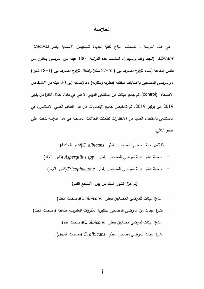
أ
الخالصة
Candida إنتاج تقنية جديدة لتشخيص االصابة بفطر تضمنتفي هذه الدراسة ،
albicans .)يعانون من عينة من المرضى 100ملت هذه الدراسة اشت)الجلد والفم والمهبل
شهر( 18-1سنة( واطفال تتراوح اعمارهم بين ) 57-55)نساء تتراوح اعمارهم بين ) المناعة نقص
عينة من االشخاص 20، والمرضى المصابين بااصابات مختلفة )فطرية وبكترية( ، باإلضافة إلى
ل الفترة من يناير في بغداد خال هليجمع عينات من مستشفى الدولي االتم (control), االصحاء
. تم تشخيص جميع اإلصابات من قبل الطاقم الطبي االستشاري في 2019 يونيوإلى 2019
كانت على دراسةالحاالت المسجلة في هذا الالمستشفى باستخدام العديد من االختبارات تظمنت
النحو التالي:
قشور الجلدية( C. albicans(ثالثون عينة للمرضى المصابين بفطر -
)قشور الجلد( .Aspergillus sppن بفطر خمسة عشر عينة لمرضى المصابي -
)قشور الجلد( Tricophactoneعينة للمرضى المصابين بفطر خمسة عشر -
{ القدم}تم عزل قشور الجلد من بين األصابع
)مسحات الجلد( C. albicansعشرة عينات للمرضى المصابين بفطر -
ببكتيريا المكورات العنقودية الذهبية )مسحات الجلد(.عشرة عينات من المرضى المصابين -
ات الفم(،)مسح C. albicansعشرة عينات لمرضى مصابين بفطر -
.)مسحات المهبل( C. albicansعشرة عينات للمرضى مصابين بفطر -

ب
فحصت العينات باستخدام التقنية الجديدة مقارنة بالطرق الروتينية للتشخيص ، وجدنا أن
ن أن بقية العينات ، في حي C. albicansالتقنية الجديدة أعطت نتيجة إيجابية للعينات المصابة بـ
لفطرية ، البكتيريا( أعطت نتيجة سلبية.) C. albicansبـ غير المصابة
٪( 100٪( ونخصصية ) 100كان أكثر تحسسية بنسبة ) Vitekوقد تبين أن اختبار
%( وتخصصية 100ذات تحسسية ) API20C) من الطرق األخرى C. albicansلتشخيص
تخصصية ٪( و 96ذات تحسسية ) (Culture)المستعمرات %(، الزرع االحيائي كمظهر 97)
٪( وتخصصية 98تحسسية )ذات (Germ tube) الجرثوميتكوين االنبوب اختبار٪( و 93)
مماثلة C. albicansكانت نتائج اختبار التقنية الجديدة في هذه الدراسة لتشخيص .٪( 95)
٪( ، فوائد هذه التقنية الجديدة 100٪( وتخصصية ) 100ذات تحسسية ) Vitekلنتائج اختبار
كلفة( وتقليل استهالك الوقت. على سبيل هي توفير معلومات عالية الدقة ، اقتصادية )منخفضة الت
المثال ، يمكن لألطباء استخدامها من خالل الزيارة التي يقوم بها المريض دون الحاجة إلى إرسالها
إلى المختبر.
جمهورية العراق وزارة التعليم العالي والبحث العلمي
جامعة بغداد كلية التربية للعلوم الصرفه/ ابن الهيثم
قسم علوم الحياة
االصابة بفطر تقنية مناعية جديدة لتشخيص اءالمبيضات البيض
رسالة مقدمة
جامعة بغداد وهي جزء من - التربية للعلوم الصرفة )ابن الهيثم(مجلس كلية الى متطلبات نيل درجة ماجستير علوم في علوم الحياة / علم المناعة
ل ب من ق
رسل اركان حسن (2016اد )جامعة بغدكلية التربية للعلوم الصرفه )ابن الهيثم( / بكالوريوس علوم الحياة/
باشراف
االستاذ المساعد الدكتورة
حازمة موسى خليل العباسي /2019م ذو الحجة/1440ھ أب
Top Related