


Languages
Pages
Legal

Nervous System

Purpose
• Communication
• Control
• Process information
• Gather information
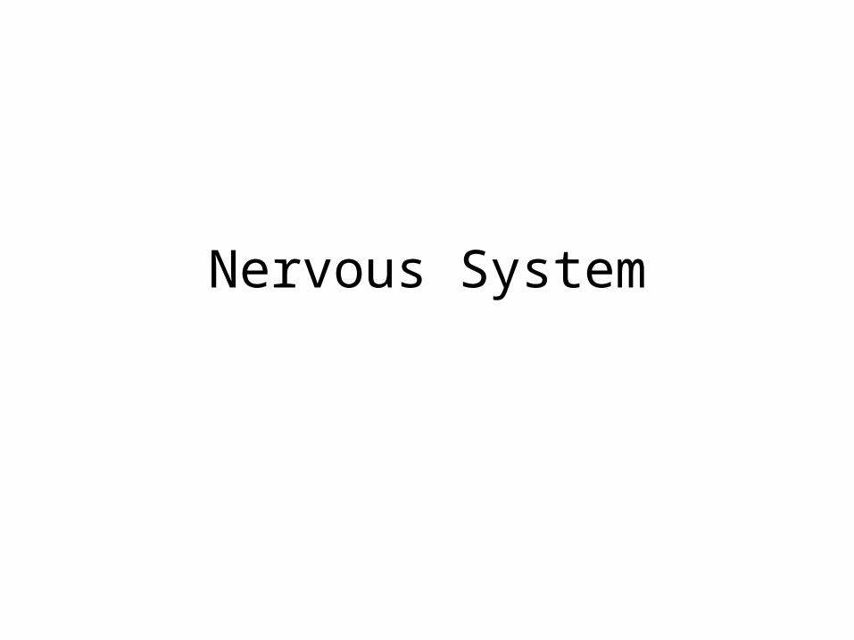
Neuron
• Cell body– Contains nucleus and most normal cell functions– Receives chemical signal from adjacent neuron
• Axon– Transmits electrical impulse to synaptic terminals
• Synaptic terminals– Transmits chemical signal to cell body of adjacent
neuron

Types of Neurons
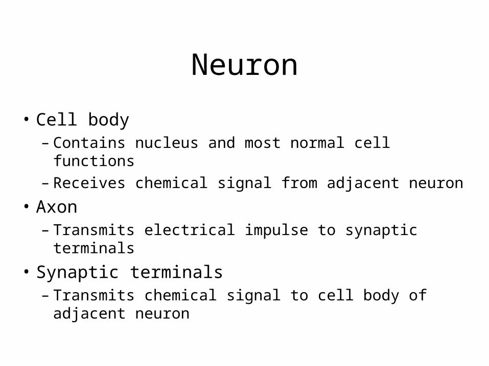
Synapse structure and function
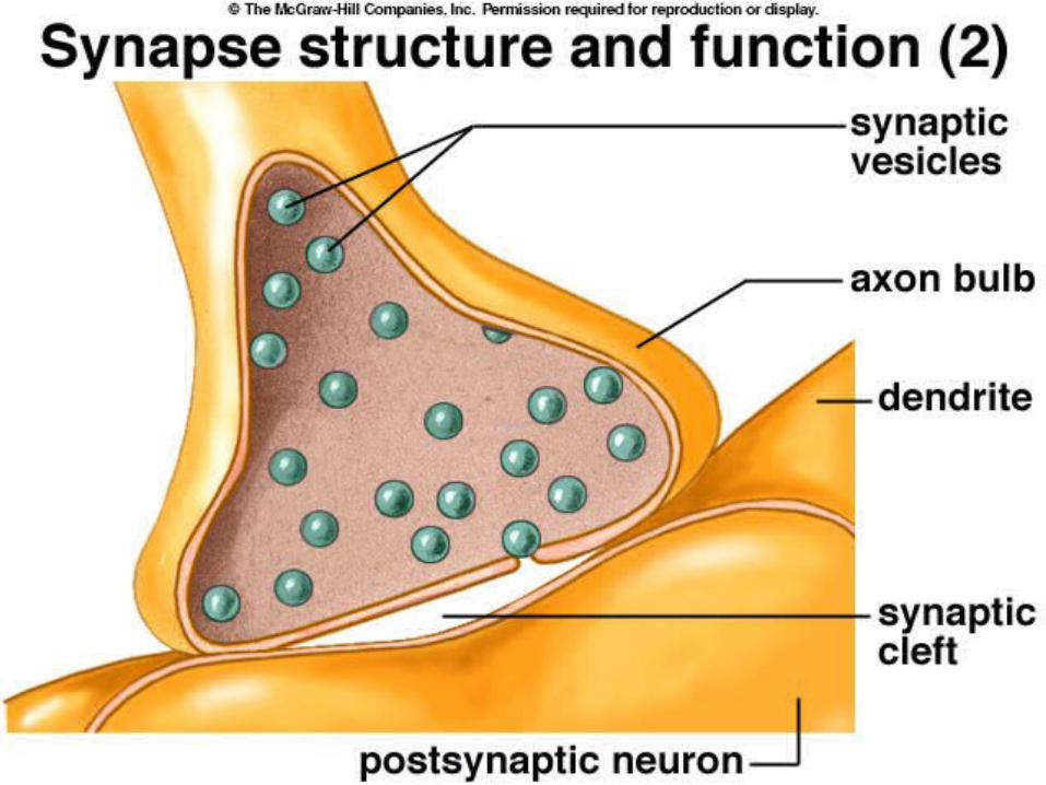
Synapse close-up

Synapse receptors
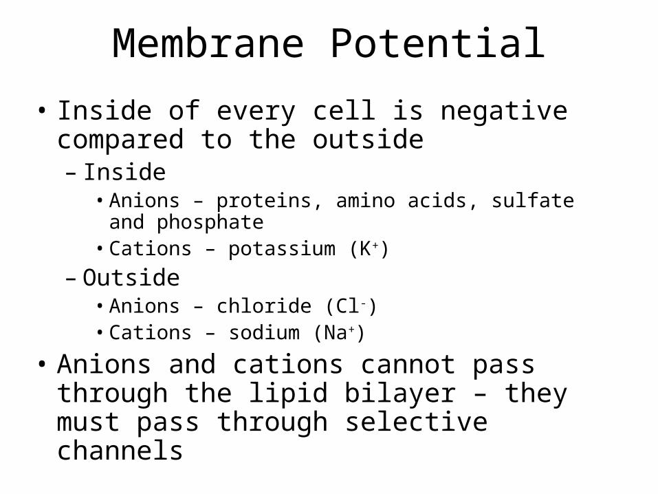
Membrane Potential
• Inside of every cell is negative compared to the outside– Inside
• Anions – proteins, amino acids, sulfate and phosphate• Cations – potassium (K+)
– Outside• Anions – chloride (Cl-)• Cations – sodium (Na+)
• Anions and cations cannot pass through the lipid bilayer – they must pass through selective channels


Action Potential
• Voltage-gated ion channels– Ions move according to gradient and charge
attraction• Potassium (more permeable)
• Sodium (less permeable)
• Sodium/potassium pump– Requires ATP– Pumps Na+ out of cell and K+ into cell


Action Potential

Myelin sheath

Saltatory Conduction

Other Cells of the Nervous System
• Support
• Nourishment
• Protection

Neuroglial Cells
• Non-neuron cells found in the nervous system– Schwann – forms myelin sheath in PNS– Microglial – protect against microbes– Astrocytes – structural and nutritive support– Oligodendroglial – forms myelin sheath in CNS– Ependymal – line CNS cavities and produce
cerebrospinal fluid

Three Neuroglial Cell Types

Nerve structure
• Bundles of myelin covered nerve fibers (axons) covered in connective tissue
• Blood vessels

Organization• Central nervous system (CNS)
– Brain– Spinal chord
• Peripheral nervous system (PNS)– Motor neurons– Sensory neurons

Path of Impulses

Central Nervous System
• Brain
• Spinal chord
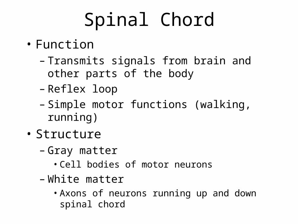
Spinal Chord• Function
– Transmits signals from brain and other parts of the body
– Reflex loop– Simple motor functions (walking, running)
• Structure– Gray matter
• Cell bodies of motor neurons
– White matter• Axons of neurons running up and down spinal chord

Spinal Chord Structure
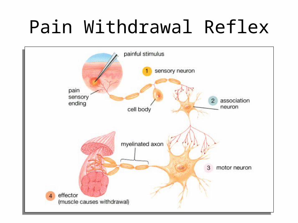
Pain Withdrawal Reflex

Reflex Loop
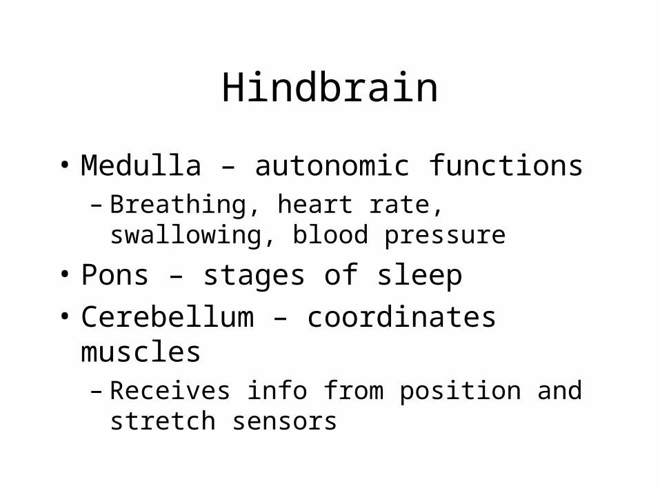
Hindbrain
• Medulla – autonomic functions– Breathing, heart rate, swallowing, blood
pressure
• Pons – stages of sleep
• Cerebellum – coordinates muscles– Receives info from position and stretch sensors

Midbrain
• Reticular formation– Sensory relays– Sensory filter

Forebrain
• Thalamus– Transmits info to and from limbic system,
senses, cerebrum and cerebellum
• Limbic system– Basic emotions, drives and behaviors
• Cerebral cortex– Thinking and information processing

Cerebrum
• Two hemispheres– Connected by corpus callosum (white matter)
• Each half divided into 4 lobes– Frontal, parietal, temporal, occipital lobes
• Cortex– 3mm layer of gray matter– Extensive folds to increase surface area

Cerebrum -- Gross Anatomy

• Frontal– Voluntary motor functions, planning, mood, smell
and social judgement
• Parietal– Sensory reception & integration of sensory
information
• Occipital– Visual center of brain
• Temporal– Hearing, smell, learning, memory and emotional
behavior
Functions of Cerebrum Lobes

Sensory Homunculus
• Demonstrates that the area of the cortex dedicated to the sensations of various body parts is proportional to how sensitive that part of the body is.

Sensory Association Areas
• Association areas interpret sensory information• Somesthetic association area (parietal lobe)
– Position of limbs, location of touch or pain, and shape, weight & texture of an object
• Visual association area (occipital lobe)– Identify the things we see– Faces are recognized in temporal lobe
• Auditory association area (temporal lobe)– Remember the name of a piece of music or identify a
person by his voice


Motor Control
• Intention to contract a muscle begins in motor association (premotor) area of frontal lobes
• Precentral gyrus (primary motor area) processes that order by sending signals to the spinal cord
• Motor homunculus is proportional to number of muscle motor units in a region (fine control)

Language• Includes reading, writing, speaking &
understanding words• Wernicke’s area
– Permits recognition of spoken & written language & creates plan of speech
– Angular gyrus processes text into a form we can speak
• Broca’s area– Generates motor program for larynx, tongue, cheeks &
lips – Transmits that to primary motor cortex for action

Language Centers

Aphasia• Any language deficit resulting from lesions in
same hemisphere as Wernicke’s & Broca’s areas• Lesion to Broca’s = nonfluent aphasia
– slow speech, difficulty in choosing words– entire vocabulary may be 2 to 3 words
• Lesion to Wernicke’s = fluent aphasia– speech normal & excessive, but makes little sense
• Anomic aphasia = speech & understanding are normal but text & pictures make no sense
• Others = understanding only 1st half of words or writing only consonants

Lateralization of Cerebral Functions

Cerebral Lateralization• Left hemisphere is categorical hemisphere
– specialized for spoken & written language, sequential & analytical reasoning (math & science), analyze data in linear way
• Right hemisphere is representational hemisphere– perceives information more holistically, perception of spatial
relationships, pattern, comparison of special senses, imagination & insight, music and artistic skill
• Highly correlated with handedness – 91% of people right-handed with left side is categorical
• Lateralization develops with age• Trauma more problems in males since females have more
communication between hemisphere (corpus callosum is thicker posteriorly)

EEG and Brain Waves
• Electroencephalogram records voltage changes from postsynaptic potentials in cerebral cortex
• Differences in amplitude & frequency distinguish 4 types of brain waves

Brain Waves & Sleep• States of consciousness can be correlated with EEG
• 4 types of brain waves– Alpha occur when awake & resting with eyes closed– Beta occur with eyes open performing mental tasks– Theta occur during sleep or emotional stress– Delta occur during deep sleep
• Sleep is temporary state of unconsciousness– Coma is state of unconsciousness with no possible
arousal

Stages of Sleep• Non-REM sleep occurs in stages
– 4 stages occurring in first 30 to 45 minutes of sleep• stage 1 is drifting sensation (would claim was not sleeping)
• stage 2 still easily aroused
• stage 3 vital signs change -- BP, pulse & breathing rates drop– reached in 20 minutes
• stage 4 is deep sleep -- difficult to arouse
– seems to have a restorative effect
• REM sleep occurs about 5 times a night– rapid eye movements under the eyelids, vital signs increase,
EEG resembles awake person dreams
– may help sort & strengthen information from memory
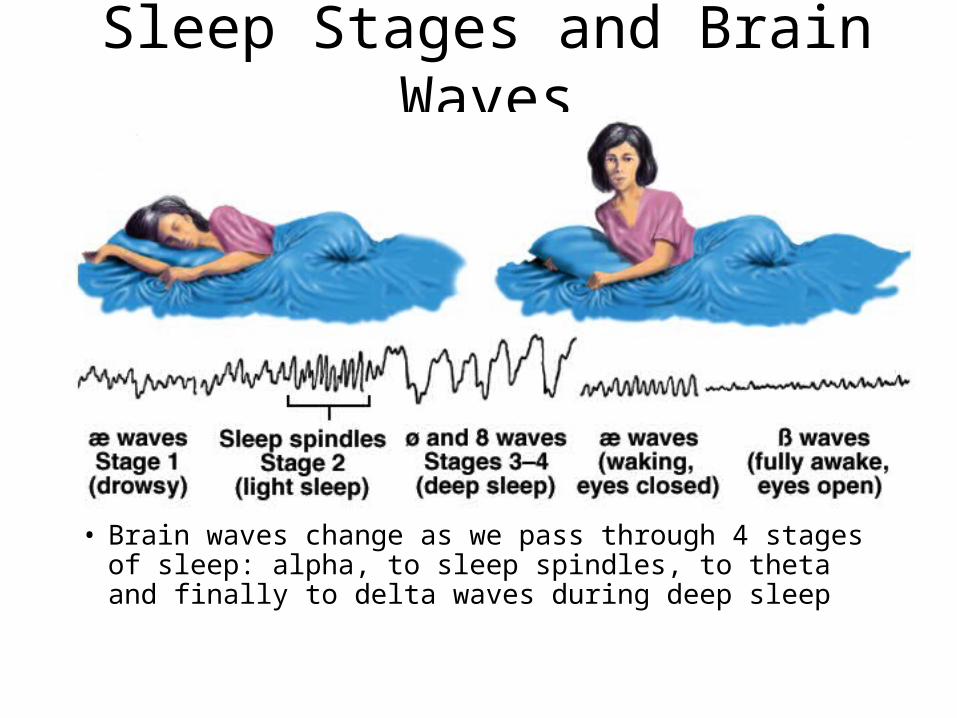
Sleep Stages and Brain Waves
• Brain waves change as we pass through 4 stages of sleep: alpha, to sleep spindles, to theta and finally to delta waves during deep sleep

Cognition
• Cognition is mental processes such as awareness, perception, thinking, knowledge & memory– 75% of brain is association areas where integration of
sensory & motor information occurs
• Examples of effects of brain lesions– temporal lobe -- inability to recognize objects or inability
to recognize faces– frontal lobe -- problems with personality

Memory• Information management requires learning,
memory & forgetting (eliminating the trivia)– Abnormalities
• pathological inability to forget have trouble with reading comprehension
• can not store new data• can not remember old data
• Hippocampus - organizes sensory & cognitive information into a memory– lesions cause inability to form new memories
• Cerebellum helps learn motor skills• Amygdala important in emotional memory

Emotion
• Prefrontal cortex controls how emotions are expressed (seat of judgement)
• Emotions form in hypothalamus & amygdala– artificial stimulation produces fear, anger, pleasure,
love, parental affection, etc.– electrode in median forebrain bundle in rat or human
and a foot pedal• press all day to the exclusion of food (report a quiet,
relaxed feeling)
• Much of our behavior is learned by rewards and punishments or responses of others to them

Peripheral Nervous System
• Motor neurons
• Sensory neurons

Organization of Nervous System

Motor Neurons
• Carry signals from the CNS to muscles and glands– Somatic – voluntary responses
• Skeletal muscles
– Autonomic – involuntary responses• Cell bodies found in ganglia
– Organs
– Glands
– Smooth muscles

Sensory Neurons
• Mechanoreceptor– Hearing, pressure, stretch, movement
• Photoreceptor– Light, vision
• Chemoreceptor– Olfactory– Taste– Pain

Sensory Receptors
• Where is the stimulus located?
• How strong is the stimulus?
• Stimulus = energy
• Sensory receptors transfer signal to other neurons
• Specialized cells or modified neurons

Receiving Sensory Information
• Sensory Transduction– Converts received signal to action potential
• Amplification– Signal transmitted contains more energy than
signal received
• Transmission– Action potential
• Integration– Information processing

Types of Sensory Receptors• Mechanoreceptor
– Stretch or bending of plasma membrane• Hearing, pressure, stretch, movement
• Chemoreceptor– Binding of specific molecules to protein receptors in
plasma membrane• Olfactory, taste, pain
• Photoreceptor– Photons hitting specific proteins in plasma membrane
• Vision

Touch
• Mechanoreceptor


Parts of the Ear
• Outer ear– Pinna and auditory canal
• Middle ear– Tympanic membrane (ear drum)– Ossicles (malleus, incus, stapes)
• Inner ear– Cochlea

Auditory Receptors• Different pitches vibrate different
parts of the basilar membrane
• The basilar membrane vibrates against hair cells in the cochlear duct
• Shape and thickness of basilar membrane effects vibrations

Pain Receptors
• Chemical receptor– Detects chemicals
released during tissue damage
– Receives chemical on specialized dentrites

Olfactory Receptors
• Chemical receptor
• Different molecules have different cells with unique receptors for that molecule


Photoreceptors• Modified neurons containing light
absorbing pigment
• Rods– Black and white– Night vision– Pigment – rhodopsin
• Cones– Color– 4 types – red, blue and green– Pigment – photopsins
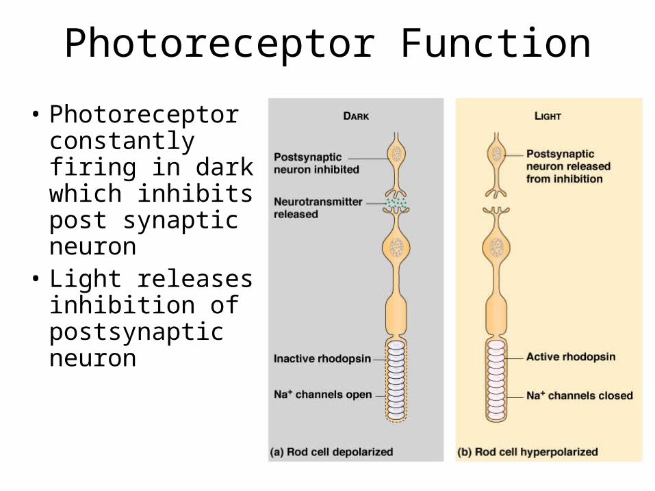
Photoreceptor Function
• Photoreceptor constantly firing in dark which inhibits post synaptic neuron
• Light releases inhibition of postsynaptic neuron

Retinal Structure
• Rods and cones are not mapped 1:1 to ganglion cells
• More cells per ganglion cell equals lower resolution but greater sensitivity
• Dogs have 5x the number of rod cells per ganglion cell
• Optic nerve exits through the fovea which creates your blind spot
Top Related