


Languages
Pages
Legal

6/29/2015
1
Nervous System:
PNS and CNS
Biology 105
Lecture 9
Chapter 8
Copyright © 2009 Pearson Education, Inc.
Outline
I. Central Nervous System vs. Peripheral Nervous System
II. Peripheral Nervous System
A. Somatic Nervous System
B. Autonomic Nervous System
III. Autonomic Nervous System
A. Parasympathetic Nervous System
B. Sympathetic Nervous System
IV. Reflex Actions
V. Central Nervous System
A. Protection of CNS
B. Spinal Cord
C. Brain
Copyright © 2009 Pearson Education, Inc.
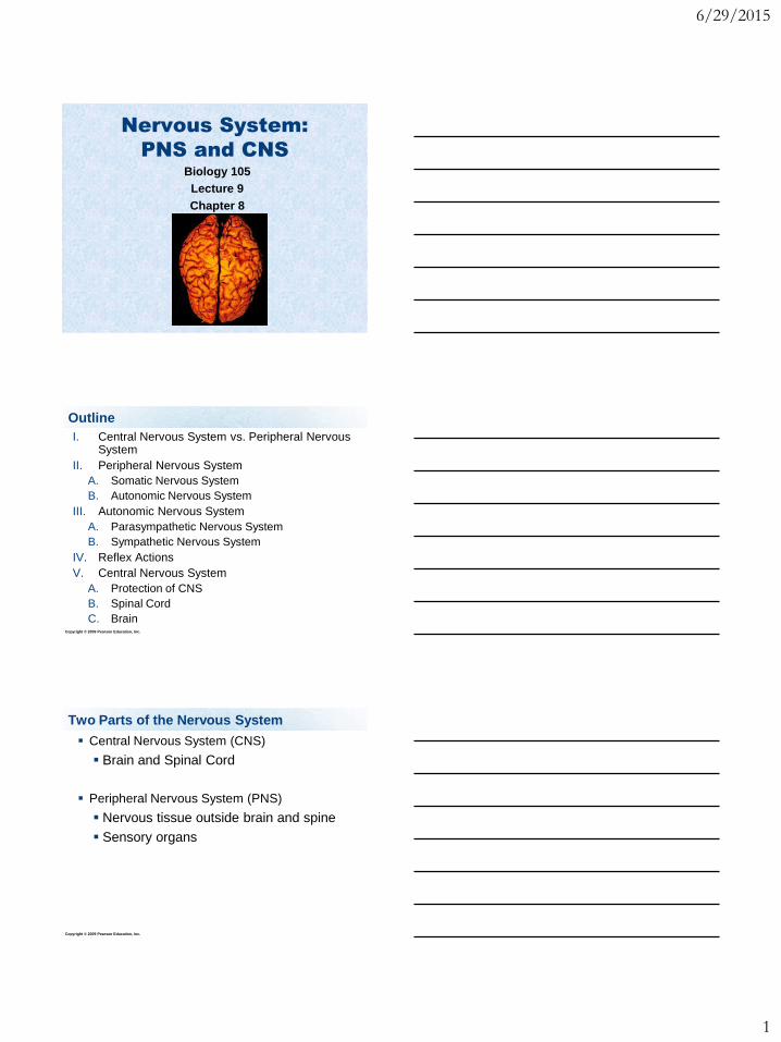
Two Parts of the Nervous System
Central Nervous System (CNS)
Brain and Spinal Cord
Peripheral Nervous System (PNS)
Nervous tissue outside brain and spine
Sensory organs
6/29/2015
2
Copyright © 2009 Pearson Education, Inc.
Somatic nervous system – division of PNS that
controls voluntary functions.
Responsible for movement, controls skeletal
muscles
Autonomic nervous system – division of PNS
that controls involuntary functions.
Controls cardiac and smooth muscles, and
glands
Two Parts of the Nervous System Peripheral Nervous System
Copyright © 2009 Pearson Education, Inc.
Autonomic Nervous System
Autonomic Nervous System is divided into
two systems:
Parasympathetic division –
“rest and digest”
Sympathetic division – stimulatory
stress responses…
“flight or fight”
Copyright © 2009 Pearson Education, Inc.
Figure 8.1 The nervous system
6/29/2015
3
Copyright © 2009 Pearson Education, Inc.
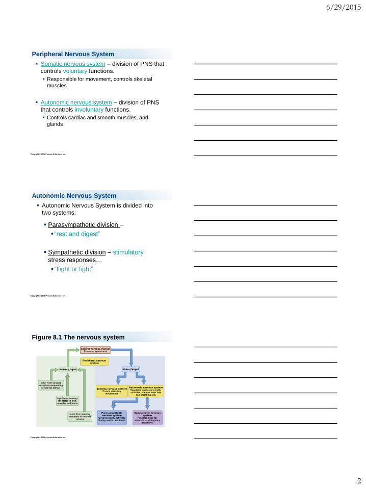
PNS – Parasympathetic Nervous System
Figure 8.12 (1 of 2)
Constricts
pupil
Increases
salivation
Decreases
breathing rate
Slows
heart rate
Widens
blood
vessels
Increases
digestive activity
Increases
digestive activity
Contracts
bladder
muscles
Stimulates
defecation
Parasympathetic Nervous System
(Cranial and sacral
regions of spinal cord)
Synapse
between
neurons
Copyright © 2009 Pearson Education, Inc.
Parasympathetic – Rest and Digest
Constricts eye
pupils
Stimulates
salivation
Slows heart rate
Constricts breathing
Dilates blood
vessels
Stimulates digestion
Constricts bladder
Stimulates sex
organs
Figure 8.12 (1 of 2)
Constricts
pupil
Increases
salivation
Decreases
breathing rate
Slows
heart rate
Widens
blood
vessels
Increases
digestive activity
Increases
digestive activity
Contracts
bladder
muscles
Stimulates
defecation
Parasympathetic Nervous System
(Cranial and sacral
regions of spinal cord)
Synapse
between
neurons
Copyright © 2009 Pearson Education, Inc.
Sympathetic – Fight or Flight
Dilates eye pupils
Inhibits salivation
Accelerates heart rate
Facilitates breathing
Stimulates secretion of epinephrine and
norepinephrine
Stimulates release of free glucose
Inhibits digestion
Relaxes bladder
Inhibits sex organs
6/29/2015
4
Copyright © 2009 Pearson Education, Inc.
Reflex Actions
Sometime the body requires a very fast
response, such as reacting to a hot stove.
We may not have time to send the message all
the way up to the brain to process the
information.
The spinal cord can process the information
and send a response back to the motor
nerves.
Copyright © 2009 Pearson Education, Inc.
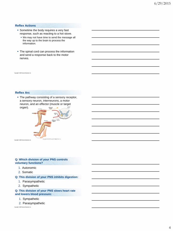
Reflex Arc
The pathway consisting of a sensory receptor,
a sensory neuron, interneurons, a motor
neuron, and an effector (muscle or target
organ).
Copyright © 2009 Pearson Education, Inc.
Q: Which division of your PNS controls
voluntary functions?
1. Autonomic
2. Somatic
Q: This division of your PNS inhibits digestion:
1. Parasympathetic
2. Sympathetic
Q: This division of your PNS slows heart rate
and lowers blood pressure:
1. Sympathetic
2. Parasympathetic
6/29/2015
5
Copyright © 2009 Pearson Education, Inc.
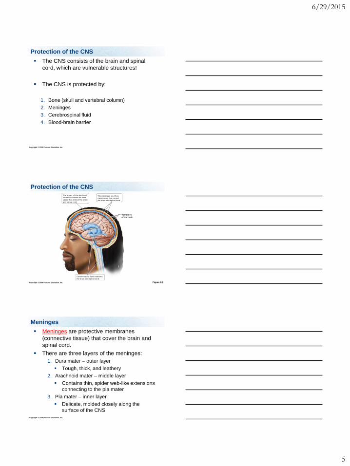
Protection of the CNS
The CNS consists of the brain and spinal
cord, which are vulnerable structures!
The CNS is protected by:
1. Bone (skull and vertebral column)
2. Meninges
3. Cerebrospinal fluid
4. Blood-brain barrier
Copyright © 2009 Pearson Education, Inc.
Protection of the CNS
Figure 8.2
Ventricles
of the brain
The bones of the skull and
vertebral column are hard
cases that protect the brain
and spinal cord.
Cerebrospinal fluid cushions
the brain and spinal cord.
The meninges are three
membranes that protect
the brain and spinal cord.
Copyright © 2009 Pearson Education, Inc.
Meninges
Meninges are protective membranes
(connective tissue) that cover the brain and
spinal cord.
There are three layers of the meninges:
1. Dura mater – outer layer
Tough, thick, and leathery
2. Arachnoid mater – middle layer
Contains thin, spider web-like extensions
connecting to the pia mater
3. Pia mater – inner layer
Delicate, molded closely along the
surface of the CNS
6/29/2015
6
Copyright © 2009 Pearson Education, Inc.
Meningitis
Meningitis – inflammation of the meninges.
It is caused by many forms of bacteria and
viruses.
If the infection spreads to the underlying brain
tissue, it can lead to encephalitis, an
inflammation of the brain.
This is a VERY serious condition!
Copyright © 2009 Pearson Education, Inc.
Cerebrospinal Fluid (CSF)
Fluid produced in the ventricles of the brain.
CSF fills:
Ventricles
In-between the meninges
Central canal of the spinal cord.
Functions:
1. Shock absorption
2. Support the weight of the brain
3. Nourishment and waste removal
Copyright © 2009 Pearson Education, Inc.
Blood-Brain Barrier
Formed by the tight junctions between the
cells lining the blood vessels of the brain.
Permits certain substances to enter the brain,
while inhibiting others from entering.
It inhibits many drugs that are not lipid-soluble
from reaching brain tissue.
6/29/2015
7
Copyright © 2009 Pearson Education, Inc.
Q: The innermost layer of the meninges is the:
1. Pia mater
2. Dura mater
3. Arachnoid mater
Q: Where is CSF found?
1. Central canal of spinal cord
2. Ventricles
3. Between meninges
4. All of the above!
Copyright © 2009 Pearson Education, Inc.
Q: The blood-brain barrier is formed by which
kind of junctions?
1. Adhesion
2. Gap
3. Tight
Copyright © 2009 Pearson Education, Inc.
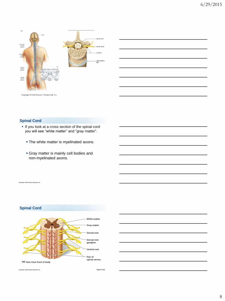
Spinal Cord
Spinal cord extends from the base of the brain
down the back, and transmits messages
between the brain and the rest of the body.
There is cerebrospinal fluid located in a
central canal in the spinal cord.
6/29/2015
8
Copyright © 2009 Pearson Education, Inc.
Spinal Cord
If you look at a cross section of the spinal cord
you will see “white matter” and “gray matter”.
The white matter is myelinated axons.
Gray matter is mainly cell bodies and
non-myelinated axons.
Copyright © 2009 Pearson Education, Inc.
Spinal Cord
Figure 8.11a
Dorsal root
Dorsal-root
ganglion
Pair of
spinal nerves
Gray matter
White matter
Ventral root
(a) View from front of body
6/29/2015
9
Copyright © 2009 Pearson Education, Inc.
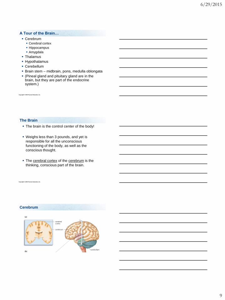
A Tour of the Brain…
Cerebrum
Cerebral cortex
Hippocampus
Amygdala
Thalamus
Hypothalamus
Cerebellum
Brain stem – midbrain, pons, medulla oblongata
(Pineal gland and pituitary gland are in the brain, but they are part of the endocrine system.)
Copyright © 2009 Pearson Education, Inc.
The Brain
The brain is the control center of the body!
Weighs less than 3 pounds, and yet is
responsible for all the unconscious
functioning of the body, as well as the
conscious thought.
The cerebral cortex of the cerebrum is the
thinking, conscious part of the brain.
Cerebrum
6/29/2015
10
Copyright © 2009 Pearson Education, Inc.
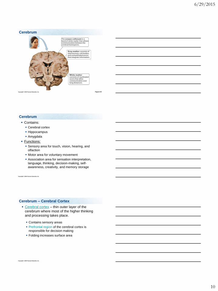
Cerebrum
Figure 8.5
Gray matter consists of interneurons, cell bodies, and unmyelinated axons that integrate information.
White matter
consists of myelinated axons that allow communication over long distances.
The corpus callosum is a band of white matter that allows communication between the cerebral hemisperes.
Copyright © 2009 Pearson Education, Inc.
Cerebrum
Contains:
Cerebral cortex
Hippocampus
Amygdala
Functions:
Sensory area for touch, vision, hearing, and
olfaction
Motor area for voluntary movement
Association area for sensation interpretation,
language, thinking, decision-making, self-
awareness, creativity, and memory storage
Copyright © 2009 Pearson Education, Inc.
Cerebrum – Cerebral Cortex
Cerebral cortex – thin outer layer of the
cerebrum where most of the higher thinking
and processing takes place.
Contains sensory areas
Prefrontal region of the cerebral cortex is
responsible for decision making
Folding increases surface area
6/29/2015
11
Copyright © 2009 Pearson Education, Inc.
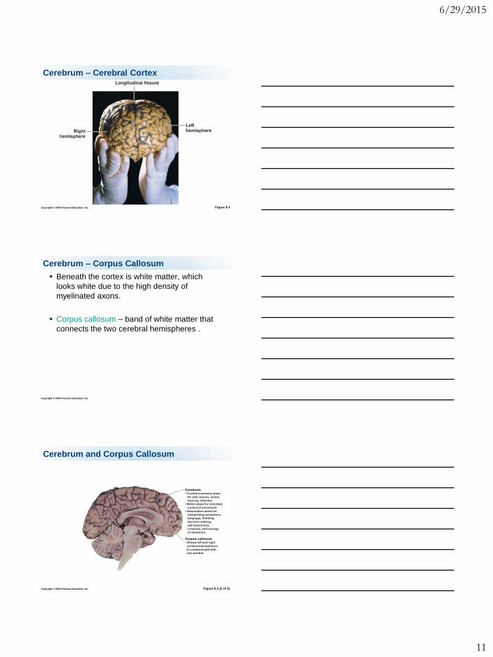
Cerebrum – Cerebral Cortex
Figure 8.4
Copyright © 2009 Pearson Education, Inc.
Beneath the cortex is white matter, which
looks white due to the high density of
myelinated axons.
Corpus callosum – band of white matter that
connects the two cerebral hemispheres .
Cerebrum – Corpus Callosum
Copyright © 2009 Pearson Education, Inc.
Cerebrum and Corpus Callosum
Figure 8.3 (2 of 2)
Cerebrum
• Contains sensory areas
for skin senses, vision,
hearing, olfaction
• Motor areas for voluntary
control of movement
• Association areas for
interpreting sensations,
language, thinking,
decision making,
self-awareness,
creativity, and storage
of memories
Corpus callosum
• Allows left and right
cerebral hemispheres
to communicate with
one another

6/29/2015
12
Copyright © 2009 Pearson Education, Inc.
Cerebrum
Hippocampus – important in long-term
memory.
Amygdala – important in remembering fear
and responding to it.
Copyright © 2009 Pearson Education, Inc.
Memory (the Limbic System)
Figure 8.8
Amygdala
Cerebrum
Hippocampus
Thalamus
Hypothalamus
Olfactory bulb
Copyright © 2009 Pearson Education, Inc.
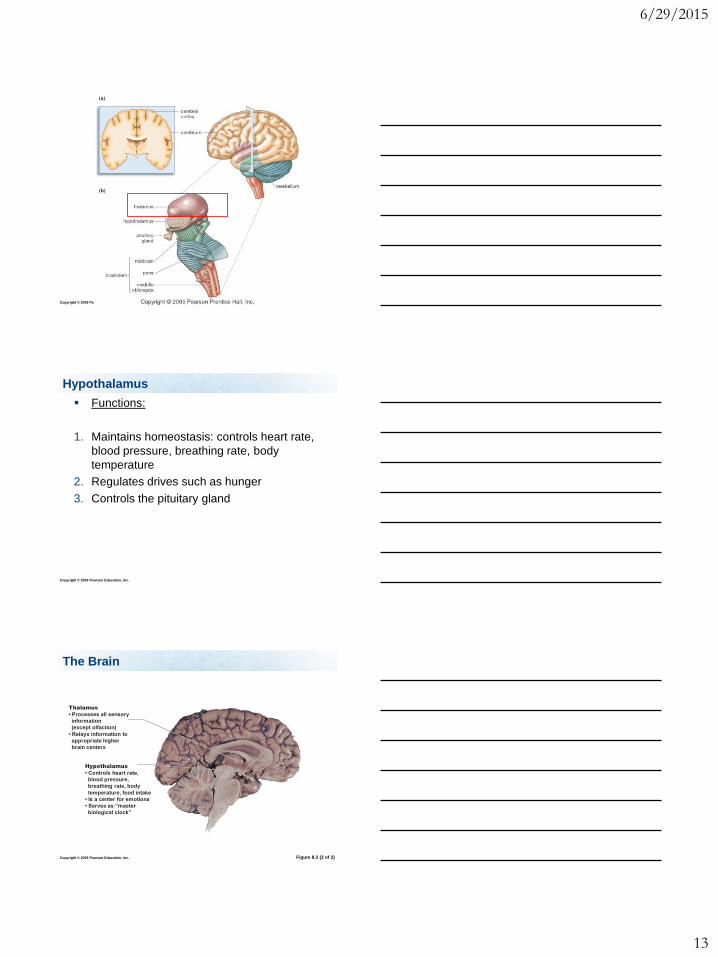
Thalamus – processes sensory information
(except smell) and relays it to other areas of
the brain.
Thalamus
6/29/2015
13
Copyright © 2009 Pearson Education, Inc.
Copyright © 2009 Pearson Education, Inc.
Functions:
1. Maintains homeostasis: controls heart rate,
blood pressure, breathing rate, body
temperature
2. Regulates drives such as hunger
3. Controls the pituitary gland
Hypothalamus
Copyright © 2009 Pearson Education, Inc.
The Brain
Figure 8.3 (2 of 2)
Hypothalamus
• Controls heart rate,
blood pressure,
breathing rate, body
temperature, food intake
• Is a center for emotions
• Serves as “master
biological clock”
Thalamus
• Processes all sensory
information
(except olfaction)
• Relays information to
appropriate higher
brain centers
6/29/2015
14
Copyright © 2009 Pearson Education, Inc.
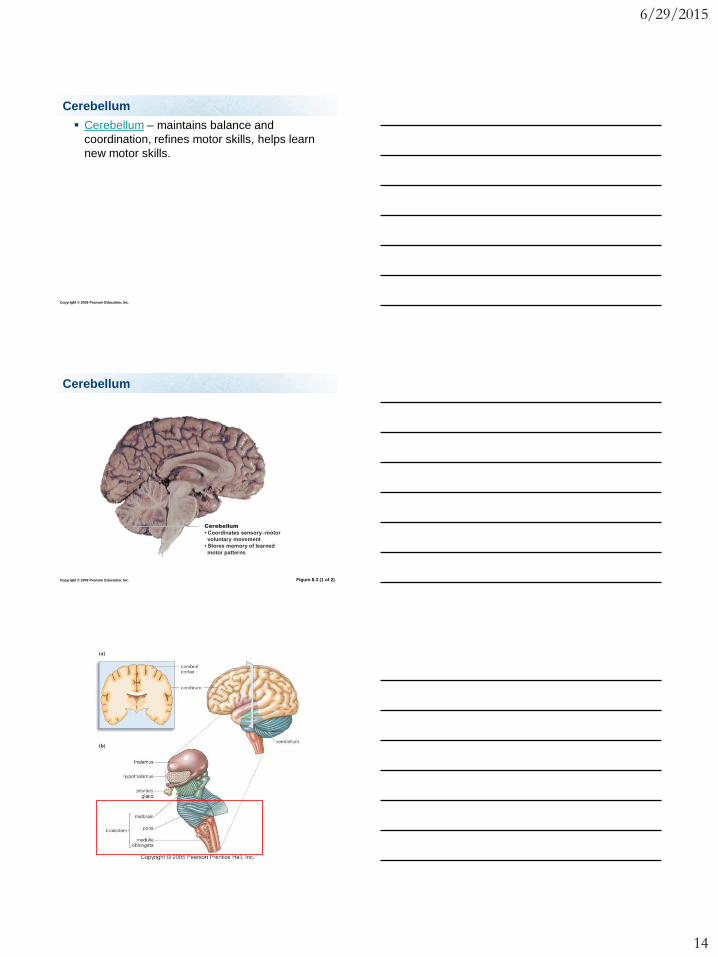
Cerebellum – maintains balance and
coordination, refines motor skills, helps learn
new motor skills.
Cerebellum
Copyright © 2009 Pearson Education, Inc.
Cerebellum
Figure 8.3 (1 of 2)
Cerebellum
• Coordinates sensory–motor
voluntary movement
• Stores memory of learned
motor patterns
6/29/2015
15
Copyright © 2009 Pearson Education, Inc.
Brain Stem – Medulla Oblongata
Functions:
Controls many vital involuntary functions including breathing, heart rate, and blood pressure
Copyright © 2009 Pearson Education, Inc.
Brain Stem – Pons
Functions:
Assists the medulla oblongata to control involuntary breathing
Relays messages between the spinal cord/cerebellum, and the cerebrum, thalamus, and hypothalamus
Copyright © 2009 Pearson Education, Inc.
Brain Stem - Midbrain
Functions:
Important in voluntary muscle control
Relay station for auditory and visual information
Relays information between the cerebellum/spinal cord and the cerebrum
Controls eye movement
6/29/2015
16
Copyright © 2009 Pearson Education, Inc.
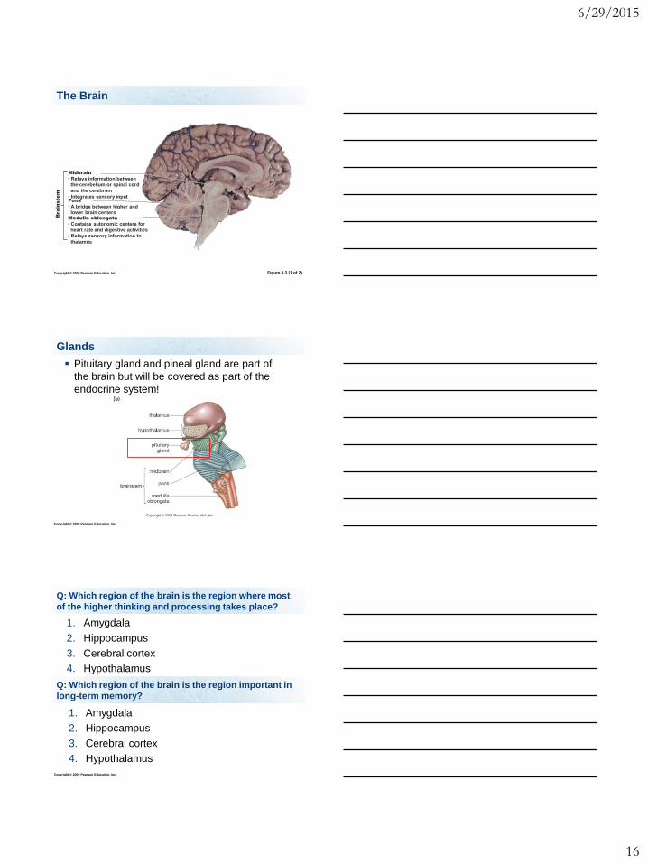
The Brain
Figure 8.3 (1 of 2)
Medulla oblongata
• Contains autonomic centers for
heart rate and digestive activities
• Relays sensory information to
thalamus
Pons
• A bridge between higher and
lower brain centers
Midbrain
• Relays information between
the cerebellum or spinal cord
and the cerebrum
• Integrates sensory input
Bra
in
ste
m
Copyright © 2009 Pearson Education, Inc.
Glands
Pituitary gland and pineal gland are part of
the brain but will be covered as part of the
endocrine system!
Copyright © 2009 Pearson Education, Inc.
Q: Which region of the brain is the region where most
of the higher thinking and processing takes place?
1. Amygdala
2. Hippocampus
3. Cerebral cortex
4. Hypothalamus
Q: Which region of the brain is the region important in
long-term memory?
1. Amygdala
2. Hippocampus
3. Cerebral cortex
4. Hypothalamus
6/29/2015
17
Copyright © 2009 Pearson Education, Inc.
Q: Which region of the brain regulates drives including
hunger, maintains homeostasis, controls the pituitary
gland?
1. Amygdala
2. Hippocampus
3. Cerebral cortex
4. Hypothalamus
Q: Which region of the brain is important in
remembering fear and responding to it?
1. Amygdala
2. Hippocampus
3. Cerebral cortex
4. Hypothalamus
Copyright © 2009 Pearson Education, Inc.
Read Chapter 8
What are the somatic nervous system and
autonomic nervous system, and what do they
each control?
What are reflex actions?
What are the parasympathetic and the
sympathetic divisions of the autonomic
nervous system?
What specifically do they control (increase heart
rate, increase respiration, etc.)?
Important Concepts
Copyright © 2009 Pearson Education, Inc.
What protects the CNS
What are the three layers of the meninges?
Be able to describe them and their locations
(which is the inner, middle, or outer layer.)
What is meningitis and what is the cause? What
is encephalitis?
What are the functions of cerebrospinal fluid?
What is the function of the blood-brain barrier,
and what does it allow to pass?
Important Concepts
6/29/2015
18
Copyright © 2009 Pearson Education, Inc.
Major regions of the brain and their functions:
cerebrum (including the cerebral cortex,
hippocampus, and amygdala), hypothalamus,
thalamus, cerebellum, brain stem (including
the midbrain, pons, and medulla oblongata)
Which parts of the brain are in the cerebrum
and which parts are in the brain stem?
What is the corpus callosum, and what is its
function?
Important Concepts
Copyright © 2009 Pearson Education, Inc.
Definitions
Long-term memory, somatic nervous system,
autonomic nervous system, voluntary,
involuntary, reflex arc, constrict, dilate, inhibit,
accelerate, facilitate, stimulate, relax, white
matter, gray matter, prefrontal region
Top Related