


![Dr. heba kalbouneh - Weeblyjumed16.weebly.com/uploads/8/8/5/1/88514776/sheet-13-2017[336].pdfconservation of water inside our body ... a lot of nerve endings that sense pain, touch,](https://static.fdocuments.in/doc/165x107/5b3afe7f7f8b9a986e8bc064/dr-heba-kalbouneh-336pdfconservation-of-water-inside-our-body-a-lot-of.jpg)

Languages
Pages
Legal


Muscle Histology
Dr. Heba Kalbouneh Assistant Professor of Anatomy and Histology
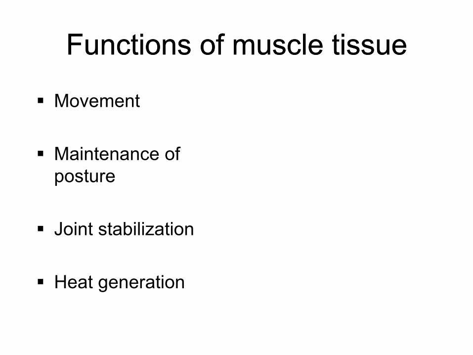
Functions of muscle tissue Functions of muscle tissue
Movement
Maintenance of
posture
Joint stabilization
Heat generation
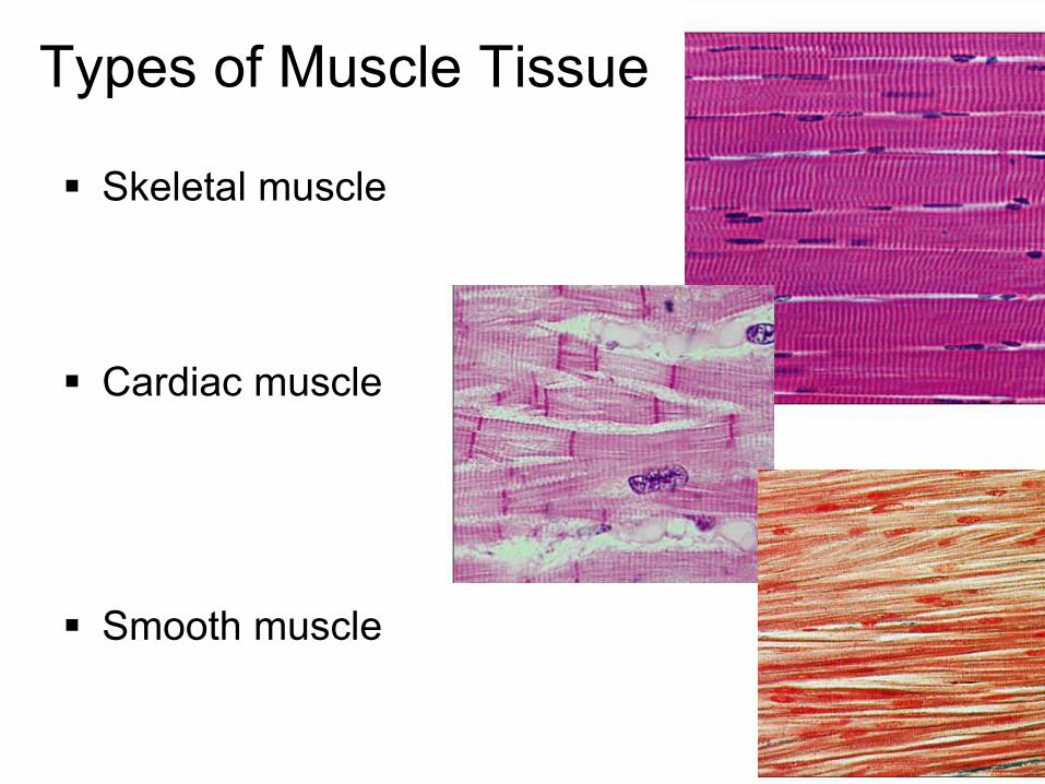
Types of Muscle Tissue
Skeletal muscle
Cardiac muscle
Smooth muscle

Types of Muscle Tissue
Skeletal •Attach to and move skeleton
•40% of body weight
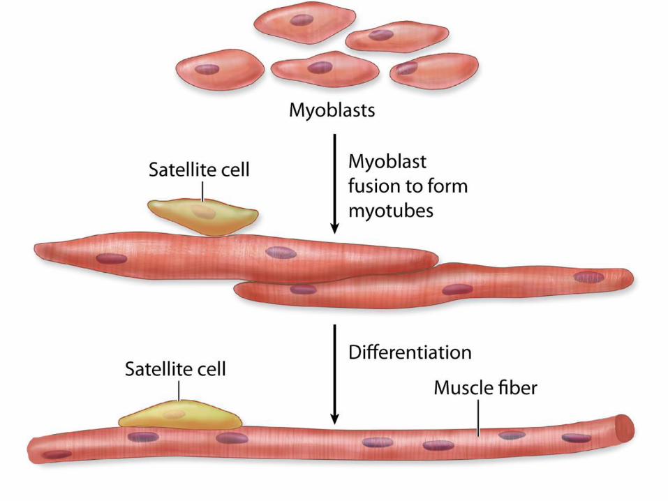
•Fibers = multinucleate cells (embryonic cells fuse)
•Cells with obvious striations
•Contractions are voluntary
Cardiac: only in the wall of
the heart
•Cells are striated
•Contractions are
involuntary (not voluntary)
Smooth: walls of hollow organs
•Lack striations
•Contractions are involuntary (not voluntary)

Similarities…
Their cells are called fibers because they
are elongated
Contraction depends on myofilaments
Actin
Myosin
Plasma membrane is called sarcolemma
Sarcos = flesh
Lemma = sheath
SKELETAL MUSCLES

Epimysium:
surrounds
whole muscle
Perimysium
is around
fascicle
Endomysium is around each
muscle fiber
Skeletal
muscle
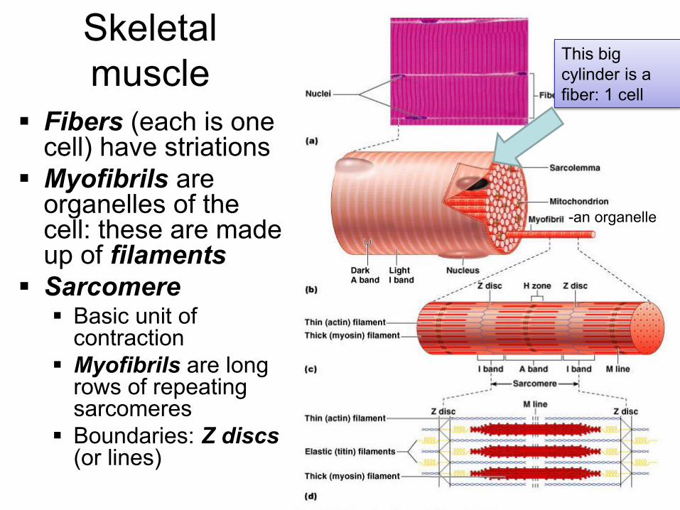
Fibers (each is one cell) have striations
Myofibrils are organelles of the cell: these are made up of filaments
Sarcomere Basic unit of
contraction
Myofibrils are long rows of repeating sarcomeres
Boundaries: Z discs (or lines)
This big
cylinder is a
fiber: 1 cell
-an organelle

M line provides an attachment to myosin filaments
Z line provides an attachment to actin filaments
A band is the darker band of the myofibril containing myosin filaments
H band is the lighter section in the middle of the A band where only myosin is present
I band is the lighter band containing only the actin filaments
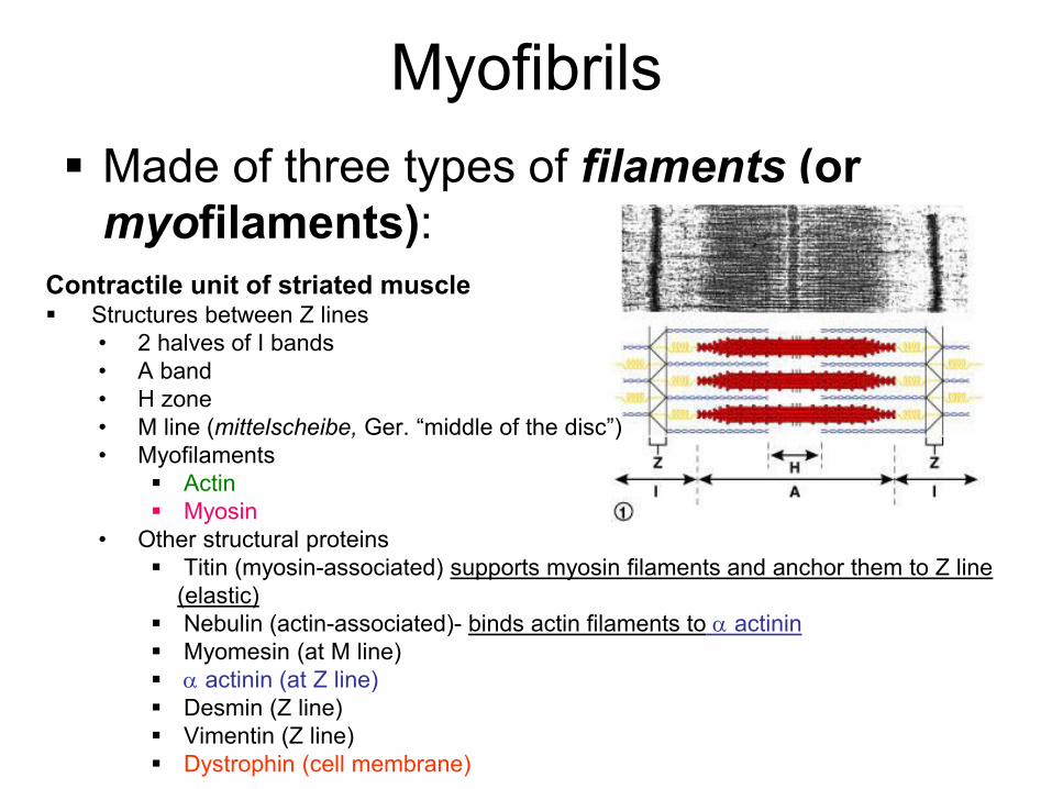
Myofibrils
Made of three types of filaments (or
myofilaments):
Contractile unit of striated muscle Structures between Z lines
• 2 halves of I bands
• A band
• H zone
• M line (mittelscheibe, Ger. “middle of the disc”) • Myofilaments
Actin
Myosin
• Other structural proteins
Titin (myosin-associated) supports myosin filaments and anchor them to Z line
(elastic)
Nebulin (actin-associated)- binds actin filaments to actinin
Myomesin (at M line)
actinin (at Z line)
Desmin (Z line)
Vimentin (Z line)
Dystrophin (cell membrane)

Myosin is composed of 2 identical heavy chains and two pairs of light chains
Heavy chains are twisted together as tail
The four light chains form a head at one end of each heavy chains

Actin filaments are composed of two thin helical twisted strands composed of
G-actin monomers
Contain a myosin binding site
Are anchored to the Z line by alpha-actinin
Associated with:
A- Tropomyosin: coil of two peptide chains located in the groove between the
two twisted actin strands
B- Troponin a complex of 3 subunits :
Tropomyosin
Calcium ion
Regulatory subunit
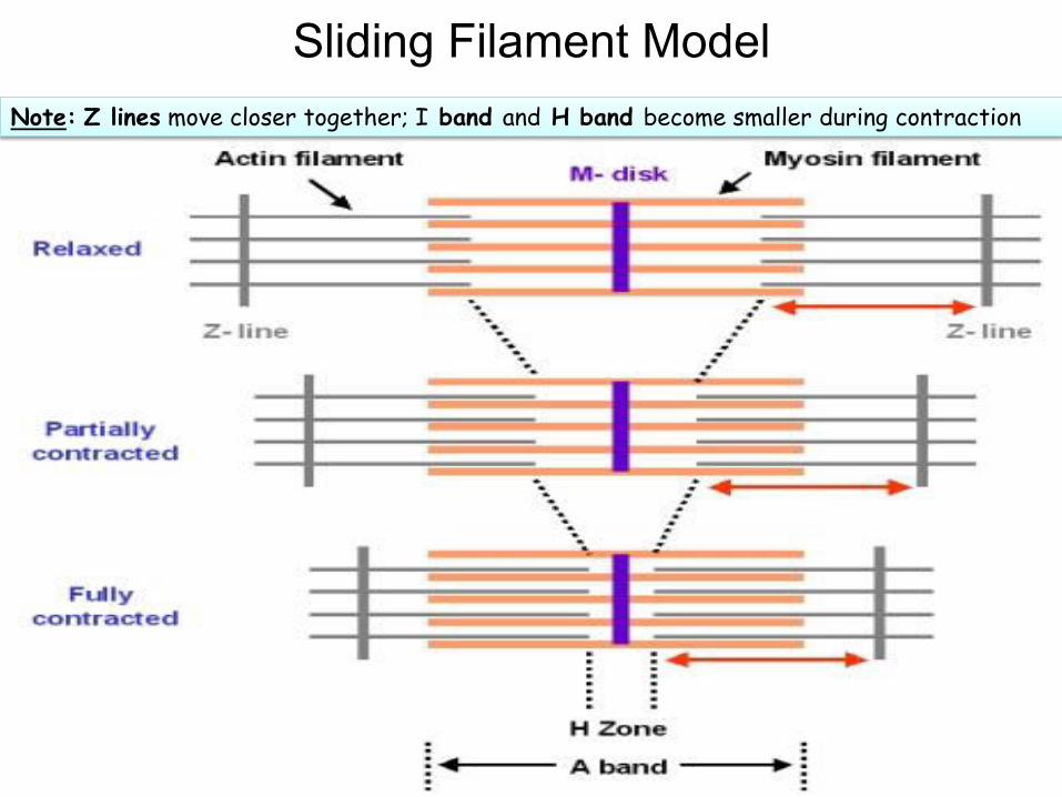
Sliding Filament Model
Note: Z lines move closer together; I band and H band become smaller during contraction

Titin resists overstretching “A” band constant because it is caused by myosin, which doesn’t change length
Sarcomere shortens because actin pulled towards its middle
by myosin cross bridges
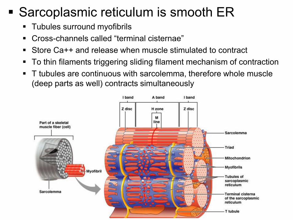
Sarcoplasmic reticulum is smooth ER Tubules surround myofibrils
Cross-channels called “terminal cisternae” Store Ca++ and release when muscle stimulated to contract
To thin filaments triggering sliding filament mechanism of contraction
T tubules are continuous with sarcolemma, therefore whole muscle
(deep parts as well) contracts simultaneously

A T-tubule (or transverse
tubule) is a deep
invagination of the
sarcolemma
T-tubules permit the
conduction of electrical
impulses

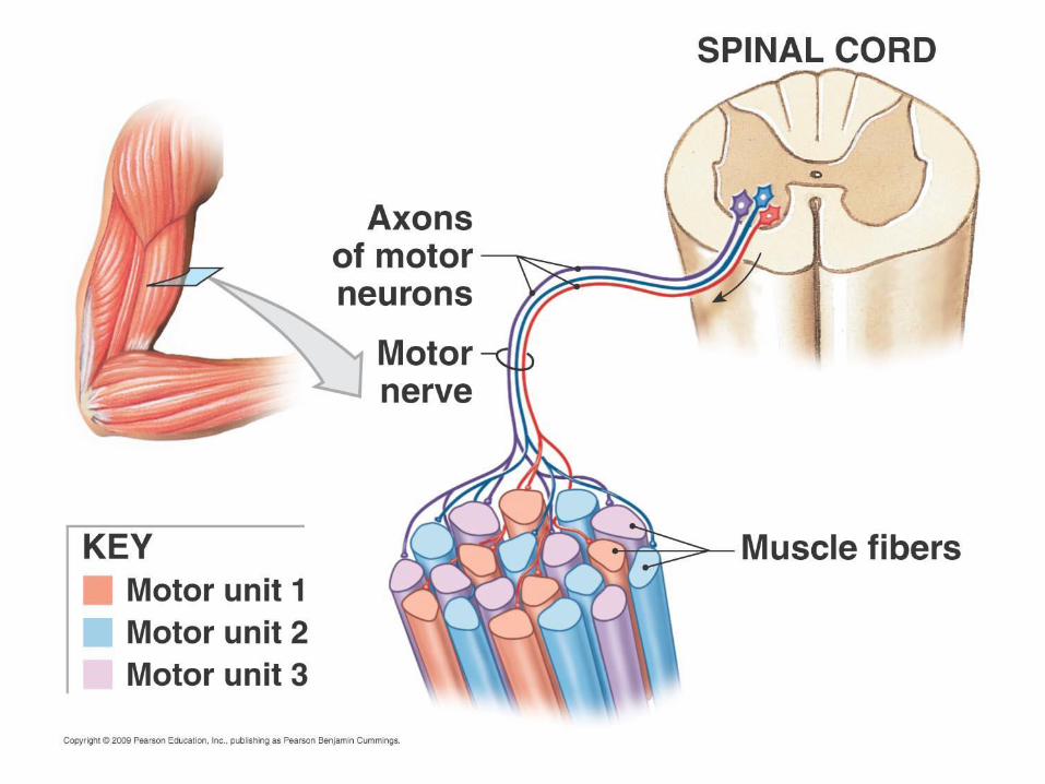
Neuromuscular
Junction
Motor neurons innervate muscle
fibers
Motor end plate is where they
meet
Neurotransmitters are released
by nerve signal: this initiates
calcium ion release and muscle
contraction
Motor Unit: a motor neuron and all the muscle fibers it innervates (these all
contract together)
•Average is 150, but range is one to several hundred muscle fibers in a motor unit
•The finer the movement, the fewer muscle fibers /motor unit
•The fibers are spread throughout the muscle, so stimulation of a single motor
unit causes a weak contraction of the entire muscle

Each motor neuron branches to innervate a variable # of muscle fibers
A motor unit includes each motor neuron and all fibers it innervates
Motor Unit
12-12

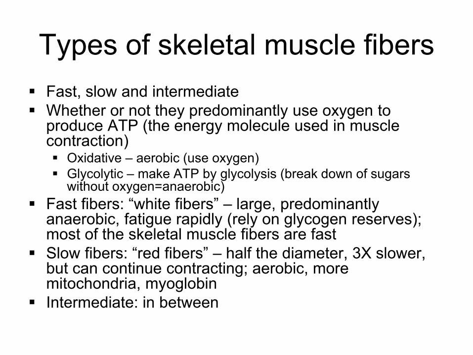
Types of skeletal muscle fibers
Fast, slow and intermediate
Whether or not they predominantly use oxygen to produce ATP (the energy molecule used in muscle contraction) Oxidative – aerobic (use oxygen)
Glycolytic – make ATP by glycolysis (break down of sugars without oxygen=anaerobic)
Fast fibers: “white fibers” – large, predominantly anaerobic, fatigue rapidly (rely on glycogen reserves); most of the skeletal muscle fibers are fast
Slow fibers: “red fibers” – half the diameter, 3X slower, but can continue contracting; aerobic, more mitochondria, myoglobin
Intermediate: in between
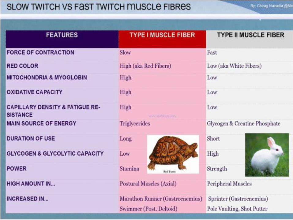

Left – Red Fiber Dominant, Marathoner
Right – White fiber Dominant, Sprinter,
Middle – Perfect, Bodybuilder
All muscle fibers of a
motor unit are of the
same type.

A skeletal muscle contracts when its motor units are stimulated
All or none principle: each muscle fiber either contracts completely or not at all
Amount of force: depends on how many motor units are activated
Muscle tone Even at rest, some motor units are active: tense the
muscle even though not causing movement: “resting tone”

Muscle hypertrophy Weight training (repeated intense workouts): increases diameter and
strength of “fast” muscle fibers by increasing production of Mitochondria
Actin and myosin protein
Myofilaments containing these contractile proteins
The myofibril organelles these myofilaments form
Fibers enlarge (hypertrophy) as number and size of myofibrils increase
[Muscle fibers (=muscle cells) don’t increase in number but increase in diameter producing large muscles]
Endurance training (aerobic): doesn’t produce hypertrophy
Muscle atrophy: loss of tone and mass from lack of stimulation Muscle becomes smaller and weaker
Note on terminology: in general, increased size is hypertrophy; increased number
of cells is hyperplasia

Muscle spindles are sensory
receptors within the belly of a
muscle that primarily detect
changes in the length of this
muscle.
They convey length information
to the central nervous system
via sensory neurons
This information can be
processed by the brain to
determine the position of body
parts
Each muscle spindle consists of an
encapsulated cluster of small striated
muscle fibers ("intrafusal muscle fibers")
with somewhat unusual structure (e.g.,
nuclei may be concentrated in a cluster
near the middle of the fiber's length).

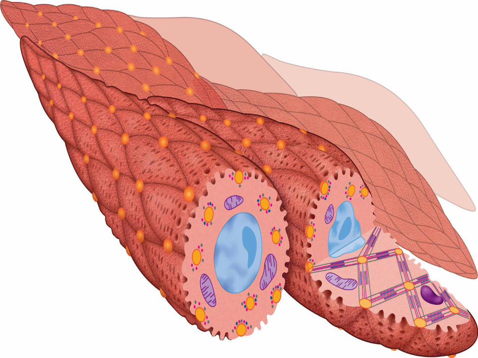
Cardiac Muscle Tissue Features:
Striated (same contractile machinery)
Self-excitatory and electrically coupled
Rate of contractions modulated by autonomic nervous system
innervation is neuroendocrine in nature (i.e. no “motor end plates”)
Cell Features:
1 or 2 centrally placed nuclei
Branched fibers with intercalated discs
Numerous mitochondria (up to 40% of cell volume)
Sarcoplasmic reticulum & T-tubules appear as diads at Z lines
Sarcoplasmic reticulum does not form terminal cisternae
T tubules are about 2x larger in diameter than in skeletal muscle

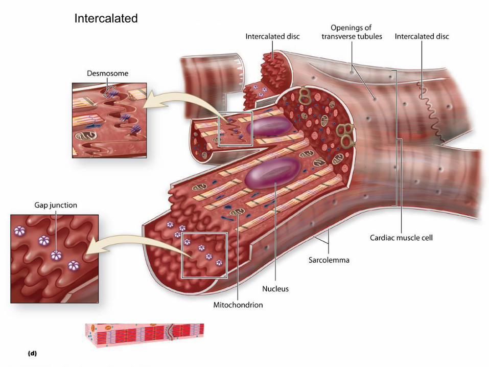
Cardiac muscle
Bundles form thick myocardium
Cardiac muscle cells are single cells (not called fibers)
Cells branch
Cells join at intercalated discs
1-2 nuclei in center
Here “fiber” = long row of joined cardiac muscle cells
Inherent rhythmicity: each cell! (muscle cells beat separately without any stimulation)
Intercalated
disc__________

Intercalated discs - junctions between
cells where force is delivered. It is a fascia
adherens like site (like zonula adherens-
disc).
Macula adherens (desmosomes) -
anchor intermediate filaments in the same
orientation as the fascia adherens
Gap junctions - allow cells to contract
simultaneously. Lined up side by side

Cardiac muscle does not
contain cells equivalent to the
satellite cells of skeletal
muscle. Therefore cardiac
muscle cannot regenerate
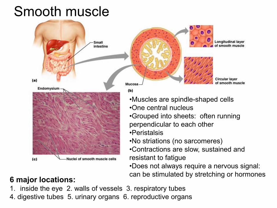
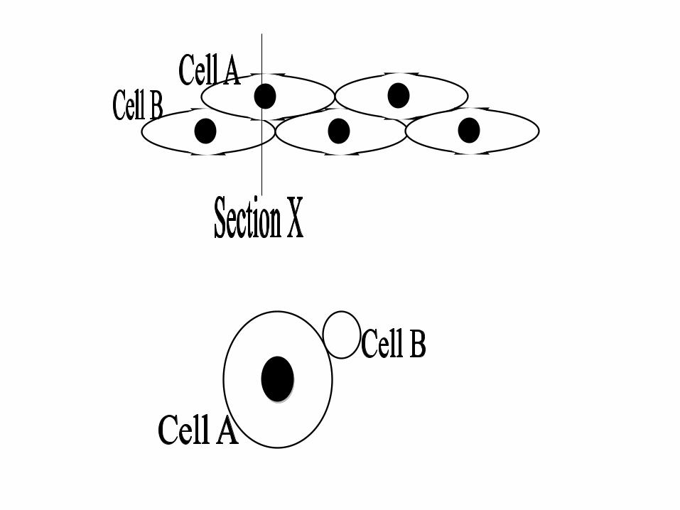
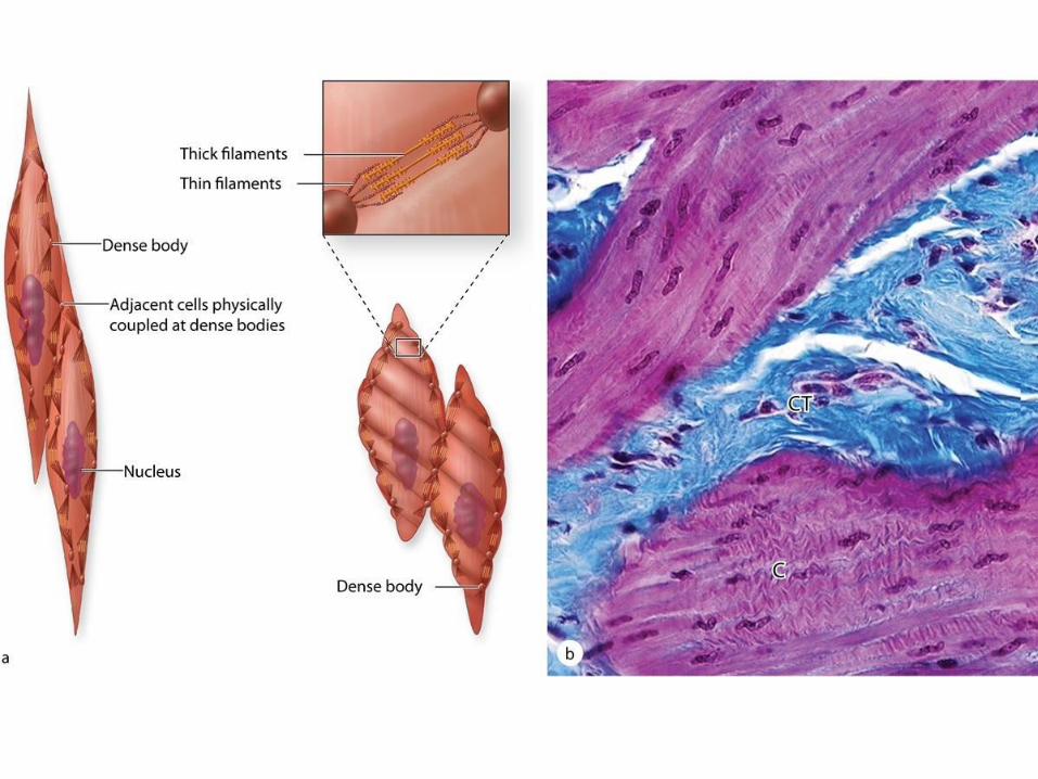
Smooth muscle
•Muscles are spindle-shaped cells
•One central nucleus
•Grouped into sheets: often running
perpendicular to each other
•Peristalsis
•No striations (no sarcomeres)
•Contractions are slow, sustained and
resistant to fatigue
•Does not always require a nervous signal:
can be stimulated by stretching or hormones 6 major locations: 1. inside the eye 2. walls of vessels 3. respiratory tubes
4. digestive tubes 5. urinary organs 6. reproductive organs

Smooth Muscle
• Fusiform, non-striated cells
• Single, centrally-placed nucleus
• Contraction is non-voluntary
• Contraction is modulated in a neuroendocrine manner
• Found in blood vessels, GI and urogenital organ walls, dermis of skin

• actin and myosin filaments
• intermediate filaments of desmin (also vimentin in vascular smooth muscle)
• membrane associated and cytoplasmic dense bodies containing actinin (similar to Z lines)
• relatively active nucleus (smooth muscle cells make collagen, elastin, and proteoglycans)
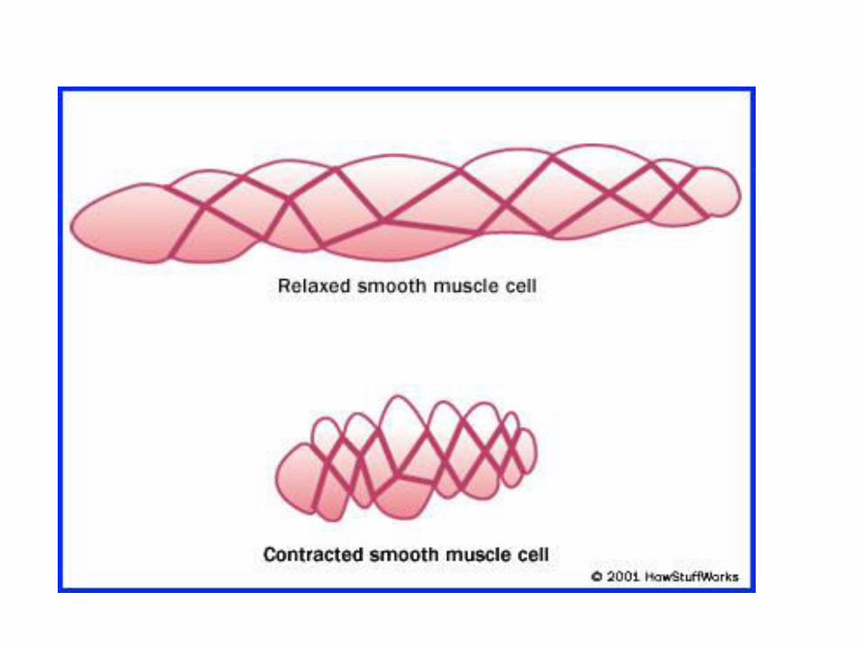
Ultrastructure of Smooth Muscle:

The myofilaments of smooth muscle are
arranged differently and appear less
organized
Thin filaments attach to dense bodies
located on the cytoplasmic surface of
the plasma membrane and deep in the
cytoplasm (intracytoplasmatic dense
bodies)
Dense bodies contain α-actinin for thin
filament attachment
Dense bodies at the membrane are also attachment sites for intermediate
filaments and for adhesive junctions between cells. This arrangement of both
the cytoskeleton and contractile apparatus allows the multicellular tissue to
contract as a unit, providing better efficiency and force
Smooth muscles can
undergo
Hypertrophy and
Hyperplasia

Top Related