


Languages
Pages
Legal


7
Murad Ibrahim & Dina bahader
Zeina Sinnokrot Asmaa Ghassan
Raneen Alshareef
Tayma Ahmad
Shayma Deeb
Bacteria can be categorized into 3 different groups according to:
⮚ Oxygen requirement:
1. Obligate Aerobes: Bacteria which need the presence of oxygen to survive.
Mycobaterium tuberculosis which causes Tuberculosis (TB) is an example.
2. Obligate Anaerobes: Bacteria that can't live with the presence of oxygen or they
require an absence of oxygen. Examples of this type of bacteria are Clostridia
such as Clostridium Tetani and Clostridium Perfringens.
Oxygen is considered toxic to this type of bacteria but the extent of toxicity varies among
different species. Some can survive but are unable to grow, whereas others are killed rapidly.
▪ How do Obligate Anaerobic Bacteria die in the presence of oxygen?
Answer: Normally, when bacteria consume oxygen, they produce toxic
oxygen radicals such as superoxide and oxygen peroxide. Aerobes and
facultative anaerobes have specific enzymes such as superoxide dismutase
and catalase that detoxify these products by breaking them into oxygen and
water. But anaerobes lack those enzymes.
3. Facultative Anaerobes: can survive with the presence and absence of oxygen.
They are able to generate ATP by aerobic respiration if oxygen is available and they are capable
of switching to fermentation if oxygen is not available.
A very common example of Facultative anaerobes is E-coli which live in the colon anaerobically
and in the surrounding environment aerobically.
4. Microaerophilic bacteria: Bacteria that require environments containing lower
levels of oxygen and elevated concentration of carbon dioxide (8 -10%).
Examples of Microaerophillic bacteria are
a. Microaerphillic streptococcus.
b. Helicobacter pylori cause ulcers in lining of the stomach or the upper part of the small
intestine.
c. Campylobacter jejuni causes severe diarrhea.
⮚ Temperature Requirement:
1. Mesophilic bacteria: Bacteria that grow in moderate temperature typically between
20-40 degree centigrade. Mesophiles grow best at human body temperature which is
usually 37 degree, that’s why all human pathogens are mesophiles.

2. Psychrophiles: Bacteria that live in temperatures lower than 20 degrees. They are
present in high altitudes and in deep ocean waters.
3. Thermophiles: Bacteria that require high temperature. (Temperature higher than 60
degrees).
4. Hyperthermophiles: Bacteria that require temperature between (80 - 105) degrees.
They live in volcanic ash and extremely hot springs. This type of bacteria has enzymes
that have developed advanaced structural properties of high thermostability due to
disulfide bonds in their structures.
NOTE: Since Thermus aquaticus bacteria is a hyperthermophile and can tolerate high
temperatures, It can the source of the heat resistant enzyme Taq DNA polymerase (DNA
polymerase that remains stable even at very high temperatures.). This DNA polymerase is used
in a reaction called Polymerase Chain Reaction (PCR) (a technique used to amplify DNA.)
Taq (T stands for thermus and aq stands for aquaticus).
5. Barophiles: Bacteria that can stand or survive under very high pressure.
6. Halophiles: Bacteria that thrive in high salt concentrations. They are found in very
salty lakes such as the Dead Sea where the percentage of salt is (11%-12%).
Question: How do these bacteria survive in these extremely salty conditions?
Answer: Through a process called “Salting Out” where they use up energy to
exclude salt from their cytoplasm to avoid protein aggregation.
7. Osmophiles: Bacteria that can adapt to environments with high osmotic pressure. (E.g.
Staphylococcus aureus which can tolerate 7.5% NaCL and streptococcus feacalis which
can tolerate 6.5 % NaCL.)
Question: What's the difference between Halophillic and Osmophillic bacteria?
Answer: Halophilic bacteria: require high salt concentration while Osmophilic
bacteria can tolerate high salt concentration.
⮚ Dynamics of bacterial growth:
NOTES:
1. Bacteria reproduce by binary fission. However, Bacteria should duplicate its cellular
division before dividing.
2. Bacteria are used widely in our industries, such as: cheese, pickles, antibiotics,
interferon (a protein that is produced by animal cells which have the property of
inhibiting virus replication), and human insulin.
3. Chemostat (a special apparatus causing unrestricted bacterial growth) it is a bioreactor
into which fresh nutrients are continuously added, while toxins and metabolic end
products (waste products) are removed continuously. This process causes the Bacteria
to remain in the log phase and never enter the stationary phase.
4. Isolated colonies = pure culture
⮚ Generation time:
Generation time is the time taken by bacteria to double in number during a specified time
period. Generation time varies with different organisms and it depends on many factors
including nutritional and environmental conditions.
NOTE: Some types of bacteria are obligate intracellular pathogens, here are some
examples:
1. Mycobaterium leprae: An intracellular bacteria (doesn't grow on bacteriological
media). It causes leprosy
2. Treponema pallidum: A host requiring bacteria that causes syphilis.
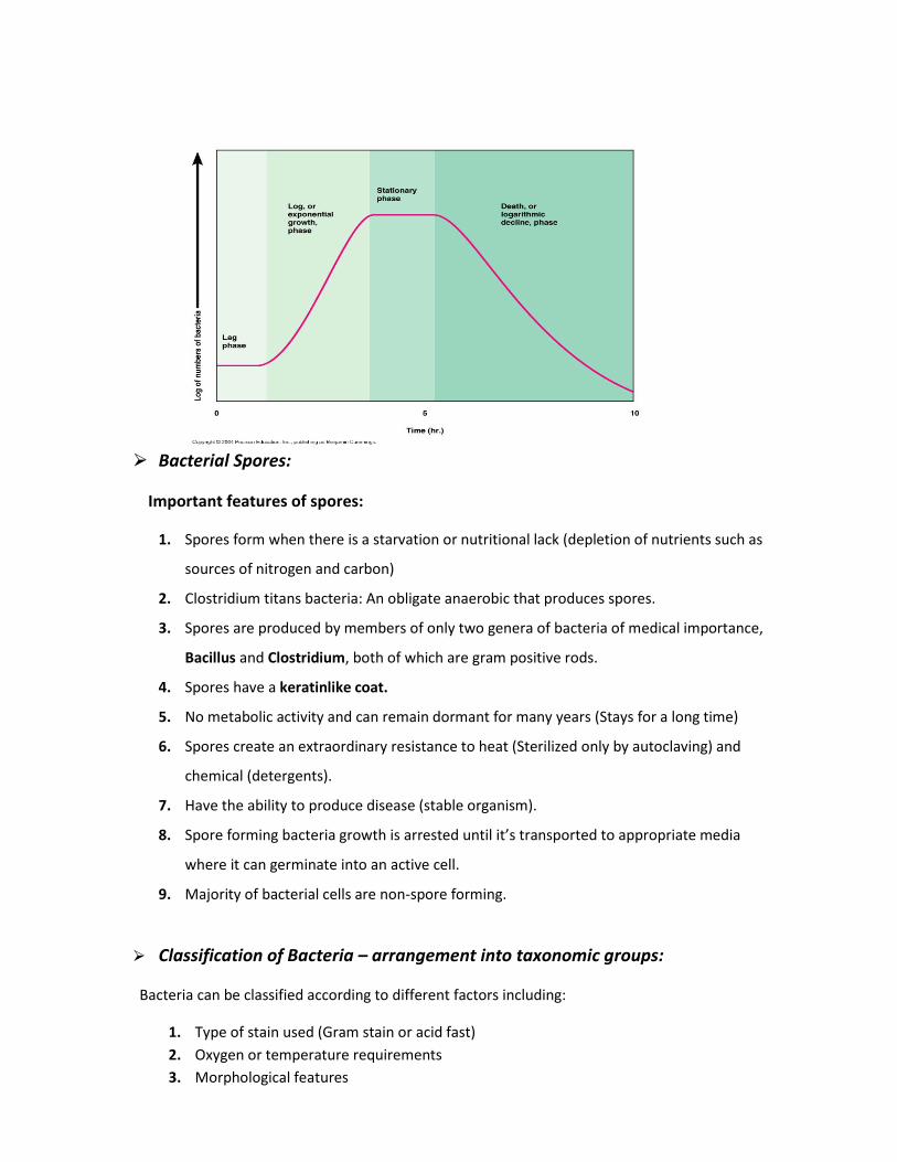
⮚ Bacterial growth curve:
1. Lag phase: In this phase, bacteria duplicate their cell mass. High metabolic activity is a
character of this phase. (CELLS DO NOT DIVIDE)
2. Log phase (Exponential phase): Phase where rapid cell division occurs. Penicillin acts on
this phase because drugs are usually effective when cells are making peptidoglycan (i.e.,
when they are dividing). And so, Penicillin acts on actively growing bacteria (active cells)
to inhibit cell wall synthesis.
3. Stationary phase: Number of cells produced = Number of cells that die.
4. Death phase: Logarithmic decline because of the reduction of nutrients and
accumulation of toxins, Spore forming bacteria will form spores during this phase.
⮚ Bacterial Spores:
Important features of spores:
1. Spores form when there is a starvation or nutritional lack (depletion of nutrients such as
sources of nitrogen and carbon)
2. Clostridium titans bacteria: An obligate anaerobic that produces spores.
3. Spores are produced by members of only two genera of bacteria of medical importance,
Bacillus and Clostridium, both of which are gram positive rods.
4. Spores have a keratinlike coat.
5. No metabolic activity and can remain dormant for many years (Stays for a long time)
6. Spores create an extraordinary resistance to heat (Sterilized only by autoclaving) and
chemical (detergents).
7. Have the ability to produce disease (stable organism).
8. Spore forming bacteria growth is arrested until it’s transported to appropriate media
where it can germinate into an active cell.
9. Majority of bacterial cells are non-spore forming.
⮚ Classification of Bacteria – arrangement into taxonomic groups:
Bacteria can be classified according to different factors including:
1. Type of stain used (Gram stain or acid fast)
2. Oxygen or temperature requirements
3. Morphological features
4. Biochemical characteristics (reactions)
These methods of classification are important to identify causative pathogens (for diagnosis). An
example of bacterium which is classified according the type of stain used is Mycobacterium
pneumonia.
Mycoplasma pneumonia: These bacteria are characterized by the following:
1. They lack a cell wall and that’s why it cannot be seen in the gram stain, instead we use
the acid fast stain.
2. We can’t use antibiotics that inhibit cell wall synthesis such as penicillin, cephalosporin
and Vancomycin, because they don’t have a cell wall.
3. They have sterols in their cell membranes (Eukaryotic cells also have sterols in their
membranes).
4. They are difficult to grow in cultures.
NOTE: Under the light microscope:
● We can see gram positive and gram negative bacteria
● We cannot see other types such as:
1. Mycoplasma pneumonia.
2. Treponema Pallidum: a spirochete bacterium that causes syphilis.
3. Mycobacteria, including M. tuberculosis
Laboratory diagnosis of infectious diseases:
1. Microscopic Examination: For examination we use either gram stain or acid fast
stain.
2. Macroscopic or Culture based tests: Using pure cultures for diagnosing bacterial or
fungal infections. (We use In Vitro culture which is the technique or process of
maintaining or cultivating cells or tissues derived from a living organism in
a culture medium).
3. Immunologic tests/ Serologic test: Using known antibody to identify the
microorganisms or using known antigens to detect antibodies in the patient’s
serum. (We use it to diagnose congenital or acquired immune diseases)
4. Molecular (nucleic acid and protein based) tests: An example is PCR=polymerase
cell reaction.
⮚ Importance of Laboratory Diagnosis:
Laboratory diagnosis is plays a fundamental role in:
1. Providing definitive, significant and relevant information to the case under
consideration.
2. Guiding clinical decisions and treatment options.
3. Assessment of clinical feature (signs and symptoms), How?
Answer: When we determine the signs and symptoms, we can determine the location of
infection. And then we can identify the suspected bacteria that caused this infection by taking a
sample of the infected area for testing.
The theory of pathogeneses is that single organism cause single disease
Sample:
● Direct: taken by the nurse or doctor
Use needle or surgical biopsy
More accurate and useful
From the place of infection
Aspect techniques, no contamination, clean
Ex. CSF, blood …
● In direct: taken by the patient
From the products
More convenient
Ex. Urine
But sometimes in direct sample become direct sample such as urine
If we cannot take urine as in direct sample because of an obstruction by pregnancy in the
last months, or a tumor, or stones. We should take it by using a needle.
Sometimes we stain the sample with gram stain and it give us a false result the causes are (the
personal false such as use very high cons. Of the dye or don’t do the right steps or in the
washing step, color)
(The cell may have no complete cell wall, we may use antibiotics because it degenerate the cell
wall, the cells may be death)
● Culture media:
Definition: It is a special medium used in microbiological laboratories to grow different kinds of
microorganisms. We use these cultures to identify the phenotype and gross appearance of
different bacteria ( For example: Identification of bacteria according to change in color).
○ Types of culture according to physical state:
1. Semi solid medium(petri dish)
2. Liquid Nutrient medium (nutrient broth)
3. Solid medium
NOTE: Cultures can't be used to grow obligate intracellular parasites such as Chlamydia and
Rickettsia.
General media: nutrient agar and trypticase soy agar
Appearance in different colonies without differentiation
We say that it is positive or negative growth without know the type
Selective media: distinguish, special components that inhibited one type and promote the
other
macConkey agar (inhibited gram+ and promote gram-)
mannitol salt (specific for staphylococcus gram+ )
Deferential media: distinguish in the same group
Enriched media: addition of fresh RBC to the trypticase soy agar to make blood agar
Addition of heated RBC to the trypticase soy agar to make chocolate agar
In macConkey agar sometimes it give 2 colors one is the pink for (lactose fermenter such as the
e.coli) and the other for the (nonlactosepinkenter such as salmonella or shigella) if we want to
know exactly whether it is a shigella or salmonella we transfer them to new media (subculture)
Types of hemolytic bacteria on blood agar:
The ability of bacterial colonies to induce hemolysis (The breakdown of red blood cells) when
grown on blood agar is used to classify certain microorganisms:
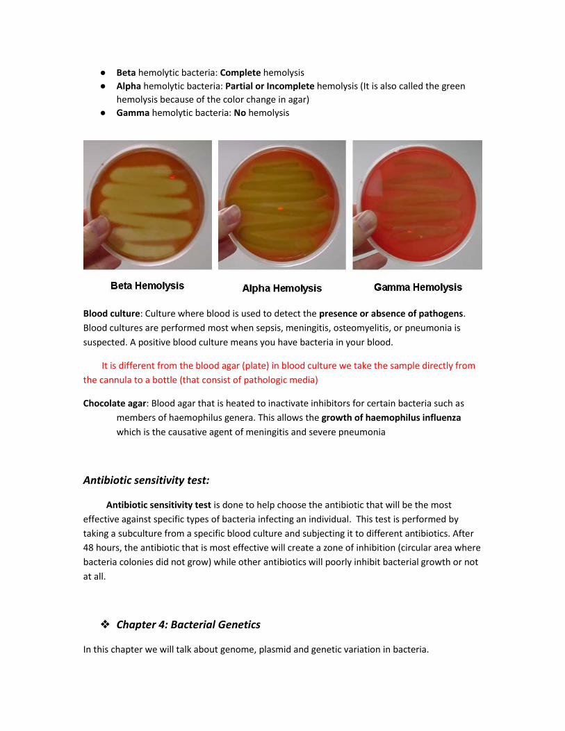
● Beta hemolytic bacteria: Complete hemolysis
● Alpha hemolytic bacteria: Partial or Incomplete hemolysis (It is also called the green
hemolysis because of the color change in agar)
● Gamma hemolytic bacteria: No hemolysis
Blood culture: Culture where blood is used to detect the presence or absence of pathogens.
Blood cultures are performed most when sepsis, meningitis, osteomyelitis, or pneumonia is
suspected. A positive blood culture means you have bacteria in your blood.
It is different from the blood agar (plate) in blood culture we take the sample directly from
the cannula to a bottle (that consist of pathologic media)
Chocolate agar: Blood agar that is heated to inactivate inhibitors for certain bacteria such as
members of haemophilus genera. This allows the growth of haemophilus influenza
which is the causative agent of meningitis and severe pneumonia
Antibiotic sensitivity test:
Antibiotic sensitivity test is done to help choose the antibiotic that will be the most
effective against specific types of bacteria infecting an individual. This test is performed by
taking a subculture from a specific blood culture and subjecting it to different antibiotics. After
48 hours, the antibiotic that is most effective will create a zone of inhibition (circular area where
bacteria colonies did not grow) while other antibiotics will poorly inhibit bacterial growth or not
at all.
❖ Chapter 4: Bacterial Genetics
In this chapter we will talk about genome, plasmid and genetic variation in bacteria.

Plasmids replicate in step with cell division also it can replicate independently of the
chromosome.
There's different sizes and types of plasmid within the cell. The large size plasmids which usually
present in fewer copies, have more genetic material. And it have the genes that code for the sex
pills (Transmissibility genes) which also code for the enzymes which are required for the
conjugation and it's essential for Fertility and establishment of the bacterial donor state.
F+ donor cell _ male donor_ Fertility plasmid.
F- recipient cell_female
At the end of conjugation, the recipient cell is transformed to a donor ( male).
Some plasmids carry genes for toxins and others carry genes for antibiotics resistance
Designed plasmids are very much used in E.coli as Cloning vectors
Cloning Vector: A DNA molecule (Plasmid) that has the capability to replicate an integrated
foreign DNA to give birth to numerous clones of the recombinant DNA.
How is cloning a foreign gene done?
A foreign gene could be human growth hormone, insulin gene which codes for human insulin or
human interferon, etc.
● In one tube a solution of human DNA (could be taken from blood cells) is prepared and
in another tube plasmids that will be used as cloning vectors are prepared.
● Then both tubes are treated with the same specific restricting enzyme which recognizes
a specific region or sequence on the human DNA and on plasmids. For example EcoR 1
enzyme recognizes GAATTC sequence and makes a neck cut between (G and A) resulting
in sticky ends.
● The two tubes then are put together in the presence of DNA ligase which leads to the
ligation of the ends of DNA with the ends of plasmids resulting in a recombinant
plasmid (genes from two different sources).
Then this recombinant plasmid (cloning vector) is used to introduce the foreign DNA into a
bacterial cell by Transformation is a term used when a foreign DNA is introduced into a
bacterial cell and Transfection is a term used for introducing foreign DNA into
eukaryotic cell (yeasts, fungi…).
● The last step is to screen if the cell has transformed or not. In this case the screening is
done to check if cloning vector carries gene for ampicillin resistance, the original cell is
not resistant for penicillin but after introducing the foreign DNA that carries the
ampicillin resistance gene it becomes resistance to penicillin. Screening is done by
plating on a medium which contains ampicillin, the colonies that will grow are only the
one containing ampicillin resistance gene.
● If the gene is expressed, it can be then purified from the medium.

*Before using human insulin, cow insulin was used and that caused the production of antibodies
in humans leading to several problems. Human interferon is the only one functional in human
bodies although interferon could be extracted from animals such as mice but it is non functional
when introduced to human cells.
Interferon interferes with viral proliferation and activates our immune system so it’s used in MS
(multiple sclerosis) and treatment of some cancers by using bacterial cells which divide very
quickly.
Bacterial chromosome is a haploid chromosome so if a slight change occurred in protein for
example it might not do a mutation or it might produce a non functional protein and if that
protein is so important then it may cause cell death.
Mutation is a permanent change in DNA sequence generally occurs due to radiation, chemical
exposure, viral infection or it could be spontaneous. It could require only one base pair
substitution.
A silent mutation is a change in the sequence of nucleotide bases which constitutes DNA,
without a subsequent change in the amino acid or the function of the overall protein.
A missense mutation is a point mutation in which a single nucleotide change results in a codon
that codes for a different amino acid. (Could affect or not affect the overall protein)
A nonsense mutation is a mutation in a sequence of DNA that results in a premature stop codon
which will stop the translation of messenger RNA that will produce a nonfunctional protein.
Frame shift mutation is due to insertion or deletion of more than one base pair, this type of mutation
results in complete change in the sequence of the reading frame in DNA.
A conditional lethal mutation is a mutation that a cell can survive with under some conditions but
could turn lethal under other conditions. E.g. Temperature-sensitive mutation like influenza virus
vaccine which is a living temperature-sensitive virus used as a nasal spray that can replicate at
32 °C in the nose by producing a specific protein but it can’t replicate at 37 °C in the body.
Mutation by transposons (jumping genes that could move from plasmid to chromosome or from
plasmid to plasmid that could cause frame change in DNA).
Gene transfer from a cell to another:
● Transformation
● Conjugation
● Transduction
Transformation is exogenous: uptake of a foreign DNA into a recipient cell and this foreign DNA has
to recombine with the cell chromosome which leads to its transformation
Only competent cells can do transformation (competence is the last stage of logarithmic phase
and)because at that stage they have receptors that can bind the foreign DNA and then it has to be
internalize, (only one strand of DNA will be internalized even if it was two stranded DNA), then it has to
be taken up by bacterial chromosome and recombined with it then expressed then the cell will be
transformed.
● Back in 1928 Frederick Griffith worked with streptococcus pneumoniae (polysaccharide
capsule containing is more virulent and without a capsule is avirulent-not virulent)
The capsule is antiphagocytic (preventing the action of phagocytes)
Usually capsule containing bacteria grown on a plate as nice and smooth colonies (mucoid
capsule) but non capsule containing grows as rough colonies.
● Frederick used a mouse for the experiment usually by Intraperitoneal injection of a
virulent, smooth capsule containing strep pneumoniae (strep pneumoniae is
pathogenic and lethal to the mouse)
● When he injected the mouse with a non virulent rough strep pneumonia the mouse
wasn’t affected.
● Heat-killed Streptococcus pneumoniae also did not affect the mouse, but injecting the
mouse with Heat-killed Streptococcus pneumonia and living rough non virulent strep
pneumonia (which are individually not affective) it lead to the death of the mouse.
● Somehow the living rough strep pneumonia turns to a virulent smooth capsule
containing bacteria from the Heat-killed Streptococcus.
● After 12 year it was discovered that the change was initially in DNA itself by exposing
the bacteria to amylase then to a protease then to RNAse which had no different
results.
● Another experiment done in 1952 using T2 bacterialphage (bacterial virus can affect
E.coli) containing protein tale fibers and protein coat and the double stranded genome
DNA, then grew it in a medium which is either radio labeled with amino acids so the
radioactivity occurs in the protein coat or grew it in a medium with radioactive
phosphorus PO4 which will be inserted in DNA, then the bacteriophage to infect E.coli
by adhering then injecting DNA only in bacteria, then they did centrifugation and
measured the reactivity in the supernatant or in precipitate (containing bacteria), they
found out that the DNA was found in the bacteria and the protein coat was found in the
supernatant.
Transduction: bacteriophage is needed to bring a double stranded DNA from a donor cell to
a recipient cell. E.g. the gene for B-lactamase (enzyme breaks down the antibiotics) can be
transferred between staph.aureus cells by transduction.
Bacteriophage attaches to the bacterial cell wall and then inject its DNA which will control
the bacterial cell, then it uses bacterial ribosomes to produce viral DNA and proteins for the
head and tale, meanwhile the bacterial chromosome is broken. then the DNA inter the
phage heads to produce bacteriophages.
During the process a bacteriophage containing parts of bacterial DNA called the transducing
phage could be produced which is able later to transfer these genes to another bacterial
cell.

Top Related