


Languages
Pages
Legal


S1
Electronic Supplementary Information (ESI) for
Multiplexed Femtomolar Quantitation of Human Cytokines in a
Fluoropolymer Microcapillary Film
Ana P. Castanheira,a Ana I. Barbosa,b Alexander D. Edwardsac and Nuno M. Reis*ab
a Capillary Film Technology Ltd, West Sussex RH14 9SJ, United Kingdom b Department of Chemical Engineering, Loughborough University, Leicestershire LE11 3TU, United Kingdom. E-mail: [email protected]; Tel: +44 (0)1509 222505; Fax: +44 (0)1509 223923 c Reading School of Pharmacy, University of Reading, Reading RG6 6AD, United Kingdom. E-mail: [email protected]; Fax: +44 (0)118 931 4404; Tel: +44 (0)118 378 4253
Electronic Supplementary Material (ESI) for Analyst.This journal is © The Royal Society of Chemistry 2015

S2
TABLE OF CONTENTS
SUPPLEMENTARY EXPERIMENTAL DESIGN
Multi-syringe aspirator (MSA) device
Fig. S1 Overview of Multi-Syringe Aspirator (MSA) device and miniaturised
fluoropolymer MCF platform.
Singleplex IL-1 Immunoassay Optimisation
Table S1 Optimised assay conditions for human IL-1 singleplex measurement
Fig. S2 Cytokine test strips imaged after OPD conversion, showing the relevance
of washing before addition of antigen. The strips were incubated with decreasing
concentration of antigens from the left to right hand side of the picture.
Selection of enzymatic amplification system
Fig. S3 Comparison of different Peroxide enzymes tested for colorimetric
amplification:
Assay Variability
Qualitative Duplex Assay
Quantitative Triplex Assay.
SUPPLEMENTARY RESULTS
Assay Optimisation
Fig. S4 Optimisation of IL-1 cytokine immunoassay measurement in the
fluoropolymer MCF.
Limit of Quantitation of the Colorimetric Detection Device
Fig. S5 Response curves for DAP detection in the MCF with flatbed scanner and
96 well MTP with microplate reader (450 nm).
Qualitative Duplex Detection
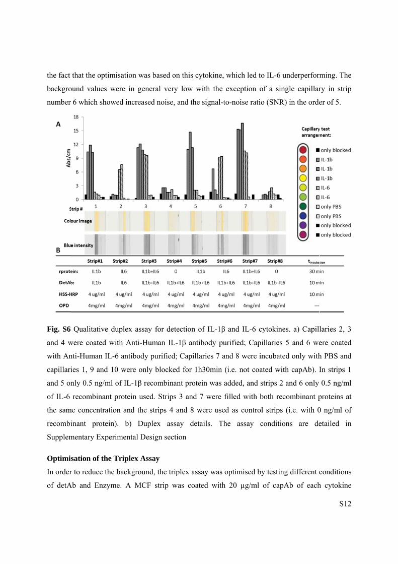
Fig. S6 Qualitative duplex assay for detection of IL-1β and IL-6 cytokines.
Optimisation of the Triplex Assay.
Fig. S7 Comparison of background in Triplex Cytokine Assay.
SUPPLEMENTARY REFERENCES

SUPPL
Multi-s
This wa
an array
fit seals
Fig. S1
MCF p
disposab
MicroC
Singlep
Several
detectio
flatbed s
volume
well, all
that cou
tested a
respect t
LEMENTAR
yringe aspi
as first prese
y of 8, 1 ml
and a custo
Overview
latform. a)
ble 1ml syr
apillary Film
plex IL-1 I
parameters
on and quan
scanner. Be
(SAV) rati
l immunoas
uld be detec
and shortlis
to maximum
RY EXPER
irator (MSA
ented by Ba
plastic syri
omised samp
of Multi-S
MSA devi
ringes and p
m (MCF). c
Immunoass
s were teste
ntification i
ecause of the
o and light
ssay conditi
cted using a
sted from a
m signal-to-
RIMENTA
A) device.
arbosa et al
inges that is
ple well, as
Syringe Asp
ice capable
pre-coated
c) Push-fit se
say Optimis
ed in order
in the MCF
e significant
path distan
ions had to
flatbed sca
an extensive
-noise ratio o
L DESIGN
l,.1 and cons
s interfaced
shown in F
pirator (MS
of doing 8
30 mm flu
eals holding
sation
to optimise
F based on
t difference
nce in small
be optimise
anner. Table
e initial sc
or signal-to
N
sists of a se
d with the pr
ig. S1.
A) device
80 simultan
uoropolymer
g 8 cytokine
e and impr
n colorimetr
s in the volu
capillaries
ed in order
e S1 summa
reening. Al
-background
emi-disposab
re-coated M
and miniatu
neous tests
r MCF strip
e test strips
rove the sen
ric ELISA
ume of reag
compared t
to produce
arises the fu
ll paramete
d ratio.
ble device c
MCF strips u
urised fluor
using an a
ps. b) Fluor
nsitivity of
quantitation
gents, surfac
to 96-micro
a colorimet
ull range for
er were opt
S3
containing
using push
ropolymer
array of 8
ropolymer
cytokines
n using a
ce-area-to-
otiter plate
tric signal
r variables
timised in

Th
length w
was incu
concent
1β capA
200 pg/
were co
was blo
Bovine
respect
concent
It
the goo
optimiza
the bloc
he optimum
with 0, 10, 2
ubated with
tration of 0,
Ab. On both
/ml. To test
oated with 1
ocked using
Serum (FB
to the initia
trations and
Table S1.
is importan
od perform
ation the tw
cking and th
m capAb con
20 and 40 μ
h PBS only.
5, 10 and 2
h experimen
the perform
10 μg/ml of
Bovine Se
BS) and 0.0
al rate of ab
blocking so
Optimised a
nt to mention
mance and
wo approach
he addition o
ncentration w
μg/ml of IL
On a separ
20 μg/ml usi
ntal sets, the
mance of dif
f IL-1β capA
erum Album
2% of Sodi
sorbance ge
olutions Ext
assay condit
n that a was
reduced v
hes were tes
of the antige
was determi
-1β mAb. F
rate experim
ing MCF str
e concentrat
fferent bloc
Ab. Followi
min 1% (BS
ium Azide
eneration. D
trAvidin wa
tions for hu
shing step b
variability
sted and it w
en was very
ined by coa
For the cont
mental set, d
rips coated w
tion of IL-1
cking solutio
ing 2 hours
SA) and the
(NaN3). Th
During the o
s used as en
uman IL-1
before the an
of the ass
was verified
important (
ating in dupl
trol (0 μg/m
detAb was te
with 10 μg/m
1β recombin
ons, two 12
s of incubat
second on
he results w
optimisation
nzyme.
singleplex m
ntigen incub
say. During
d that the w
(Figure S2).
licate 8 strip
ml of capAb
ested in dup
ml or 20 μg
nant protein
2 cm long M
tion, one of
e with 2.5%
were then an
n of capAb a
measuremen
bation was c
g prelimina
washing step
S4
ps of 3 cm
b) the strip
plicate at a
g/ml of IL-
n used was
MCF strips
f the strips
% of Fetal
nalysed in
and detAb
nt
crucial for
ary assay
p between
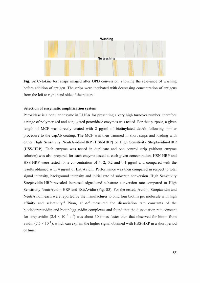
Fig. S2
before a
from the
Selectio
Peroxid
a range
length o
procedu
either H
(HSS-H
solution
HSS-HR
results o
signal in
Streptav
Sensitiv
NeutrAv
affinity
biotin/st
for strep
avidin (
of time.
Cytokine t
addition of
e left to righ
on of enzym
dase is a pop
of polymer
of MCF w
ure to the ca
High Sensiti
HRP). Each
n) was also
RP were tes
obtained wi
ntensity, ba
vidin-HRP r
vity NeutrAv
vidin each w
and selec
treptavidin
ptavidin (2.
7.5 × 10⁻⁸),
test strips im
antigen. Th
ht hand side
matic ampli
pular enzym
ized and co
was directly
apAb coatin
ivity NeutrA
enzyme w
prepared fo
sted for a c
th 4 μg/ml
ackground i
revealed in
vidin-HRP
were reporte
ctivity.2 Pir
and biotin/e
.4 × 10−6 s−
which can
maged after
he strips wer
of the pictu
fication sys
me in ELISA
njugated pe
coated wi
ng. The MC
Avidin–HR
was tested
or each enzy
concentratio
of ExtrAvid
ntensity and
ncreased sig
and ExtrAv
ed by the m
ran, et al2
egg avidin c−1) was abo
explain the
r OPD conv
re incubated
ure.
stem
A for presen
eroxidase en
ith 2 μg/m
CF was then
RP (HSN-HR
in duplicat
yme tested a
on of 4, 2,
din. Perform
d initial rat
gnal and su
vidin (Fig. S
manufacturer2 measured
complexes a
out 30 time
higher sign
version, sho
d with decr
nting a very
nzymes was
ml of biotiny
n trimmed
RP) or Hig
e and one
at each give
0.2 and 0.1
mance was t
te of substra
ubstrate con
S3). For the
r to bind fou
d the disso
and found th
s faster tha
nal obtained
owing the r
reasing conc
high turnov
tested. For
ylated detA
in short str
gh Sensitivi
control str
en concentr
1 μg/ml and
then compar
ate convers
nversion rat
tested, Avi
ur biotins pe
ociation rat
hat the disso
an that obse
with HSS-H
relevance o
centration o
ver number,
that purpos
Ab followin
rips and loa
ity Streptav
rip (withou
ation. HSN
d compared
red in respe
ion. High S
te compared
idin, Strepta
er molecule
te constant
ociation rat
erved for bi
HRP in a sh
S5
f washing
of antigens
, therefore
se, a given
ng similar
ading with
vidin–HRP
ut enzyme
-HRP and
d with the
ect to total
Sensitivity
d to High
avidin and
with high
ts of the
e constant
iotin from
hort period

Fig. S3
Sensitiv
ExtrAvi
Assay V
The assa
the lev
Absorba
samples
the met
%Accur
For each
values p
calculat
first valu
Th
of recom
was obt
the Intra
Quantifi
Compariso
vity NeutrAv
idin-HRP (E
Variability
ay sensitivit
vel above w
ance (Abs)
s. The Limit
thod descri
racy betwee
h experimen
plot was dra
ted. The LoQ
ue in which
he linear ran
mbinant pro
tained. The p
a-assay pre
fication, LLo
on of differe
vidin-HRP
EA-HRP). T
ty was asses
which sam
of the nega
t of Quantit
ibed by Ed
en the exper
nt, the expe
awn to find
Q was defin
h the Accura
nge was def
tein (IL-1β)
precision is
cision three
oQ, Middle
ent Peroxid
(HSN-HRP
The enzyme
ssed as LoD
mples were
ative contro
tation (LoQ)
derveen,3 w
rimental and
ected values
the linear r
ned as the co
acy was betw
fined as the
) for which
s herein refe
e samples w
e range, MR
de enzymes
P), High Sen
s were comp
D or “Cut-of
considered
ol plus three
) was also d
which uses
d expected
s were calcu
range. From
oncentration
ween 80-12
interval bet
an appropri
erred to as In
with differen
R; and near
tested for c
nsitivity Str
pared at sam
ff value” of
d positive,
e times the
determined
the best c
values calc
ulated and a
m this, the a
n correspon
0% and Pre
tween the lo
ate level of
ntra and Int
nt concentra
the Higher
colorimetric
reptavidin-H
me concentr
f the assay, w
and determ
standard d
for all the c
combination
culated using
an experime
accuracy and
nding to the
ecision <20%
ower and the
f precision, a
ter-assay pre
ations (near
Limit of Q
c amplificat
HRP (HSS-H
ration of 4 μ
which was d
mined as
eviation of
cytokines te
n of %Prec
g a calibrati
ental versus
d precision
Abs that rev
%.
e upper con
accuracy and
ecision. To
r the Lower
Quantificatio
S6
tion: High
HRP) and
μg/ml
defined by
the mean
the blank
sted using
cision and
ion curve.
s expected
were then
vealed the
ncentration
d linearity
determine
r Limit of
on, HLoQ,

S7
all determined from the calibration curve) were tested in six replicates (6 strips with 10
capillaries; 60 capillaries in total). The average Abs values and the standard deviation for each
sample were calculated and the Coefficient of Variation (CV) was also determined for each
concentration within a given assay run. The Inter-Assay Calibration was determined by running
three assays in different days and using different MSA devices, in which samples were analysed
in duplicate, using the same three concentrations of recombinant proteins (corresponding to
LLoQ, MR and HLoQ). The average and the standard deviation for each sample were calculated
and the CV determined for each concentration between the assay runs. Typical CVs for ELISA
in 96-well MTP are in the range of 10–20%. 3,4
Accuracy or Recovery was also determined from the calibration curve by comparing the
expected value with the actual cytokine concentration in the assay. The expected versus the
average of the measured values was determined for each sample, by calculating the %Recovery=
assay value/expected value x 100. The typical range for accepted accuracies is 80-120%.3
Qualitative Duplex Assay
In order to demonstrate the ability of the new miniaturized platform to detect simultaneously
more than one cytokine a simple qualitative duplex assay was developed with of IL-1β and IL-6
reagents. For this purpose, solutions containing IL-1β or IL-6 capAb were injected into each
individual capillary using a small syringe needle. The MCF strip was then incubated for 2 hours
at room temperature, and then further incubated for 1.5 h with the blocking solution and washed
with PBS-T. The strips were then trimmed into 30 mm long individual test strips and attached
onto the MSA. Equal concentration of recombinant proteins (0.5 ng/ml) and detAb (10 g/ml)
were then used. All subsequent steps followed same sandwich ELISA procedure described in the
main manuscript.
Quantitative Triplex Assay
A quantitative triplex assay consisting of full response curve for each cytokine was performed
for simultaneous quantitation of IL-1β, IL-12 and TNFα. Individual capillaries on a 25 cm long
fluoropolymer MFC strip (containing 10 capillaries) were injected and incubated into one pair of
capillaries each with one of the following solutions: PBS (overall negative control), 3% BSA
(blocking solution control) or IL-1β, IL-12 capAb or TNFα at 20 µg/ml. All subsequent ELISA

S8
steps were as already described for the singleplex and duplex assays, with the exception that
standard curves were prepared using a 1:3 dilution series of recombinant protein. All
recombinant proteins and detAb solutions for each cytokine were combined at same
concentration, which ultimately represents 3 times higher protein content on each solution well
when compared to singleplex detection. Combining different biotinylated detAb for multiplex
ELISA detection was found to significantly affect the individual cytokine performance by
increasing the background, therefore it was necessary to re-optimise the multiplex assay, in
respect to detAb and enzyme concentration, to maintain similar signal-to-noise ratios to
singleplex assays. This is described in Supplementary Results section.
SUPPLEMENTARY RESULTS
Assay Optimisation
The first two parameters tested were the incubation times of the capAb and recombinant
proteins. Two hours incubation at room temperature was sufficient to fully immobilize the capAb
by passive adsorption, as no differences were detected in signal strength and signal-to-noise ratio
(Fig. S4a). This had the advantage of saving the typical overnight incubation required for MTP
sandwich ELISAs. Equally, the incubation of recombinant protein for sensitive detection could
be reduced to 30 min in the fluoropolymer MCF without compromising sensitivity (Fig. S4b).
This is linked to the very short diffusion distances in the plastic microcapillaries. A parameter
found paramount in controlling the signal-to-noise ratio in the fluoropolymer MCF ELISA was
the blocking solution, for that reason few different formulations were tested. BSA and FBS are
commonly used for blocking non-specific binding sites in plastic surfaces. Although no
significant difference could be detected in respect to Abs signal intensity, the kinetic analysis of
the OPD conversion in the capillaries for different cytokine concentrations revealed poor
performance for both 1% BSA or 2.5% FBS in respect of background development. Fig. S4c
shows the initial rates of assay; in that plot the initial velocity v0 corresponded to the rate of
generation of absorbance in the MCF during the first few minutes of OPD conversion in the full
cytokine sandwich ELISA. BSA and FBS have similar proteins in size and molar rations on their
composition, since BSA is the main compound present in FBS; however on both cases a high
background was detected (Abs0≈0.05). In order to reduce the background which directly controls

S9
to the sensitivity of the assays, a synthetic SuperBlock blocking solution from Thermo Scientific
was tested, which revealed lower backgrounds (Abs0≈0.02) (data not shown).
The use of higher capAb concentrations of 40 μg/ml and above resulted in increased
background and reduced signal (Fig. S4d), suggesting FEP antibody adsorption and/or
orientation was not favored by the presence of a very high capAb concentration. This is
presumably linked to the orientation of the surface adsorbed capAb molecules.5
The effect of detAb concentration was tested for a range between 0 and 20 μg/ml, and it
was also observed a benefit in using 10 μg/ml (Fig. S4e). Again, this is significantly higher than
the concentrations normally used for sensitive sandwich ELISA in MTPs and the increase in the
signal can be explained based on the same binding equilibrium principle. A large solution excess
of detAb favors the formation of the complex capAb-Ag-detAb at the surface of the plastic
capillaries, which is linked to the larger SAV ratio in small bore microcapillaries.

S10
Fig. S4 Optimisation of IL-1 cytokine immunoassay measurement in the fluoropolymer MCF.
a) and b) show effect of of incubation time of capAb and recombinant protein, respectively. c)
Initial rates of colourimetric signal generation in the MCF strips for different blocking solutions
(BSA 1% and FBS 2.5%). d) Effect of capAb concentration (20 μg/ml). e) Effect of detAb
concentration for two different capAb concentration coatings. Assays conditions are detailed in
0.000
0.005
0.010
0.015
0.020
0.025
0.030
0.0 0.5 1.0
v0 (
ng
/ml.
s)
IL-1beta concentration (ng/ml)
BSA 1% TMB
2.5% FBS & 0.02%NaN3 TMB
C
0
0.1
0.2
0.3
0.4
0.5
0.6
0.0 2.5 5.0
Ab
s
IL-1beta concentration (ng/ml)
22.05.2013
23.05.2013
Overnight (fridge)
2 hours (RT)
A B
0.000
0.015
0.030
0.045
0.060
0 5 10 15 20 25
Ab
s
Detection Antibody concentration (ug/ml)
CapAb=10 ug/ml
CapAb=20 ug/ml
E
0
0.1
0.2
0.3
0.4
0.5
0.6
0.0 2.5 5.0
Ab
s
IL-1beta concentration (ng/ml)
1 hour
30 min
0.00
0.05
0.10
0.15
0.20
0.25
0 10 20 40 80 100
Ab
s
Capture Antibody concentration (μg/ml)
Exp Controls
D

S11
Experimental Design section in the manuscript. The optimised concentrations for capAb and
detAb were considered 20 μg/ml and 10 μg/ml, respectively
Limit of Quantitation of the Colorimetric Detection Device
A series of dilutions of 2,3-diaminophenazine (DAP), the final product of the conversion of the
substrate OPD by the immunoassay enzyme HRP, were scanned in MCF with a flatbed scanner
starting at a concentration of 2 mg/ml, and in parallel peak absorbance (450 nm) of the same
dilutions was measured in a 96-well MTP using a microplate reader (Fig. S5). The DAP
absorbance in the blue channel of the scanned image was calculated by image analysis.
Fig. S5 Response curves for DAP detection in the MCF with flatbed scanner and 96 well MTP
with microplate reader (450 nm). Only concentrations corresponding to the range of DAP
concentration versus normalised Abs are presented for the MTP and fluoropolymer MCF
Qualitative Duplex Detection
To demonstrate the capability of simultaneous detection of two or more cytokines on each
fluoropolymer MCF strip, a duplex qualitative assay using IL-1β and IL-6 was performed and
analysed. All capillaries showed a positive color signal according to the capAb coating pattern
(Fig. S6), which confirms the possibility of detecting simultaneously more than one cytokine
from a single sample. The main assay conditions used on each MCF strip in the 8-channel MSA
device are shown in Fig. S6b. The higher signal observed for IL-1β cytokine was possibly due to
0.00024
0.0625
0.01
0.1
1
10
100
0.0001 0.001 0.01 0.1 1 10
Ab
s/c
m
DPA conc (mg/ml)
DAP MTP
DAP MCF
the fact
backgro
number
Fig. S6
and 4 w
with An
capillari
and 5 on
of IL-6
the sam
recombi
Supplem
Optimis
In order
of detA
that the opt
ound values
6 which sh
Qualitative
were coated
nti-Human I
ies 1, 9 and
nly 0.5 ng/m
recombinan
me concentra
inant prote
mentary Exp
sation of th
r to reduce t
Ab and Enzy
timisation w
s were in ge
owed increa
e duplex ass
with Anti-
IL-6 antibod
d 10 were o
ml of IL-1β
nt protein u
ation and th
ein). b) D
perimental D
he Triplex A
the backgrou
yme. A MC
was based on
eneral very
ased noise, a
say for dete
Human IL-
dy purified;
only blocked
recombinan
used. Strips
he strips 4 a
Duplex assa
Design secti
Assay
und, the trip
CF strip w
n this cytoki
y low with t
and the sign
ection of IL
-1β antibody
Capillaries
d for 1h30m
nt protein w
3 and 7 we
and 8 were
ay details.
ion
plex assay w
as coated w
ine, which l
the exceptio
nal-to-noise
L-1β and IL
y purified;
s 7 and 8 we
min (i.e. not
was added, a
ere filled w
used as con
The assa
was optimise
with 20 µg
led to IL-6 u
on of a sin
ratio (SNR
L-6 cytokine
Capillaries
ere incubate
t coated wit
and strips 2
ith both rec
ntrol strips
ay conditio
ed by testing
/ml of capA
underperform
ngle capillar
R) in the orde
es. a) Capill
5 and 6 we
ed only with
th capAb). I
and 6 only
combinant p
(i.e. with 0
ons are de
g different c
Ab of each
S12
ming. The
ry in strip
er of 5.
laries 2, 3
ere coated
h PBS and
In strips 1
0.5 ng/ml
proteins at
0 ng/ml of
etailed in
conditions
h cytokine

followin
Design
tested fo
Enzyme
cytokine
concent
detAb a
Fig. S7
assay co
HSS-HR
SUPPL
1 A2
2 U
3 J
4 J
ng the proce
section. Du
for the diffe
e, using 333
es tested).
trations givi
and 4 µg/ml
Comparison
onditions. a)
RP
LEMENTAR
A. I. Barbosa2928.
U. Piran and
. Ederveen,
. R. Crowth
edure descr
uplicated str
rent combin
3 pg/ml of
Several fu
ing the high
HSS-HRP
n of backgro
) 10 µg/ml d
RY REFER
a, A. P. Cas
d W. Riordan
A Practical
her, The ELI
ibed for qua
rips plus a
nations of 2
f recombina
ull response
her signal-to
(Fig. S7).
ound in Trip
detAb and 4
RENCES
stanheira, A
n, J. Immun
l Approach
ISA guidebo
antitative tr
blank strip
2.5, 5 and 1
ant protein
e curves w
o-noise ratio
plex Cytoki
4 µg/ml HSS
. D. Edward
nol. Methods
to Biologica
ook., 2000, v
riplex assay
(with no a
10 µg/ml of
(which fitte
were built
o. The optim
ine Assay us
S-HRP and
ds and N. M
s, 1990, 133
al Assay Va
vol. 149.
in Supplem
dded recom
f detAb and
ed the linea
using the
mised cond
sing optimis
b) 5 µg/ml d
M. Reis, Lab
3, 141–143.
alidation, Ho
mentary Exp
mbinant prot
d 1, 2 and 4
ar range of
detAb and
itions were
sed a) and in
detAb and 2
Chip, 2014
oofddorp, 2
S13
perimental
tein) were
4 µg/ml of
f all there
d Enzyme
10 µg/ml
nitial b)
2 µg/ml
4, 2918–
010.

S14
5 M. E. Wiseman and C. W. Frank, Langmuir, 2012, 28, 1765–74.
Top Related