


Languages
Pages
Legal

Endocrine-Related Cancer (1999) 6 449-473
b ofontednofhed
s aslopdll orenap’eay
r,ayilyaseisnd
m
Multiple endocrine neoplasia type 1
A A J Pannett and R V ThakkerMolecular Endocrinology Group, Nuffield Department of Medicine, John Radcliffe Hospital, Headington, Oxford OX3 9D4, UK
(Requests for offprints should be addressed to R V Thakker; Email: [email protected])
AbstractCombined clinical and laboratory investigations of multiple endocrine neoplasia type 1 (MEN1) haveresulted in an increased understanding of this disorder which may be inherited as an autosomaldominant condition. Defining the features of each disease manifestation in MEN1 has improved patientmanagement and treatment, and has also facilitated a screening protocol to be instituted. Theapplication of the techniques of molecular biology has enabled the identification of the gene causingMEN1 and the detection of mutations in patients. The function of the protein encoded by the MEN1gene has been shown to be in the regulation of JunD-mediated transcription but much still remains tobe elucidated. However, these recent advances provide for the identification of mutant MEN1 genecarriers who are at a high risk of developing this disorder and thus require regular and biochemical
Endocrine-Related Cancer (1999) 6 449-473
screening to detect the development of endocrine tumours.
IntroductionMultiple endocrine neoplasia (Thakker & Ponder 1988,Thakker 1995, Marx 1998) is characterised by theoccurrence of tumours involving two or more endocrineglands within a single patient. The disorder has previouslybeen referred to as multiple endocrine adenopathy (MEA)or the pluriglandular syndrome. However, glandularhyperplasia and malignancy may also occur in somepatients and the term multiple endocrine neoplasia (MEN)is now preferred. There are two major forms of multipleendocrine neoplasia referred to as type 1 and type 2 andeach form is characterised by the development of tumourswithin specific endocrine glands (Table 1). Thus, thecombined occurrence of tumours of the parathyroidglands, the pancreatic islet cells and the anterior pituitaryis characteristic of multiple endocrine neoplasia type 1(MEN1), which is also referred to as Wermer’s syndrome(Wermer 1954). In addition to these tumours, adrenalcortical, carcinoid, facial angiofibromas, collagenomasand lipomatous tumours have also been described inpatients with MEN1 (Trump et al. 1996, Marx 1998).However, in multiple endocrine neoplasia type 2 (MEN2),which is also called Sipple’s syndrome (Sipple 1961),medullary thyroid carcinoma (MTC) occurs in associationwith phaeochromocytoma, and three clinical variants,referred to as MEN2a, MEN2b and MTC-only, arerecognised (Thakker & Ponder 1988, Thakker 1998). InMEN2a, which is the most common variant, thedevelopment of MTC is associated with phaeochromo-
cytoma and parathyroid tumours. However, in MEN2parathyroid involvement is absent and the occurrenceMTC and phaeochromocytoma is found in associatiwith a marfanoid habitus, mucosal neuromas, medullacorneal fibres and intestinal autonomic gangliodysfunction leading to a megacolon. In the variant MTC-only, medullary thyroid carcinoma appears to be tsole manifestation of the syndrome. Although MEN1 anMEN2 usually occur as distinct and separate syndromeoutlined above, some patients occasionally may devetumours which are associated with both MEN1 anMEN2. For example, patients suffering from islet cetumours of the pancreas and phaeochromocytomasfrom acromegaly and phaeochromocytoma have bedescribed and these patients may represent ‘overlsyndromes. All these forms of MEN may either binherited as autosomal dominant syndromes, or they moccur sporadically i.e. without a family history. Howevethis distinction between sporadic and familial cases msometimes be difficult as in some sporadic the famhistory may be absent because the parent with the disemay have died before developing symptoms. In threview, the main clinical features, molecular genetics, arecent progress in the study of MEN1 will be discussed
Clinical features of MEN1
The incidence of MEN1 has been estimated frorandomly chosen post mortem studies to be 0.25%, and to
Endocrine-Related Cancer (1999) 6 449-473 Online version via http://www.endocrinology.org1351-0088/99/006-449 © 1999 Society for Endocrinology Printed in Great Britain

Pannett and Thakker: MEN1
nsp
be % amongst patients with primary hyperpara-thyroidism(Marx et al. 1982, Thakker 1995). The disorder affects allage groups, with a reported age range of 8-81 years, and
>95% of patients have developed clinical manifestatioof the disorder by the fifth decade (Thakker 1995, Trumet al. 1996, Bassett et al. 1998). The clinical
Table 1 The multiple endocrine neoplasia (MEN) syndromes, their characteristic tumours and associated genetic abnormalities
Type (chromosomal location) Tumours
Gene: most frequently (%) mutated codons
MEN1 Parathyroids MEN1:
(11q13) Pancreatic islets 83/84, 4 bp del (≈6%)
Gastrinoma 119, 3 bp del (≈2%)
Insulinoma 209-211, 4 bp del (≈4%)
Glucagonoma 514-516, del or ins (=7%)
VIPoma
PPoma
Pituitary (anterior)
Prolactinoma
Somatotrophinoma
Corticotrophinoma
Non-functioning
Associated tumours
Adrenal cortical
Carcinoid
Lipoma
Angiofibromas
Collagenomas
MEN2
(10 cen-10q.11.2)
MEN2a MTC ret: 634, missense e.g. Cys→Arg (≈85%)
Phaeochromocytoma
Parathyroid
MTC-only MTC ret: 618, missense (>50%)
MEN2b MTC ret: 918, Met →Thr (>95%)
Phaeochromocytoma
Associated abnormalities
Mucosal neuromas
Marfanoid habitus
Medullated corneal nerve fibres
Megacolon
Autosomal dominant inheritance of the MEN syndromes has been established.Del, deletion, ins, insertion.(Adapted from Thakker (1998), with permission)
450

Endocrine-Related Cancer (1999) 6 449-473
er-lehedrinsalal
esstall
caltha-ore
altinnd ofsful
e-er
ts,ntscndnsionfbelin
ofaysas
ithhyrtalied. aialnts.or
s,er
manifestations of MEN1 are related to the sites of tumoursand to their products of secretion. In addition to the triadof parathyroid, pancreatic and pituitary tumours, whichconstitute the major components of MEN1, adrenalcortical, carcinoid, facial angiofibromas, collagenomasand lipomatous tumours have also been described (Trumpet al. 1996, Marx 1998).
Parathyroid tumours
Primary hyperparathyroidism is the most common featureof MEN1 and occurs in more than 95% of all MEN1patients (Benson et al. 1987, Marx et al. 1986,Thakker1995, Trump et al. 1996). Patients may present withasymptomatic hypercalcaemia, or nephrolithiasis, orosteitis fibrosa cystica or vague symptoms associated withhypercalcaemia, for example polyuria, polydipsia,constipation, malaise or occasionally with peptic ulcers.Biochemical investigations reveal hypercalcaemia usuallyin association with raised circulating parathyroid hormone(PTH) concentrations. No effective medical treatment forprimary hyperparathyroidism is generally available andsurgical removal of the abnormally overactive para-thyroids is the definitive treatment. However, all fourparathyroid glands are usually affected with multipleadenomas or hyperplasia, although this histologicaldistinction may be difficult, and total parathyroidectomyhas been proposed as the definitive treatment for primaryhyperparathyroidism in MEN1, with the resultant life-long hypocalcaemia being treated with oral calcitriol(1,25-dihydroxy vitamin D3) (Rizzoli et al. 1985). It isrecommended that such total parathyroidectomy should bereserved for the symptomatic hypercalcaemic patient withMEN1, and that the asymptomatic hypercalcaemic MEN1patient should not have parathyroid surgery but haveregular assessments for the onset of symptoms andcomplications, when total parathyroidectomy should beundertaken.
Pancreatic tumours
The incidence of pancreatic islet cell tumours in MEN1patients varies from 30 to 80% in different series (Thakker& Ponder 1988, Thakker 1995, Trump et al. 1996). Themajority of these tumours produce excessive amounts ofhormone, for example gastrin, insulin, glucagon orvasoactive intestinal polypeptide (VIP), and are associatedwith distinct clinical syndromes.
GastrinomasThese gastrin-secreting tumours represent over 50% of allpancreatic islet cell tumours in MEN1, and are the majorcause of morbidity and mortality in MEN1 patients. Thisis due to the recurrent severe multiple peptic ulcers whichmay perforate. This association of recurrent peptic
ulceration, marked gastric acid production and non β-isletcell tumours of the pancreas is referred to as the ZollingEllison syndrome (Zollinger & Ellison 1955). Additionaprominent clinical features of this syndrome includdiarrhoea and steatorrhoea. The diagnosis is establisby demonstration of a raised fasting serum gastconcentration in association with an increased bagastric acid secretion (Wolfe & Jensen 1987). Medictreatment of MEN1 patients with the Zollinger-Ellisonsyndrome is directed at reducing basal acid output to lthan 10 mmol/l, and this may be achieved by the pariecell H+-K+-ATPase inhibitor, omeprazole. The ideatreatment for a non-metastatic gastrinoma is surgiexcision of the gastrinoma. However, in patients wiMEN1 the gastrinomas are frequently multiple or extrpancreatic and surgery has not been successful (Delcet al. 1989, Sheppard et al. 1989). The treatment ofdisseminated gastrinomas is difficult and hormontherapy with octreotide, which is a human somatostaanalogue, chemotherapy with streptozotocin a5-fluoroaracil, hepatic artery embolisation, and removalall resectable tumour have all occasionally been succes(Thakker 1995).
InsulinomaThese β-islet cell tumours secreting insulin represent onthird of all pancreatic tumours in MEN1 patients (Thakk1995, Trump et al. 1996). Insulinomas also occur inassociation with gastrinomas in 10% of MEN1 patienand the two tumours may arise at different times. Patiewith an insulinoma present with hypoglycaemisymptoms which develop after a fast or exertion aimprove after glucose intake. Biochemical investigatioreveal raised plasma insulin concentrations in associatwith hypoglycaemia. Circulating concentrations oC-peptide and proinsulin, which are also raised, may useful in establishing the diagnosis, as may an insusuppression test. Medical treatment, which consistsfrequent carbohydrate feeds and diazoxide, is not alwsuccessful and surgery is often required. Most insulinomare multiple and small and preoperative localisation wcomputed tomography scanning, coeliac axis angiograpand pre-perioperative percutaneous transhepatic povenous sampling is difficult and success rates have varSurgical treatment, which ranges from enucleation ofsingle tumour to a distal pancreatectomy or partpancreatectomy, has been curative in some patieChemotherapy, which consists of streptozotocin octreotide, is used for metastatic disease.
GlucagonomaThese α-islet cell, glucagon-secreting pancreatic tumourhave been reported in a few MEN1 patients (Thakk1995, Trump et al. 1996, Bassett et al. 1997, Marx 1998).
451

Pannett and Thakker: MEN1
ncetsofhepy
sus-en
5,
nal
etheffer thee
aligh
geng’sn’s
Inur
es5%r,althets
eeen
The characteristic clinical manifestations of a skin rash(necrolytic migratory erythyema), weight loss, anaemiaand stomatitis may be absent and the presence of thetumour is indicated only by glucose intolerance andhyperglucagonaemia. The tail of the pancreas is the mostfrequent site for glucagonomas and surgical removal ofthese is the treatment of choice. However, treatment maybe difficult as 50% of patients have metastases at the timeof diagnosis. Medical treatment of these with octreotide,or with streptozotocin has been successful in somepatients.
VIPomaPatients with VIPomas, which are vasoactive intestinalpeptide (VIP)-secreting pancreatic tumours, developwatery diarrhoea, hypokalaemia and achlorhydria,referred to as the WDHA syndrome (Marx et al. 1967).This clinical syndrome has also been referred to as theVerner-Morrison syndrome (Verner & Morrison 1958) orthe VIPoma syndrome (Bloom et al. 1973). VIPomas havebeen reported in only a few MEN1 patients and thediagnosis is established by documenting a markedly raisedplasma VIP concentration (Thakker 1995). Surgicalmanagement of VIPomas, which are mostly located in thetail of the pancreas, has been curative. However, inpatients with unresectable tumour, treatment withstreptozotocin, octreotide, corticosteroids, indomethacin,metoclopramide and lithium carbonate has provedbeneficial.
PPomaThese tumours, which secrete pancreatic polypeptide (PP)are found in a large number of patients with MEN1(Friesen et al. 1980, Skogseid et al. 1987, Thakker 1995).No pathological sequelae of excessive PP secretion areapparent and the clinical significance of PP is unknown,although the use of serum PP measurements has beensuggested for the detection of pancreatic tumours inMEN1 patients.
Pituitary tumoursThe incidence of pituitary tumours in MEN1 patientsvaries from 15 to 90% in different series (Thakker 1995,Trump et al. 1996). Approximately 60% of MEN1-associated pituitary tumours secrete prolactin, 25%secrete growth hormone (GH), 3% secreteadenocorticotrophin (ACTH) and the remainder appear tobe non-functioning. The clinical manifestations dependupon the size of the pituitary tumour and its product ofsecretion. Enlarging pituitary tumours may compressadjacent structures such as the optic chiasm or normalpituitary tissue and cause bitemporal hemianopia orhypopituitarism respectively. The tumour size andextension are radiologically assessed by computed
tomography scanning and nuclear magnetic resonaimaging. Treatment of pituitary tumours in MEN1 patienis similar to that in non-MEN1 patients and consists medical therapy or selective hypophysectomy by ttransphenoidal approach if feasible, with radiotherabeing reserved for residual unresectable tumour.
Associated tumoursPatients with MEN1 may have tumours involving glandother than the parathyroids, pancreas and pituitary. Thcarcinoid, adrenal cortical, facial angiofibromas, collagenomas, thyroid and lipomatous tumours have bedescribed in association with MEN1 (Thakker 199Trump et al. 1996, Marx 1998).
Carcinoid tumoursCarcinoid tumours which occur more frequently ipatients with MEN1 may be inherited as an autosomdominant trait in association with MEN1 (Duh et al.1987). The carcinoid tumour may be located in thbronchi, the gastrointestinal tract, the pancreas, or thymus. Most patients are asymptomatic and do not sufrom the flushing attacks and dyspnoea associated withcarcinoid syndrome, which usually develops after thtumour has metastasised to the liver.
Adrenal corticol tumoursThe incidence of asymptomatic adrenal cortic
tumours in MEN1 patients has been reported to be as has 40% (Skogseid et al. 1992). The majority of thesetumours are non-functioning. However, functioninadrenal cortical tumours in MEN1 patients have bedocumented to cause hypercortisolaemia and Cushinsyndrome, and primary hyperaldosteronism, as in Consyndrome (Thakker 1995, Trump et al. 1996).
LipomasLipomas may occur in 20%-30% of patients (Darling et al.1997, Marx 1998), and frequently they are multiple. addition, pleural or retroperitoneal lipomas may also occin patients with MEN1.
Thyroid tumours Thyroid tumours, consisting of adenomas, colloid goitrand carcinomas have been reported to occur in over 2of MEN1 patients (Thakker 1995, Marx 1998). Howevethe prevalence of thyroid disorders in the generpopulation is high and it has been suggested that association of thyroid abnormalities in MEN1 patienmay be incidental and not significant.
Facial angiofibromas and collagenomasMultiple facial angiofibromas, which are identical to thosobserved in patients with tuberous sclerosis, have b
452

Endocrine-Related Cancer (1999) 6 449-473
secell
observed in 88% of MEN1 patients (Darling et al. 1997,Marx 1998), and collagenomas have been reported in>70% of MEN1 patients (Darling et al. 1997, Marx 1998).
Molecular genetics of MEN1
Models of tumour development
The development of tumours may be associated withmutations or inappropriate expression of specific normalcellular genes, which are referred to as oncogenes(reviewed in Thakker & Ponder 1988, Thakker 1993, 19941995, Brown & Solomon 1997). Two types of oncogenes,referred to as dominant and recessive oncogenes, havebeen described. An activation of dominant oncogenesleads to transformation of the cells containing them, andexamples of this are the chromosomal translocationsassociated with the occurrence of chronic myeloidleukaemia and Burkitt’s lymphoma. In these conditions,the mutations which lead to activation of the oncogene aredominant at the cellular level, and therefore only one copyof the mutated gene is required for the phenotypic effect.Such dominantly acting oncogenes may be assayed in cellculture by first transferring them into recipient cells andthen scoring the numbers of transformed colonies, and thisis referred to as the transfection assay. However, in someinherited neoplasms which may also arise sporadically,such as retinoblastoma, tumour development is associatedwith two recessive mutations which inactivate oncogenes,and these are referred to as recessive oncogenes. In theinherited tumours, the first of the two recessive mutationsis inherited via the germ cell line and is present in all thecells. This recessive mutation is not expressed until asecond mutation, within a somatic cell, causes loss of thenormal dominant allele. The mutations causing theinherited and sporadic tumours are similar but the celltypes in which they occur are different. In the inheritedtumours the first mutation occurs in the germ cell, whereasin the sporadic tumours both mutations occur in thesomatic cell. Thus, the risk of tumour development in anindividual who has not inherited the first germlinemutation is much smaller, as both mutational events mustcoincide in the same somatic cell. In addition, the apparentparadox that the inherited cancer syndromes are due torecessive mutations but dominantly inherited at the levelof the family is explained because, in individuals whohave inherited the first recessive mutation, a loss of asingle remaining wild type allele is almost certain to occurin at least one of the large number of cells in the targettissue. This cell will be detected because it forms a tumour,and almost all individuals who have inherited the germlinemutation will express the disease, even though theyinherited a single copy of the recessive gene. This modelinvolving two (or more) mutations in the development oftumours is known as the ‘two hit’ or Knudson’s hypothesis
(Knudson 1971, 1993). The normal function of therecessive oncogenes appears to be in regulating
Figure 1 Loss of heterozygosity (LOH) involving polymorphic loci from chromosome 11 in a parathyroid tumour from a patient with familial MEN1. The micro-satellite polymorphisms obtained from the patient’s leucocyte (L) and parathyroid tumour (T) DNA at the PTH, D11S480, phosphorylase glycogen muscle (PYGM), D11S970 and apolipoprotein C-III (APOCIII) loci are shown. These microsatellite polymorphisms have been identified using specific primers for each of the loci which have been localised to chromosome 11, and are shown juxtaposed to their region of origin on the short (p) and long (q) arms of chromosome 11. The microsatellite polymorphisms are assigned alleles (see Fig. 2). For example D11S480 yielded a 197 bp product (allele 1) and a 189 bp product (allele 2) following PCR amplification of leucocyte DNA, but the tumour cells have lost the 197 bp product (allele 1) and are hemizygous (alleles -,2). Similar losses of alleles are detected using the other DNA markers, and an extensive loss of alleles involving the whole of chromosome 11 is observed in the parathyroid tumour of this patient with MEN1. In addition, the complete absence of bands suggests that this abnormality has occurred within all the tumour cells studied, and indicates a monoclonal origin for this MEN1 parathyroid tumour. (From Pang & Thakker (1994), with permission.)
453

Pannett and Thakker: MEN1
eledsse
growth and differentiation, and these genes have also beenreferred to as anti-oncogenes or tumour suppressor genes.An important feature which has facilitated the investi-gation of these genetic abnormalities associated with
tumour development is that the loss of the remaining all(i.e. the ‘second hit’), which occurs in the somatic cell angives rise to the tumour, often involves a large scale loof chromosomal material. This ‘second hit’ may b
Figure 2 Segregation of D11S480, a polymorphic locus from chromosome 11q13, and MEN1 in a family. Genomic DNA from the family members (upper panel) was used with [γ-32P]adenosine triphosphate (ATP) for PCR amplification of the polymorphic repetitive element (CA)n at this locus. The PCR amplification products were separated on a polyacrylamide gel and detected by autoradiography (lower panel); these ranged in size from 189 to 199 bp. Alleles were designated for each PCR product and are indicated on the right. For example, individuals II.1, II.2 and III.1 reveal 2 pairs of bands on autoradiography. The upper pair of bands is designated allele 1 and the lower pair of bands is designated allele 3; and these 3 individuals are therefore heterozygous (alleles 1, 3). A pair of bands for each allele is frequently observed in the PCR detection of microsatellite repeats. The upper band in the pair is the ‘true’ allele and the lower band in the pair is its associated ‘shadow’ which results from slipped-strand mispairing during the PCR. The segregation of these bands and their respective alleles together with the disease can be studied in the family members whose alleles and ages are shown. In some individuals, the inheritance of paternal and maternal alleles can be ascertained; the paternal allele is shown on the left. Individuals are represented as unaffected male (open square), affected male (filled square), unaffected female (open circle), and affected female (filled circle). Individual II.2 is affected and heterozygous (alleles 3, 1) and an examination of her affected child (III.1), grandchild (IV.2), sibling (II.5) and niece (III.4) reveals inheritance of allele 3 with the disease. The unaffected individuals II.3, II.4, and III.3 have not inherited this allele 3. However, the daughter (IV.1) of individual III.1 has inherited allele 3, but remains unaffected at the age of 20 years; this may either be a representation of age-related penetrance (see Fig. 5), or a recombination between the disease and D11S480 loci. Thus, in this family, the disease and D11S480 loci are co-segregating in 8 out of the 9 children, but in one individual (IV.1), assuming a 100% penetrance (see below) in early childhood, recombination is observed. Thus, MEN1 and D11S480 are co-segregating in 8/9 of the meioses and not segregating in 1/9 meioses, and the likelihood that the two loci are linked at θ=0.11, i.e. 11% recombination, is (8/9)8× (1/9)1. If the disease and the D11S480 loci were not linked, then the disease would be associated with allele 1 in one half (1/2) of the children and with allele 3 in the remaining half (1/2) of the children, and the likelihood that the two loci are not linked is (1/2)9. Thus, the odds ratio in favour of linkage between the MEN1 and D11S480 loci at θ=0.11, i.e. 11% in this family, is therefore (8/9)8× (1/9)1÷(1/2)9=22.17:1, and the LOD score (i.e. log10 of the odds ratio favouring linkage)=1.34 (i.e. log10 22.17). A LOD score of +3 which indicates a probability in favour of linkage of 1000:1 establishes linkage. LOD scores from individual families can also be summated, and such studies revealed that the peak LOD score between MEN1 and the DS11S480 locus was >+3 (Pang et al. 1996), thereby establishing linkage between MEN1 and D11S480 loci.
454

Endocrine-Related Cancer (1999) 6 449-473
henpa
at as
theat
).
detected by a comparison of the DNA sequencepolymorphisms in the leucocytes and tumour obtainedfrom a patient, and observing a loss of heterozygosity(LOH) in the tumour (Fig. 1).
Identification of the MEN1 gene The gene causing MEN1 was localised to chromosome11q13 by genetic mapping studies which investigatedMEN1-associated tumours for LOH (Fig. 1) and bysegregation studies in MEN1 families (Fig. 2) (Larssonet al. 1988, Friedman et al. 1989, Thakker et al. 1989,1993, Byström et al. 1990). The results of these studies,which were consistent with Knudson’s model for tumourdevelopment, indicated that the MEN1 gene represented aputative tumour suppressor gene. Further genetic mappingstudies defined a <300 kb region as the minimal criticalsegment that contained the MEN1 gene and
characterisation of genes from this region led to tidentification, in 1997, of the MEN1 gene (The EuropeaConsortium on MEN1 1996, 1997, Chandrasekharapet al. 1997, Debelenko et al. 1997a), which consists of10 exons with a 1830 bp coding region (Fig. 3) thencodes a novel 610 amino acid protein, referred toMENIN (Chandrasekharappa et al. 1997). Mutations ofthe MEN1 gene (Figs 3 and 4) have been identified and total number of germline mutations of the MEN1 gene thhave been identified by 21 studies (Agarwal et al. 1997,Debelenko et al. 1997b, Mayr et al. 1997, 1998, Shimizuet al. 1997, Toliat et al. 1997, Zhuang et al. 1997a, Bartschet al. 1998, Bassett et al. 1998, Chico et al. 1998, Fujimoriet al. 1998, Giraud et al. 1998, Kishi et al. 1998, Sakuraiet al. 1998, Sato et al. 1998, Tanaka et al. 1998, Teh et al.1998a,b,c, Gortz et al. 1999, Poncin et al. 1999) during thepast 2 years in MEN1 patients is 262 (Table 2
Figure 3 Schematic representation of the genomic organisation of the MEN1 gene illustrating germline (panel A) and somatic (panel B) mutations. The human MEN1 gene consists of 10 exons that span >9 kb of genomic DNA and encodes a 610 amino acid protein (Chandrasekharappa et al. 1997). The 1.83 kb coding region is organised into 9 exons (exons 2 to 10) and 8 introns (indicated by a line but not to scale). The sizes of the exons (boxes) range from 88 to 1312 bp and those of the introns range from 41 to 1564 bp. The start (ATG) and stop (TGA) sites in exons 2 and 10, respectively, are indicated. Exon 1, the 5' part of exon 2 and 3' part of exon 10 are untranslated (indicated by the hatched boxes). The locations of the two nuclear localisation sites (NLS), which are at codons 479 to 497, and 588 to 608 at the C-terminus, are represented by the thick horizontal lines, and the locations of the 3 domains, which are formed by codons 1 to 40 (exon 2), 139 to 242 (exons 2, 3 and 4) and 323 to 428 (exons 7, 8 and 9), that interact with JunD are indicated by the stippled boxes. The sites of the 262 germline mutations (panel A) and 67 somatic mutations (panel B) are indicated by the vertical lines; the missense and in-frame mutations are represented above the gene and the nonsense, frameshift and splice site mutations are represented below the gene. The detailed descriptions of these 329 mutations are given in Table 2. Mutations which have occurred more than 4 times (scale shown on the right) are indicated.
455

Pannett and Thakker: MEN1
Figure 4 Detection of mutation in exon 3 in family 8/89 by restriction enzyme analysis. DNA sequence analysis of individual II.1 revealed a 1 bp deletion at the second position (GGT) of codon 214 (panel a). The deletion has caused a frameshift which continues to codon 223 before a stop codon (TGA) is encountered in the new frame. The 1 bp deletion results in the loss of an MspI restriction enzyme site (C/CGG) from the normal (wild type, WT) sequence (panel a) and this has facilitated the detection of this mutation in the other affected members (II.4, III.3, and III.4) of this family (panel b). The mutant (m) PCR product is 190 bp whereas the wild type (WT) products are 117 and 73 bp (panel c). The affected individuals were heterozygous, and the unaffected members were homozygous for the wild type sequence. Individuals III.6 and III.10, who are 40 and 28 years old respectively, are mutant gene carriers who are clinically and biochemically normal and this is due to the age-related penetrance of this disorder (see Fig. 5). Individuals are represented as: male (square); female (circle); unaffected (open); affected with parathyroid tumours (filled upper right quadrant), with gastrinoma (filled lower right quadrant), with prolactinoma (filled upper left quadrant); and unaffected mutant gene carriers (dot in the middle of the open symbol). Individual I.2 who is deceased but was known to be affected (tumour details not known) is shown as a filled symbol. The age is indicated below for each individual at diagnosis or at the time of the last biochemical screening. The standard size marker (S) in the form of the 1 kb ladder is indicated. Co-segregation of this mutation with MEN1 in family 8/89 and its absence in 110 alleles from 55 unrelated normal individuals (N1-3 shown) indicates that it is not a common DNA sequence polymorphism. (Adapted from Bassett et al. (1998), with permission.)
220bp134bp
m
WT
456

Endocrine-Related Cancer (1999) 6 449-473
ndadin ofeich&nse1
ryrerstheg
ofHedice
dic
rs
l
ionareens,sonns7%der
Approximately 22% are nonsense mutations, ≈48% areframeshift deletions or insertions, 8% are in-framedeletions or insertions, 5% are donor-splice site mutationsand ≈17% are missense mutations. More than 10% of theMEN1 mutations arise de novo and may be transmitted tosubsequent generations (Agarwal et al. 1997, Bassett et al.1998, Teh et al. 1998a). It is also important to note thatbetween 5% to 10% of MEN1 patients may not harbourmutations in the coding region of the MEN1 gene(Agarwal et al. 1997, Chandrasekharappa et al. 1997, TheEuropean Consortium on MEN1 1997, Bassett et al. 1998,Giraud et al. 1998, Teh et al. 1998a), and that theseindividuals may have mutations in the promoter oruntranslated regions (UTRs), which remain to beinvestigated.
The majority (75%) of the MEN1 mutations areinactivating, and are consistent with those expected in atumour suppressor gene. The mutations are not onlydiverse in their types but are also scattered (Fig. 3)throughout the 1830 bp coding region of the MEN1 genewith no evidence for clustering as observed in MEN2(Table 1) (Gagel & Cotes 1996). However, some of themutations have been observed to occur several times inunrelated families (Table 2 and Fig. 3), and the4 deletional and insertional mutations involving codons83 and 84, codon 119, codons 209 to 211, and codons 514to 516, account for approximately 19% of all the germlineMEN1 mutations, and thus these may represent potential‘hot’ spots. Such deletional and insertional hot spots maybe associated with DNA sequence repeats that may consistof long tracts of either single nucleotides or shorterelements, ranging from dinucleotides to octanucleotides.Indeed, the DNA sequence in the vicinity of codons 83 and84 in exon 2, and codons 209 to 211 in exon 3, containsCT and CA dinucleotide repeats, respectively, flanking the4 bp deletions (Table 2); these would be consistent with areplication-slippage model in which there is misalignmentof the dinucleotide repeat during replica-tion, followed byexcision of the 4 bp single-stranded loop (Bassett et al.1998). A similar replication-slippage model may also beinvolved at codons 119 to 120 which both consist of AAGnucleotides encoding a lysine (K) residue (Table 2). Thedeletions and insertions of codon 516 involve a poly(C)7tract, and a slipped-strand mispairing model is also themost likely mechanism to be associated with thismutational hot spot (Bassett et al. 1998). Thus, the MEN1gene appears to contain DNA sequences that may renderit susceptible to deletional and insertional mutations.
Correlations between the MEN1 mutations and theclinical manifestations of the disorder appear to be absent.For example, a detailed study of 5 unrelated families withthe same 4 bp deletion in codons 210 and 211 (Table 3)revealed a wide range of MEN1-associated tumours(Bassett et al. 1998); all the affected family members had
parathyroid tumours, but members of families 1, 3, 4 a5 had gastrinomas whereas members of family 2 hinsulinomas. In addition, prolactinomas occurred members of families 2, 3, 4 and 5 but not membersfamily 1, which were affected with carcinoid tumours. Thapparent lack of genotype-phenotype correlations, whcontrasts with the situation in MEN2 (Table 1) (Gagel Cotes 1996), together with the wide diversity of mutatioin the 1830 bp coding region of the MEN1 gene, will makmutational analysis for diagnostic purposes in MENtime-consuming and expensive (Thakker 1998).
MEN1 mutations in sporadic non-MEN1 endocrine tumours
Parathyroid, pancreatic islet cell, and anterior pituitatumours may occur either as part of MEN1 or, mocommonly, as sporadic, non-familial, tumours. Tumoufrom MEN1 patients have been observed to harbour germline mutation together with a somatic LOH involvinchromosome 11q13 (Larsson et al. 1988, Friedman et al.1989, Thakker et al. 1989, 1993, Byström et al. 1990), asexpected from Knudson’s model and the proposed rolethe MEN1 gene as a tumour suppressor. However, LOinvolving chromosome 11q13, which is the location of thMEN1, has also been observed in 5% to 50% of sporaendocrine tumours, implicating the MEN1 gene in thaetiology of these tumours (Byström et al. 1990, Thakkeret al. 1993). Somatic MEN1 mutations (Fig. 3 anTable 2) have been detected in 13% of sporadparathyroid tumours (total number, n=150) (Heppneret al. 1997, Carling et al. 1998, Farnebo et al. 1998, Shanet al. 1998), 39% of gastrinomas (n=54) (Zhuang et al.1997a, Wang et al. 1998, Mailman et al. 1999), 17% ofinsulinomas (n=18) (Zhuang et al. 1997a, Shan et al.1998), 66% of VIPomas (n=3) (Shan et al. 1998, Wanget al. 1998), 13% of non-functioning pancreatic tumou(n=15) (Hessman et al. 1998), 100% of glucagonomas(n=2) (Hessman et al. 1998), 1.5% of adrenal corticatumours (n=68) (Gortz et al. 1999), 36% of bronchialcarcinoid tumours (n=11) (Debelenko et al. 1997b), 40%of anterior pituitary adenomas (n=117) (Zhuang et al.1997b, Prezant et al. 1998, Tanaka et al. 1998), 10.5% ofangiofibromas (n=19) (Boni et al. 1998) and 17% oflipomas (n=6) (Vortmeyer et al. 1998). These 67 somaticmutations are scattered through the 1830 bp coding reg(Fig. 3), and 9% are nonsense mutations, 45% frameshift deletions or insertions, 6% are in-framdeletions or insertions, 4% are donor-splice site mutatioand 36% are missense mutations (Table 2). A compariof the locations of the somatic and germline mutatiorevealed a higher frequency (43% (somatic) versus 2(germline), P<0.001) of somatic mutations in exon 2, anthe significance of this observation (Pannett & Thakk
457
Pannett and Thakker: MEN1
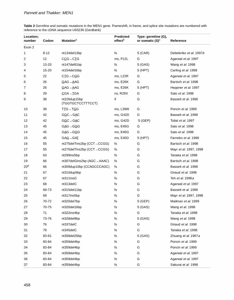
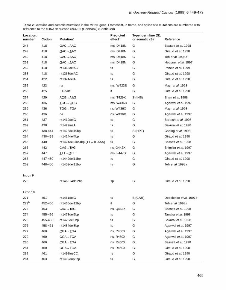
Table 2 Germline and somatic mutations in the MEN1 gene. Frameshift, in frame, and splice site mutations are numbered with reference to the cDNA sequence U93236 (GenBank)
Location; number Codon Mutation a
Predicted effect b
Type: germline (G), or somatic (S) c Reference
Exon 2
1 8-12 nt134del13bp fs S (CAR) Debelenko et al. 1997b
2 12 CCG→CTG ms, P12L G Agarwal et al. 1997
3 13-33 nt147del61bp fs S (GAS) Wang et al. 1998
4 15-20 nt154del16bp fs S (HPT) Carling et al. 1998
5 22 CTG→CGG ms, L22R G Agarwal et al. 1997
6 26 GAG→AAG ms, E26K G Bartsch et al. 1998
7 26 GAG→AAG ms, E26K S (HPT) Heppner et al. 1997
8 29 CGA→TGA ns, R29X G Sato et al. 1998
9 38 nt226dup15bp (TGGTGCTCCTTTCCT)
if G Bassett et al. 1998
10 39 TTG→TGG ms, L39W G Poncin et al. 1999
11 42 GGC→GAC ms, G42D G Bassett et al. 1998
12 42 GGC→GAC ms, G42D S (GEP) Toliat et al. 1997
13 45 GAG→GGG ms, E45G G Sato et al. 1998
14 45 GAG→GGG ms, E45G G Sato et al. 1998
15 45 GAG→GAT ms, E45D S (HPT) Farnebo et al. 1998
16 55 nt275delTins2bp (CCT→CCGG) fs G Bartsch et al. 1998
17 55 nt275delTins2bp (CCT→CCGG) fs G Mayr et al. 1997, 1998
18 63 nt299ins5bp fs G Tanaka et al. 1998
19 66 nt307delGins2bp (AGC→AAAC) fs G Bartsch et al. 1998
20d 66 nt309dup10bp (CCAGCCCAGC) fs G Bassett et al. 1998
21 67 nt310dup5bp fs G Giraud et al. 1998
22 67 nt311insG fs G Teh et al. 1998a
23 68 nt313delC fs G Agarwal et al. 1997
24 69-73 nt315del11bp fs G Bassett et al. 1998
25 69 nt317ins5bp fs G Mayr et al. 1997, 1998
26 70-72 nt320del7bp fs S (GEP) Mailman et al. 1999
27 70-75 nt320del16bp fs S (GAS) Wang et al. 1998
28 71 nt322ins4bp fs G Tanaka et al. 1998
29 73-76 nt328del9bp fs S (GAS) Wang et al. 1998
30 76 nt337delC fs G Giraud et al. 1998
31 79 nt345delC fs G Tanaka et al. 1998
32 83-91 nt358del25bp fs S (GAS) Zhuang et al. 1997a
33 83-84 nt359del4bp fs G Poncin et al. 1999
34 83-84 nt359del4bp fs G Poncin et al. 1999
35 83-84 nt359del4bp fs G Agarwal et al. 1997
36 83-84 nt359del4bp fs G Agarwal et al. 1997
37 83-84 nt359del4bp fs G Sakurai et al. 1998
458

Endocrine-Related Cancer (1999) 6 449-473
38 83-84 nt359del4bp fs G Bassett et al. 1998
39 83-84 nt359del4bp fs G Bassett et al. 1998
40 83-84 nt359del4bp fs G Bassett et al. 1998
41 83-84 nt359del4bp fs G Giraud et al. 1998
42 83-84 nt359del4bp fs G Giraud et al. 1998
43 83-84 nt359del4bp fs G Giraud et al. 1998
44 83-84 nt359del4bp fs G Teh et al. 1998a
45 83-84 nt359del4bp fs G Teh et al. 1998a
46 83-84 nt359del4bp fs G Teh et al. 1998a
47 83-84 nt359del4bp fs G Teh et al. 1998a
48 83-84 nt359del4bp fs G Teh et al. 1998a
49 83-84 nt359del4bp fs S (GAS) Zhuang et al. 1997a
50 83-84 nt359del4bp fs S (GAS) Wang et al. 1998
51 83-84 nt359del4bp fs S (LIP) Vortmeyer et al. 1998
52 84 nt360dupGT fs G Giraud et al. 1998
53 86 ATC→TTC ms, I86F S (GAS) Zhuang et al. 1997a
54 86-87 nt368del4bp fs S (HPT) Carling et al. 1998
55 89 CTC→CGC ms, L89R S (GLU) Hessman et al. 1998
56 90 nt379delAT fs G Bassett et al. 1998
57 96 CAG→TAG ns, Q96X G Giraud et al. 1998
58 98 CAG→TAG ns, R98X G Mayr et al. 1997, 1998
59 98 CAG→TAG ns, R98X G Bassett et al. 1998
60 98 CAG→TAG ns, R98X G Giraud et al. 1998
61 98 CAG→TAG ns, R98X G Giraud et al. 1998
62 98 CAG→TAG ns, R98X G Teh et al. 1998a
63 102-113 nt416del32bp fs G Mayr et al. 1997, 1998
64e 102 nt416delC fs G Agarwal et al. 1997
65 102 nt416delC fs G Teh et al. 1998a
66 108 CGA→TGA ns, R108X G Giraud et al. 1998
67 108 CGA→TGA ns, R108X S (HPT) Heppner et al. 1997
68 109 GAA→TAA ns, E109X S (ADR) Gortz et al. 1999
69 110 nt437insGG fs G Giraud et al. 1998
70 119 K119del if G Bassett et al. 1998
71 119 K119del if G Shimizu et al. 1997
72 119 K119del if G Sakurai et al. 1998
73 119 K119del if G Mayr et al. 1998
74 119 K119del if G Agarwal et al. 1997
75 119 K119del if G Agarwal et al. 1997
76 119 K119del if S (HPT) Farnebo et al. 1998
Table 2 Germline and somatic mutations in the MEN1 gene. Frameshift, in frame, and splice site mutations are numbered with reference to the cDNA sequence U93236 (GenBank) (Continued)
Location; number Codon Mutation a
Predicted effect b
Type: germline (G), or somatic (S) c Reference
459

Pannett and Thakker: MEN1
77 119 K119del if S (GEP) Toliat et al. 1997
78 120 AAG→TAG ns, K120X G Agarwal et al. 1997
79 125 nt483delAT fs G Tanaka et al. 1998
80 125 nt483delAT fs G Shimizu et al. 1997
81 125 nt483delAT fs S (GAS) Zhuang et al. 1997a
82 126 TGG→GGG ms, W126G S (GAS) Zhuang et al. 1997a
83d 126 TGG→TAG ns, W126X G Bassett et al. 1998
84 131 nt501delC fs G Bassett et al. 1998
85 133 AAG→TAG ns, Y133X G Giraud et al. 1998
86f 134 nt512delC fs G Agarwal et al. 1997
87 134 nt512delC fs G Bassett et al. 1998
88 135 AAG→TAG ns, K135X S (ANG) Boni et al. 1998
89 135 nt515delG fs S (HPT) Carling et al. 1998
90 138 nt522delG fs S (VIP) Wang et al. 1998
91 139 CAC→GAC ms, H139D G Agarwal et al. 1997
92 139 CAC→GAC ms, H139D G Zhuang et al. 1997b
93 139 CAC→GAC ms, H139D S (HPT) Carling et al. 1998
94 139 CAC→TAC ms, H139Y G Agarwal et al. 1997
95 141 CAG→CGG ms, Q141R S (GAS) Wang et al. 1998
96 144 TTC→GTC ms, F144V G Agarwal et al. 1997
97 145 nt545insT fs S (INS) Zhuang et al. 1997a
98 145 AGC→AGG ms, S145R S (NF) Wang et al. 1998
99 147 I147del if S (GAS) Wang et al. 1998
Intron 2
100 nt556-3 (c→g) sp G Teh et al. 1998b
Exon 3
101 150 nt560insA fs G Teh et al. 1998a
102 152 TTG→TGG ms, L152W S (HPT) Carling et al. 1998
103 153 nt569delC fs G Sato et al. 1998
104 159 TTT→TGT ms, F159C S (GAS) Zhuang et al. 1997a
105 160 GCT→CCT ms, A160P G Bassett et al. 1998
107 160 GCT→ACT ms, A160T G Teh et al. 1998a
108 162 GTT→TGT ms, V162C S (GAS) Wang et al. 1998
109d 163 nt597delG fs G Teh et al. 1998a
110 163 nt598dup5bp (GTTGG) fs G Bassett et al. 1998
111 163 nt599insA fs G Teh et al. 1998a
112 163 nt599insA fs G Teh et al. 1998a
Table 2 Germline and somatic mutations in the MEN1 gene. Frameshift, in frame, and splice site mutations are numbered with reference to the cDNA sequence U93236 (GenBank) (Continued)
Location; number Codon Mutation a
Predicted effect b
Type: germline (G), or somatic (S) c Reference
460

Endocrine-Related Cancer (1999) 6 449-473
113 164 GCC→GAC ms, A164D G Bassett et al. 1998
114 166 CAG→TAG ns, Q166X G Tanaka et al. 1998
115 168 CTG→CCG ms, L168P G Bartsch et al. 1998
116 171-173 nt621del9bp if G Sakurai et al. 1998
117 171-173 nt621del9bp if G Sakurai et al. 1998
118 172 GAT→TAT ms, D172Y G Poncin et al. 1999
119 172 GAT→TAT ms, D172Y G Poncin et al. 1999
120 172 GAT→TAT ms, D172Y G Poncin et al. 1999
121 172 GAT→TAT ms, D172Y G Giraud et al. 1998
122d 174 nt630delC fs G Bassett et al. 1998
123 175 CTC→CGC ms, L175R S (HPT) Farnebo et al. 1998
124 176 GCC→CCC ms, A176P G Agarwal et al. 1997
125 178 nt643insC fs S (GAS) Mailman et al. 1999
126 179 GAG→GAT ms, E179D G Poncin et al. 1999
127 183 TGG→TCG ms, W183S G Bassett et al. 1998
128 183 na ns, W183X G Agarwal et al. 1997
129 183 TGG→TGA ns, W183X G Bassett et al. 1998
130g 184 GTA→GAA ms, V184E G Fujimori et al. 1998
131 191 GAG→TAG ns, E191X G Bassett et al. 1998
132 191 GAG→TAG ns, E191X G Teh et al. 1998a
133 197 nt699insA fs S (HPT) Farnebo et al. 1998
134 197 T197del if G Giraud et al. 1998
135 198 na ns, W198X G Agarwal et al. 1997
136 198 TGG→TGA ns, W198X S (VIP) Shan et al. 1998
137 201 nt711delA fs G Sato et al. 1998
138 201 nt713delG fs G Agarwal et al. 1997
139 208 nt734delC fs G Teh et al. 1998a
140i 209-210 nt735del4bp fs G Agarwal et al. 1997
141i 209-210 nt735del4bp fs G Agarwal et al. 1997
142 210-211 nt738del4bp fs G Sakurai et al. 1998
143 210-211 nt738del4bp fs G Bassett et al. 1998
144 210-211 nt738del4bp fs G Bassett et al. 1998
145 210-211 nt738del4bp fs G Bassett et al. 1998
146 210-211 nt738del4bp fs G Bassett et al. 1998
147 210-211 nt738del4bp fs G Bassett et al. 1998
148 210-211 nt738del4bp fs G Giraud et al. 1998
149 210-211 nt738del4bp fs G Teh et al. 1998a
150 210-211 nt738del4bp fs G Teh et al. 1998a
151 214 nt751delG fs G Bassett et al. 1998
Table 2 Germline and somatic mutations in the MEN1 gene. Frameshift, in frame, and splice site mutations are numbered with reference to the cDNA sequence U93236 (GenBank) (Continued)
Location; number Codon Mutation a
Predicted effect b
Type: germline (G), or somatic (S) c Reference
461

Pannett and Thakker: MEN1
152 218 nt764G→T sp G Giraud et al. 1998
Intron 3
153 nt764+1 (g→t) sp G Teh et al. 1998b
154 nt764+1 (g→t) sp G Teh et al. 1998a
155 nt764+3 (a→g) sp S (CAR) Debelenko et al. 1997b
156 nt765-1 (g→t) sp G Mayr et al. 1998
Exon 4
157 220 na ns, W220X G Teh et al. 1998b
158 220 na ns, W220X G Teh et al. 1998a
159 220-223 nt770del9bp if S (GAS) Wang et al. 1998
160 222 nt776delC fs G Giraud et al. 1998
161 223 nt778delT fs G Giraud et al. 1998
162 223 CTG→CCG ms, L223P G Giraud et al. 1998
163 224-225 nt781del5bp fs S (HPT) Farnebo et al. 1998
164 227 na ns, Y227X G Teh et al. 1998a
165 236-239 nt817del9bp if G Bassett et al. 1998
166 236 nt818insT fs G Bartsch et al. 1998
167 242 GCC→GTC ms, A242V G Agarwal et al. 1997
168 255 nt875insA fs S (GAS) Zhuang et al. 1997a
169g 255 GAG→AAG ms, E255K G Teh et al. 1998c
170 257 nt879ins7bp (GGAGCTT) fs G Poncin et al. 1999
171 258 CAG→TAG ns, Q258X G Poncin et al. 1999
172 260 CAG→TAG ns, Q260X G Shimizu et al. 1997
173 260 CAG→TAG ns, Q260X G Agarwal et al. 1997
174 261 CAG→TAG ns, Q261X G Giraud et al. 1998
Intron 4
175 nt893+1(g→c) sp G Poncin et al. 1999
176 nt893+1(g→t) sp G Giraud et al. 1998
177 nt893+1(g→t) sp G Teh et al. 1998a
178 nt894-9 (g→a) sp G Gortz et al. 1999
179 nt894-1 (g→c) sp G Giraud et al. 1998
Exon 5
180 262 nt894delAinsGG fs G Bassett et al. 1998
181 262 AAG→TAG ns, K262X G Bassett et al. 1998
182 264 CTC→CCC ms, L264P G Poncin et al. 1999
Table 2 Germline and somatic mutations in the MEN1 gene. Frameshift, in frame, and splice site mutations are numbered with reference to the cDNA sequence U93236 (GenBank) (Continued)
Location; number Codon Mutation a
Predicted effect b
Type: germline (G), or somatic (S) c Reference
462

Endocrine-Related Cancer (1999) 6 449-473
183 265 nt904delG fs S (GEP) Toliat et al. 1997
184d 265 na ns, W265X G Agarwal et al. 1997
185 265 na ns, W265X G Giraud et al. 1998
186g 267 CTC→CCC ms, L267P G Poncin et al. 1999
187 268 nt912insT fs G Chico et al. 1998
188 268 TAT→TAG ns, T268X G Bartsch et al. 1998
189 269 nt916del22bp fs S (GH/PRL) Tanaka et al. 1998
190 272 nt924insC fs G Giraud et al. 1998
Intron 5
191 nt934+1(g→a) sp S (HPT) Heppner et al. 1997
192 nt935-2 (a→g) sp G Toliat et al. 1997
Exon 6
193d 284 GCA→GAA ms, A284E G Bassett et al. 1998
194 286 CTA→CCA ms, L286P G Agarwal et al. 1997
Exon 7
195 308 na ns, S308X G Agarwal et al. 1997
196 309 GCC→CCC ms, A309P G Agarwal et al. 1997
197 312 na ns, Y312X G Sato et al. 1998
198 312 na ns, Y312X G Agarwal et al. 1997
199 314 CGG→CCG ms, R314P G Giraud et al. 1998
200 315 nt1054insA fs G Giraud et al. 1998
201 315 nt1055insA fs G Poncin et al. 1999
202 320 CCC→CTC ms, P320L G Tanaka et al. 1998
203 323 na ns, Y323X G Agarwal et al. 1997
204 327 nt1089delT fs G Bassett et al. 1998
205 327 nt1089delT fs G Teh et al. 1998a
206 330 CGC→CCC ms, R330P S (GAS) Wang et al. 1998
207 330 CGC→CCC ms, R330P S (GAS) Wang et al. 1998
208 337 GCC→GAC ms, A337D G Giraud et al. 1998
209 340 GCC→ACC ms, A340T S (HPT) Shan et al. 1998
210 341 nt1132delG fs G Agarwal et al. 1997
211 341 TGG→CGG ms, W341R G Giraud et al. 1998
212 341 TGG→TAG ns, W341X G Bassett et al. 1998
213 341 na ns, W341X G Teh et al. 1998a
214 341 na ns, W341X G Teh et al. 1998a
215 344 ACG→AGG ms, T344R G Agarwal et al. 1997
Table 2 Germline and somatic mutations in the MEN1 gene. Frameshift, in frame, and splice site mutations are numbered with reference to the cDNA sequence U93236 (GenBank) (Continued)
Location; number Codon Mutation a
Predicted effect b
Type: germline (G), or somatic (S) c Reference
463

Pannett and Thakker: MEN1
216 345 nt1143delG fs G Sakurai et al. 1998
217 345 nt1144delC fs S (HPT) Farnebo et al. 1998
218 349 CAG→TAG ns, Q349X G Bassett et al. 1998
219 349 CAG→TAG ns, Q349X G Bassett et al. 1998
Intron 7
220d nt1159+1 (g→a) sp G Bassett et al. 1998
221 nt1160-5del7bp sp S (GEP) Toliat et al. 1997
222 nt1160-2 (a→g) sp G Tanaka et al. 1998
Exon 8
223g 353 TAC→TAA ns, Y353X G Shimizu et al. 1997
224 358 E358del if G Bassett et al. 1998
225 358-359 nt1184transvGG→AA ms,E359K S (ANG) Boni et al. 1998
226 363 E363del if G Agarwal et al. 1997
227 363 E363del if G Agarwal et al. 1997
228 363 E363del if G Zhuang et al. 1997a
229 364-365 nt1202del2bp G Agarwal et al. 1997
230 368 GCC→GAC ms, A368D G Giraud et al. 1998
231 368-370 nt1212del7bp fs S (GAS) Zhuang et al. 1997a
232 373 nt1229delC fs G Bassett et al. 1998
233 387 nt1269delG fs S (GAS) Wang et al. 1998
234 388 GAG→TAG ns, E388X G Bassett et al. 1998
235 390 nt1280delG fs G Agarwal et al. 1997
236 391 nt1279ins11bp (GAGGAGCGGCC)
fs S (HPT) Heppner et al. 1997
237 392 GAG→TAG ns, E392X S (GLU) Hessman et al. 1998
Exon 9
238 397 nt1300delC fs G Giraud et al. 1998
239 405 nt1325delG fs G Giraud et al. 1998
240 406-408 nt1328del5bp fs G Bassett et al. 1998
241 410 na ms, F410L S (ACTH) Zhuang et al. 1997b
242g 414 L414del if G Sato et al. 1998
243 415 CGA→TGA ns, R415X G Giraud et al. 1998
244 415 CGA→TGA ns, R415X G Teh et al. 1998b
245 415 CGA→TGA ns, R415X S (NFpanc) Hessman et al. 1998
246 418-421 nt1362del12bp if G Giraud et al. 1998
247 418 D418del if G Agarwal et al. 1997
Table 2 Germline and somatic mutations in the MEN1 gene. Frameshift, in frame, and splice site mutations are numbered with reference to the cDNA sequence U93236 (GenBank) (Continued)
Location; number Codon Mutation a
Predicted effect b
Type: germline (G), or somatic (S) c Reference
464
Endocrine-Related Cancer (1999) 6 449-473
248 418 GAC→AAC ms, D418N G Bassett et al. 1998
249 418 GAC→AAC ms, D418N G Giraud et al. 1998
250 418 GAC→AAC ms, D418N G Teh et al. 1998a
251 418 GAC→AAC ms, D418N G Heppner et al. 1997
252 418 nt1363delAC fs G Poncin et al. 1999
253 418 nt1363delAC fs G Giraud et al. 1998
254 422 nt1374delA fs G Giraud et al. 1998
255 423 na ms, W423S G Mayr et al. 1998
256 425 E425del if G Giraud et al. 1998
257 429 ACG→AAG ms, T429K S (INS) Shan et al. 1998
258 436 TGG→CGG ms, W436R G Agarwal et al. 1997
259 436 TGG→TGA ns, W436X G Mayr et al. 1998
260 436 na ns, W436X G Agarwal et al. 1997
261 437 nt1419delG fs G Bartsch et al. 1998
262 438 nt1422insA fs G Sakurai et al. 1998
263 438-444 nt1423del19bp fs S (HPT) Carling et al. 1998
264 438-439 nt1424del4bp fs G Giraud et al. 1998
265 440 nt1424del2ins4bp (TT�1GAAA) fs G Bassett et al. 1998
266 442 CAG→TAG ns, Q442X G Shimizu et al. 1997
267 447 TTT→CTT ms, F447S G Agarwal et al. 1997
268 447-450 nt1449del11bp fs G Giraud et al. 1998
269 448-450 nt1452del11bp fs G Teh et al. 1998a
Intron 9
270 nt1460+4del2bp sp G Giraud et al. 1998
Exon 10
271 451 nt1461delG fs S (CAR) Debelenko et al. 1997b
272h 452-456 nt1466del12bp if G Teh et al. 1998a
273 453 CAG→TAG ns, Q453X G Bassett et al. 1998
274 455-456 nt1473del5bp fs G Tanaka et al. 1998
275 455-456 nt1473del5bp fs G Sakurai et al. 1998
276 458-461 nt1484del8bp fs G Agarwal et al. 1997
277 460 CGA→TGA ns, R460X G Agarwal et al. 1997
279 460 CGA→TGA ns, R460X G Agarwal et al. 1997
280 460 CGA→TGA ns, R460X G Bassett et al. 1998
281 460 CGA→TGA ns, R460X G Giraud et al. 1998
282 461 nt1491insCC fs G Giraud et al. 1998
284 463 nt1499dup8bp fs G Giraud et al. 1998
Table 2 Germline and somatic mutations in the MEN1 gene. Frameshift, in frame, and splice site mutations are numbered with reference to the cDNA sequence U93236 (GenBank) (Continued)
Location; number Codon Mutation a
Predicted effect b
Type: germline (G), or somatic (S) c Reference
465

Pannett and Thakker: MEN1
285 467 nt1508ins fs G Giraud et al. 1998
286 467 nt1509ins2bp fs G Agarwal et al. 1997
287 477 nt1539insG fs G Poncin et al. 1999
288 493 nt1587delCCinsG (CCG→GG) fs G Bartsch et al. 1998
289 499 nt1607delA fs G Giraud et al. 1998
290 502 AAG→ATG ms, K502M S (NFpit) Zhuang et al. 1997b
291 507 nt1630insC fs G Giraud et al. 1998
292 510 nt1639delCA fs G Bartsch et al. 1998
293 516 nt1656delC fs G Agarwal et al. 1997
294 516 nt1656delC fs G Agarwal et al. 1997
295 516 nt1656delC fs G Bassett et al. 1998
296 516 nt1656delC fs G Giraud et al. 1998
297 516 nt1656delC fs G Giraud et al. 1998
298 516 nt1657insC fs G Agarwal et al. 1997
299 516 nt1657insC fs G Sakurai et al. 1998
300 516 nt1657insC fs G Sakurai et al. 1998
301 516 nt1657insC fs G Sakurai et al. 1998
302 516 nt1657insC fs G Giraud et al. 1998
303 516 nt1657insC fs G Bassett et al. 1998
304 516 nt1657insC fs G Bassett et al. 1998
305 516 nt1657insC fs G Giraud et al. 1998
306 516 nt1657insC fs G Giraud et al. 1998
307 516 nt1657insC fs G Giraud et al. 1998
308 516 nt1657insC fs G Teh et al. 1998a
309 516 nt1657insC fs G Teh et al. 1998a
310 516 nt1657insC fs S (CAR) Debelenko et al. 1997b
311 519 nt1666delC fs G Poncin et al. 1999
312 527 CGA→TGA ns, R527X G Agarwal et al. 1997
313 527 CGA→TGA ns, R527X G Bassett et al. 1998
314 527 CGA→TGA ns, R527X G Teh et al. 1998a
315 527 CGA→TGA ns, R527X G Giraud et al. 1998
316 527 CGA→TGA ns, R527X G Giraud et al. 1998
317 530-531 nt1699del3ins2bp (AAGG→AGC) fs G Debelenko et al. 1997b
318 535 GCT→GTT ms, A535V S (INS) Zhuang et al. 1997a
319 536 CAG→TAG ns, Q536X G Giraud et al. 1998
320 543 TCA→TTA ms, S543L S (GAS) Zhuang et al. 1997a
321 550 nt1768delT fs G Giraud et al. 1998
322 550 nt1768delT fs G Giraud et al. 1998
323 555 AGT→AAT ms, S555N G Giraud et al. 1998
Table 2 Germline and somatic mutations in the MEN1 gene. Frameshift, in frame, and splice site mutations are numbered with reference to the cDNA sequence U93236 (GenBank) (Continued)
Location; number Codon Mutation a
Predicted effect b
Type: germline (G), or somatic (S) c Reference
466

Endocrine-Related Cancer (1999) 6 449-473
gedod in
se
1999) in the context of the Knudson two-hit hypothesisremains to be elucidated. The tumours harbouring asomatic MEN1 mutation had chromosome 11q13 LOH asthe other genetic abnormality, or ‘hit’, consistent withKnudson’s hypothesis. These studies (Debelenko et al.1997b, Heppner et al. 1997, Toliat et al. 1997, Zhuanget al. 1997a,b, Boni et al. 1998, Carling et al. 1998,Farnebo et al. 1998, Hessman et al. 1998, Prezant et al.1998, Shan et al. 1998, Vortmeyer et al. 1998, Wang et al.1998, Gortz et al. 1999, Mailman et al. 1999) indicate thatalthough inactivation of the MEN1 gene may have a rolein the aetiology of some sporadic endocrine tumours, theinvolvement of other genes with major roles in theaetiology of such sporadic endocrine tumours is highlylikely.
Function of MEN1 protein (MENIN)Initial analysis of the predicted amino acid sequenceencoded by the MEN1 gene did not reveal homologies toany other proteins, sequence motifs, signal peptides, orconsensus nuclear localisation signals(Chandrasekharappa et al. 1997), and thus the putativefunction of the protein (MENIN) could not be deduced.However, studies based on immunofluorescence, Westernblotting of subcellular fractions, and epitope tagging withenhanced green fluorescent protein, revealed that MENIN
was located primarily in the nucleus (Guru et al. 1998).Furthermore, enhanced green fluorescent protein-tagMENIN deletional constructs identified at least twindependent nuclear localisation signals that are locatethe C-terminal quarter of the protein (Fig. 3) (Guru et al.1998). Interestingly, none of the MEN1 germline missenor in-frame deletions (Agarwal et al. 1997, Debelenkoet al. 1997b, Mayr et al. 1997, 1998, Shimizu et al. 1997,Toliat et al. 1997, Zhuang et al.1997a, Bartsch et al. 1998,
324 556 nt1777delA fs S (GH/PRL) Tanaka et al. 1998
325 558 M558del if G Giraud et al. 1998
326 558 nt1782delA fs G Giraud et al. 1998
327 561 ATG→ACG ms, M561T S (HPT) Heppner et al. 1997
328 568-569 nt1812del5bp fs S (HPT) Heppner et al. 1997
329 whole gene
G Kishi et al. 1998
aMutation: na=mutation not available.bPredicted effect: fs=frameshift mutation; ns=nonsense mutation; ms=missense mutation; if=in-frame deletion or insertion mutation; sp=splice site mutation.cSomatic mutation - origin of tumour: HPT= parathyroid tumour; GAS= gastrinoma; INS= insulinoma; VIP=VIPoma; GLU=glucagon; GEP= neuroendocrine gastroenteropancreatic tumour; NFPpanc= non-functioning pancreatic tumour;PRL=prolactinoma; GH= somatotrophinoma; ACTH=corticotrophinoma; NFpit=non-functioning pituitary tumour; ADR=adrenal cortical tumour ; CAR=lung carcinoid; ANG=angiofibroma; LIP=lipoma; NF=non-functioning.dOccurrence of de novo mutation demonstrated by familial analysis.eMutation reported in 5 related USA families.fMutation reported in 6 related USA families.gFamilial isolated primary hyperparathyroidism (FIHP) mutation.hMutation reported in 8 Finnish families; likely to be due to a founder effect.iThese mutations may also involve codons 210-211.ins, insertion; del, deletion; dup, duplication; transv, transversion.
Table 2 Germline and somatic mutations in the MEN1 gene. Frameshift, in frame, and splice site mutations are numbered with reference to the cDNA sequence U93236 (GenBank) (Continued)
Location; number Codon Mutation a
Predicted effect b
Type: germline (G), or somatic (S) c Reference
Table 3 MEN1-associated tumours in five unrelated families with a 4 bp deletion at codons 210 and 211
Family
Tumour 1 2 3 4 5
Parathyroid + + + + +
Gastrinoma + − + + +
Insulinoma − + − − −
Glucagonoma − − − − +
Prolactinoma − − − − +
Carcinoid + − − − −
+, presence; -, absence of tumours.(Adapted from Thakker (1998), with permission).
467

Pannett and Thakker: MEN1
ctn inhe be
l byich),
isaterN-ll
ine
ynde
Bassett et al. 1998, Chico et al. 1998, Fujimori et al. 1998,Giraud et al. 1998, Kishi et al. 1998, Sakurai et al. 1998,Sato et al. 1998, Tanaka et al. 1998, Teh et al. 1998a,b,c,Gortz et al. 1999, Poncin et al. 1999) alter either of theseputative nuclear localisation signals (Fig. 3). However, allof the truncated MEN1 proteins that would result from thenonsense and frameshift mutations, if expressed, wouldlack at least one of these nuclear localisation signals(Fig. 3). The nuclear localisation of MENIN suggestedthat it may act either in the regulation of transcription, orDNA replication, or the cell cycle. In order to investigatethis further and to identify proteins that may interact withMENIN, the yeast two hybrid system was utilised. Thisrevealed that MENIN directly interacts with the N-terminus of the AP-1 transcriptional factor JunD, torepress JunD-activated transcription (Agarwal et al.1999). Analysis of several MEN1 missense and deletionalmutations indicated that the N-terminus and centraldomains of MENIN (Fig. 3) have a critical role inMENIN-JunD interaction. However, JunD inhibits cellgrowth (Hirai et al. 1989, Ryder et al. 1989, Pfarr et al.1994), an action that differs from that of other AP-1proteins, and thus the repressive effect of MENIN on
JunD-mediated transcriptional activation would predienhanced growth rather than the observed suppressiogrowth. This seeming paradox may be due to tinvolvement of other target genes and proteins that mayinvolved in cell proliferation and that may haveinteractions with the MENIN-JunD complex (Agarwaet al. 1999). These suggestions are further supportedthe observation that disease-associated mutations whoccur outside the domains interacting with JunD (Fig. 3are associated with normal MENIN-JunD binding; thsuggests that MENIN may interact with other proteins thmay influence JunD-mediated transcription. Furthinvestigations are needed to elucidate the role of MENIJunD interactions in the control of endocrine ceproliferation.
Screening in MEN1
MEN1 is inherited as an autosomal dominant disorderthe majority of patients. Occasionally, MEN1 may arissporadically (i.e. without family history), although it mabe difficult to make the distinction between sporadic afamilial forms; in some cases the family history may b
468
A
Cum
ulat
ive
%
Age (years)
Pen
etra
nce
(cum
ulat
ive)
%
Age (years)
B
Figure 5 The age distributions (panel A) and age-related penetrances (panel B) of MEN1 determined from an analysis of 174 mutant gene carriers. The age distributions were determined for 3 groups of MEN1 mutant gene carriers from 40 families in whom mutations were detected (Bassett et al. 1998). The 91 members of group A presented with symptoms, whereas the 40 members of group B were asymptomatic and were detected by biochemical screening. The 43 members of group C represent those individuals who are MEN1 mutant gene carriers (Fig. 4) and who remain asymptomatic and biochemically normal. The ages included for members of groups A, B and C are those at the onset of symptoms, at the finding of the biochemical abnormality, and at the last clinical and biochemical evaluation respectively. Groups B and C contained members who were significantly younger than those in group A (P<0.001). The younger age of the group C mutant gene carriers is consistent with an age-related penetrance for MEN1, and this was calculated (panel B) for the first 5 decades. The age-related penetrances (i.e. the proportion of mutant gene carriers with manifestations of the disease by a given age) rose steadily from 7% in the <10 years group, to 52%, 87%, 98% and 100%, by the ages of 20, 30, 40, and 60 years respectively. (Adapted from Bassett et al. (1998), with permission.)
100
80
60
40
20
00 10 20 30 50 6040 70
100
80
60
40
20
0<5 <10 <15 <20 <25 <30 <35 <40 <45 <50 <55 <60

Endocrine-Related Cancer (1999) 6 449-473
ndityhensg. 5)thes aage
eof
ofn
ingalnd
y.ostising
skld
um,ng,10rlymeforasecaleis,m,aly, ofn-mdrin,bens
or
absent because the parent with MEN1 is not available, andhas perhaps already died before developing anymanifestations, and other cases may be due to de novomutations, which will be transmitted in an autosomaldominant manner in future generations (Agarwal et al.1997, Bassett et al. 1998, Teh et al. 1998a). MEN1 is anuncommon disorder, but because of its autosomaldominant inheritance, the finding of MEN1 in a patient hasimportant implications for other family members; first-degree relatives have about a 50% risk of developing thedisease. Screening for MEN1 in patients involves thedetection of tumours and the ascertainment of thegermline genetic state i.e. normal or mutant gene carrier.The detection of tumours entails clinical, biochemical andradiological investigations for MEN1-associated tumoursin patients. The recent cloning of the MEN1 gene hasfacilitated the identification of individuals who havemutations and hence are at a high risk of developing thedisease (Figs 3 and 4).
Genetic analysisMutational analysis for MEN1 could now be introduced toidentify those individuals who are mutant carriers and thusat a high risk of developing tumours. The advantages ofDNA analysis are that it requires a single blood sampleand does not need, in theory, to be repeated. This isbecause the analysis is independent of the age of theindividual and provides an objective result. Suchmutational analysis could be undertaken in childrenaround the first decade, as some children have developedtumours by the age of 8 years (Thakker 1995, Trump et al.1996), and appropriate intervention in the form ofbiochemical testing and/or treatment has been considered.However, the great diversity together with the widelyscattered locations of the MEN1 mutations (Fig. 3), and alack of genotype-phenotype correlation (Table 3) willmake such mutational screening time-consuming, arduousand expensive, and as yet this is not widely available.Nevertheless, an integrated programme of both mutationalanalysis, to identify mutant gene carriers, and biochemicalscreening, to detect the development of tumours, would beof advantage. Thus, a DNA test identifying an individualas a mutant gene carrier is likely to lead not to immediatemedical or surgical treatment but to earlier and morefrequent biochemical and radiological screening, whereasa DNA result that indicates that an individual is not at riskwill lead to a decision for no further clinical investigations.Thus, the identification of MEN1 mutations may be ofhelp in the clinical management of patients and theirfamilies with this disorder.
Detection of MEN1 tumoursBiochemical screening for the development of MEN1tumours in asymptomatic members of families with
MEN1 is of great importance, as earlier diagnosis atreatment of these tumours may help to reduce morbidand mortality. The age-related penetrance (i.e. tproportion of gene carriers manifesting symptoms or sigof the disease by a given age) has been ascertained (Fiand the mutation appears to be non-penetrant below age of 8 years. Thereafter, the mutant MEN1 gene hahigh penetrance, being >50% penetrant by 20 years of and >95% penetrant by 40 years (Bassett et al. 1998).Screening for MEN1 tumours is difficult because thclinical and biochemical manifestations in members any one family are not uniformly similar (Trump et al.1996). The attempts to screen for the developmentMEN1 tumours in the asymptomatic relatives of aaffected individual have depended largely on measurthe serum concentrations of calcium, gastrointestinhormones and prolactin, and also on ultrasound aradiological imaging of the abdomen and pituitarParathyroid overactivity causing hypercalcaemia is alminvariably the first manifestation of the disorder and thhas become a useful and easy biochemical screeninvestigation (Benson et al. 1987, Marx et al. 1986).
At present, it is suggested that individuals at high riof expressing MEN1 (i.e. mutant gene carriers) shouundergo biochemical screening at least once per annand also have baseline pituitary and abdominal imagiwhich should then be repeated infrequently (at 5 to yearly intervals). Screening should commence in eachildhood, as the disease has developed in soindividuals by the age of 8 years, and should continue life, as some individuals have not developed the diseuntil the eighth decade. Screening history and physiexamination should be directed towards eliciting thsymptoms and signs of hypercalcaemia, nephrolithiaspeptic ulcer disease, neuroglycopaenia, hypopituitarisgalactorrhoea and amenorrhoea in women, acromegCushing’s disease, visual field loss and the presencesubcutaneous lipomata, angiofibromas, and collageomas. Biochemical screening should include serucalcium and prolactin estimations in all individuals, anmeasurement of gastrointestinal hormones e.g. gastand more specific endocrine function tests should reserved for individuals who have symptoms or sigsuggestive of a clinical syndrome.
AcknowledgementsWe are grateful to the Medical Research Council, UK fsupport; and to S Kingsley for typing the manuscript.
ReferencesAgarwal SK, Kester MB, Debelenko LV, Heppner C,
Emmert-Buck MR, Skarulis MC, Doppman JL, Kim YS, Lubensky IA, Zhuang Z, Green JS, Guru SC, Manickam P,
469
Pannett and Thakker: MEN1
e
, S,
al
K, C
e in
8 d
P,
Olufemi S-E, Liotta LA, Chandrasekharappa SC, Collins FS, Spiegel AM, Burns AL & Marx SJ 1997 Germline mutations of the MEN1 gene in familial multiple endocrine neoplasia type 1 and related states. Human Molecular Genetics 6 1169-1175.
Agarwal SK, Guru SC, Heppner C, Erdos MR, Collins RM, Park SY, Saggar S, Chandrasekharappa SC, Collins FS, Spiegel AM, Marx SJ & Burns AL 1999 Menin interacts with the AP1 transcription factor JunD and represses JunD-activated transcription. Cell 84 730-735.
Bartsch D, Kopp I, Bergenfelz A, Rieder H, Munch K, Jager K, Deiss Y, Schudy A, Barth P, Arnold R, Rothmund M & Simon B 1998 MEN1 gene mutations in 12 MEN1 families and their associated tumors. European Journal of Endocrinology 139 416-420.
Bassett JHD, Williamson C, Pang J, Bishop A, Lynn J, Polak JM, Bloom SR & Thakker RV 1997 Glucagonomas in multiple endocrine neoplasia type 1. Journal of Endocrinology 152 P68.
Bassett JHD, Forbes SA, Pannett AAJ, Lloyd SE, Christie PT, Wooding C, Harding B, Besser GM, Edwards CR, Monson JP, Sampson J, Wass JAH, Wheeler MH & Thakker RV 1998 Characterisation of mutations in patients with multiple endocrine neoplasia type 1 (MEN1). American Journal of Human Genetics 62 232-244.
Benson L, Ljunghall S, Akerstrom G & Oberg K 1987 Hyperparathyroidism presenting as the first lesion in multiple endocrine neoplasia type 1. American Journal of Medicine 82 731-737.
Bloom SR, Polak JM & Pearse AGE 1973 Vasoactive intestinal peptide and watery diarrhoea syndrome. Lancet 2 14-16.
Boni R, Vortmeyer AO, Pack S, Park WS, Burg G, Hofbauer G, Darling T, Liotta L & Zhuang ZP 1998 Somatic mutations of the MEN1 tumor suppressor gene detected in sporadic angiofibromas. Journal of Investigative Dermatology 111 539-540.
Brown MA & Solomon E 1997 Studies on inherited cancers: outcomes and challenges of 25 years. Trends in Genetics 13 202-206.
Byström MC, Larsson C, Blomberg C, Sandelin F, Falkmer U, Skogseid B, Oberg K, Werner S & Nordenskjöld M 1990 Localisation of the MEN1 gene to a small region within chromosome 11q13 by deletion mapping in tumours. Proceedings of the National Academy of Sciences of the USA 87 1968-1972.
Carling T, Correea P, Hessman O, Hedberg J, Skogseid B, Lindberg D, Rastad J, Westin G & Akerström G 1998 Parathyroid MEN1 gene mutations in relation to clinical characteristics of non-familial primary hyperparathyroidism. Journal of Clinical Endocrinology and Metabolism 83 2951-2954.
Chandrasekharappa SC, Guru SC, Manickam P, Olufemi S-E, Collins FS, Emmert-Buck MR, Debelenko LV, Zhuang Z, Lubensky IA, Liotta LA, Crabtree JS, Wang Y, Roe BA, Weisemann J, Boguski MS, Agarwal SK, Kester MB, Kim YS, Heppner C, Dong Q, Spiegel AM, Burns AL & Marx SJ 1997 Positional cloning of the gene for multiple endocrine neoplasia-type 1. Science 276 404-407.
Chico A, Gallart L, Mato E, Mayoral C, Martin-Campos J, Catasus L, Rodriguez-Espinosa J, Matias-Guiu X, Blanco-Vaca F & de Leiva A 1998 A novel germline mutationin exon 5 of the multiple endocrine neoplasia type 1 gene. Journal of Molecular Medicine 76 837-839.
Darling TN, Skarulis MC, Steinberg SM, Marx SJ, Spiegel AM& Turner M 1997 Multiple facial angiofibromas and collagenomas in patients with multiple endocrine neoplasiatype 1. Archives of Dermatology 133 853-861.
Debelenko LV, Emmert-Buck MR, Manickam P, Kester M, Guru SC, DiFranco EM, Olufemi SE, Agarwal S, Lubensky IA, Zhuang Z, Burns AL, Spiegel AM, Liotta LA, Collins FS, Marx SJ & Chandrasekharappa SC 1997a Haplotype analysis defines a minimal interval for the multiplendocrine neoplasia type 1 (MEN1) gene. Cancer Research 57 1039-1042.
Debelenko LV, Brambilla E, Agarwal SK, Swalwell JI, Kester MB, Lubensky IA, Zhuang Z, Guru SC, Manickam POlufemi S-E, Chandrasekharappa SC, Crabtree JS, Kim YHeppner C, Burns AL, Spiegel AM, Marx SJ, Liotta LA, Collins FS, Travis WD & Emmert-Buck MR 1997b Identification of MEN1 gene mutations in sporadic carcinoidtumors of the lung. Human Molecular Genetics 6 2285-2290.
Delcore R, Hermreck AS & Friesen SR 1989 Selective surgicmanagement of correctable hypergastrinemia. Surgery 106 1094-1102.
Duh QY, Hybarger CD, Geist R, Gamsu G, Goodman PC, Gooding GA & Clark OH 1987 Carcinoids associated with multiple endocrine neoplasia syndromes. American Journal of Surgery 154 142-148.
Farnebo F, Teh BT, Kytölä S, Svensson A, Phelan C, SandelinThompson NW, Höög A, Weber G, Farnebo L-O & Larsson1998 Alterations of the MEN1 gene in sporadic parathyroid tumors. Journal of Clinical Endocrinology and Metabolism 83 2627-2630.
Friedman E, Sakaguchi K, Bale AE, Falchetti A, Steeten E, Zimering MB, Weinstein LS, McBride WO, Nakamura Y, Brandi ML, Norton JA, Aurbach GD, Spiegel AM & Marx SJ1989 Clonality of parathyroid tumours in familial multiple endocrine neoplasia type 1. New England Journal of Medicine 321 213-218.
Friesen SR, Kimmel JR & Tomita T 1980 Pancreatic polypeptidas screening marker for pancreatic polypeptide apudomasmultiple endocrinopathies. American Journal of Surgery 139 61-72.
Fujimori M, Shirahama S, Sakurai A, Hashizume K, Hama Y, Ito K, Shingu K, Kobayashi S, Amano J & Fukushima Y 199Novel V184E MEN1 germline mutation in a Japanese kindrewith familial hyperparathyroidism. American Journal of Medical Genetics 80 221-222.
Gagel RF & Cotes GJ 1996 Ret protooncogene mutations in multiple endocrine neoplasia type 2. In Principles of Bone Biology, pp 799-807. Eds JP Bilezikian, LG Raisz & GA Rodan. San Diego: Academic Press.
Giraud S, Zhang CX, Serova Sinilnikova O, Wautot V, Salandre J, Buisson N, Waterlot C, Bauters C, Porchet N, Aubert JP, Emy P, Cadiot G, Delemer B, Chabre O, NiccoliLeprat F, Duron F, Emperauger B, Cougard P, Goudet P,
470

Endocrine-Related Cancer (1999) 6 449-473
6 ry
, 3
s a
8
ne
Sarfati E, Riou JP, Guichard S, Rodier M, Meyrier A, Caron P, Vantyghem MC, Assayag M, Peix JL, Pugeat M, Rohmer V, Vallotton M, Lenoir G, Gaudray P, Proye C, ConteDevolx B, Chanson P, Shugart YY, Goldgar D, Murat A & Calender A 1998 Germ-line mutation analysis in patients with multiple endocrine neoplasia type 1 and related disorders. American Journal of Human Genetics 63 455-467.
Gortz B, Roth J, Speel EJM, Krahenmann A, De Kruger RR, Matias Guiu X, Muletta Feurer S, Rutmann K, Saremaslani P, Heitz PU & Komminoth P 1999 MEN1 gene mutation analysis of sporadic adrenocortical lesions. International Journal of Cancer 80 373-379.
Guru SC, Goldsmith PK, Burns AL, Marx SJ, Spiegel AM, Collins FS & Chandrasekharappa SC 1998 Menin, the product of the MEN1 gene, is a nuclear protein. Proceedings of the National Academy of Sciences of the USA 95 1630-1634.
Heppner C, Kester MB, Agarwal SK, Debelenko LV, Emmert-Buck MR, Guru SC, Manickam P, Olufemi S-E, Skarulis MC, Doppman JL, Alexander RH, Kim YS, Saggar SK, Lubensky IA, Zhuang Z, Liotta LA, Chandrasekharappa SC, Collins FS, Spiegel AM, Burns AL & Marx SJ 1997 Somatic mutation of the MEN1 gene in parathyroid tumours. Nature Genetics 16 375-378.
Hessman O, Lindberg D, Skogseid B, Carling T, Hellman P, Rastad J, Akerstrom G & Westin G 1998 Mutation of the multiple endocrine neoplasia type 1 gene in nonfamilial, malignant tumors of the endocrine pancreas. Cancer Research 58 377-379.
Hirai SI, Ryseck RP, Mechta F, Bravo R & Yaniv M 1989 Characterization of JunD: a new member of the jun proto-oncogene family. EMBO Journal 8 1433-1439.
Kishi M, Tsukada T, Shimizu S, Futami H, Ito Y, Kanbe M, Obara T & Yamaguchi K 1998 A large germline deletion of the MEN1 gene in a family with multiple endocrine neoplasia type 1. Japanese Journal of Cancer Research 89 1-5.
Knudson AG 1971 Mutation and cancer: statistical study of retinoblastoma. Proceedings of the National Academy of Sciences of the USA 68 820-823.
Knudson AG 1993 Antioncogenes and human cancer. Proceedings of the National Academy of Sciences of the USA 90 10914-10921.
Larsson C, Skogseid B, Oberg K, Nakamura Y & Nordenskjold MC 1988 Multiple endocrine neoplasia type I gene maps to chromosome 11 and is lost in insulinoma. Nature 332 85-87.
Mailman MD, Muscarella P, Schrimer WJ, Ellison EC, O’Dorisio T & Prior TW 1999 Identification of MEN1 mutations in sporadic enteropancreatic neuroendocrine tumors by analysis of paraffin-embedded tissue. Clinical Chemistry 45 29-34.
Marx IN, Bank S & Louw JH 1967 Islet cell tumour of the pancreas with reversible watery diarrhoea and achlorhydria. Gastroenterology 52 695-708.
Marx SJ 1998 Multiple endocrine neoplasia type 1. In Genetic Basis of Human Cancer, pp 489-506. Eds B Vogelstein & KW Kinzler. New York: McGraw Hill.
Marx SJ, Spiegel AM, Levine MA, Rizzoli RE, Lasker RD, Santora AC, Downs RV Jr & Aurbach GD 1982 Familial
hypocalciuric hypercalcaemia: the relation to primary parathyroid hyperplasia. New England Journal of Medicine 307 416-426.
Marx SJ, Vinik AI, Santen RJ, Floyd JC, Mills JL & Green J 198Multiple endocrine neoplasia type 1: assessment of laboratotests to screen for the gene in a large kindred. Medicine 65 226-241.
Mayr B, Apenberg S, Rothamel T, vonzurMuhlen A & Brabant G1997 Menin mutations in patients with multiple endocrine neoplasia type 1. European Journal of Endocrinology 137 684-687.
Mayr B, Brabant G & vonzurMuhlen A 1998 Menin mutations inMEN1 patients. Journal of Clinical Endocrinology and Metabolism 83 3004-3005.
Pang JT & Thakker RV 1994 Multiple endocrine neoplasia type 1. European Journal of Cancer 30 1961-1968.
Pang JT, Lloyd S, Wooding C, Farren B, Pottinger B, Harding BLeigh SEA, Pook MA, Benham FJ, Gillett GT, Taggart RT &Thakker RV 1996 Genetic mapping studies of 40 loci and 2cosmids in chromosome 11p13-11q13, and exclusion of µ-calpain as the multiple endocrine neoplasia type 1 gene.Human Genetics 97 732-741.
Pannett AAJ & Thakker RVT 1999 Meta-analysis of 31 studiereporting 344 mutations in the multiple endocrine neoplasitype 1 (MEN1) gene. Journal of Bone and Mineral Research 14 (Abstract) 5321.
Pfarr CM, Mechta F, Spyrou G, Lallemand D, Carillo S & Yaniv M 1994 Mouse JunD negatively regulates fibroblast growth and antagonizes transformation by ras. Cell 76 747-760.
Poncin J, Abs R, Velkeniers B, Bonduelle M, Abramowicz M, Legros JJ, Verloes A, Meurisse M, Van Gaal L, Verellen C,Koulischer L & Beckers A 1999 Mutation analysis of the MEN1 gene in Belgian patients with multiple endocrine neoplasia type 1 and related diseases. Human Mutation 13 54-60.
Prezant TR, Levine J & Melmed S 1998 Molecular characterization of the Men 1 tumor suppressor gene in sporadic pituitary tumors. Journal of Clinical Endocrinology and Metabolism 83 1388-1391.
Rizzoli R, Green J & Marx S J 1985 Primary hyperpara-thyroidism in familial multiple endocrine neoplasia type 1. Long term follow-up of serum calcium levels after parathyroidectomy. American Journal of Medicine 78 467-474.
Ryder K, Lanahan A, Perez-Albuerne & Nathans D 1989 junD: a third member of the jun gene family. Proceedings of the National Academy of Sciences of the USA 86 1500-1503.
Sakurai A, Shirahama S, Fujimori M, Katai M, Itakura Y, Kobayashi S, Amano J, Fukushima Y & Hashizume K 199Novel MEN1 gene mutations in familial multiple endocrine neoplasia type 1. Journal of Human Genetics 43 199-201.
Sato M, Matsubara S, Miyauchi A, Ohye H, Imachi H, Murao K& Takahara J 1998 Identification of five novel germline mutations of the MEN1 gene in Japanese multiple endocrineoplasia type 1 (MEN1) families. Journal of Medical Genetics 35 915-919.
471

Pannett and Thakker: MEN1
).
a.
f
e
B ne
let
0
e
e
C, ,
Shan L, Nakamura Y, Nakamura M, Yokoi T, Tsujimoto M, Arima R, Kaeya T & Kakudo K 1998 Somatic mutations of multiple endocrine neoplasia type 1 gene in the sporadic endocrine tumors. Laboratory Investigation 78 471-475.
Sheppard BC, Norton JA, Dopmann JL, Maton PN, Gardner JD & Jensen RT 1989 Management of islet cell tumours in patients with multiple endocrine neoplasia: a prospective study. Surgery 106 1108-1118.
Shimizu S, Tsukada T, Futami H, Ui K, Kameya T, Kawanaka M, Uchiyama S, Aoki A, Yasuda H, Kawano S, Ito Y, Kanbe M, Obara T & Yamaguchi K 1997 Germline mutations of the MEN1 gene in Japanese kindred with multiple endocrine neoplasia type 1. Japanese Journal of Cancer Research 88 1029-1032.
Sipple JH 1961 The association of pheochromocytoma with carcinoma of the thyroid gland. American Journal of Medicine 31 163-166.
Skogseid B, Oberg K, Benson L, Lindgren PS, Lörelius LE, Lundquist G, Wide L & Wilander E 1987 A standardised meal stimulation test of the endocrine pancreas for early detection of pancreatic endocrine tumours in multiple endocrine neoplasia type 1 syndrome: five years experience. Journal of Clinical Endocrinology and Metabolism 64 1233-1240.
Skogseid B, Larsson C, Lindgren PG, Kvanta E, Rastad J, Theodorsson E, Wide L, Wilander E & Oberg K 1992 Clinical and genetic features of adrenocortical lesions in multiple endocrine neoplasia type 1. Journal of Clinical Endocrinology and Metabolism 75 76-81.
Tanaka C, Kimura T, Yang P, Moritani M, Yamaoka T, Yamada S, Sano T, Yoshimoto K & Itakura M 1998 Analysis of loss of heterozygosity on chromosome 11 and infrequent inactivation of MEN1 gene in sporadic pituitary adenomas. Journal of Clinical Endocrinology and Metabolism 83 2631-2634.
Teh BT, Kytola S, Farnebo F, Bergman L, Wong FK, Weber G, Hayward N, Larsson C, Skogseid B, Beckers A, Phelan C, Edwards M, Epstein M, Alford F, Hurley D, Grimmond S, Silins G, Walters M, Stewart C, Cardinal J, Khodaei S, Parente F, Tranebjaerg L, Jorde R, Salmela P et al. 1998a Mutation analysis of the MEN1 gene in multiple endocrine neoplasia type 1, familial acromegaly and familial isolated hyperpara-thyroidism. Journal of Clinical Endocrinology and Metabolism 83 2621-2626.
Teh BT, Zedenius J, Kytola S, Skogseid B, Trotter J, Choplin H, Twigg S, Farnebo F, Giraud S, Cameron D, Robinson B, Calender A, Larsson C & Salmela P 1998b Thymic carcinoids in multiple endocrine neoplasia type 1. Annals of Surgery 228 99-105.
Teh BT, Esapa CT, Houlston R, Grandell U, Farnebo F, Nordenskjold M, Pearce CJ, Carmichael D, Larsson C & Harris PE 1998c A family with isolated hyperparathyroidism segregating a missense MEN1 mutation and showing loss of the wild-type alleles in the parathyroid tumors. American Journal of Human Genetics 63 1544-1549.
Thakker RV 1993 The molecular genetics of the multiple endocrine neoplasia syndromes. Clinical Endocrinology 39 1-14.
Thakker RV 1994 Molecular mechanisms of tumor formation in hereditary and sporadic tumors of the MEN1 type: the impact
of genetic screening in the management of MEN1. In Endocrinology and Metabolism Clinics of North America, pp 117-135. Ed RF Gagel. Philadelphia: WB Saunders.
Thakker RV 1995 Multiple endocrine neoplasia type 1 (MEN1In Endocrinology, pp 2815-2831. Eds LJ DeGroot, GK Besser, HG Burger, JL Jameson, DL Loriaux, JC Marshall,WD Odell, JT Potts & AH Rubinstein. Philadelphia: WB Saunders.
Thakker RV 1998 Multiple endocrine neoplasia-syndromes ofthe twentieth century. Journal of Clinical Endocrinology and Metabolism 83 2617-2620.
Thakker R V & Ponder BAJ 1988 Multiple endocrine neoplasiIn Clinical Endocrinology and Metabolism, pp 1031-1068. Ed MC Sheppard. London: Balliere Tindall.
Thakker RV, Bouloux P, Wooding C, Chotai K, Broad PM, Spurr NK, Besser GM & O’Riordan JLH 1989 Association oparathyroid tumors in multiple endocrine neoplasia type 1 with loss of alleles on chromosome 11. New England Journal of Medicine 321 218-224.
Thakker RV, Pook MA, Wooding C, Boscaro M, Scanarini M &Clayton RN 1993 Association of somatotrophinomas with loss of alleles on chromosome 11 and with gsp mutations. Journal of Clinical Investigation 91 2815-2821.
The European Consortium on MEN1 1996 Definition of the minimal MEN1 candidate area based on a 5-Mb integratedmap of proximal 11q13. Genomics 37 354-360.
The European Consortium on MEN1 1997 Identification of thmultiple endocrine neoplasia type 1 (MEN1) gene. Human Molecular Genetics 6 1177-1183.
Toliat M-R, Berger W, Ropers HH, Neuhaus P & Wiedenmann1997 Mutations in the MEN1 gene in sporadic neuroendocritumours of gastroenteropancreatic system. Lancet 350 1223.
Trump D, Farren B, Wooding C, Pang JT, Besser GM, Buchanan KD, Edwards CR, Heath DA, Jackson CE, Jansen S, Lips K, Monson J, O’Halloran D, Sampson J, ShaSM, Wheeler MH, Zink A & Thakker RV 1996 Clinical studies of multiple endocrine neoplasia type 1 (MEN1) in 22patients. Quarterly Journal of Medicine 89 653-669.
Verner JV & Morrison AB 1958 Islet cell tumour and a syndromof refractory watery diarrhoea and hypocalcaemia. American Journal of Medicine 25 374-380.
Vortmeyer AO, Böni R, Pak E, Pack S & Zhuang Z 1998 Multiple endocrine neoplasia type 1 alterations in MEN1-associatedand sporadic lipomas. Journal of the National Cancer Institute 90 398-399.
Wang EH, Ebrahimi SA, Wu AY, Kashefi C, Passaro E Jr & Sawicki MP 1998 Mutation of the MENIN gene in sporadicpancreatic endocrine tumors. Cancer Research 58 4417-4420.
Wermer P 1954 Genetic aspects of adenomatosis of endocringlands. American Journal of Medicine 16 363-371.
Wolfe MM & Jensen RT 1987 Zollinger-Ellison syndrome. Current concepts in diagnosis and management. New England Journal of Medicine 317 1200-1209.
Zhuang Z, Vortmeyer AO, Pack S, Huang S, Pham TA, Wang Park W-S, Agarwal SK, Debelenko LV, Kester MB, Guru SCManickam P, Olufemi S-E, Yu F, Heppner C, Crabtree JS, Skarulis MC, Venzon DJ, Emmert-Buck MR, Spiegel AM, Chandrasekharappa SC, Collins FS, Burns AL, Marx SJ,
472

Endocrine-Related Cancer (1999) 6 449-473
as.
Jensen RT, Liotta LA & Lubensky IA 1997a Somatic mutations of the MEN1 tumor suppressor gene in sporadic gastrinomas and insulinomas. Cancer Research 57 4682-4686.
Zhuang Z, Ezzat SZ, Vortmeyer AO, Weil R, Oldfield EH, Park W-S, Pack S, Huang S, Agarwal SK, Guru SC, Manickam P, Debelenko L, Kester MB, Olufemi S-E, Heppner C, Crabtree JS, Burns AL, Spiegel AM, Marx, SJ,
Chandrasekharappa SC, Collins FS, Emmert-Buck MR, Liotta LA, Asa SL & Lubensky IA 1997b Mutations of the MEN1 tumor suppressor gene in pituitary tumors. Cancer Research 57 5446-5451.
Zollinger RM & Ellison EH 1955 Primary peptic ulcerations ofthe jejunum associated with islet cell tumours of the pancreAnnals of Surgery 142 709-728.
473
Top Related