


Languages
Pages
Legal

Multimodal Brain Imaging
Will D. Penny FIL, London
Guillaume Flandin, CEA, ParisNelson Trujillo-Barreto, CNC, Havana

ExperimentalManipulation
Neuronal Activity
MEG,EEGOpticalImaging
PETfMRI
Single/multi-unitrecordings
Spatialconvolution via Maxwell’sequations
Temporal convolutionvia Hemodynamic/Balloon models
FORWARD MODELS
Sensorimotor MemoryLanguageEmotionSocial cognition
ExperimentalManipulation
Neuronal Activity
MEG,EEG
fMRI
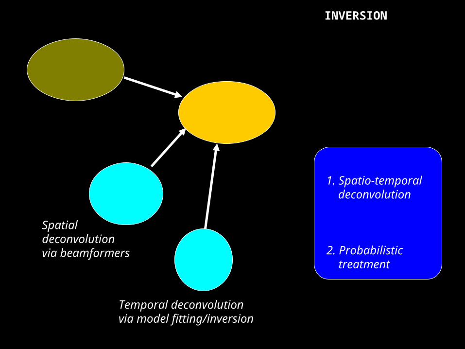
Spatialdeconvolution via beamformers
Temporal deconvolutionvia model fitting/inversion
INVERSION
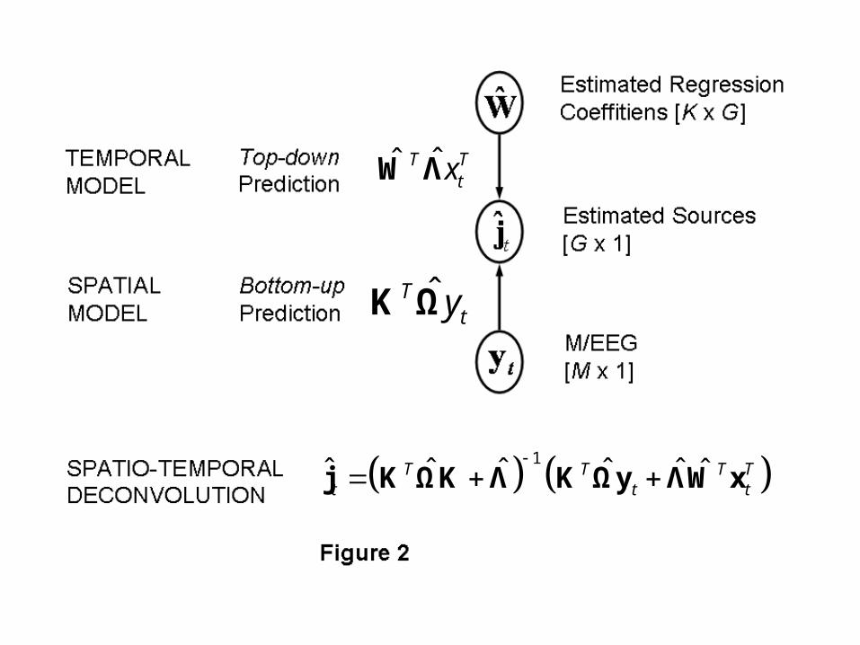
1. Spatio-temporal deconvolution
2. Probabilistic treatment

OverviewOverview
Spatio-temporal deconvolution for M/EEG
Spatio-temporal deconvolution for fMRI
Towards models for multimodal imaging

Spatio-temporal deconvolution for M/EEG
Add temporal constraints in the form of a General Linear Model to describe the temporal evolution of the signal.
Puts M/EEG analysis into same framework as PET/fMRIanalysis.
Work with Nelson. Described in chapter of new SPMbook.


1
1
1
:, ,
:, ,
1,: ,
tM T M G G T M T
TtG T K T G K G T
TT TkK G K G
k
t N
t N
k N
Y K J E e E 0 Ω
J X W Z z Z 0 Λ
W β β Β 0 D D
, 1, ,
, 1, ,
m
g
diag m M
diag g G
Ω
Λ
,g gg Ga b c ,m Ga b c ,k Ga b c
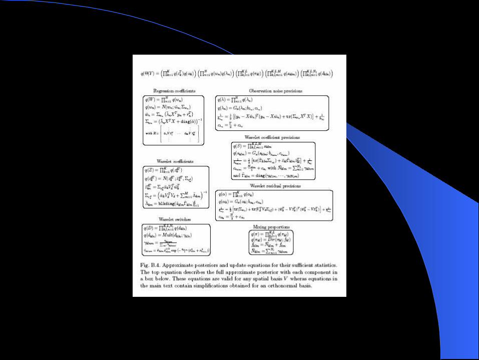
Generative Model:
Hyperpriors:
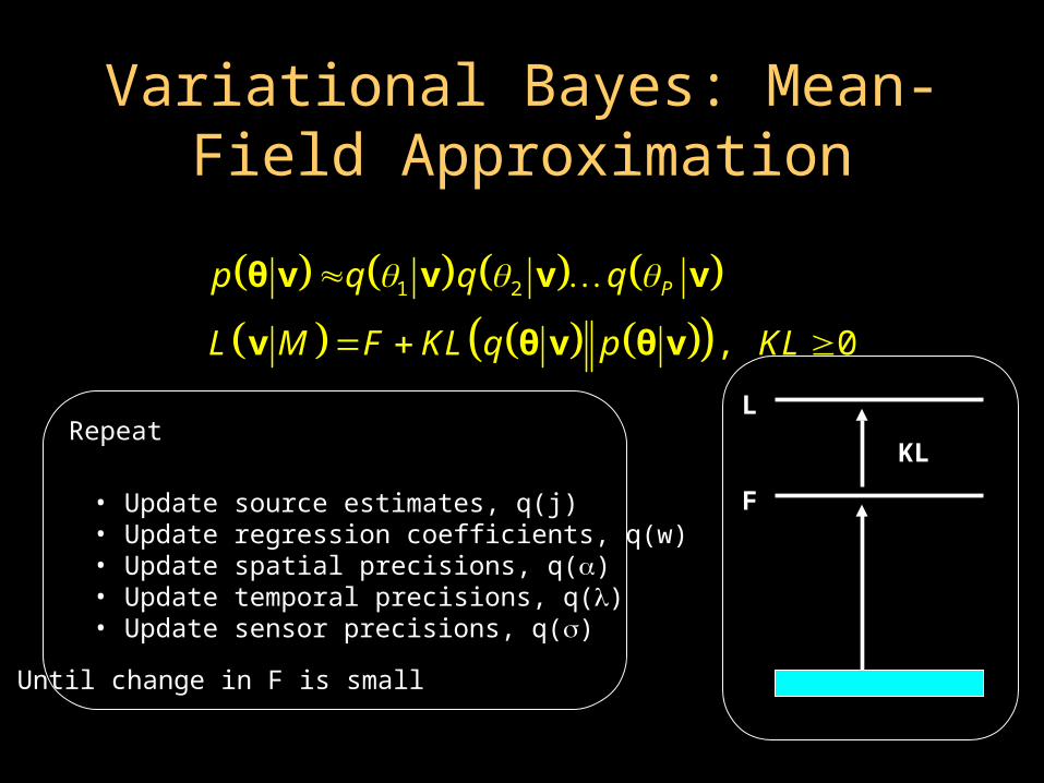
Variational Bayes: Mean-Field Variational Bayes: Mean-Field ApproximationApproximation
Repeat
• Update source estimates, q(j)• Update regression coefficients, q(w)• Update spatial precisions, q()• Update temporal precisions, q()• Update sensor precisions, q()
Until change in F is small
L
F
KL
1 2
, 0
Pp q q q
L M F KL q p KL
θ v v v v
v θ v θ v
1 11
, , , ,G
T g Kg
q q q q q q
θ j j w Ω Λ
11
, ,T
T tt
q q
j j j ˆ ˆ,tt tq N jj j Σ
ˆˆ ,gg gq N ww w Σ 1
1
, ,G
G gg
q q
w w w
1
G
gg
q q
Λ
1
M
mm
q q
Ω ˆ ˆ,m post postq Ga b c
ˆ ˆ,g post postq Ga b c
11
, ,K
K kk
q q
ˆ ˆ,kq Ga b c
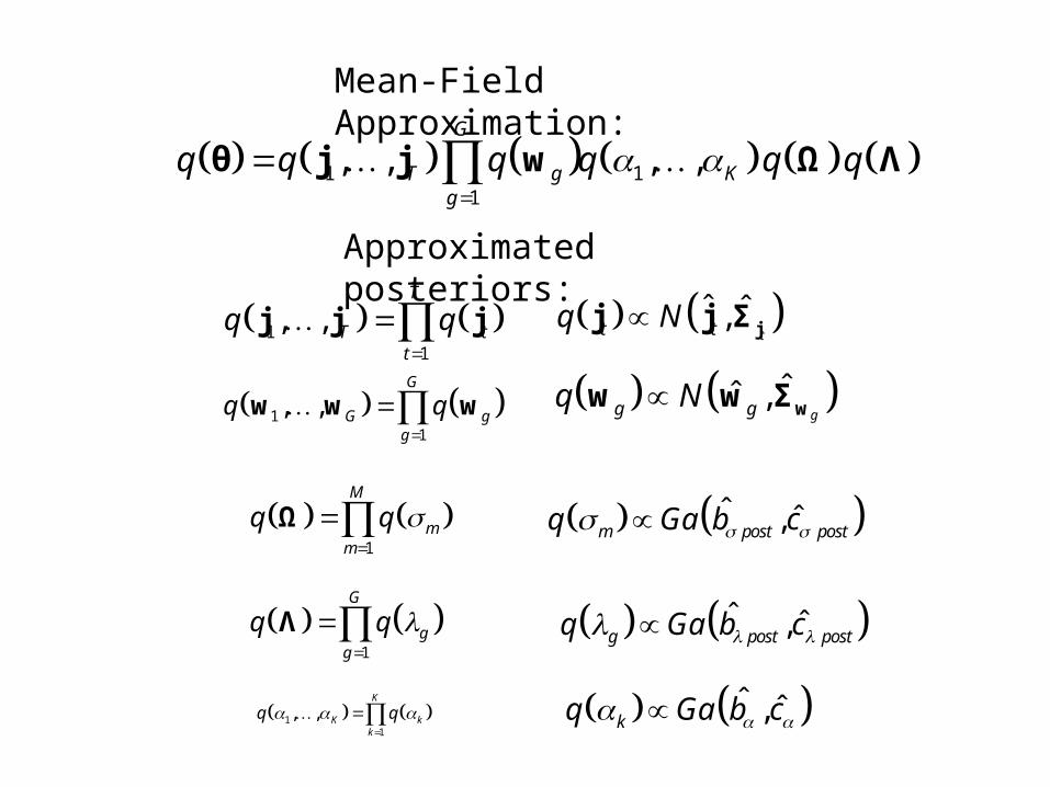
Mean-Field Approximation:
Approximated posteriors:
1ˆ ˆ ˆ ˆ ˆ ˆT T T Tt t t
j K ΩK Λ K Ωy ΛW x
ˆTtyK Ω
ˆˆ T TtxW Λ
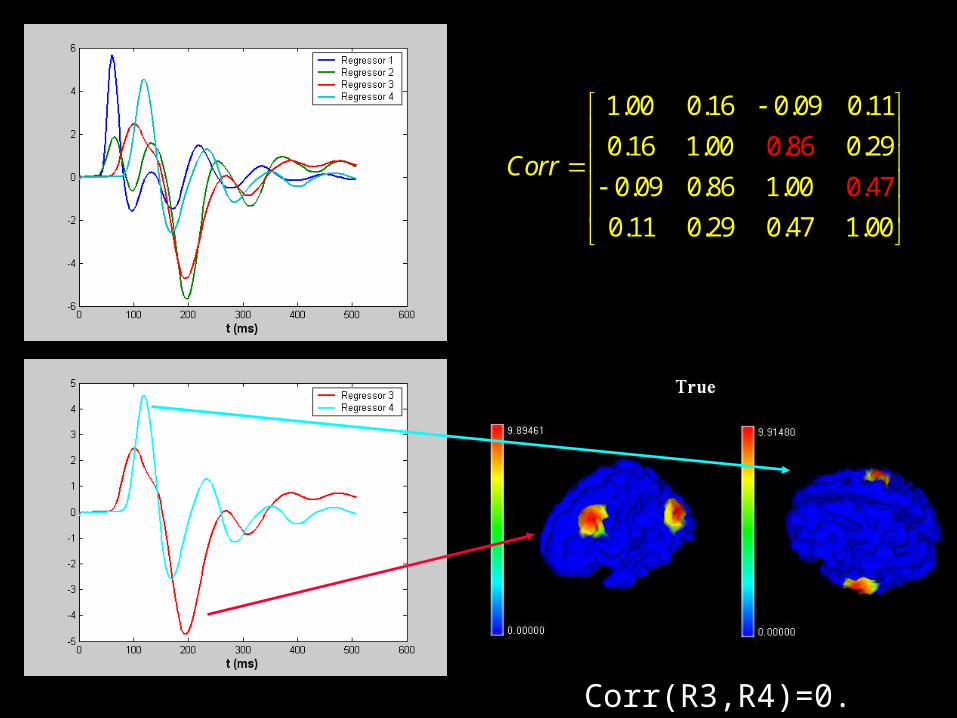
Corr(R3,R4)=0.47
1.00 0.16 0.09 0.11
0.16 1.00 0.29
0.09 0.86 1.00
0.11 0.29 0.47 1.
0.86
0.4
00
7Corr


o
+
500ms
LowSymmetry
LowAsymmetry
HighSymmetry
HighAsymmetry
Phase 1
Time
600ms
+700ms
+
o
2456ms
+
Fa
+
Sb
Ub
+
Sa
Henson R. et al., Cerebral Cortex, 2005
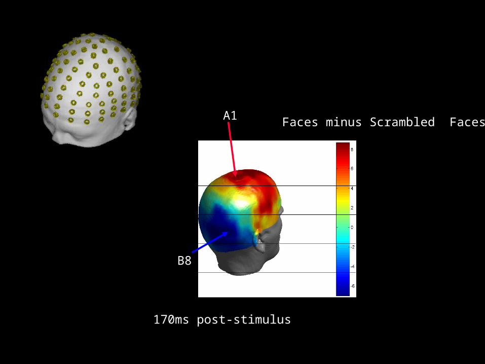
B8
A1 Faces minus Scrambled Faces
170ms post-stimulus
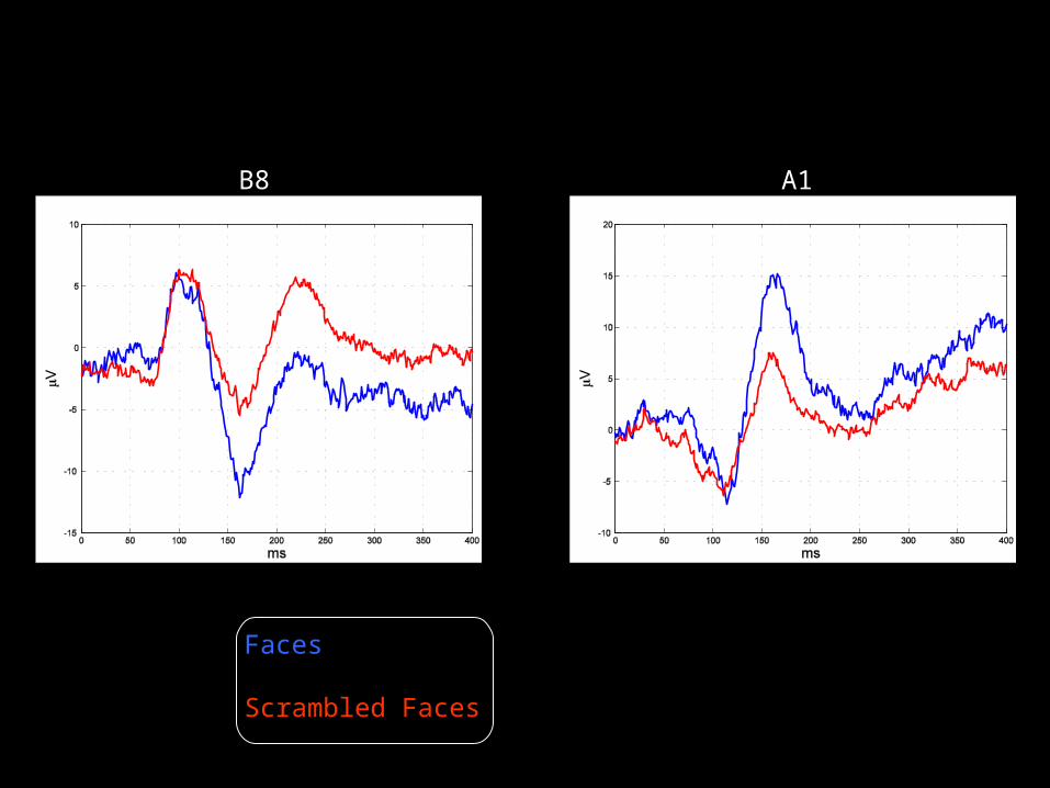
B8 A1
Faces
Scrambled Faces
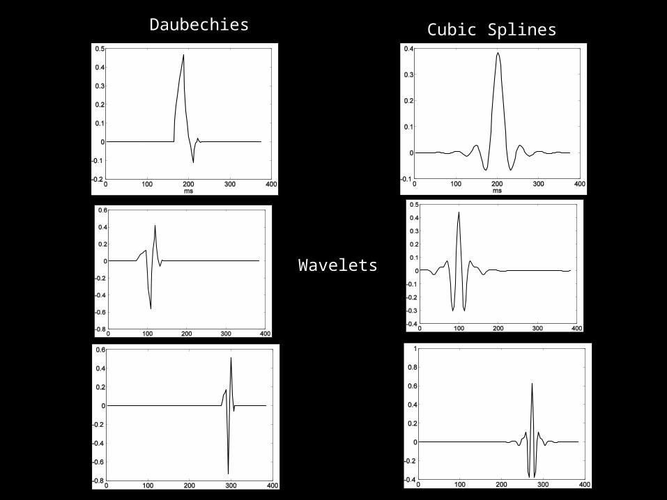
Daubechies Cubic Splines
Wavelets

28 Basis Functions 30 Basis Functions
Daubechies-4
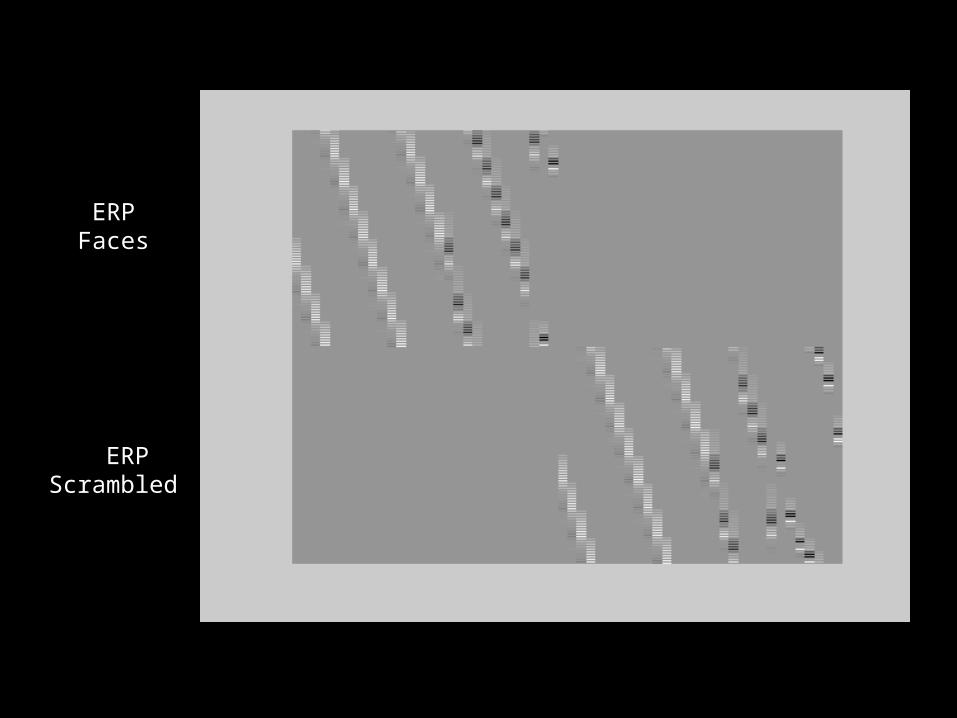
ERP Faces
ERPScrambled
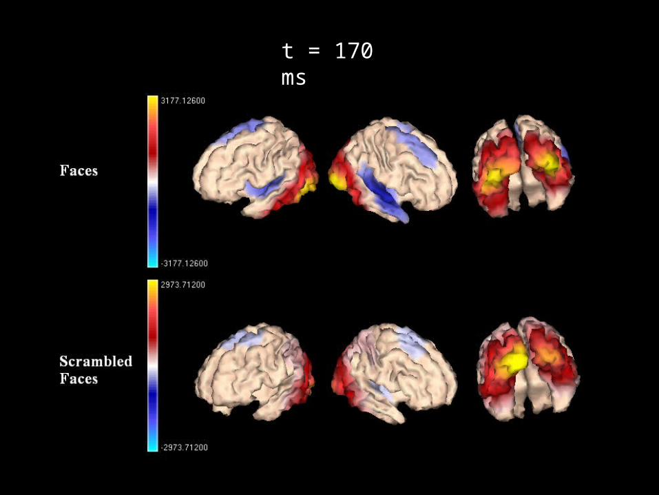
t = 170 ms
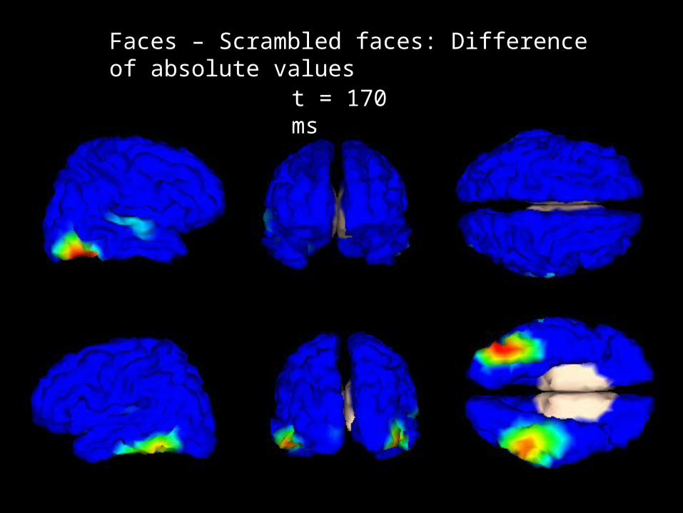
t = 170 ms
Faces – Scrambled faces: Difference of absolute values

Spatio-temporal deconvolution for fMRI
Temporal evolution is described by GLM in the usual way.
Add spatial constraints on regression coefficients in the form of a spatial basis set eg. spatial wavelets.
Automatically select the appropriate basis subset using a mixture prior which switches off irrelevant bases.
Embed this in a probabilistic model.
Work with Guillaume. To appear in Neuroimage very soon.

Spatial Model eg. Wavelets
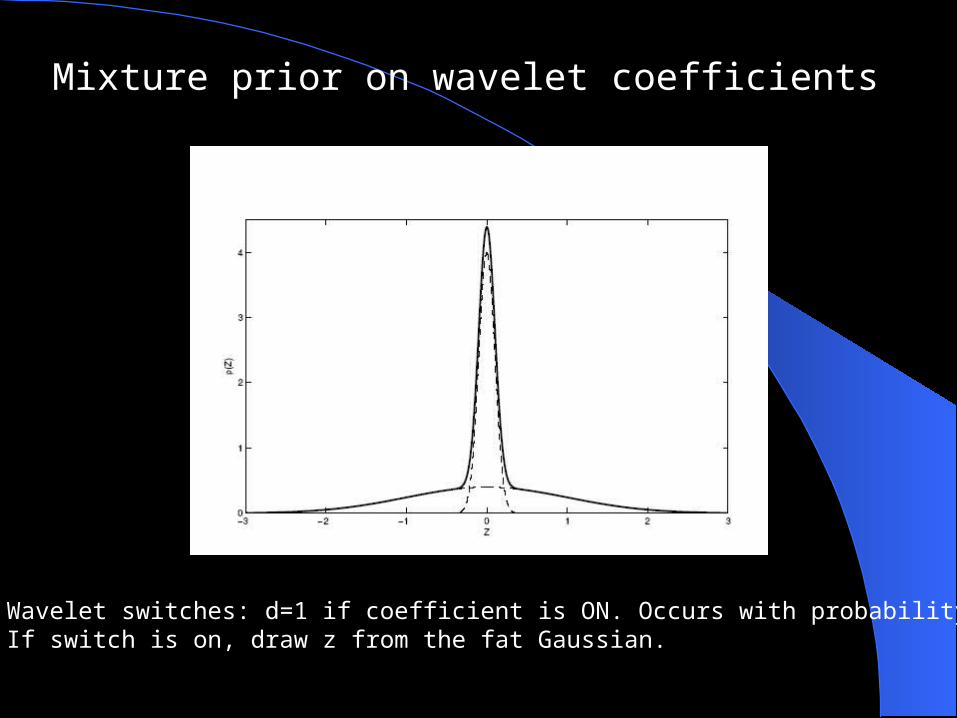
Mixture prior on wavelet coefficients
(1) Wavelet switches: d=1 if coefficient is ON. Occurs with probability (2) If switch is on, draw z from the fat Gaussian.

Probabilistic Generative Model
fMRI data
General LinearModel
Waveletcoefficients
TemporalModel
Spatial Model
Waveletswitches
Switchpriors

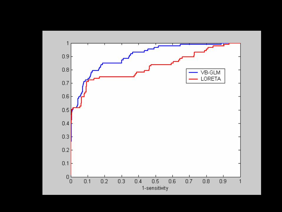
Compare to (i) GMRF prior used in M/EEG and (ii) no prior
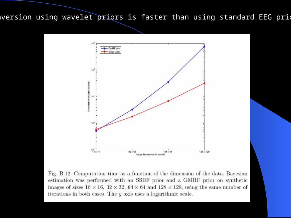
Inversion using wavelet priors is faster than using standard EEG priors

Results on face fMRI data

Towards multimodal imaging
Use simultaneous EEG- fMRI to identify relationship Between EEG and BOLD (MMN and Flicker paradigms)
EEG is compromised -> artifact removal
Testing the `heuristic’
Start work on specifying generative models
Ongoing work with Felix Blankenburg and James Kilner
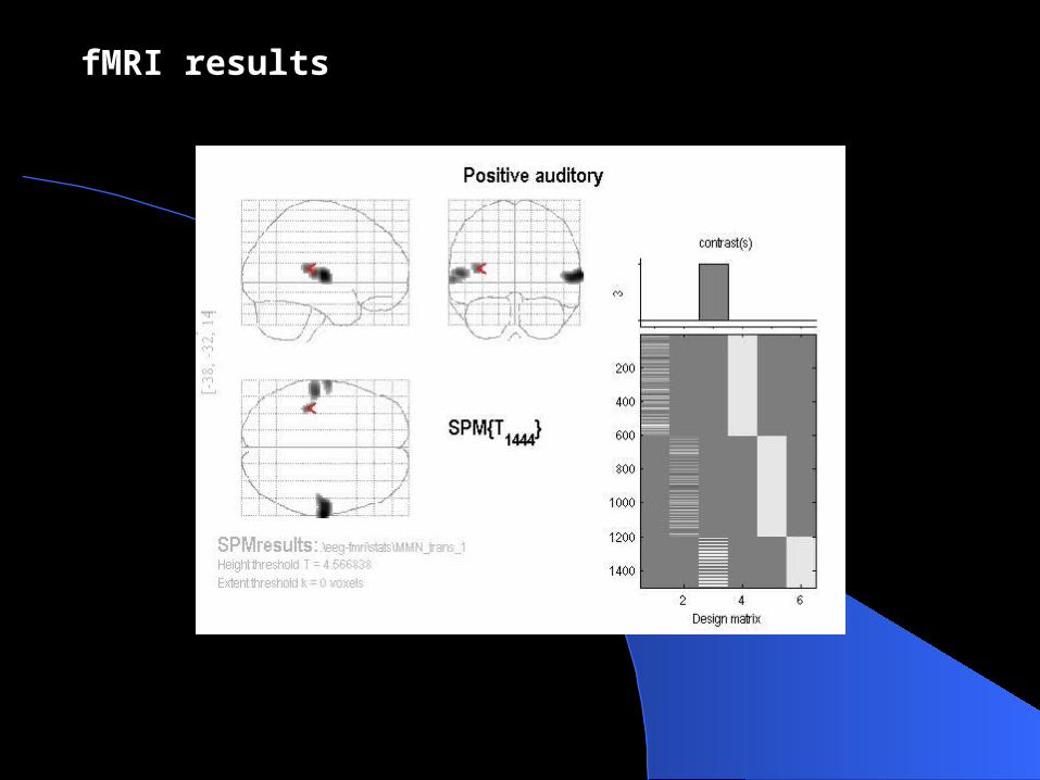
fMRI results

fMRI results
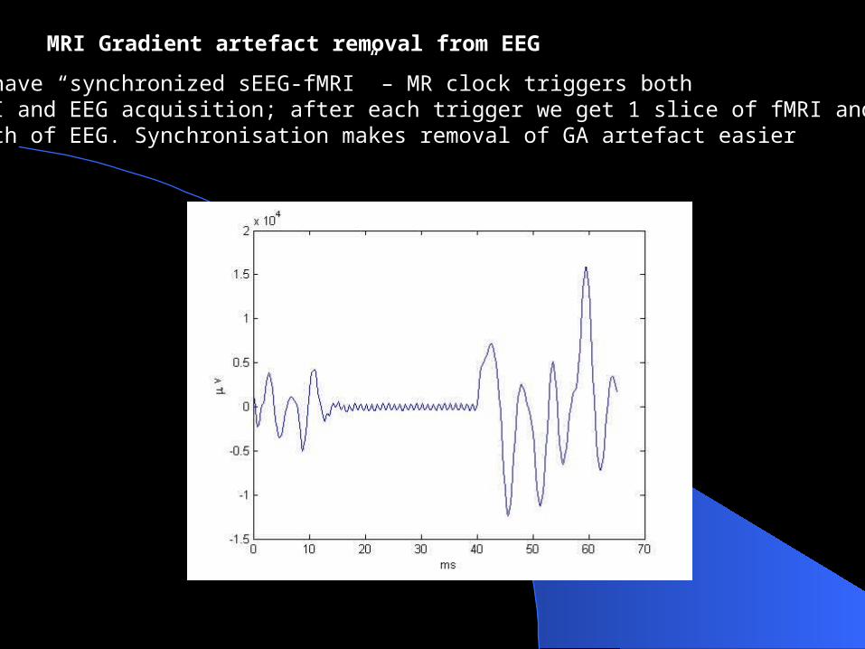
We have “synchronized sEEG-fMRI” – MR clock triggers both fMRI and EEG acquisition; after each trigger we get 1 slice of fMRI and 65ms worth of EEG. Synchronisation makes removal of GA artefact easier
MRI Gradient artefact removal from EEG
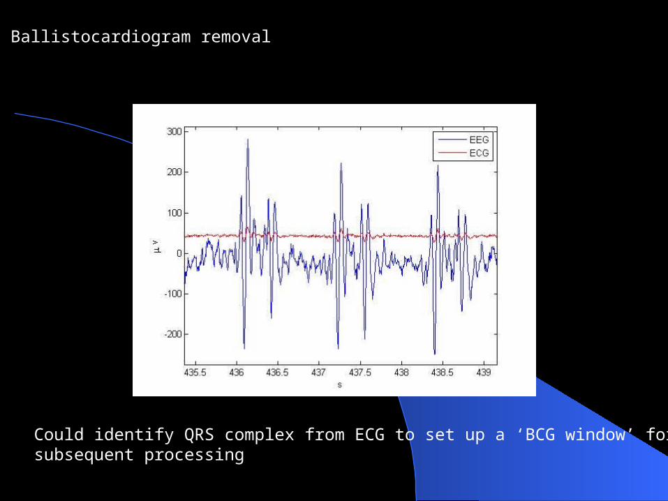
Ballistocardiogram removal
Could identify QRS complex from ECG to set up a ‘BCG window’ for subsequent processing
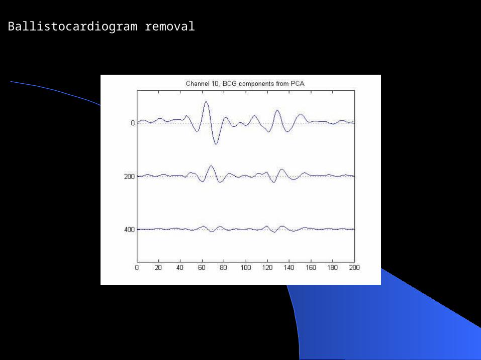
Ballistocardiogram removal
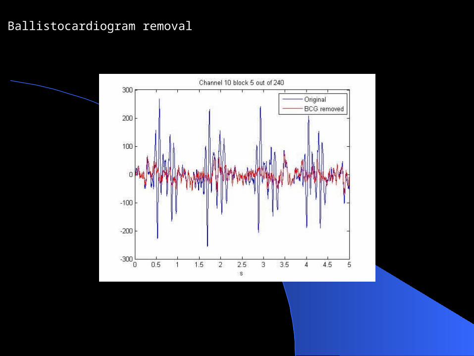
Ballistocardiogram removal

The EEG-BOLD heuristic (Kilner, Mattout, Henson & Friston) contends that increases in average EEG frequency predict BOLD activation.
g(w) = spectral density
Testing the heuristic

RMSF for Marta’s data at Cz
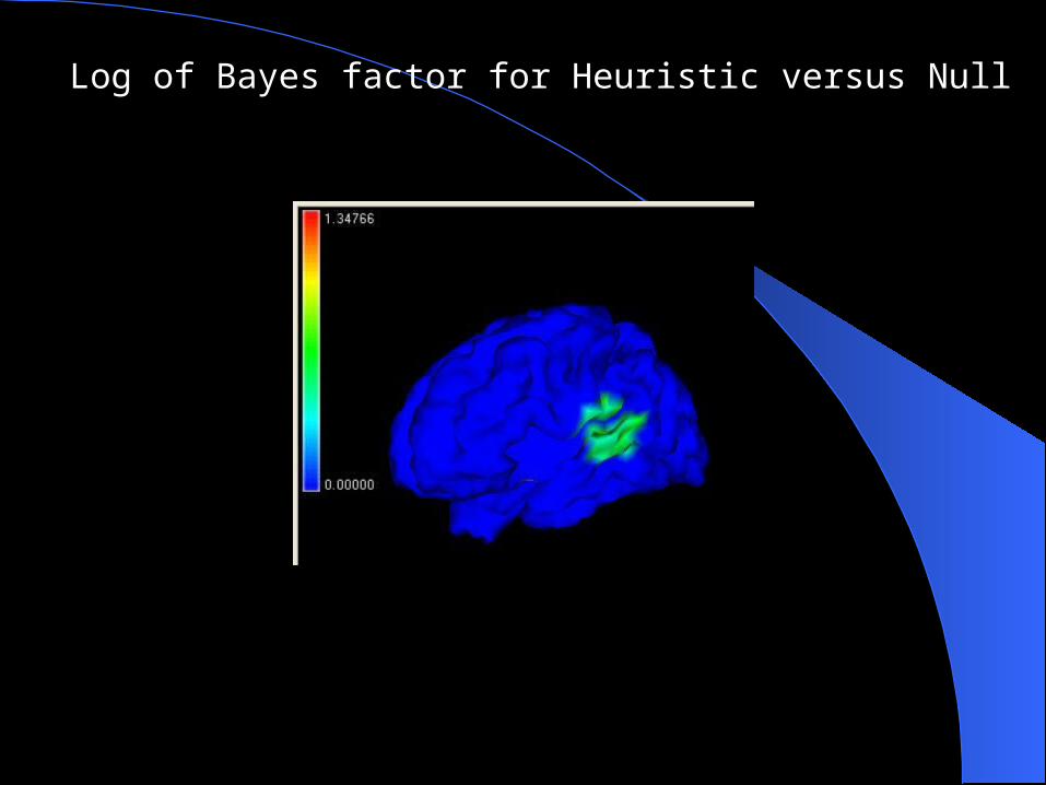
Log of Bayes factor for Heuristic versus Null
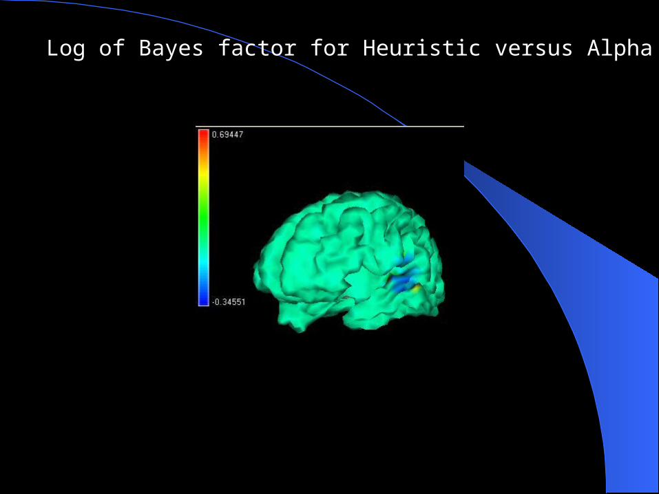
Log of Bayes factor for Heuristic versus Alpha

Tentative probabilistic generative model
THANK-YOU FOR
YOUR ATTENTION !
Top Related