


Languages
Pages
Legal


European Scientific Journal January 2020 edition Vol.16, No.3 ISSN: 1857 – 7881 (Print) e - ISSN 1857- 7431
177
Mold Occurrence in Fresh Chilli Pepper
(Capsicum spp.) Harvested Directly in the
Field in Benin Republic
Nicéphore M. Glodjinon, Msc
Pacôme A. Noumavo, PhD
Kifouli Adéoti, PhD
Hermann Savi, Msc
Kamal Garba, PhD
Sonangnon S. Kouhoundé, PhD
Fatiou Toukourou, PhD Université d’Abomey-Calavi, Laboratoire de Microbiologie et des
Technologies Alimentaires, Bénin
Lamine Baba-Moussa, PhD Université d’Abomey-Calavi, Laboratoire de Biologie et de Typage
Moléculaire en Microbiologie, Faculté des Sciences et Techniques, Bénin
Aly Savadogo, PhD Université Ouaga I Pr Joseph Ki-ZERBO, Laboratoire de Biochimie et
Immunologie Appliquée, Burkina Faso
Farid Baba-Moussa, PhD Université d’Abomey-Calavi, Laboratoire de Microbiologie et des
Technologies Alimentaires, Bénin
Doi:10.19044/esj.2020.v16n3p177 URL:http://dx.doi.org/10.19044/esj.2020.v16n3p177
Abstract
Introduction: The chilli pepper (Capsicum spp.), ranked among the
world's leading spices or food additives, is now Benin's second most-important
vegetable crop after tomatoes. Unfortunately, chilli peppers are likely to be
contaminated with mold which produces dangerous mycotoxins due to
cultural practices, transport, and post-harvest storage. Objective: The purpose
of this study is to isolate and identify the molds that contaminate chilli pepper
varieties in open fields according to the cultivation methods used in the
Republic of Benin. Materials and Methods: A total of 240 samples of three
varieties of chilli peppers were taken directly from six districts of Benin. The
molds were isolated and purified on a PDA (Potato Dextrose Agar) medium
for identification. The identification focused on the morphological and cultural

European Scientific Journal January 2020 edition Vol.16, No.3 ISSN: 1857 – 7881 (Print) e - ISSN 1857- 7431
178
characteristics of isolated strains. Results: Nine (9) fungal genera from
different taxonomic groups were detected. The genera that have been
represented are Aspergillus (34%), Fusarium (21%), Penicillium (16%),
Alternaria (7%), Cladosporium (7%), Mucor (7%), Scytalidium (4%),
Trichophyton (3%), and Rhizopus (1%). Conclusion: The present study shows
that chilli pepper is being contaminated in Benin. The genera Aspergillus,
Fusarium, Penicillium, and Alternaria are respectively the main toxinogenic
molds that contaminate peppers in the field. The mold control in chilli pepper
against pathogenic agents became urgently required to reduce a consumer
disease caused by chilli pepper in Benin.
Keywords: Benin, Chilli Pepper, Molds, Mycotoxins, Occurrence
1. Introduction
Around the world, chilli pepper is classified among the leading spices
or food additives. The chilli pepper (Capsicum spp.) is now the second in
market garden crops after tomatoes in Benin (INRAB, 2009). It is rich in
vitamins (A, B6, C, E, K); in trace elements such as copper, iron, manganese,
and potassium; and also in ascorbic acid, flavonoids, oligosaccharides, and
carotenoid compounds (Orobiyi, 2015). In Benin, chilli pepper is one of the
most produced and consumed vegetable crops undeniably occupying a
prominent place in the Beninese household. Several varieties of chilli pepper
are used in fresh fruit directly from the fields, in dried fruit from the
conservation, and transformed into paste or powder for the preparation of
different foods. In addition to food, chilli pepper is also used for its medicinal
properties (FAO, 2016). In phyto-medicine, the fruit is used orally as an
infusion to improve blood circulation, treat nausea, high blood pressure,
diabetes, and overweight (Lejeune et al., 2003; Koffi et al., 2014). The main
interest of chilli pepper is in its physicochemical composition (Tilahun et al.,
2013) which varies according to its varieties and environmental conditions
(Bae et al., 2012; Toffa, 2015). Its richness in antioxidants among which
capsaicin is found gives it a great nutraceutical value (nutritional and
medicinal). Unfortunately, the presence of fungal contaminants on chilli
pepper varieties is effective. Chilli pepper is likely to be contaminated with
Xerophilous xerophilic toxigenic fungi in the field, after harvest, during
transport, and during storage (FAO/WHO, 2017). Indeed, the cultural
practices used could have a significant impact on the chilli pepper
contamination in the field, especially in the humid lowlands. Also, the
improper storage or preservation methods used and the unsuitability of certain
varieties for conservation would also lead to attacks by micro-organisms
including molds of the genus Aspergillus, Penicillium, Fusarium, and
Alternaria, etc. (Zinedine, 2004; Nguyen, 2007; Debodé et al., 2016). They

European Scientific Journal January 2020 edition Vol.16, No.3 ISSN: 1857 – 7881 (Print) e - ISSN 1857- 7431
179
are heterotrophic eukaryotic unicellular or multicellular fungi of complex
structure, endowed with the power of sporulation, which grow on the surface
of foodstuffs because of their aerobic nature (Toffa, 2015; Meilé, 2017). They
are the most known contaminants of chilli pepper. Once on the chilli pepper,
these micro-organisms alter their sanitary quality causing, thus, enormous
economic losses (INRAB, 2009; Fondio et al., 2015). Generally,
contamination evolves after harvest, storage, or storage based on a
combination of intrinsic and extrinsic factors (Royer & Tap, 2004; Toffa,
2015). There is no doubt that molds have a remarkable ability to adapt.
Therefore, a change in a technological process can lead to a quantitative and
qualitative change in mycoflora. A large number of mold species belonging
mainly to the three very common genera, Aspergillus, Penicillium, and
Fusarium (Zinedine 2004; Nguyen, 2007; Debodé et al., 2016), present in
ambient air and soil, of agricultural crops are able, for example, to develop on
certain substrates such as peanuts, spices, coffee, and cereals to synthesize and
excrete mycotoxins (Garba et al., 2014; Ba et al., 2016; Tovidé et al., 2018).
Basically, these are products of their secondary metabolism, and they are very
toxic for humans and animals (Tozlovanu, 2008; Toffa, 2015; Debodé et al.,
2016; Meilé, 2017). Productive mold may also disappear from the food, while
mycotoxins, which are very stable, may persist even after heat treatment
(Toffa, 2015; Debodé et al., 2016). Therefore, a better knowledge of chilli
pepper infective mold in Benin and mycotoxin production potential of mold
strains will facilitate better risk prediction and corrective measures in Hazard
Analysis Critical Control Point (HACCP) system. Therefore, the objective of
this work is to study the variability of the contaminating molds of chillies
according to the technical production, storage, or conservation routes.
2. Material and Methods
2.1. Sampling
The chilli peppers were sampled in southern Benin in the district of
Ouémé, Plateau, Zou, Collines, Atlantique, and Littoral (Figure 1). The three
main varieties most produced and consumed in Benin were the subject of this
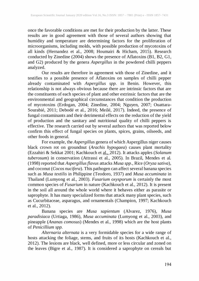
study. These varieties are: variety of round form called Gbotakin in the local
language of Fon (Capsicum chinense), long-sized variety called Afundja in the
local language of Fon (Capsicum annuum), and the very small variety called
Danhometakin in the local language of Fon (Capsicum frutescens) (Photo 1).
A total of 240 samples of three varieties of chilli peppers were collected
directly in the communes of the six districts mentioned.

European Scientific Journal January 2020 edition Vol.16, No.3 ISSN: 1857 – 7881 (Print) e - ISSN 1857- 7431
180
Figure 1. Figure showing the location of communes surveyed by district
Photo 1. Chilli pepper varieties used in this study.
[A] Fresh Gbotakin variety, [B] Fresh Afundja variety, [C] fresh Danhometakin variety.
2.2. Enumeration of Aerobic Total Mesophilic Flora
The total mesophilic aerobic flora was counted by the bulk inoculation
technique on Plate Count Agar (PCA) culture medium from the decimal
dilutions (10-5, 10-6, and 10-7) prepared. This was done by adding 10g of
sample to 90ml of a 0.1% sterile buffered peptone water solution. Then, 1ml
of each dilution was put into two boxes kneaded by dilution. Since the
inoculum was already in the dishes, about 15ml of the super-cooled culture

European Scientific Journal January 2020 edition Vol.16, No.3 ISSN: 1857 – 7881 (Print) e - ISSN 1857- 7431
181
medium was then poured and the whole was then incubated at 30°C for 72
hours after solidification. After culture, only boxes with a colony count
between 30 and 300 were taken into account for the different statistical
analysis.
2.3. Enumeration of Molds
Dilutions 10-1, 10-2, and 10-3 were prepared by adding 10g of sample to
90ml of a sterile 0.1% peptone water solution. Then, 0.1ml of each dilution
was inoculated on Sabouraud surface with Chloramphenicol at the rate of two
Petri dishes by dilution and then incubated at 25°C. Counting was performed
from the third day of incubation by evaluating the number of colony-forming
units per gram of chilli pepper (CFU/g) for 5 days.
2.4. Characterization of Molds
2.4.1. Isolation, Purification, and Conservation of Strains
From colonies obtained on Sabouraud agar Chloramphenicol, those
with mold features were separated by isolation. The latter were placed directly
on a standard selective medium such as Chloramphenicol PDA (Potato
Dextrose Agar) agar and the petri dishes were incubated at 25°C for 7 days.
The strains formed were isolated again and then purified according to the
technique of successive subcultures in peak by exhaustion of fungi. The last
colonies pushed on the successive subculturing points by exhaustion
constituted the pure strains. The pure strains obtained were stored in buffered
peptone water at 4°C for later identification.
2.4.2. Morphological Identification of Isolated Strains
The morphological identification of mold strains was based on the
cultural characteristics and the morphology of fructifications and spores. Also,
the identification keys used were those of Raper and Fennell (1965) and Pitt
and Hocking (1985) recently described and reported with some modifications
by Campbell et al. (2013) and Dufresne and St Germain (2018). These
methods have been used by several authors for the morphological
(macroscopic and microscopic) identification of molds of the genus
Aspergillus, Penicillium, Fusarium, and thermophilic filamentous fungi
(Cooney & Emerson, 1964; Chabasse et al., 2002; Lecellier, 2013; Houmairi
& Hicham, 2015; Compaore et al., 2017).
2.4.2.1. Macroscopic Characteristics
The macroscopic characteristics of isolated and purified mold strains
on PDA and MEA agar were determined with the naked eye with cultures of
at least 7 days incubated at 25°C. This is mainly the shape, size, color,
appearance, contour, and growth rate (fast, medium, slow) of fungal colonies.

European Scientific Journal January 2020 edition Vol.16, No.3 ISSN: 1857 – 7881 (Print) e - ISSN 1857- 7431
182
2.4.2.2. Microscopic Identification
The superficial part of the fungal mycelium was removed with a sterile
platinum loop and placed on a slide to which lactophenol blue was added as a
diluent. The characteristics noted were: the color, appearance and size of
conidiophores, the number of conidia, the shape of the head (aspergillary,
penicillary, and fusariare), differentiation of thallospores, mode of branching
of septa, and the presence of metulae. The observation was made with the
Optical Microscope-LCD Digital coupled to a camera for the capture of
images. Magnifications (× 10) (× 40) and immersion magnification (× 100)
were used.
2.5. Statistical Analysis
The data collected from the results of manipulations on the collected
samples were captured using the Excel 2016 software. The results of the
enumeration of the aerobic total mesophilic flora (TAMF) and that of the
yeasts and molds (YM) were submitted to the LSD-Test (Fisher's Least
Significant Difference-Test) and the Student-Newman-Keuls test using
Statistical Analysis System (SAS) version 8.1. This was performed in order to
compare the averages between the contamination loads of the studied peppers
and to determine the significant differences between the means of the districts.
The difference was considered statistically significant when the value of p <
0.05. The geographical map of the sampling zones was carried out using Qgis
software version 2.14.
3. Results
3.1. Global Contamination of Chilli Peppers Studied by Microorganisms
Figure 2. Microbial load in total mesophilic Figure 3. Microbial load in yeasts
and molds (YM) aerobic flora (TAMF)

European Scientific Journal January 2020 edition Vol.16, No.3 ISSN: 1857 – 7881 (Print) e - ISSN 1857- 7431
183
Figure 4. Degree of contamination in TAMF and YM of studied chilli peppers
Figure 5. Microbial load in mold of studied chilli peppers
The means between districts with the same letters are not significantly different at the 5%
probability level according to Fisher's LSD test and = p ˃ 0.05 (no difference); * = p <0.05
(significant); ** = p <0.01 (highly significant); *** = p <0.001 (very highly significant).
3.2. Morphological Characterization of Isolated Strains
Purified molds were identified by macroscopic examination that was
performed after incubation for at least 7 days on PDA medium with
choramphenicol at a temperature of 25°C, and slide microscopic examination
with lactophenol added as diluent, gives results which have been mentioned
below (Table 1).
A total of seventy-six (76) mold strains were isolated, purified, and
characterized from 240 chilli pepper samples. Thus, Table 1 summarizes the
appearance of the mycelium of some isolated fungal strains, the consistency
of the colonies, the color of the back of the box, and the presence or absence
of pigments characteristic of each strain. Table 2 presents the representative
descriptive characteristics of each of the isolated fungal genera and their
reference images.

European Scientific Journal January 2020 edition Vol.16, No.3 ISSN: 1857 – 7881 (Print) e - ISSN 1857- 7431
184
Table 1. Macroscopic and microscopic characteristic (G X 40) of the molds grew at 250C on PDA medium
CODES MACROSCOPIC OBSERVATION
(BOTH & SIDES)
MICROSCOPIC OBSERVATION CHARACTERISTICS
SPECIES
FUNGAL
GENERA
M9R3
*Color
-revers: Beige
-mycelium: white
-spores: Yellowish green
* diameter: 4 cm
* growth: rapid
* appearance : powdery
Aspergillus sp.
M109R1
*Color
-revers: yellow rack
-mycelium: white
-spores: black
* diameter: 5cm
* growth: rapid
* appearance: granular
and floury
Aspergillus sp.

European Scientific Journal January 2020 edition Vol.16, No.3 ISSN: 1857 – 7881 (Print) e - ISSN 1857- 7431
185
M46R3
*Color
-revers: beige
-mycelium: green
-spores: green
* diameter: 1, 2cm
* growth: rapid
* appearance: powdery
Aspergillus
sp.
M38R3
*Color
-revers: mustard yellow
-mycelium: light pink
-spores: light pink
* diameter: 7.1cm
* growth: rapid
* appearance : ovoid and
cottony
Fusarium sp.
M89R5
*Color
-revers: pure beige
-mycelium: white
-spores: white
* diameter: 5.5 cm
-growth: average
* appearance: cottony
Fusarium sp.

European Scientific Journal January 2020 edition Vol.16, No.3 ISSN: 1857 – 7881 (Print) e - ISSN 1857- 7431
186
M26R5
*Color
-revers: yellow
-mycelium: white
-spores: White, green
* diameter: 3.7 cm
* growth: average
* aspect: Wool
Penicillium
sp.
M1R3
*Color
-revers: Yellow
-mycelium: Green
-spores: green
* diameter: 1, 2 cm
* growth: rapid
* appearance : powdery
and invasive
Pénicillium sp.
M107R1
*Color
-revers: Beige, black
-mycelium: white
-spores: White
* diameter: 5, 8cm
* growth: rapid
* appearance: cottony
Alternaria sp.

European Scientific Journal January 2020 edition Vol.16, No.3 ISSN: 1857 – 7881 (Print) e - ISSN 1857- 7431
187
M59R4
*Color
-revers: Beige
-mycelium: White
-spores: Yellow
* diameter: 2, 3 cm
* growth: Average
* appearance: Cottony
and powdery
Cladosporium sp.
M125R4
*Color
-revers: yellow
-mycelium: yellow
-spores: yellow
* diameter: 8.2cm
* growth: rapid
* appearance : Cottony
Mucor sp.
M103R1
*Color
-revers: Black
-mycelium: white
-spores: white
* diameter: 1cm
* growth: slow
* appearance: cottony and
pleated
Mucor sp.

European Scientific Journal January 2020 edition Vol.16, No.3 ISSN: 1857 – 7881 (Print) e - ISSN 1857- 7431
188
M29R5
*Color
-revers: yellow
-mycelium: White
-spores: Beige
* diameter: 2, 9 cm
* growth: average
* appearance: Cottony
Trichophyton sp.
M122R1
*Color
-revers: White
-mycelium: white
-spores: white
* diameter: 8, 4 cm
* growth: rapid
* appearance : cottony
Scytalidium
sp.
M25R5
*Color
-revers: Beige
-mycelium: White
-spores: beige
* diameter: 3.9cm
* growth: average
* appearance: ovoid and
cottony
Rhizopus sp.

European Scientific Journal January 2020 edition Vol.16, No.3 ISSN: 1857 – 7881 (Print) e - ISSN 1857- 7431
189
Table 2. References of isolated fungal genera
Species Reference Picture (x40) Descriptive Characteristics
Aspergillus
spp.
Demonstration of aspergillary heads on
microscopic examination of colonies.
A vegetative thallus formed of hyaline
filaments, of fine and regular diameter,
septate and branched.
(Campbell et al., 2013)
Fusarium spp.
Conidiophores short and often
branched. They carry phialides that
may have one or more budding sites for
the production of conidia.
(Campbell et al., 2013)
Pénicillium
spp.
Hyphae septa, hyaline, bear
conidiophores simple or branched, are
sometimes grouped in bush or coremia.
The phialides are arranged in whorls at
the end of the conidiophores.
(Campbell et al., 2013)
Alternaria spp.
Hyphae, septate, are branched and later
some filaments are pigmented brown.
Conidiophores are septate, brown,
septate, simple or branched, more or
less straight or flexuous (geniculate).
(Dufresne & St Germain, 2018)

European Scientific Journal January 2020 edition Vol.16, No.3 ISSN: 1857 – 7881 (Print) e - ISSN 1857- 7431
190
Cladosporium
spp.
Hyphae, septate, are pigmented. Plain
or multicellular blastospores arranged
in acropetal chains.
(Campbell et al., 2013)
Mucor spp.
Cloisonne filaments, globular
sporocyst, round spores.
Broad filaments little gold no septate.
No stolons nor rhizoids.
(Campbell et al., 2013)
Scytalidium
spp.
The hyphae are regular, septate, and
hyaline.
(Chabasse et al., 2002)
Trichophyton
spp.
Macroconidia absent;
Microconidia club-shaped, formed
along the sides of the hyphae.
(Dufresne & St Germain, 2018)

European Scientific Journal January 2020 edition Vol.16, No.3 ISSN: 1857 – 7881 (Print) e - ISSN 1857- 7431
191
Rhizopus spp.
Mushroom with non-septate filaments.
Wide filaments not or slightly septate.
Stolons, rhizoids and sporocystophores
are well differentiated. The spores are
ovoid.
(ANOFEL, 2014)
Table 3. Relative number and frequency of isolated and characterized mold species on the
chilli peppers studied.
Fungal species Number Frequency Mold species
Aspergillus spp.
26
34%
Aspergillus niger
Aspergillus flavus
Aspergillus
ochraceus
Aspergillus
parasiticus
Aspergillus fumigatus
Aspergillus nidulans
Aspergillus flavipes
Aspergillus oryzae
Aspergillus spp.
Fusarium spp.
16
21%
Fusarium solani
Fusarium
moniliforme
Fusarium oxysporum Fusarium
proliferatum
Fusarium spp.
Pénicillium spp.
12
16%
Pénicillium expansum
Pénicillium
cyclopium
Pénicillium citrinum
Pénicillium notatum
Pénicillium
citreonigrum
Pénicillium spp.
Alternaria spp.
5 7% Alternaria alternata
Alternaria infectoria
Alternaria spp.
Cladosporium
spp. 5 7% Cladosporium spp.

European Scientific Journal January 2020 edition Vol.16, No.3 ISSN: 1857 – 7881 (Print) e - ISSN 1857- 7431
192
Mucor spp. 5 7% Mucor spp.
Trichophyton spp. 3 4% Trichophyton spp.
Scyatalidium spp. 2 3% Scytalidium
dimidiatum Scyatalidium spp.
Rhizopus spp. 1 1% Rhizopus spp.
Total number : 76 100% -
4. Discussion
The level of contamination of chilli peppers by the aerobic total
mesophilic flora, yeasts, and molds are presented in Figures 2 and 3.
According to these results, it can be seen that 34% of the chilli peppers studied,
all origins combined, have a mean microbial load in aerobic mesophilic flora.
It is quite high and is above the threshold recommended by the Quebec
standard (108 CFU/g) (CQIASA, 2009) with a maximum microbial load of
4.93.108 CFU/g. It also emerges from these results that there is a high
significance at the 5% threshold between the samples coming from the same
district and between certain district in a specific way. Thus, between the
district of Zou and that of the hills, there is a highly significant difference in
the aerobic total mesophilic flora. Also, there is also a highly significant
difference between these two districts and those of the Littoral, Atlantic, and
Plateau between where there is no differentiating significance.
Yeasts and molds are also above the recommended threshold (105
CFU/g) for spices, fresh fruits, and vegetables by the Quebec standard
(CQIASA, 2009) for all samples of chilli peppers studied. It should be noted
that there is no significant difference at the level of 5% threshold between
samples analyzed from the same district and from the six districts for yeast
and mold. Of the samples analyzed, Figure 4 shows that all districts are more
contaminated by yeasts and molds with the exception of Zou district which is
more contaminated by aerobic total mesophilic flora. These values are similar
and consistent with those reported by Kumar et al. (2008) and Fondio et al.
(2015) who have shown that the microorganisms that contaminated chilli
pepper varieties are more fungi, including yeasts and especially molds, than
bacteria.
Figure 5 shows the molds contamination of chilli peppers studied.
These results show that the district of Ouémé is the most contaminated area
by the mold. Opposite to Ouémé, the district of Zou is the least contaminated.
Also, there is a very high significance at the 5% threshold for the six study
districts with regard to the mold species contaminating the chilli peppers
studied. This high level of mold contamination observed in the district of
Ouémé is explained by the fact that the cultural practices carried out in this
district are those of a recession culture and of the lowland. Here, the humidity

European Scientific Journal January 2020 edition Vol.16, No.3 ISSN: 1857 – 7881 (Print) e - ISSN 1857- 7431
193
is quite important and it easily favors contamination and development of
microorganisms including fungi and especially molds. In fact, low-lying or
low-lying wetland crops are generally found in market gardens just at the
beginning of the flood, where the various types of waste left by the river such
as dead fish and other rotten debris are still there.
These results are in good agreement with those of several authors who show
that humidity and temperature are determining factors for the proliferation of
microorganisms, including molds with possible production of mycotoxins
(Pardo et al., 2005; Hernandez et al., 2008).
The characterization of the local strains of the mold species studied by
the present study reveals a contamination of three varieties of chilli pepper
(Capsicum chinense, Capsicum annuum, and Capsicum frutescens) produced
in Benin by seventy-six (76) fungal species from nine (9) taxonomic genera.
The results reveal a variety of fungal agents developing on chillies, thus,
affecting its health status and consequently its aesthetics. The genera that were
most represented are Aspergillus (34%), Fusarium (21%), Penicillium (16%),
Alternaria (7%), Cladosporium (7%), Mucor (7%), Scytalidium (4%),
Trichophyton (3%), and Rhizopus (1%). The dominant species in the genus
Aspergillus are Aspergillus niger (47%), Aspergillus flavus (26%), and
Aspergillus ochraceus (16%). In the genus Fusarium, Fusarium solani (41%),
Fusarium moniliforme (30%), and Fusarium oxysporum (19%) are the most
frequently isolated. On the other hand, Penicillium was Penicillium expansum
(43%), Penicillium cyclopium (36%), and Penicillium citrinum (23%).
Specific diversity was higher in the genus Aspergillus (twenty-six different
species) followed by the genus Fusarium (sixteen species) and the genus
Penicillium (twelve species).
The predominance of telluric genera such as Aspergillus, Penicillium,
Fusarium, or Rhizopus with other less common genera such as Cladosporium
in spice samples, fresh fruits, and vegetables such as chilli pepper has been
reported by several authors (Erdogan, 2004; Kumar, 2008; Houmairi &
Hicham, 2015). Several studies have pointed out that species that affect spices
are storage xerophiles such as Aspergillus spp. and Penicillium spp. or pre-
harvest contaminants such as Fusarium (Kachkouch et al., 2012; Houmairi &
Hicham, 2015). They are a group of molds normally confined to the soil but
found on spices, fresh fruits, and vegetables under precarious hygienic
conditions in relation to harvesting, drying, storage, and transport procedures
depending on whether they have undergone or not preventive treatments (Abu
Donia, 2008; Kachkouch et al., 2012).
The most contaminated chilli pepper samples would certainly have the
highest moisture levels and come from lowland or lowland wetland crops.
Also, poor farming practices are an undeniable cause of contamination of the
chilli pepper in the open field by molds and, therefore, later by mycotoxins
European Scientific Journal January 2020 edition Vol.16, No.3 ISSN: 1857 – 7881 (Print) e - ISSN 1857- 7431
194
once the favorable conditions are met for their production by the latter. These
results are in good agreement with those of several authors showing that
humidity and temperature are determining factors for the proliferation of
microorganisms, including molds, with possible production of mycotoxins of
all kinds (Hernandez et al., 2008; Houmairi & Hicham, 2015). Research
conducted by Zinedine (2004) shows the presence of Aflatoxins (B1, B2, G1,
and G2) produced by the genera Aspergillus in the powdered chilli peppers
analyzed.
Our results are therefore in agreement with those of Zinedine, and it
testifies to a possible presence of Aflatoxins on samples of chilli pepper
already contaminated with Aspergillus spp. in Benin. However, this
relationship is not always obvious because there are intrinsic factors that are
the constituents of each species of plant and other extrinsic factors that are the
environmental and geographical circumstances that condition the production
of mycotoxins (Erdogan, 2004; Zinedine, 2004; Nguyen, 2007; Ouattara-
Sourabié, 2011; Debodé et al., 2016; Meilé, 2017). Indeed, the presence of
fungal contaminants and their detrimental effects on the reduction of the yield
of production and the sanitary and nutritional quality of chilli peppers is
effective. The research carried out by several authors that was reported below
confirm this effect of fungal species on plants, spices, grains, oilseeds, and
other foods in general.
For example, the Aspergillus genera of which Aspergillus niger causes
black crown rot on groundnut (Arachis hypogaea) causes plant mortality
(Ezzahiri & Sekkat 2001; Kachkouch et al., 2012). It attacks apples (Solanum
tuberosum) in conservation (Attrassi et al., 2005). In Brazil, Mendes et al.
(1998) reported that Aspergillus flavus attacks Musa spp., Rice (Oryza sativa),
and coconut (Cocos nucifera). This pathogen can affect several banana species
such as Musa textilis in Philippine (Teodoro, 1937) and Musa accuminata in
Thailand (Lumyong et al., 2003). Fusarium oxysporum is certainly the most
common species of Fusarium in nature (Kachkouch et al., 2012). It is present
in the soil all around the whole world where it behaves either as parasite or
saprophyte. It has many specialized forms that attack many plant species, such
as Cucurbitaceae, asparagus, and ornamentals (Champion, 1997; Kachkouch
et al., 2012).
Banana species are Musa sapientum (Alvarez, 1976), Musa
paradisiaca (Urtiaga, 1986), Musa accuminata (Lumyong et al., 2003), and
pineapple (Ananas comosus) (Mendes et al., 1998) which are the host plants
of Penicillium spp.
Alternaria alternata is a very formidable species for a wide range of
hosts attacking the foliage, stems, and fruits of its hosts (Kachkouch et al.,
2012). The lesions are black, well defined, more or less circular and zoned on
the leaves (Bigre et al., 1987). It is considered a saprophyte on cereals but

European Scientific Journal January 2020 edition Vol.16, No.3 ISSN: 1857 – 7881 (Print) e - ISSN 1857- 7431
195
Wrather and Sweets (1999) reported that this species causes ear rot (Berber,
2008; Kachkouch et al., 2012).
Recent studies in Benin have revealed the presence of these molds in
seeds and groundnut cake (Arachis hypogaea) (Garba et al., 2014), on Maize
(Zea mays) (Ba et al., 2016), on millet (Pennisetum glaucum), and on sorghum
(Sorghum bicolor) grains (Tovidé et al., 2018). Other studies have also
confirmed the presence of molds including Aspergillus flavus in groundnut
seeds in Burkina-Faso (Ouattara-Sourabié, 2011) and, very recently, the
contamination of smoked and dried fish from Lake Fitri in the Batha region of
Chad by fungal strains has also been proven (Abdoullahi et al., 2019). Today,
these molds are the most well-known contaminants in chilli peppers and
unfortunately deteriorate to its sanitary quality, causing huge economic losses
(INRAB, 2009). Generally, contamination evolves after harvest, storage, or
storage based on all intrinsic and extrinsic factors (Royer & Tap 2004;
Nguyen, 2007).
It is certain that the present work constitutes a true base for the
evaluation of the current state of the chilli peppers produced in Benin.
Furthermore, it reveals, for the first time, the presence of nine (9) fungal
genera on the varieties of chilli pepper (Capsicum chinense, Capsicum
annuum, and Capsicum frutescens) including Alternaria spp., Cladosporium
spp., Mucor spp., Scytalidium spp., Trichophyton spp., and Rhizopus spp. in
addition to the best-known fungal species of agricultural and food products
such as Aspergillus spp., Penicillium spp., and Fusarium spp. Thus,
appropriate measures must be taken to improve the sanitary quality of chillies
before harvest in order to protect consumers against possible diseases caused
by molds and their toxins.
5. Conclusion
The present study included two hundred and forty (240) samples of
three chilli pepper varieties (Capsicum chinense, Capsicum annuum and
Capsicum frutescens) directly from the cultivated fields of six districts of
Benin. The results of the analysis showed that the majority of these are of
unsatisfactory sanitary quality and have a fairly high average mold load
predominantly dominated by several species of the genus Aspergillus (34%),
Fusarium (21%), Penicillium (16%), Alternaria (7%), Cladosporium (7%),
Mucor (7%), Scytalidium (4%), Trichophyton (3%), and a species of the genus
Rhizopus (1%). A total of seventy-six (76) fungal strains were isolated. Indeed,
no real study has been done until now on the fungal contamination of chilli
peppers produced and consumed in Benin. Thus, it is for the first time that the
fungal species Aspergillus spp., Fusarium oxysporum, Penicillium spp.,
Alternaria alternata, Cladosporium spp., Mucor spp., Scytalidium
dimitadium, Trichophyton spp., and Rhizopus spp. were isolated from chilli

European Scientific Journal January 2020 edition Vol.16, No.3 ISSN: 1857 – 7881 (Print) e - ISSN 1857- 7431
196
pepper varieties produced in Benin. Since some of these molds produce
mycotoxins that can cause serious illness in consumers at certain
concentrations, it is necessary to improve the cultural practices and to respect
the technical production routes of chilli pepper recommended by the
competent structures for the cultivation of chilli pepper as well, in the humid
lowlands which is only on the mainland.
Acknowledgment
We thank the France Campus who through the Service Cooperation
and Cultural Action (SCCA) of the Embassy of France provided the financial
support we needed for this research.
References:
1. Abou-Arab, A.A.K., Soliman, A.K., EL Tantawy, M.E., Ismail, B.R.,
& Khayria, N. (1999). Estimation quantitative de certains
contaminants dans les plantes médicinales d'usage courant sur le
marché égyptien. Food Chemistry, 67: 357-363.
2. Abou Donia, M.A. (2008). Métaux lourds dans les épices et les plantes
médicinales égyptiennes et effet du traitement sur leur teneur. Global
veterinaria, 2(4): 175-181.
3. Alvarez, M.G. (1976). Catalogue de projets d'enquêtes de plantes
mexicaines. Fitofilo, 71 : 1-169.
4. Association Française des Enseignants de Parasitologie et Mycologie
(ANOFEL). (2014) : Aspergilloses et autres champignons filamenteux
opportunistes. 1-19 pp.
5. Attrassi, K., Selmaoui, K., Ouazzani-touhami, A., Badoc, A. & Douira,
A. (2005). Biologie et physiologie des principaux agents fongiques de
la pourriture des pommes en conservation et lutte chimique par
l’azoxystrobine. Bull. Soc. Pharm. Bordeaux, 144: 47-62.
6. Ba, R., Monteiro, N.M.F., Hounguè, U., Donou Hounsodé, M.T.,
Gbaguidi, F. & Baba-Moussa, L. 2016. Perception des producteurs et
impact des facteurs socio-économiques sur la connaissance des
mycotoxines du maïs en stockage au Bénin. Int. J. Biol. Chem. Sci.
10(1): 155-166.
7. Bae, H., Jayaprakasha, G.K., Jifon, J. & Patil, B.S. (2012). Variation
of antioxidant activity and the levels of bioactive compounds in
lipophilic and hydrophilic extracts from hot pepper (Capsicum spp.)
cultivars. Food Chemistry, 134: 1912-1918.
8. Berber, F., Ouazzani-Touhami, A. & Douira, A. (2008). Identification
de la mycoflore pathogène de Sorghum bicolor (L.) Moench, cultivé
dans le Gharb et le Loukkos (Nord-ouest du Maroc). Bulletin de
l’Institut Scientifique, Rabat. Section Sciences de la Vie, 30: 2-18.

European Scientific Journal January 2020 edition Vol.16, No.3 ISSN: 1857 – 7881 (Print) e - ISSN 1857- 7431
197
9. Bigre, JP., Morand, JC. & Tharaud, M. (1987). Pathologie des cultures
florales et ornementales. Ed. Lavoisier, Paris, 233 pp.
10. Centre québécois d’inspection des aliments et de santé animale
(CQIASA). (2009). Méthodes analytiques accréditées selon ISO/CEI
17025 par le Conseil canadien des normes, 131: 1-59.
11. Campbell, C. K., Johnson, E. M., Warnock, D. W. (2013).
Identification of pathogenic fungi. Health Protection Agency. Wiley-
Blackwell, seconde édition. Hong Kong.1-337 pp.
12. Champion, R. (1997). Identifier les champignons transmis par les
semences. INRA, Paris, 398 pp.
13. Chabasse, D., Bouchara, J.P., Gentile, L., Brun, S., Cimon, B., & Penn,
P. (2002). Les moisissures d’intérêt médical. (edn) Bioforma. Paris.
160 pp.
14. Compaore, H., Sawadogo-Lingani, H., Samandoulougou, S., Guira, F.,
Savadogo A., & Traore A. S. (2017). Aptitude de trois souches de
moisissures à produire des enzymes extracellulaires en milieu solide
au Burkina Faso. Journal of Applied Biosciences, 110 : 10776-10782.
15. Cooney, D. G. & Emerson, R. (1964). Champignons thermophiles. 1-
188pp.
16. Debode, F., Lateur, M., Van Damme, J. & CRA, W. (2016). Les
mycotoxines. Dossier spécial : Conservation des fruits et légumes.
Itinéraires BIO, 30: 30-32.
17. Dufresne, P. & St-Germain, G. (2018). Identification des champignons
d’importance médicale. Institut National de santé publique. Québec. 1-
64 pp.
18. Erdogan, A. (2004). The aflatoxin contamination of some pepper types
sold in Tur‐key. Chemosphere, 56: 321-325.
19. Ezzahiri, B. & Sekkat, A. (2001). Maladies et ravageurs de l’arachide
identification et moyens de lutte. Bulletin Mensuel d’Information et de
Liaison du PNTTA. Transfert de Technologie en Agriculture, 86: 1-4.
20. FAO (2016). FAOSTAT Database. Food and Agriculture
Organization, Roma, Italy. Available online at URL: www.fao.org.
21. FAO/OMS (2007). Programme mixte FAO/OMS sur les normes
alimentaires, Commission du Codex Alimentarius. Céréales, légumes
secs, légumineuses et matières protéiques végétales, 1ère édition,
Rome, Italie. 128 pp.
22. Fondio, L, N’zi, J.C. & Kobenan, K. (2015). Comportement
agronomique et sanitaire de nouvelles lignées de piment (Capsicum sp)
dans le Sud de la Côte d’Ivoire. Journal of Applied Biosciences, 92:
8594–8609.

European Scientific Journal January 2020 edition Vol.16, No.3 ISSN: 1857 – 7881 (Print) e - ISSN 1857- 7431
198
23. Garba, K., Adeoti, K., Hodonou, A., Tidjani, A., Hounhouigan, J. &
Toukourou, F. (2014). Study of Sanitary of Groundnut Oil and Peanut
Cakes from Agonlin Plateau: Identification of Critical Control Points
During Groundnut Craft Transformation. Microbiologie Hygiène
Alimentaire, 75: 17-21.
24. Hernandez, J.M., Garcia, R.J., Rodriguez, P., & Toruño, I.V. (2008).
Analytical method development and monitoring of Aflatoxin B1, B2,
G1, G2 and Ochratoxin A in animal feed using HPLC with
Fluorescence detector and photochemical reaction device. Journal of
Agricultural and Food Chemistry, 56: 751-756.
25. Houmairi, H. & Hicham, M. (2015). Composition en mycobiota et
mycotoxines de type aflatoxines et ochratoxine A de quelques épices
dans la région centrale du Maroc. J. Mater. Environ. Sci. 6(3): 877-
884.
26. INRAB (2009). Référentiel technico-économique de la production
agricole. Production durable du piment au bénin. Institut National des
Recherches Agricoles du Bénin, Abomey-Calavi, République du
Bénin. 47pp.
27. Kachkouch, W., Ouazzani-Touhami, A., Benkirane, R., & Douira, A.
(2012). Identification de la mycoflore de l’espece ornementale,
erythrina caffra, au maroc. Rev. Ivoir. Sci. Technol, 19: 88 – 101.
28. Koffi, A.C., Koffi-Nevry, R., Kouassi, K.C., & Loukou, Y. G. (2014).
Activité des extraits de six variétés de piment (Capsicum) utilisés en
Côte d’Ivoire. Journal of Applied Biosciences, 82:7379 – 7388.
29. Kumar, V., Basu, M.S., & Rajendran, T.P. (2008). Implication de la
contamination par les aflatoxines dans les produits agricoles. Crop
Protection, 27: 891-950.
30. Lecellier, A. (2013). Caractérisation et identification des champignons
filamenteux par spectroscopie vibrationnelle. Thèse de Doctorat.
Reims, France, 1-196 pp.
31. Lejeune, M.P.G.M., Kovacs, E.M.R., & Westesterps-Plantenga, M.S.
(2003). Effect of capsaicin on substrate oxidation and weight
maintenance after modest body weight loss in human subjects.
Cambridge Journals of Nutrition, 90: 651-659.
32. Lumyong, P., Photita, W., McKenzie, E.H.C., Hyde, K.D. &
Lumyong, S. (2003). Saprobic fungi on dead wild banana. Mycotaxon,
85: 345-346.
33. Mendes, M.A.S., da-Silva, V.L. & Dianese, J.C. (1998). Fungos em
Plants no Brasil. Embrapa-SPI/Embrapa-Cenargen, Brasilia, 555 pp.
34. Meile, J.C. (2017). Risques et enjeux associés à la présence de
mycotoxines sur les cultures et aliments de l'Océan Indien. UMR

European Scientific Journal January 2020 edition Vol.16, No.3 ISSN: 1857 – 7881 (Print) e - ISSN 1857- 7431
199
Qualisud - CIRAD Réunion, Journées Qualireg, Montpellier, France.
24pp.
35. Nguyen, M.T. (2007). Identification des espèces de moisissures
potentiellement productrices de mycotoxine dans le riz commercialisé
dans cinq provinces de la région centrale de Vietman-Etude des
conditions pouvant induire la production de mycotoxines. Thèse de
doctorat, Toulouse, France. 147pp.
36. Orobiyi, A. (2015). Etude ethnobotanique, evaluation participative et
caracterisations agromorphologique et biochimique du piment
(Capsicum annuum L.) au Bénin. Thèse de doctorat. Abomey-Calavi,
République du Bénin. 119pp.
37. Ouattara-Sourabié, P.B. (2011). Contribution à la conservation des
récoltes : Cas de l’inhibition de la croissance et de la production
d’aflatoxine chez Aspergillus spp. par des extraits de plantes. Thèse de
Doctorat. Ouagadougou, Burkina-Faso. 1-135 pp.
38. Pardo, E., Marin, S., Ramosm, A.J., & Sanchis, V. (2005). Conditions
de formation de l'ochratoxine A lors du séchage, du transport et de
différentes marchandises. Journal of food protection, 68: 133-138.
39. Pitt, J.I. & Hocking, A.D. (1985). Interfaces entre genres apparentés à
Aspergillus et Penicillium. Mycologia, 77 (5): 810-824.
40. Raper, K.B. & Fennell, D.I. (1965). Le genre Aspergillus. Williams et
Wilkins, Philadelphie, 686 pp.
41. Royer, G. & TAP, J. (2004). Les mycotoxines. Université Paris XII
Année 2003/2004 Institut Universitaire Professionnalisé. Salon
International de l'Alimentation (SIAL), Paris, France. 7 pp.
42. Teodoro, N.G. (1937). An enumeration of Philippine fungi. Techn.
Bull. Dept. Agric. Comm. Manila, 4-585 pp.
43. Tilahun, S., Paramaguru, P., & Rajamani, K. (2013). Capsaicin and
ascorbic acid variability in chilli and paprika cultivars as revealed by
HPLC analysis. J. Plant Breed. Genet, 1(2): 85-89.
44. Toffa, D. D. (2015). Étude de la contamination de certains aliments
d’origine végétale de la République du Niger les moisissures
toxinogènes. Thèse de Doctorat. Niamey, Niger, 1-252 pp.
45. Tovide, N., Adéoti, K., Noumavo, P. A., Garba, K., Ohin, B.,
Soninhekpon, A., Tchobo, F., Gandonou, C., Toukourou, F. & Baba-
Moussa, F. (2018). Occurrence of Molds and Identification of
Mycoflora Contaminating Millet and Sorghum Produced and
Consumed in Benin. International Journal of Current Microbiology
and Applied Sciences, 7(8): 2319-7706.
46. Tozlovanu, M. (2008). Evaluation du risque de contamination
alimentaire en mycotoxines néphrotoxiques et cancérogènes

European Scientific Journal January 2020 edition Vol.16, No.3 ISSN: 1857 – 7881 (Print) e - ISSN 1857- 7431
200
(notamment l’ochratoxine A) : Validation de biomarqueurs
d’exposition et d’effet. Thèse de Doctorat. Toulouse, France.1-261 pp.
47. Urtiaga, R. (1986). Indice de enfermedades en plantas de Venezuela y
Cuba. Impresos en Impresos Nuevo Siglo. S.R.L., Barquisimeto,
Venezuela, 202 pp.
48. Wrather, A. & Sweets, L. (1999). Management of grain sorghum
diseases in Missouri.,4 pp.
49. Zinedine, A. (2004). Détermination des mycotoxines dans les aliments
et étude de la réduction des aflatoxines par les bactéries lactiques
isolées des ferments panaires traditionnels. Thèse de doctorat, Maroc.
120 pp.
Top Related