


Languages
Pages
Legal

MITRAL VALVE
STENOSIS:QUANTITATIVE METHODS
Robert A. Levine, MD
Judy Hung, MD

MITRAL STENOSIS
• Diagnosis
• Quantification
• Management
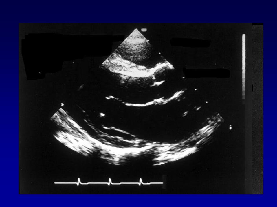
Diastolic mitral leaflet doming
concave toward the LA is seen in:
1. Only rheumatic MS
2. Rheumatic and calcific MS
3. Rheumatic and congenital MS
4. Rheumatic MS, and AI with
flow impinging on the MV
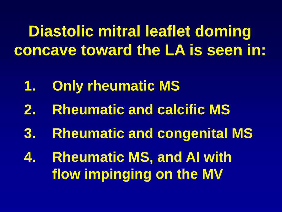
Rheumatic MS


MITRAL STENOSIS
• Diagnosis
• Quantification
• Management
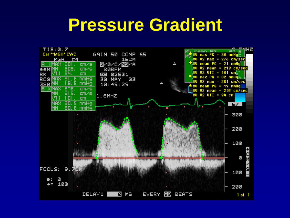
Pressure Gradient
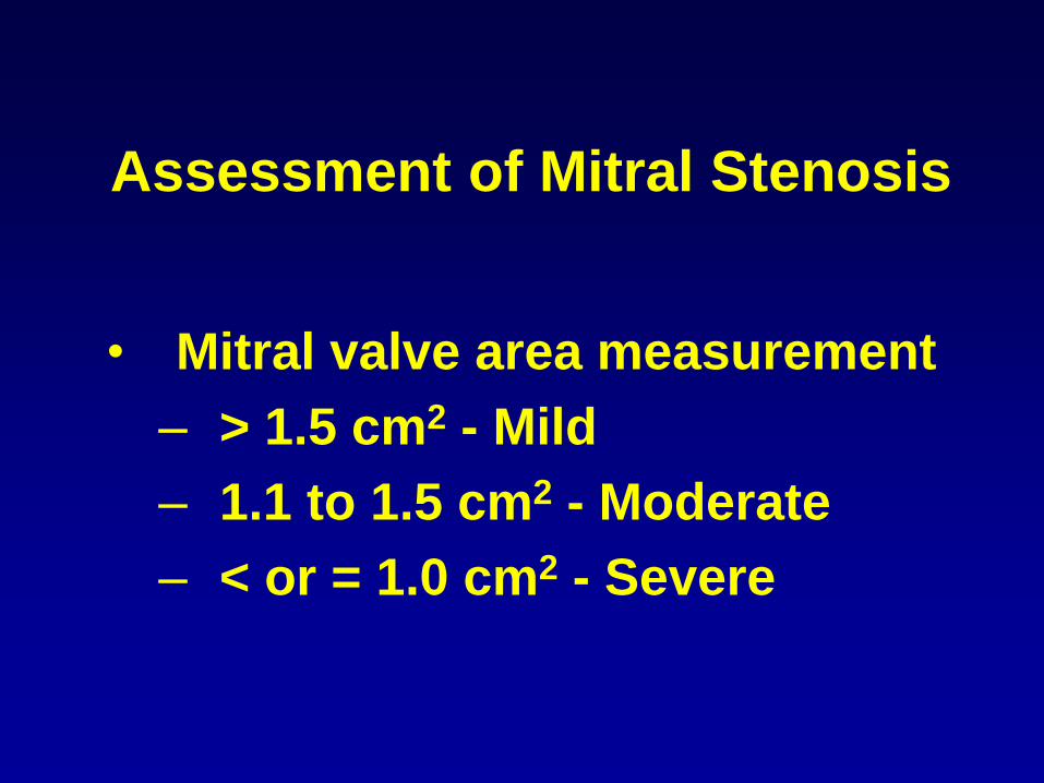
Assessment of Mitral Stenosis
• Mitral valve area measurement
– > 1.5 cm2 - Mild
– 1.1 to 1.5 cm2 - Moderate
– < or = 1.0 cm2 - Severe

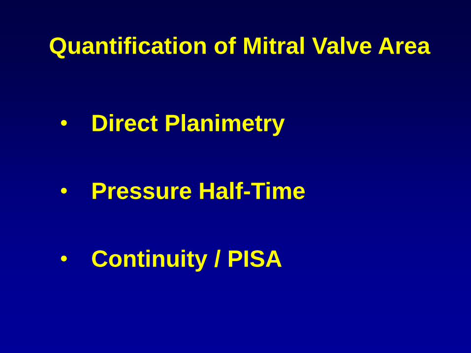
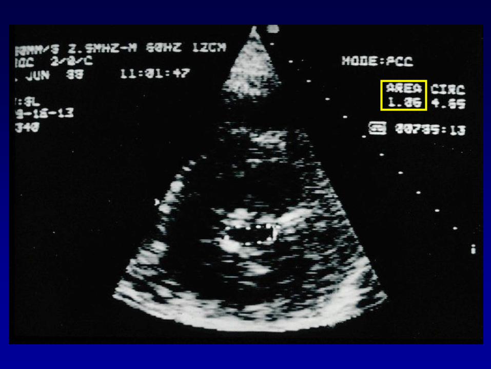
Quantification of Mitral Valve Area
• Direct Planimetry
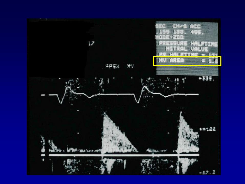
• Pressure Half-Time
• Continuity / PISA

Martin and Stamm

2005 R. Levine
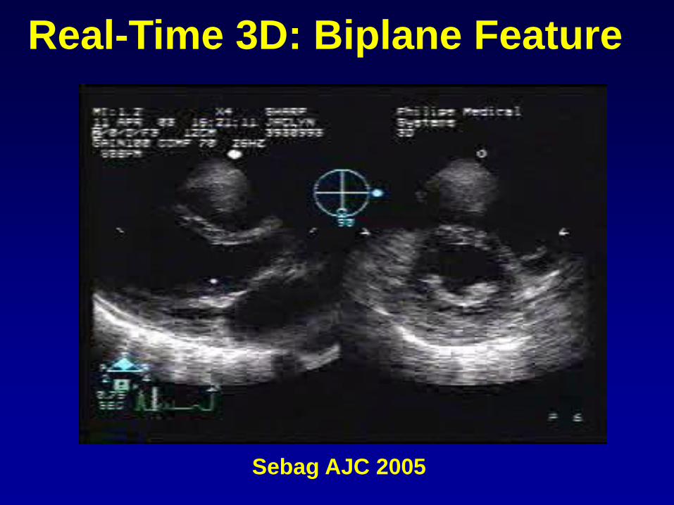
Real-Time 3D: Biplane Feature
Sebag AJC 2005
Quantification of Mitral Valve Area
• Direct Planimetry
• Pressure Half-Time
• Continuity / PISA
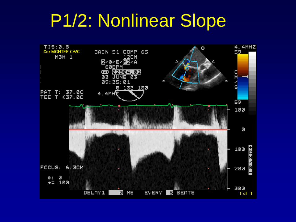
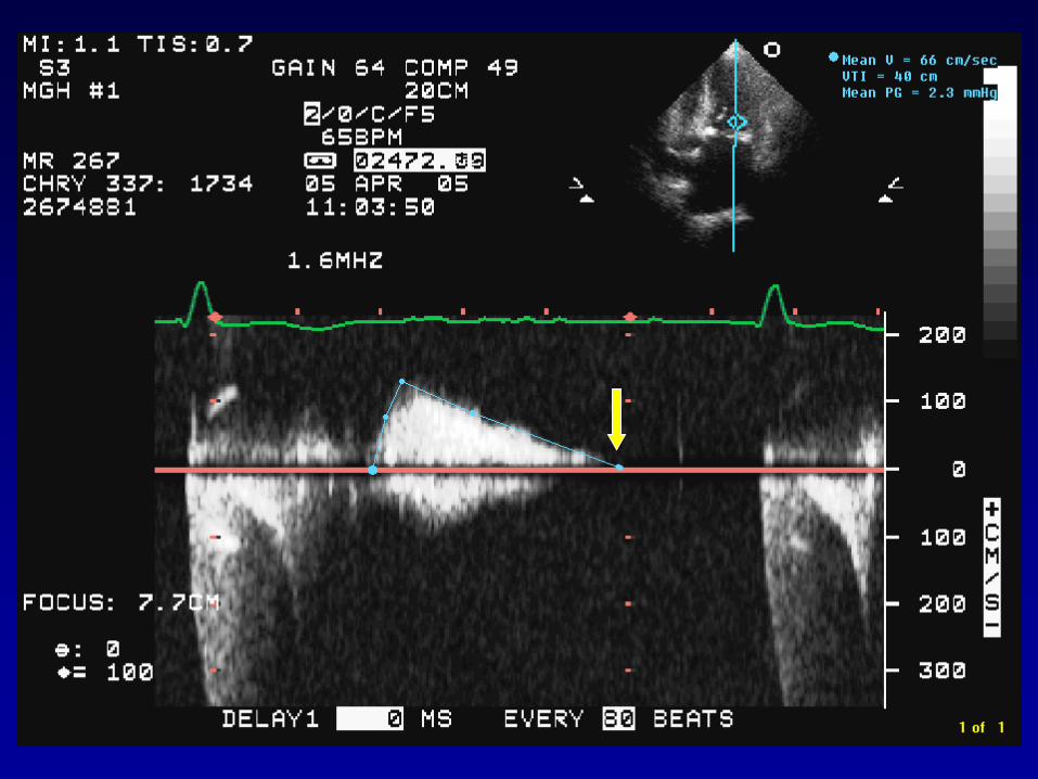
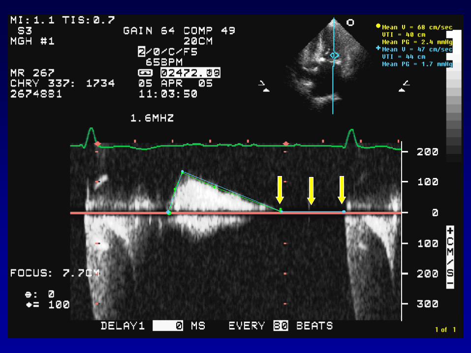
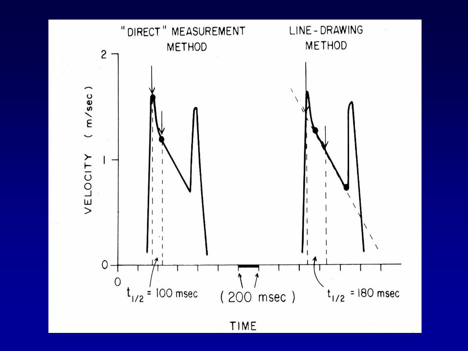
P1/2: Nonlinear Slope
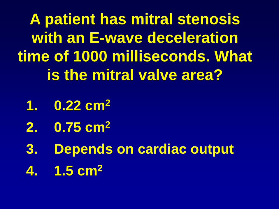
A patient has mitral stenosis
with an E-wave deceleration
time of 1000 milliseconds. What
is the mitral valve area?
1. 0.22 cm2
2. 0.75 cm2
3. Depends on cardiac output
4. 1.5 cm2
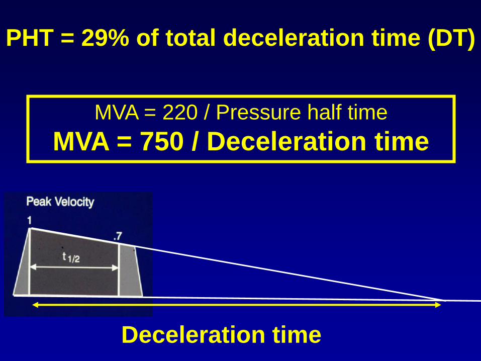
PHT = 29% of total deceleration time (DT)
MVA = 220 / Pressure half time
MVA = 750 / Deceleration time
Deceleration time

James Thomas
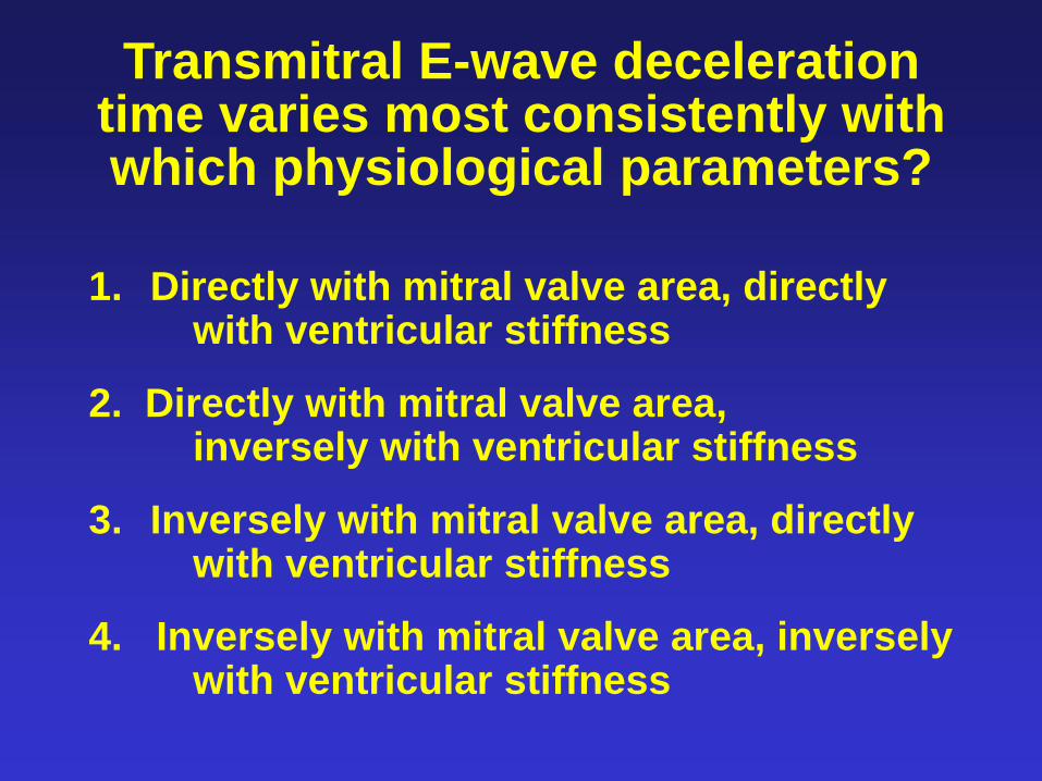
Transmitral E-wave deceleration time varies most consistently with which physiological parameters?
1. Directly with mitral valve area, directly with ventricular stiffness
2. Directly with mitral valve area, inversely with ventricular stiffness
3. Inversely with mitral valve area, directly with ventricular stiffness
4. Inversely with mitral valve area, inversely with ventricular stiffness

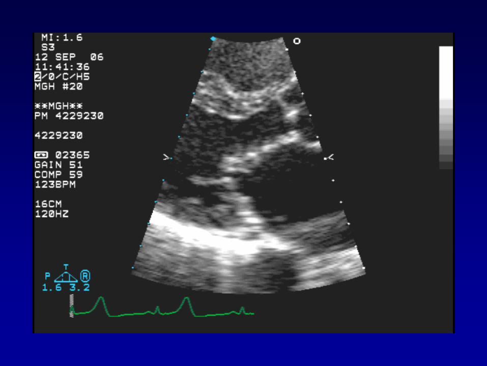
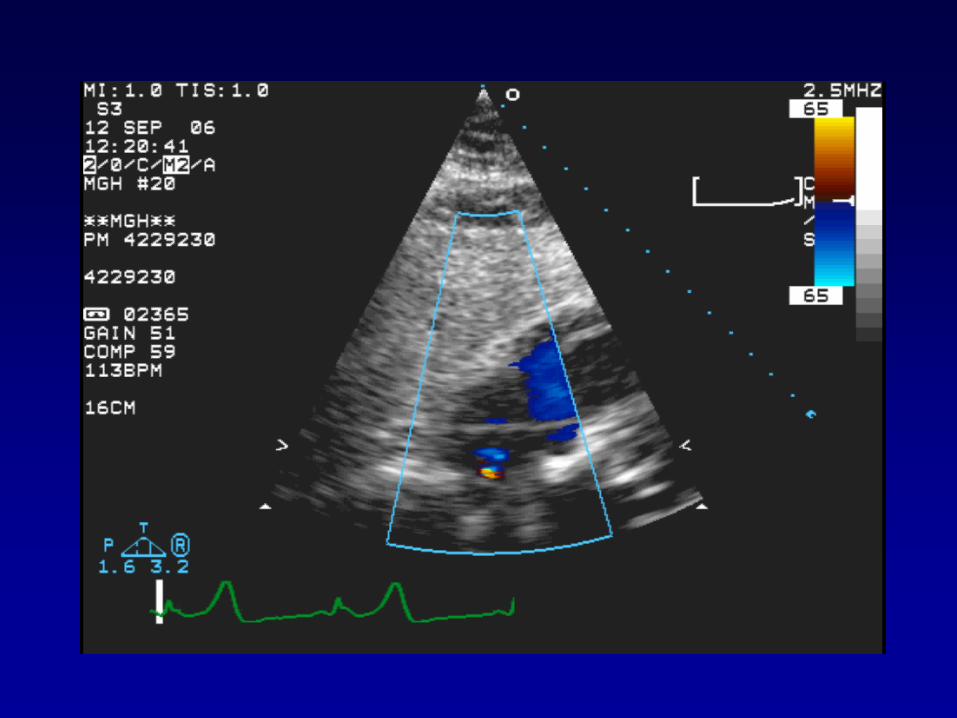
Rheumatic Mitral Valve Stenosis: Case
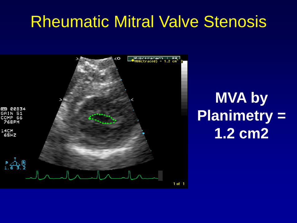
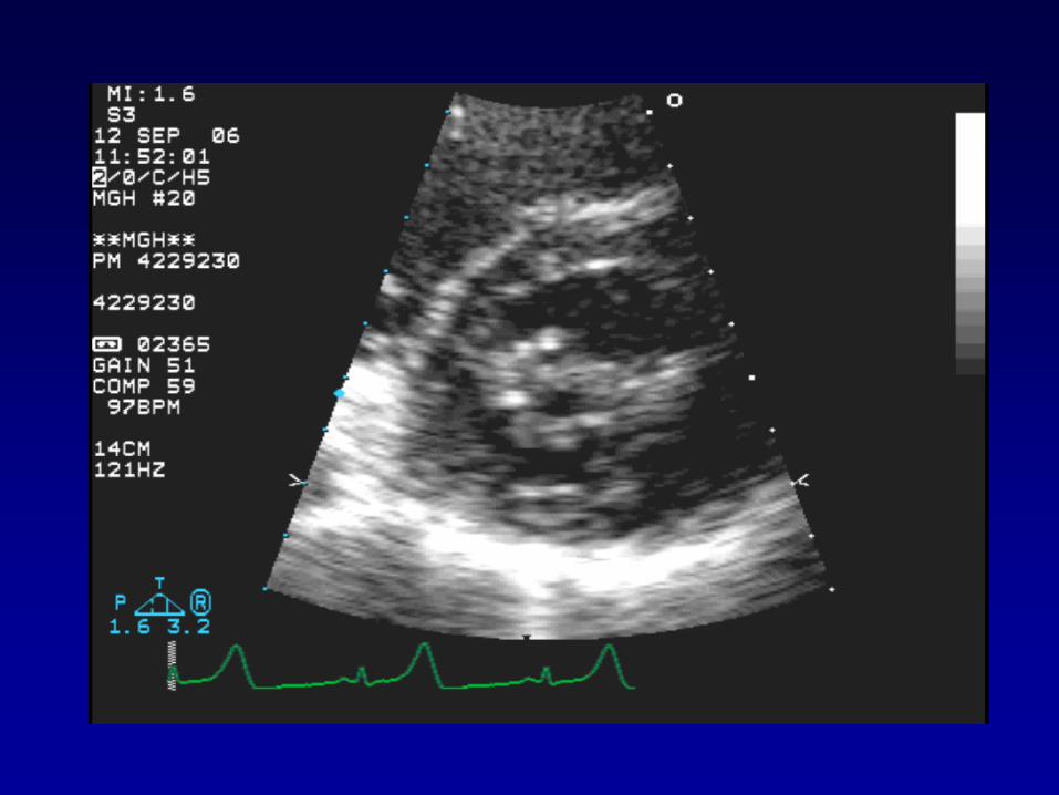
Rheumatic Mitral Valve Stenosis
MVA by
Planimetry =
1.2 cm2
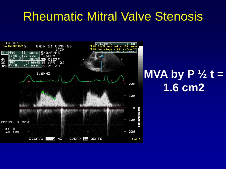
Rheumatic Mitral Valve Stenosis
MVA by P ½ t =
1.6 cm2

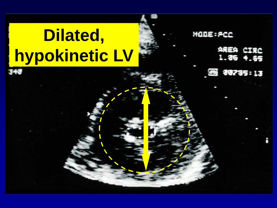
Dilated,
hypokinetic LV
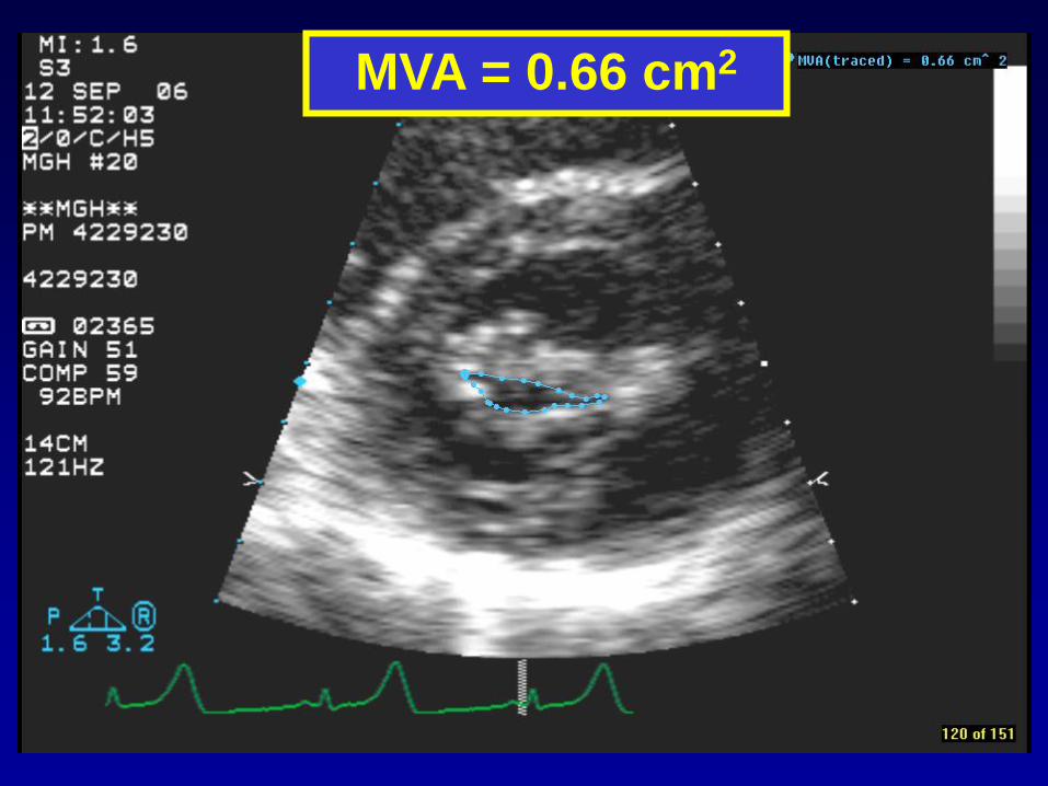
MVA = 0.66 cm2
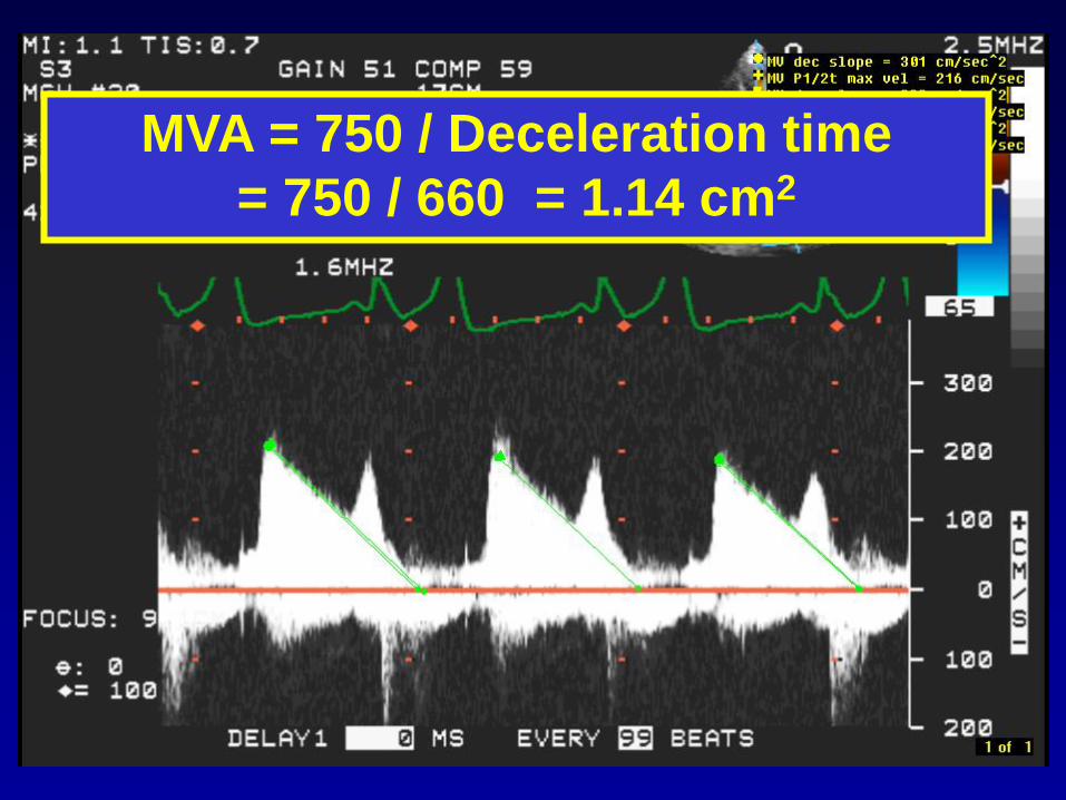
MVA = 750 / Deceleration time
= 750 / 660 = 1.14 cm2

Take Home Message
• Rely on planimetry, esp. biplane
• Pressure half time area can be
falsely elevated because of
noncompliant (stiff) LA or LV, AI
(at least moderate), or ASD.

Quantification of Mitral Valve Area
• Direct Planimetry
• Pressure Half-Time
• Continuity / PISA
AREA = Flow rate / velocity

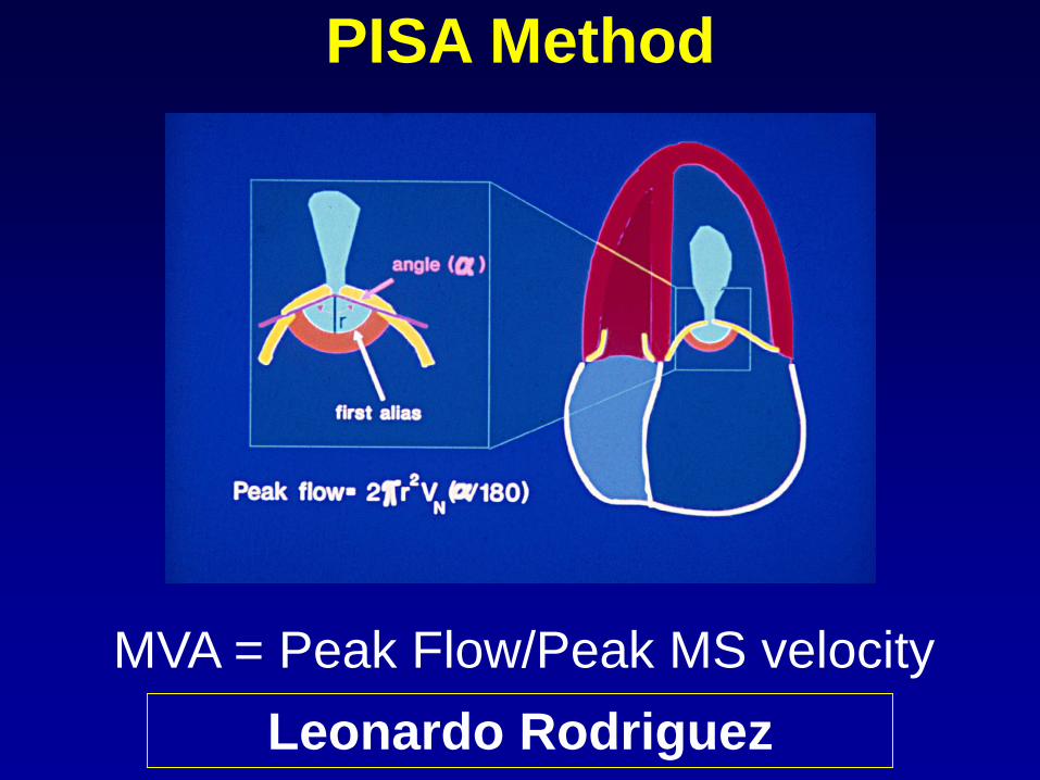
PISA Method
MVA = Peak Flow/Peak MS velocity
Leonardo Rodriguez
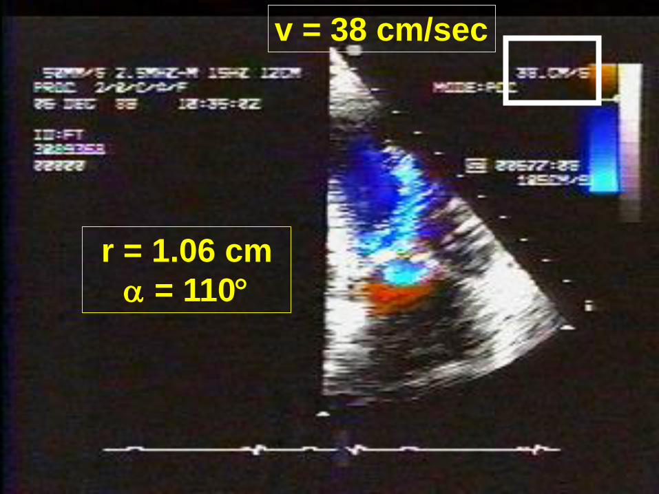
r = 1.06 cm
= 110
v = 38 cm/sec
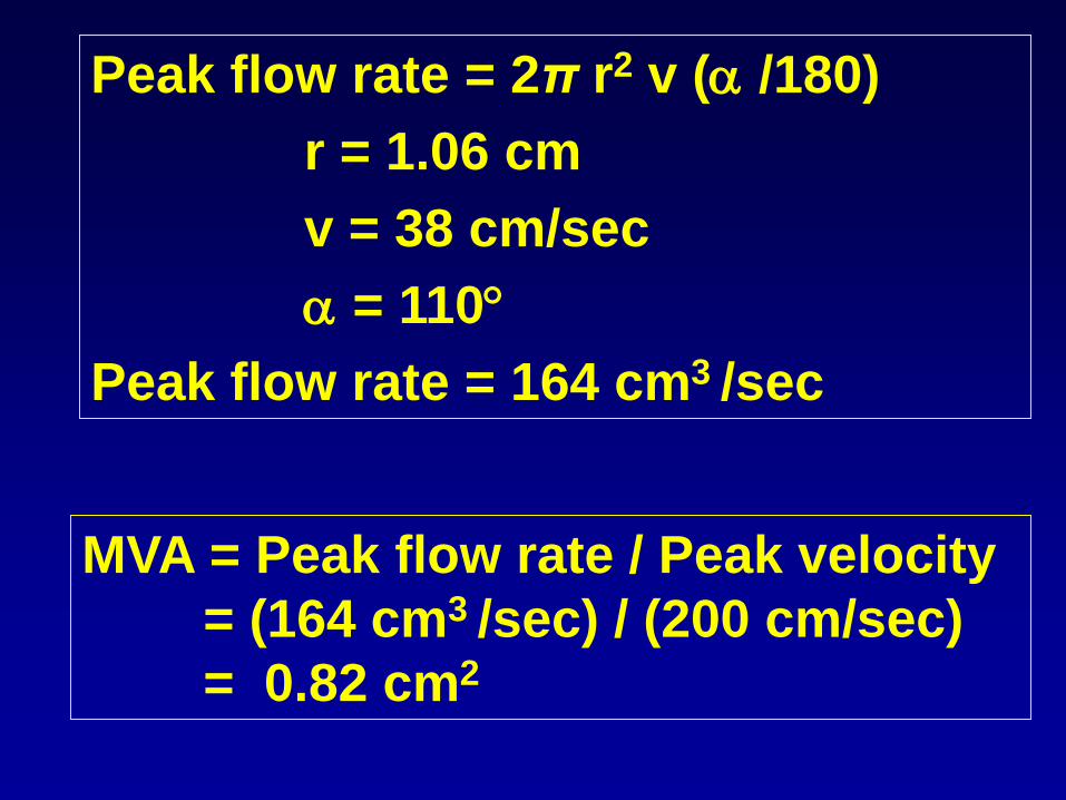
Peak flow rate = 2π r2 v ( /180)
r = 1.06 cm
v = 38 cm/sec
= 110
Peak flow rate = 164 cm3 /sec
MVA = Peak flow rate / Peak velocity
= (164 cm3 /sec) / (200 cm/sec)
= 0.82 cm2
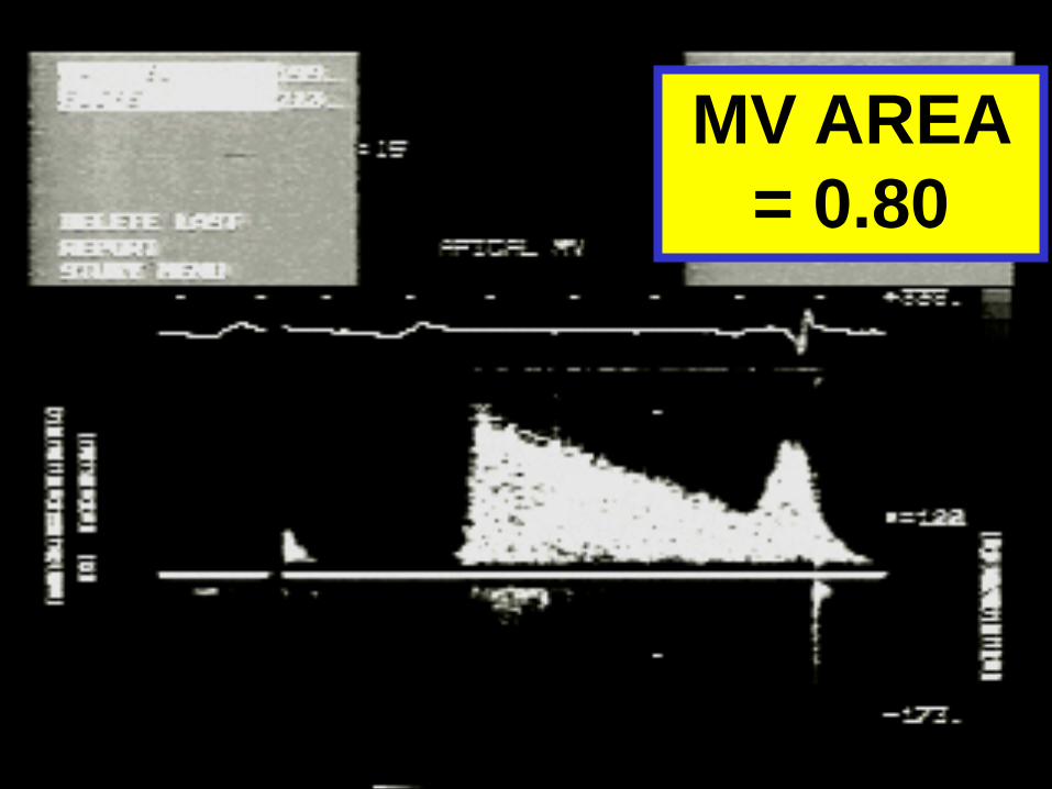
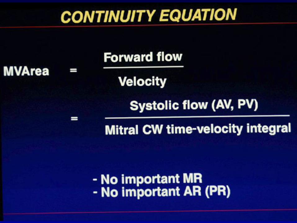
MV AREA
= 0.80
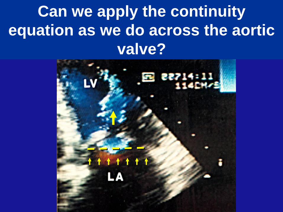
Can we apply the continuity
equation as we do across the aortic
valve?

MITRAL STENOSIS
• Diagnosis
• Quantification
• Management
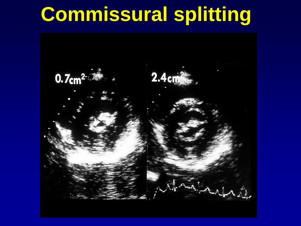
Commissural splitting
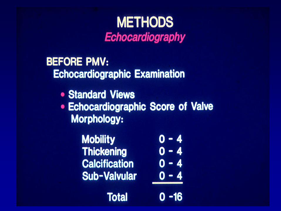

Echo score < 8 associated with
greater success of percutaneous
mitral valvuloplasty
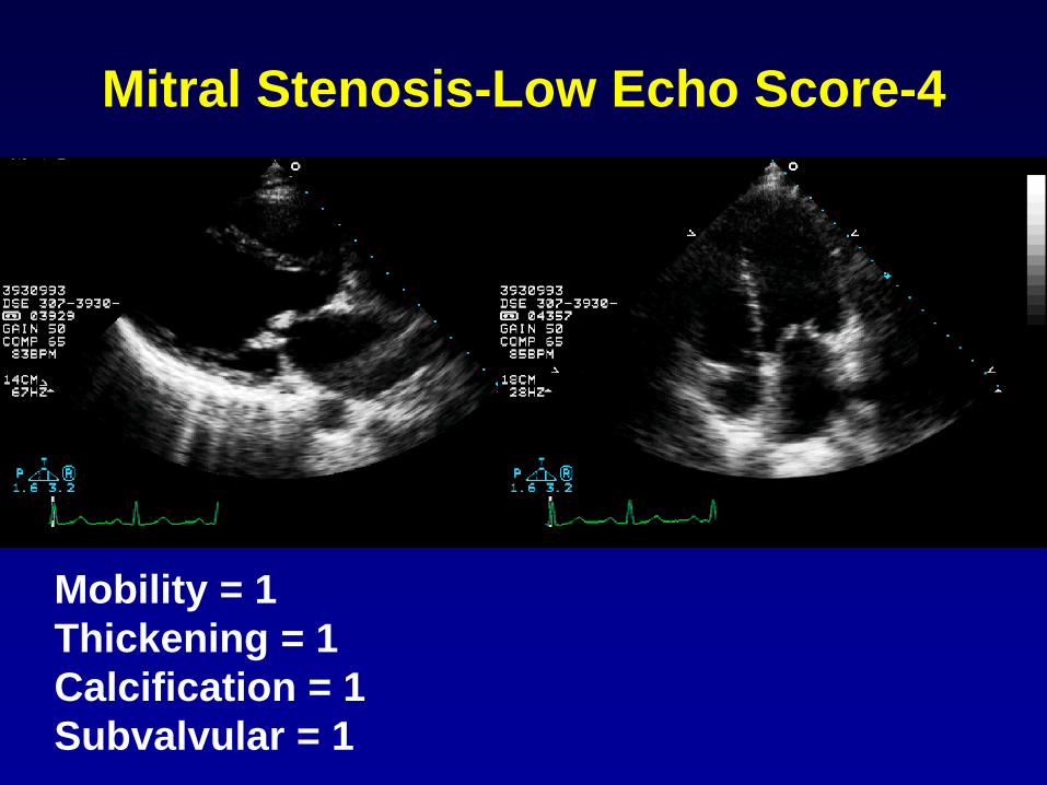
Mitral Stenosis-Low Echo Score-4
Mobility = 1
Thickening = 1
Calcification = 1
Subvalvular = 1
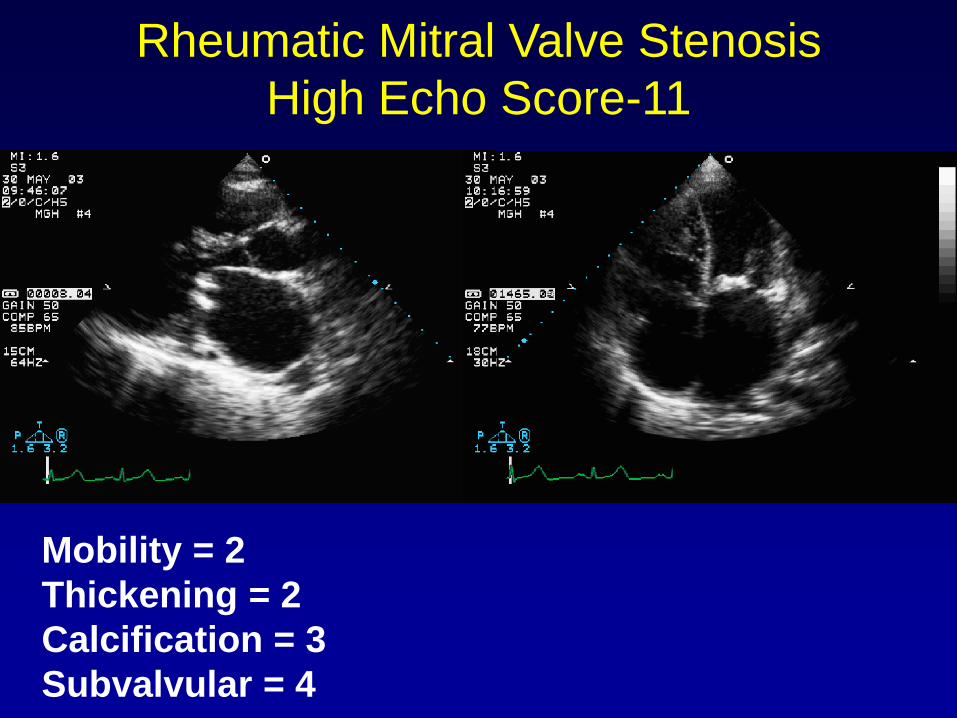
Rheumatic Mitral Valve Stenosis
High Echo Score-11
Mobility = 2
Thickening = 2
Calcification = 3
Subvalvular = 4
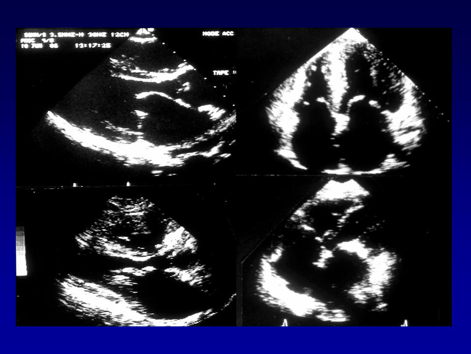

DON’T DO IT!
• Calcific MS
• Moderate MR
• High score
• LA thrombus
• Likely to tear
• Severe TR
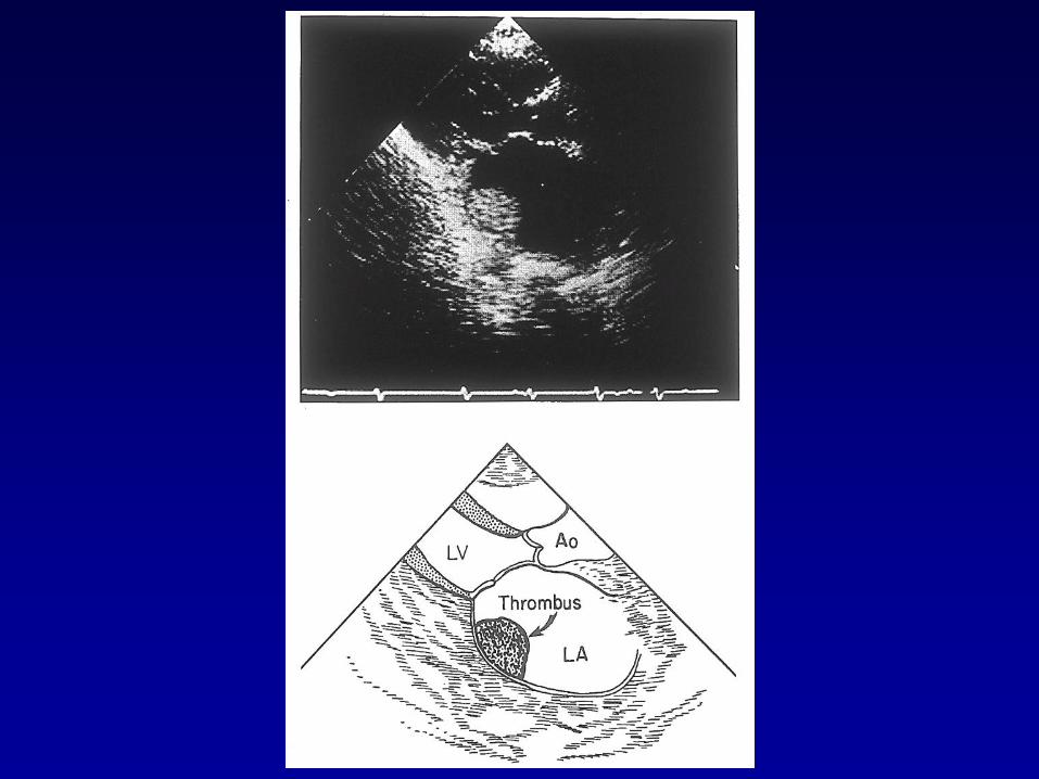

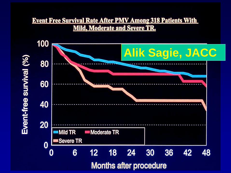
Alik Sagie, JACC

MITRAL STENOSIS
• Diagnosis
• Quantification
• Management
Top Related