


Languages
Pages
Legal


S1
Supporting Information for:
Metal ion directed metal-organic rotaxane frameworks with
intrinsic features of self-penetration and interpenetration
Jun Liang,a Xin-long Wang,*a Yan-Qing Jiao,a Chao Qin,a Kui-Zhan Shao,a Zhong-Min Su*a and Qing-Yin Wub
aInstitute of Functional Material Chemistry, Key Lab of Polyoxometalate Science of Ministry of Education, Faculty of Chemistry, Northeast Normal University, Changchun, 130024 Jilin, People’s Republic of China.
bDepartment of Chemistry, Zhejiang University
Table of Contents Page
General Comments S-3
X-ray Structure determination S-3
Details for Electrochemical Experiments. S-4
Scheme S1 for MORFs 1(Cu), 2 (Zn) and 3(Cd). S-5
Synthesis of [C6CN42+]•2[NO3]-. S-5
Synthesis of [PCN642+]•2[NO3]-. S-5
Fig. S1. 1H NMR spectrum of [C6CN42+]•2[NO3]- (500 MHz in D2O). S-6
Fig. S2. 1H NMR spectrum of [PCN642+]•2[NO3]- (500 MHz in D2O). S-6
Preparation of 1 [Cu(PCA64)(PCA642-)]•13H2O. S-7
Preparation of 2 and 3. S-7
Fig. S3. Powder-XRD results for compounds 1, 2 and 3. S-7
Fig. S4. The IR spectrum of compounds 1, 2 and 3. S-8
Fig. S5. The single three-dimensional mok structure of 1 without CB6. S-8
Fig. S6. The 3-fold interpenetration of mok frameworks in 1. S-8
Fig. S7. Thermal gravimetric analysis (TGA) curve for as-synthesized 1. S-9
Fig. S8. The 2-fold helical chains in 1. S-9
Fig. S9. Emission spectra of compounds 1, 2, 3 and PCA64. S-10
Fig. S10. Schematic view of the closest four CB[6]s around each Cu center. S-10
Fig. S11. The different outer-rotaxanic hydrogen bonding styles of PCA64 S-11
Fig. S12. Single crystal X-ray structure of PCN64 and PCA64. S-11
Electronic Supplementary Material (ESI) for Chemical CommunicationsThis journal is © The Royal Society of Chemistry 2013

S2
Fig.S13. Variable temperature PXRD data for 1. S-12
Fig. S14. The PXRD data (blue) of 1 after immersion in 1 M NaCl+H2SO4 (pH=1) for 30 minutes
and the simulated 1 (black). S-12
Fig. S15. The cyclic voltammograms of the 1-CPE in 1 M NaCl+H2SO4 (pH=1). S-12
Electronic Supplementary Material (ESI) for Chemical CommunicationsThis journal is © The Royal Society of Chemistry 2013

S3
General Comments CB[6] was synthesized according to references1. Other reagents were purchased from
commercial sources and used as received. The Fourier transform infrared (FT-IR) spectra were
recorded from KBr pellets in the range of 4000–400 cm–1 on a Mattson Alpha-Centauri
spectrometer. Elemental analyses (C, H, and N) were performed on a Perkin-Elmer 2400 elemental
analyzer. Cu was determined with a Plasma-SPEC(I) ICP atomic emission spectrometer. 1H NMR
solution experiments were performed on a Brüker Avance 500 instrument, with working frequencies
of 499.8 MHz for 1H nuclei. Chemical shifts are quoted in ppm relative to tetramethylsilane using
the residual solvent peak as a reference standard. Thermogravimetric analysis (TGA) was performed
on a Perkin-Elmer TG-7 analyzer over the temperature 40–800 °C in a nitrogen-gas atmosphere
with a heating rate of 10 °C min−1. Solid-state luminescent spectra were measured on a Cary Eclipse
spectrofluorometer (Varian) equipped with a xenon lamp and quartz carrier at room temperature.
Powder XRD measurements were recorded on a Siemens D5005 diffractometer with Cu Kα ( λ =
1.5418Å) radiation in the range 5−50°.
1. (a) Day, A.; Arnold, A. P.; Blanch, R. J. and Snushall, B. J. Org. Chem., 2001, 66, 8094–8100; (b) Kim, J.; Jung,
In-S.; Kim, S. Y.; Lee, E.; Kang, J. K.; Sakamoto, S.; Yamaguchi, K. and Kim, K. J. Am. Chem. Soc., 2000, 122,
540–541.
X-ray Structure determination X-ray diffraction data collection of the compounds was performed on a Bruker Smart Apex II
CCD diffractometer with graphitemonochromated Mo-Ka radiation (λ = 0.71073 Å) at room
temperature. All absorption corrections were performed by using the SADABS program. The crystal
structure was solved by the Direct Method of SHELXS-97 and refined with full-matrix least-squares
techniques (SHELXL-97) within WINGX. PCN64: C58H78N28O19, Mr = 1471.8, Monoclinic, C2/c,
a =28.266(5) Å, b = 15.802(5) Å, c = 18.367(5) Å, β= 119.599(5)°, V = 7133(3) Å3, Z = 4, ρcalcd.=
1.370 Mgm–3, final R1 = 0.1312 and wR2 = 0.3800 (Rint = 0.0436) for 17970 independent reflections
[I > 2σ(I)]. PCA64: C58H81N26O23.5, Mr = 1518.49, Triclinic, P-1, a = 12.485(5) Å, b = 17.202(5) Å,
c = 17.659(5) Å, α = 94.549(5)°, β = 93.303(5)°, γ = 111.017(5)°, V = 3514(2) Å3, Z = 2, ρcalcd.=
1.435 Mgm–3, final R1 = 0.1483 and wR2 = 0.4220 (Rint = 0.0640) for 18336 independent reflections
[I > 2σ(I)]. 1 [Cu(PCA64)(PCA642-)]•13H2O: CuC116H150N52O45, Mr = 3056.42, Monoclinic, C2/c,
a = 37.209(5) Å, b = 37.083(14) Å, c = 23.517(7) Å, β= 113.162(5)°, V = 29834(9) Å3, Z = 8,
ρcalcd.= 1.361 Mgm–3, final R1 = 0.1347 and wR2 = 0.4439 (Rint = 0.1009) for 77397 independent
reflections [I > 2σ(I)]. 2 [Zn(PCA64)(PCA642-)]•13H2O:ZnC116H146N52O43, Monoclinic, a = 37.93
Å, b = 37.93 Å, c = 23.61 Å, β= 113.78°, V = 31087 Å3. 3 [Cd(PCA64)(PCA642-)]•13H2O:
CdC116H146N52O43, Monoclinic, a = 37.614 Å, b = 37.630 Å, c = 23.41 Å, β= 113.71°, V = 30338 Å3.
The crystal data of compounds 2 and 3 are not good enough to obtain the crystal structures. We
have only get the cell parameters and they are very close to the compound 1. Furthermore, The
PXRD confirm that the compound 1, 2 and 3 are isostructures. CCDC 938011 (PCN64), 938012
(PCA64) and 938013 (1) contain the supplementary crystallographic data for this paper. These data
Electronic Supplementary Material (ESI) for Chemical CommunicationsThis journal is © The Royal Society of Chemistry 2013

S4
can be obtained free of charge from the Cambrige Crystallographic Data Centre via
www.ccdc.cam.ac.uk/data_request /cif for 1, PCN64 and PCA64.
Details for Electrochemical Experiments Materials. High purity graphite powder (average particle 1-2 um) was obtained from Aldrich.
Paraffin oil was purchased from Beijing Chemical Plant and used as received. Other chemicals were
of analytical grade and used without further purification.
Apparatus. A CHI 660 Electrochemical Workstation connected to a Digital-586 personal computer
was used for control of the electrochemical measurements and for data collection. A conventional
three-electrode cell, consisting a 1-modified carbon paste electrode or a glassy carbon electrode as
the working electrode, a saturated silver reference electrode and a Pt wire counter electrode was
used. All potentials were measured and reported versus the Ag+/Ag. All the experiments were
conducted at room temperature (25-30°C).
Preparation of carbon paste electrode modified by compound 1 (1-CPE) Compound 1 modified CPE (1-CPE) was fabricated by the following process: 0.33g graphite
powder was added to the solution of 8 mL ethanol containing 30mg 1 and the mixture was
ultrasonically mixed for 10 min, followed by evaporation of ethanol under vacuum, which produced
rather homogenously covered graphite particles. To the graphite 0.2 mL paraffin oil was added and
stirred with a glass rod; then the mixture was prepared to pack into 3 mm inner diameter glass tube
to a length of 0.8 cm from one of its end, and the surface was pressed tightly on smooth plastic
paper with a copper rod through the back. Electrical contact was established with a copper rod
through the back of the electrode.
Electronic Supplementary Material (ESI) for Chemical CommunicationsThis journal is © The Royal Society of Chemistry 2013

S5
Synthesis
Scheme S1. Synthetic steps involved in the preparation of MORFs 1 (Cu), 2 (Zn) and 3 (Cd). The barrel-like cartoon is a simplified depiction of CB[6].
[C6CN42+]•2[NO3]- : A solution of 4-cyanobenzaldehyde (2.00 g, 15.24 mmol) in MeOH (100 mL)
was added to a solution of 1,6 -diaminobutane (0.844g, 7.26 mmol) in MeOH (50 mL) and the
mixture was heated at reflux for 20 h. When the solution was coolled down to room temperature,
NaBH4 (1.00 g, 26.4 mmol) was added little by little before the mixture was refluxed for another 12
h.The solvent was removed in vacuo and the white residue was dissolved in a small amount of
distilled water. After the aqueous solution was basified with NaOH(s) (pH>12), the product was
extracted with dichloromethane. The extract was dried over magnesium sulfate and evaporated in
vacuo. The while solid was dissolved in 300ml EtOH and excess amount of HNO3 was added to the
solution to precipitate the product, which was then filtered, washed with EtOH and dried in vacuo
(2.71g, 80%). Mp >212°C (decomp); 1H NMR (500MHz, D2O): δ= 1.14 (s, 4H), 1.46 (s, 4H), 2.84
(br, 4H), 4.07 (s, 4H), 7.38 (d, 4H), 7.62 (d, 4H); Anal. Calcd(Found%) for C22H28N6O6.2H2O: C,
51.97 (51.10); N, 16.53 (16.42); H, 6.30 (6.43).
[PCN642+]•2[NO3]- : To a solution of [C6CN42+]•2[NO3]- (1.066 g, 2.26 mmol) in water (300 mL)
was added CB[6] (3.06 g, 2.6 mmol). Then the mixture was put in an ultrasonic instrument for 30
mins before heating at reflux for 3 h. After that, undissolved CB[6] was filtered off. When the
volume of the filtrate was reduced to ~80 mL, heating was stopped. After several hours,
considerable amount of colorless crystals came out. Ethanol (300 mL) was added to the solution to
precipitate more product, which was filtered, washed with EtOH and dried in vacuo (2.67g, 70%).
Mp >324°C (decomp); 1H NMR (500MHz, D2O): δ= 0.26 (s, 4H), 0.63 (s, 4H), 2.82 (s, 4H), 4.22 (s,
4H), 4.08 (d, 12H), 5.34 (s, 12H), 5.47(d, 12H), 7.64 (s, 8H). IR (cm-1): υ(C-H) 3072, 2928; υ(C≡N)
2231; υ(C=O) 1739; υ(CH2) 1473.
Electronic Supplementary Material (ESI) for Chemical CommunicationsThis journal is © The Royal Society of Chemistry 2013

S6
Fig. S1. 1H NMR spectrum of [C6CN42+]•2[NO3]- (500 MHz in D2O).
Fig. S2. 1H NMR spectrum of [PCN642+]•2[NO3]- (500 MHz in D2O).
Electronic Supplementary Material (ESI) for Chemical CommunicationsThis journal is © The Royal Society of Chemistry 2013
S7
Preparation of 1 [Cu(PCA64)(PCA642-)]•13H2O
[PCN642+]•2[NO3]- (0.168 g, 0.1 mmol), Cu(NO3)2·3H2O (45 mg, 0.19 mmol), triethylamine
(3drops, where 1ml injection syringe is used) were suspended in water (6 ml) in a stainless-steel
bomb, which was then sealed, kept at 170°C for 72 h, and cooled to room temperature at a constant
cooling rate of 10°C h-1. Large, green crystals of 1 were washed with water several times, collected
by manual operation and dried in air (92 mg, 60%). IR (cm-1): υ(C-H) 3000, 2935; υ(C=O) 1736;
υ(CH2) 1474. Anal. Calcd(Found%): Cu, 2.07 (1.98); C, 45.78 (44.96); N, 23.81 (23.63); H, 4.97
(4.52).
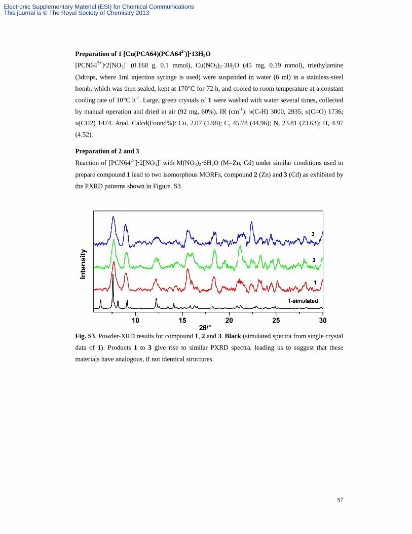
Preparation of 2 and 3
Reaction of [PCN642+]•2[NO3]- with M(NO3)2·6H2O (M=Zn, Cd) under similar conditions used to
prepare compound 1 lead to two isomorphous MORFs, compound 2 (Zn) and 3 (Cd) as exhibited by
the PXRD patterns shown in Figure. S3.
Fig. S3. Powder-XRD results for compound 1, 2 and 3. Black (simulated spectra from single crystal
data of 1). Products 1 to 3 give rise to similar PXRD spectra, leading us to suggest that these
materials have analogous, if not identical structures.
Electronic Supplementary Material (ESI) for Chemical CommunicationsThis journal is © The Royal Society of Chemistry 2013

S8
Fig. S4. The IR spectrum of compounds 1, 2, 3 and L([PCN642+]•2[NO3]-). The IR spectrum of L
exhibits a band at 2231 cm-1 attributed to υ (C≡N).
Fig. S5. Ball-and-stick representation of the single three-dimensional mok structure of 1 without
CB6. Atom color code: C black, N blue, O red, Cu or polyhedra green.
Fig. S6. Ball-and-stick representation of 3-fold interpenetration of mok framework as in 1, showing
the channels that contain the solvated water molecules.
Electronic Supplementary Material (ESI) for Chemical CommunicationsThis journal is © The Royal Society of Chemistry 2013

S9
Fig. S7. Thermal gravimetric analysis (TGA) curve for as-synthesized 1. Water of crystallization is
volatilized from room temperature to about 170℃ and organic ligand decomposed rapidly only after
300℃.
Fig. S8. a) The assembly of Cu(NO3) and PCA64/PCA642- has also produced helical chains, in
which trans-PCA64, Cu2+ and cis-PCA642- alternate, with a helical pitch of 37.08 and width of
37.06 Å. b) The entanglement of the nanotubes. c) Each helix is interwoven with other two
belonging to different parallel nanotubes in 1. CB6 is omitted for clarity.
Electronic Supplementary Material (ESI) for Chemical CommunicationsThis journal is © The Royal Society of Chemistry 2013

S10
Fig. S9. Emission spectra of compounds 1, 2, 3 and PCA64 in the solid state at room temperature,
which illustrated that only intraligand (π-π*) fluorescent emission occurred because all emissions
were quite similar compared to that of PCA64 at 403 nm (λex=309nm).
Fig. S10. a) Schematic view of the closest four CB[6]s belonging to three different networks
(colored in violet, light blue and light orange, respectively) around each Cu center. b)
Representation of the outer-rotaxanic hydrogen bonds (H…O distances ranges from 2.39 to 2.59 Å)
between CB[6]s and c)carboxylic groups and π―π stacking interactions (average distance 3.466(8)
Å) around each Cu center. Note: some hydrogen atoms are omitted for clarity. Dashed cyan and
orange lines represent hydrogen bonding and π―π interactions, respectively.
Electronic Supplementary Material (ESI) for Chemical CommunicationsThis journal is © The Royal Society of Chemistry 2013

S11
Fig. S11. The different outer-rotaxanic hydrogen bonding styles and π―π stacking interactions of
trans-PCA64 (a, c) and cis-PCA642- (b, d) in 1. Dashed cyan and orange lines represent hydrogen
bonding and π―π interactions, respectively.
Single Crystal X-ray Structure of PCN64 and PCA64
Fig. S12. A ball-and-stick representation of the X-ray structure of PCN64 and PCA64 with labeling scheme. Black, carbon; Blue, nitrogen; red, oxygen; gray, hydrogen.
Electronic Supplementary Material (ESI) for Chemical CommunicationsThis journal is © The Royal Society of Chemistry 2013
S12
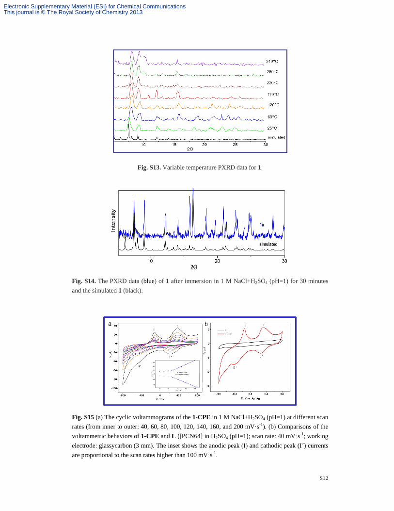
Fig. S13. Variable temperature PXRD data for 1.
Fig. S14. The PXRD data (blue) of 1 after immersion in 1 M NaCl+H2SO4 (pH=1) for 30 minutes and the simulated 1 (black).
Fig. S15 (a) The cyclic voltammograms of the 1-CPE in 1 M NaCl+H2SO4 (pH=1) at different scan rates (from inner to outer: 40, 60, 80, 100, 120, 140, 160, and 200 mV·s-1). (b) Comparisons of the voltammetric behaviors of 1-CPE and L ([PCN64] in H2SO4 (pH=1); scan rate: 40 mV·s-1; working electrode: glassycarbon (3 mm). The inset shows the anodic peak (I) and cathodic peak (I´) currents are proportional to the scan rates higher than 100 mV·s-1.
Electronic Supplementary Material (ESI) for Chemical CommunicationsThis journal is © The Royal Society of Chemistry 2013
Top Related