


Languages
Pages
Legal

MAXILLOFACIAL TRAUMATOLOGY
Department of Maxillofacial Surgery
Semmelweis University, Budapest
• isolated maxillofacial injury
• multiple injuries
• polytrauma (injury of more region or organ of the body and one of
them is life threatening)
Maxillofacial injuries
Incidence of maxillofacial injuries
• Injury of soft tissues of head and neck region (35%)
• Injury of jaws (65%)
– Mandibular fracture (71%)
– Fracture of middle face bones (25%)
– Combined fractures (4%)
male – female ratio: 2-1
• Traffic accident
• Violance
• Accident at work
• Sport injury
Causes of maxillofacial injuries
First-aid
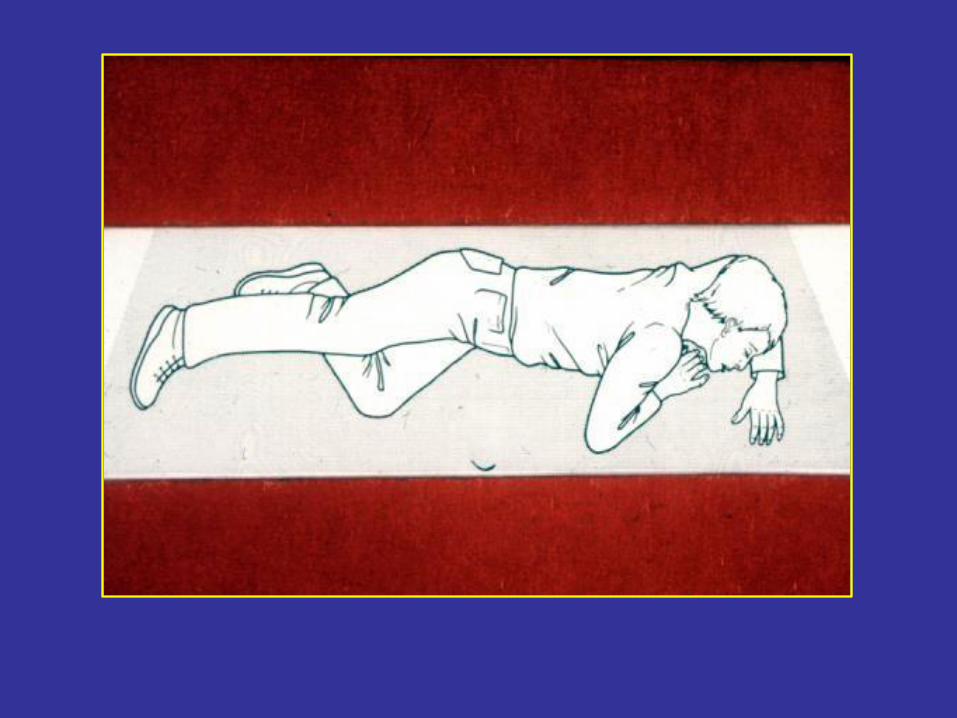
• Maintance of free respiration (saliva, blood,
prosthesis, luxated teeth, foreign body, fractured middle face, tounge stb.)
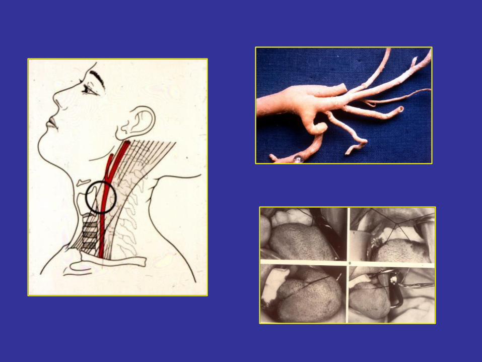
• Stop bleeding
• Maintance of circulation (volumen replacement, shock
-therapy)
• Covering of wounds
• Fixation of fractured ends
• Hospitalisation
Treatment in hospital
if it is possible immediate and definitive!!!
• diagnosis (clinical symptomes, rtg.)
• treatment of soft tissue injuries
• reposition of fractured bone ends, immobilisation
• antibiotic administration
• nutrition, rehabilitation
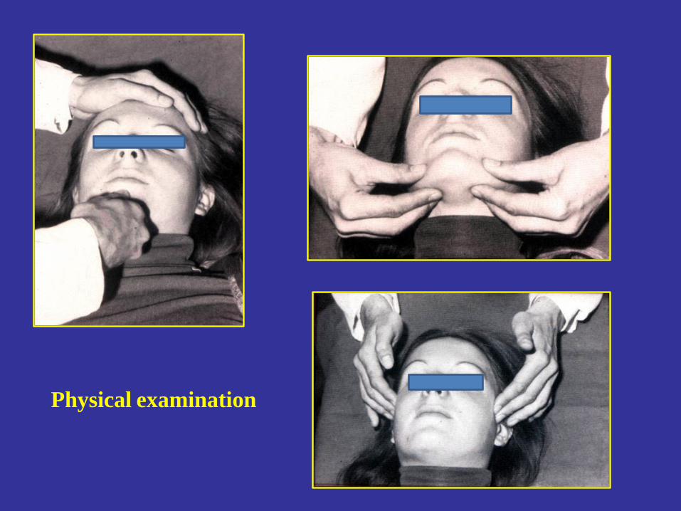
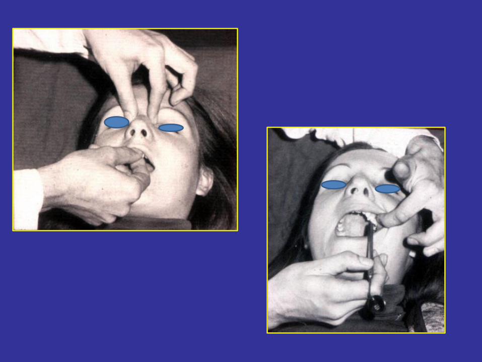
Physical examination
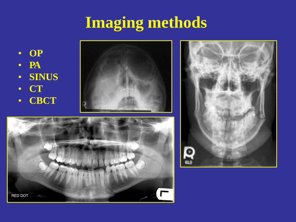
Imaging methods
• OP
• PA
• SINUS
• CT
• CBCT
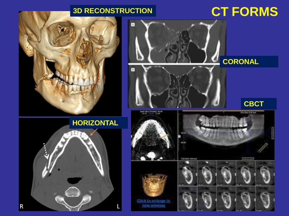
CT FORMS
CORONAL
CBCT
3D RECONSTRUCTION
HORIZONTAL
Mandibular fractures
• 75 % of jaw fractures
Classification of mandibular
fractures
• connection with outside world (open, closed)
• type (infraction, greenstick fracture, hole width fracture, multiplex fracture)
• site
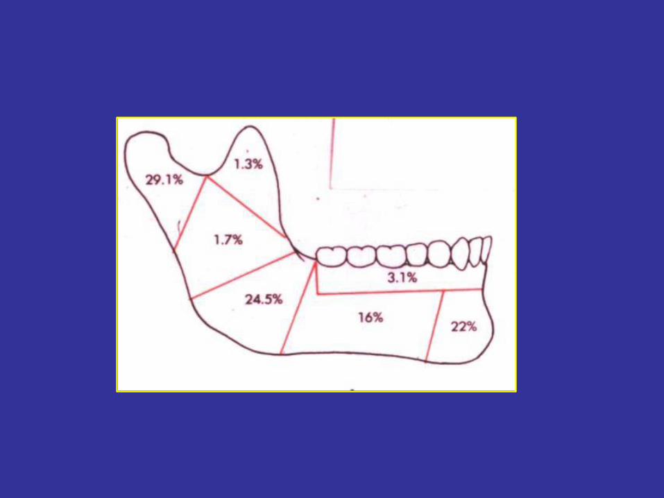
– symphiseal /childhood/
– in region of the canine tooth
– corpus (between the canine tooth and angle)
– mandibular angle (second in frequency, and the most often in case of single
fracture)
– ramus of the mandible
– muscular process
– condylar process (most often; change in the occlusion)
forms: -intracapsular (condylar)
-extracpsular (subcondylar)
Diagnosis
• anamnesis
• inspection
• physical examination
• imaging methods ( x-ray, CT, CBCT)
General (uncertain) symptoms of
jaw fractures
• Pain (spontenous, induced by palpation or move)
• Swelling
• Soft tissue injury
• Functional disorders (trismus, biting disorder,
paresthesia of the innervation site of n. mentalis)
Certain symptoms of jaw fracture
• Occlusional problems
• Pathologic moves
• Crepitation (due to moves of fractured ends)
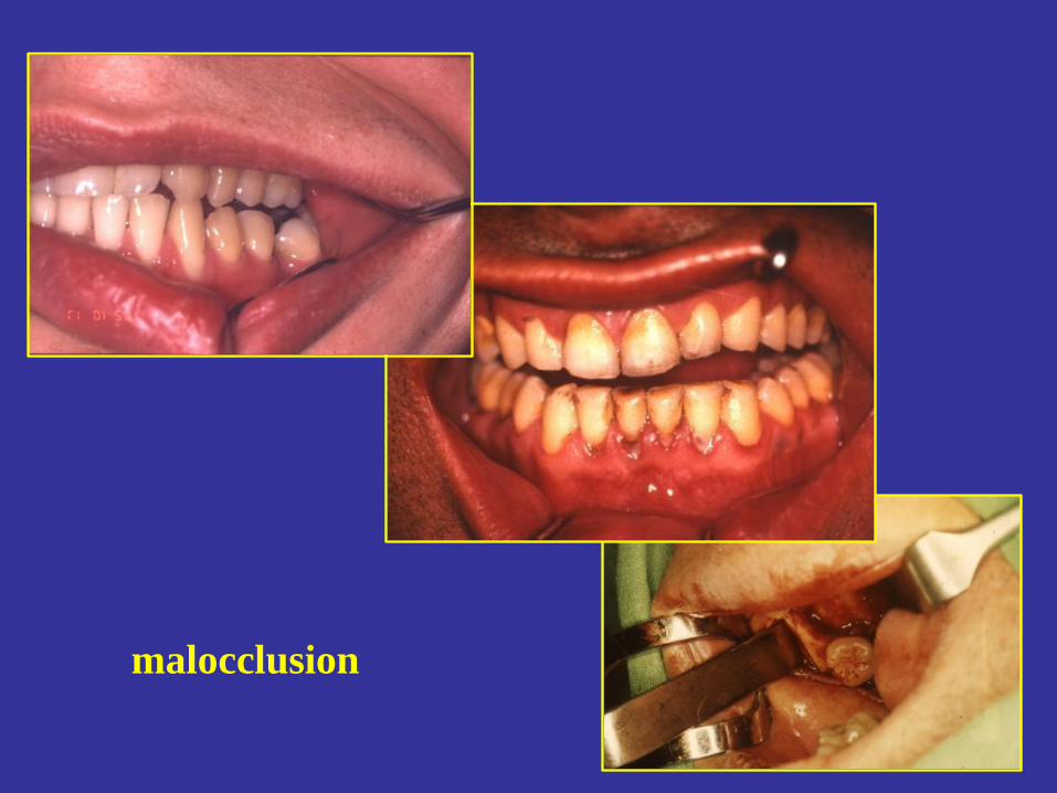
malocclusion
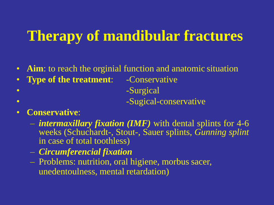
Therapy of mandibular fractures
-Conservative
-Surgical
-Sugical-conservative
• Aim: to reach the orginial function and anatomic situation
• Type of the treatment:
•
•
• Conservative:
– intermaxillary fixation (IMF) with dental splints for 4-6 weeks (Schuchardt-, Stout-, Sauer splints, Gunning splint in case of total toothless)
– Circumferencial fixation – Problems: nutrition, oral higiene, morbus sacer,
unedentoulness, mental retardation)
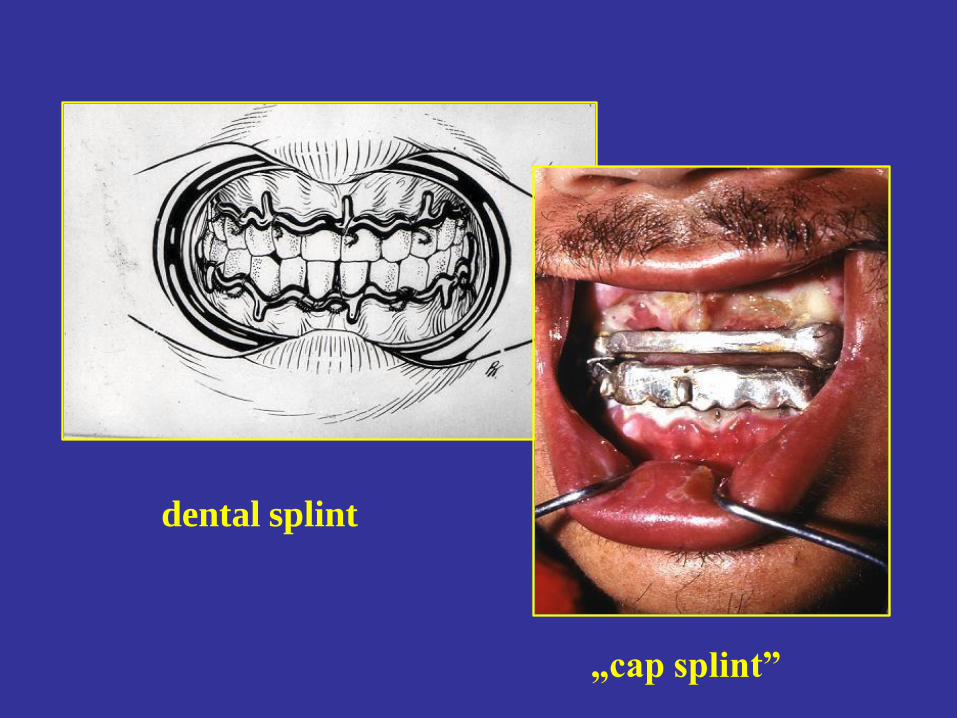
dental splint
„cap splint”
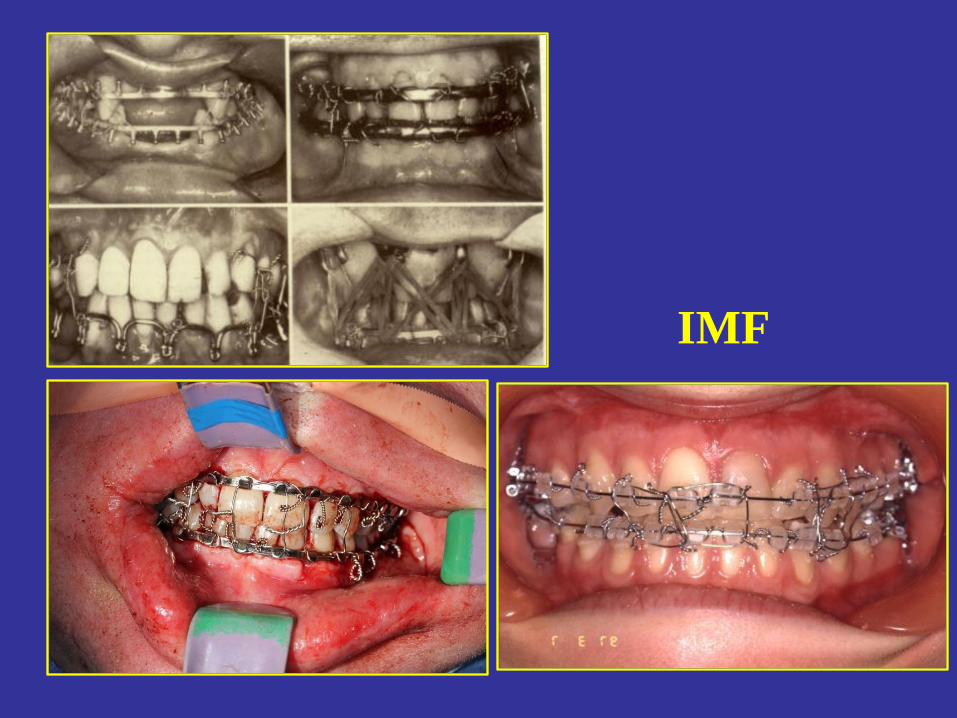
IMF
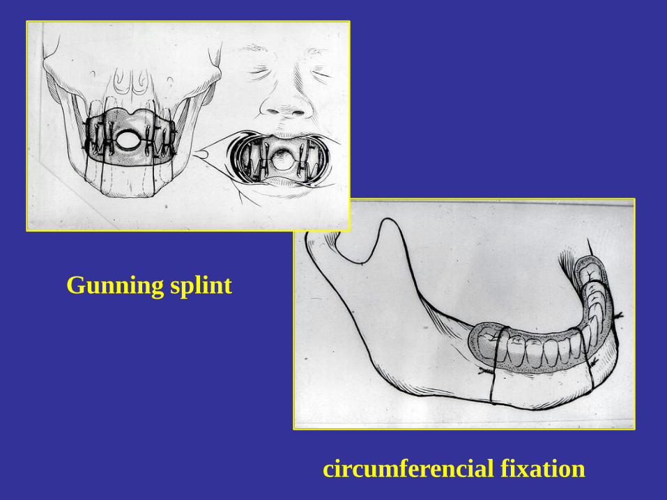
circumferencial fixation
Gunning splint
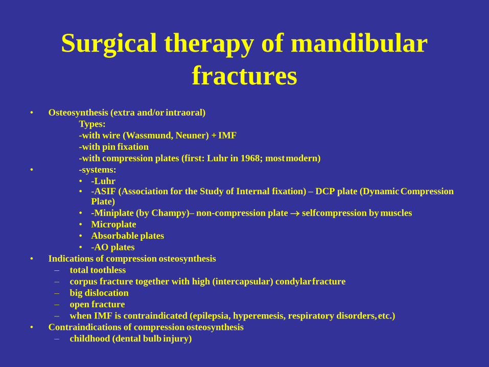
Surgical therapy of mandibular
fractures
•
• Osteosynthesis (extra and/or intraoral)
Types:
-with wire (Wassmund, Neuner) + IMF
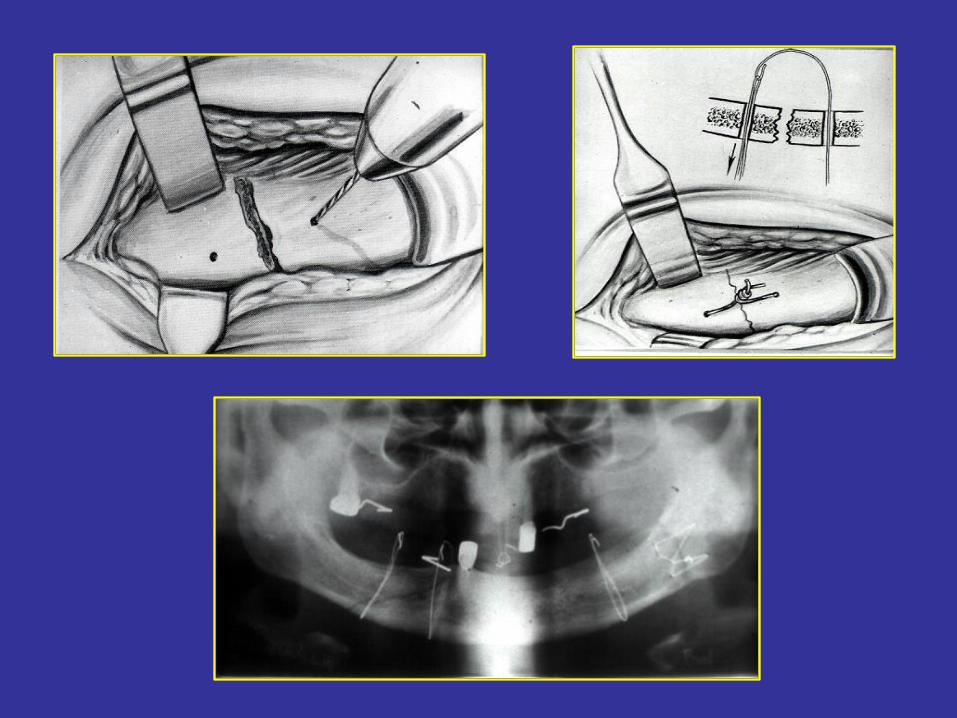
-with pin fixation
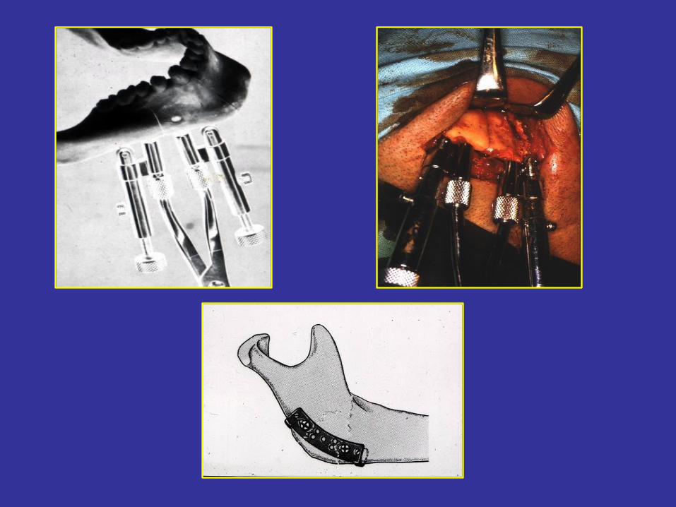
-with compression plates (first: Luhr in 1968; most modern)
-systems:
• -Luhr • -ASIF (Association for the Study of Internal fixation) – DCP plate (Dynamic Compression
Plate)
• -Miniplate (by Champy)– non-compression plate selfcompression by muscles
• Microplate
• Absorbable plates
• -AO plates
• Indications of compression osteosynthesis
– total toothless
– corpus fracture together with high (intercapsular) condylar fracture
– big dislocation
– open fracture
– when IMF is contraindicated (epilepsia, hyperemesis, respiratory disorders, etc.)
• Contraindications of compression osteosynthesis
– childhood (dental bulb injury)
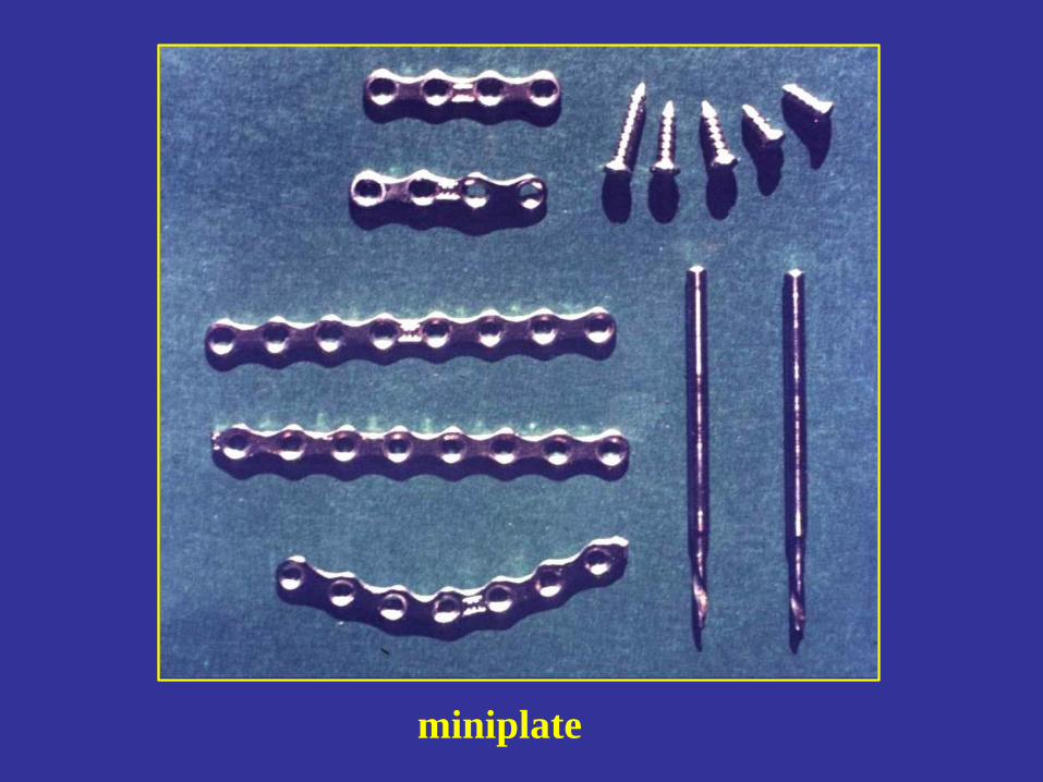
miniplate
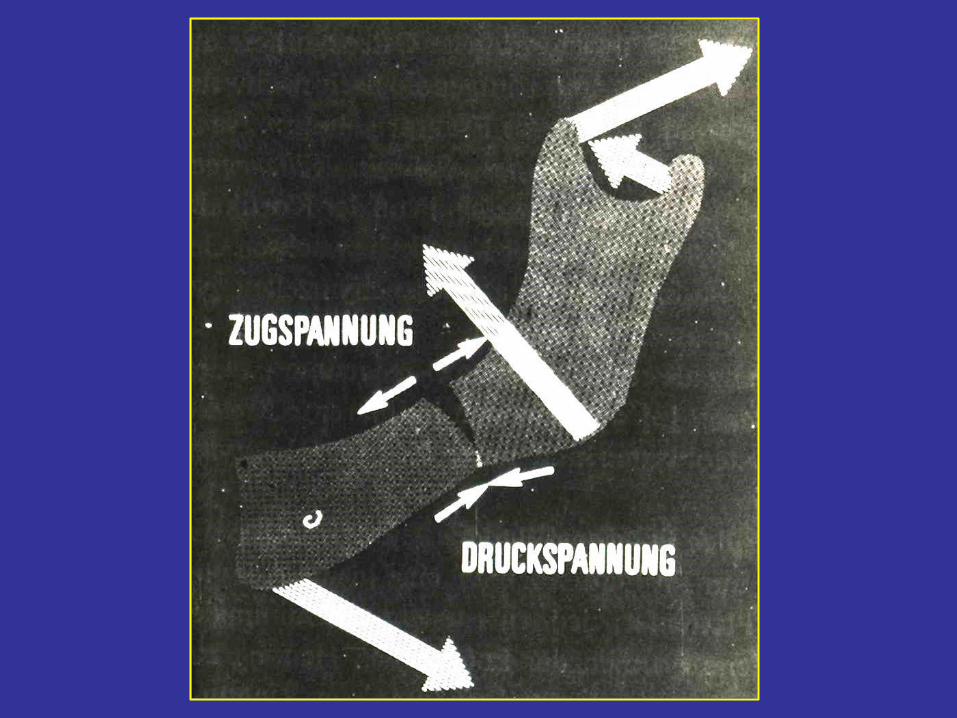
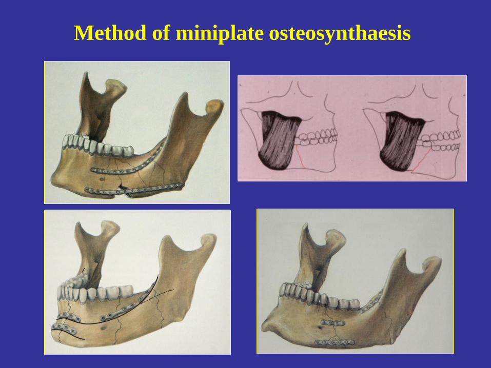
Method of miniplate osteosynthaesis
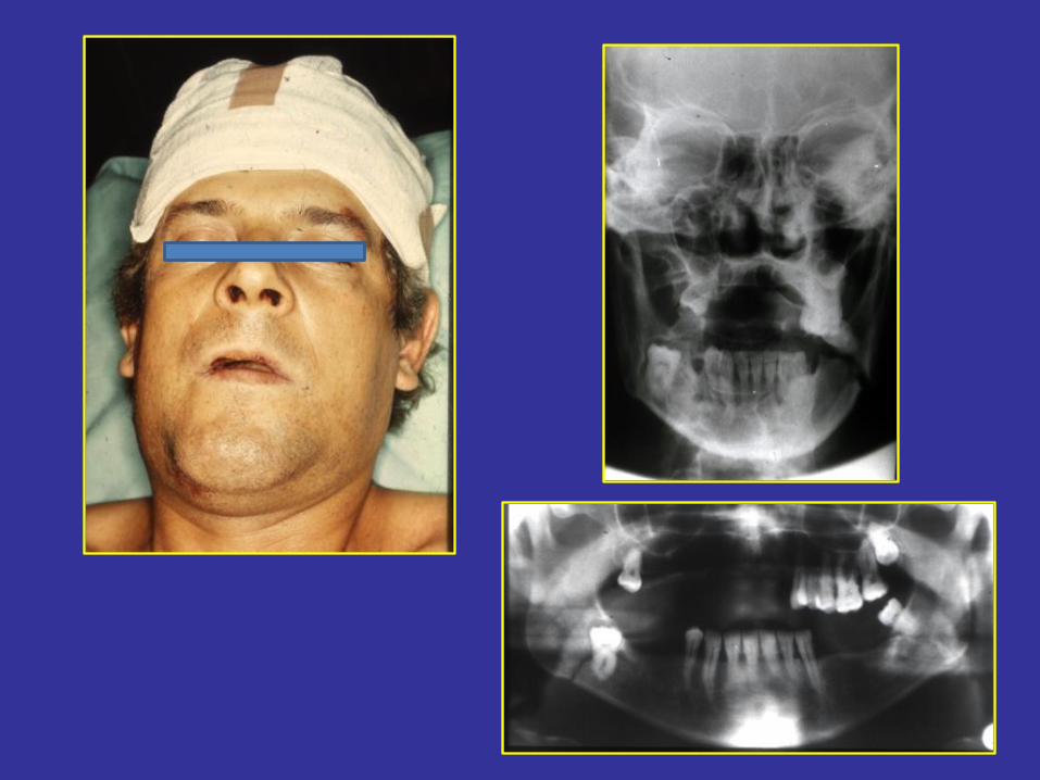
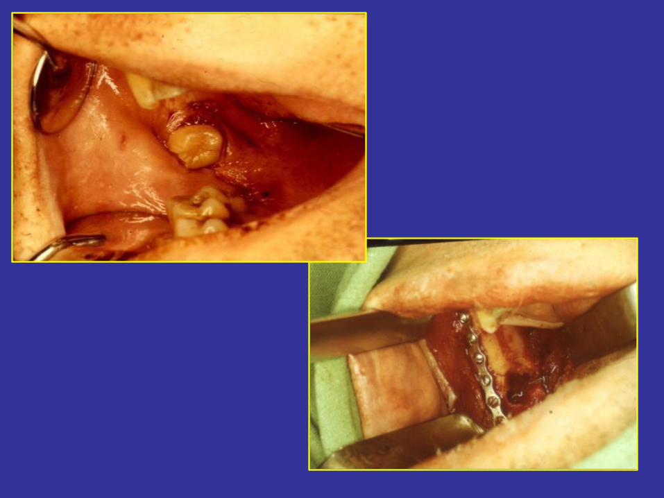
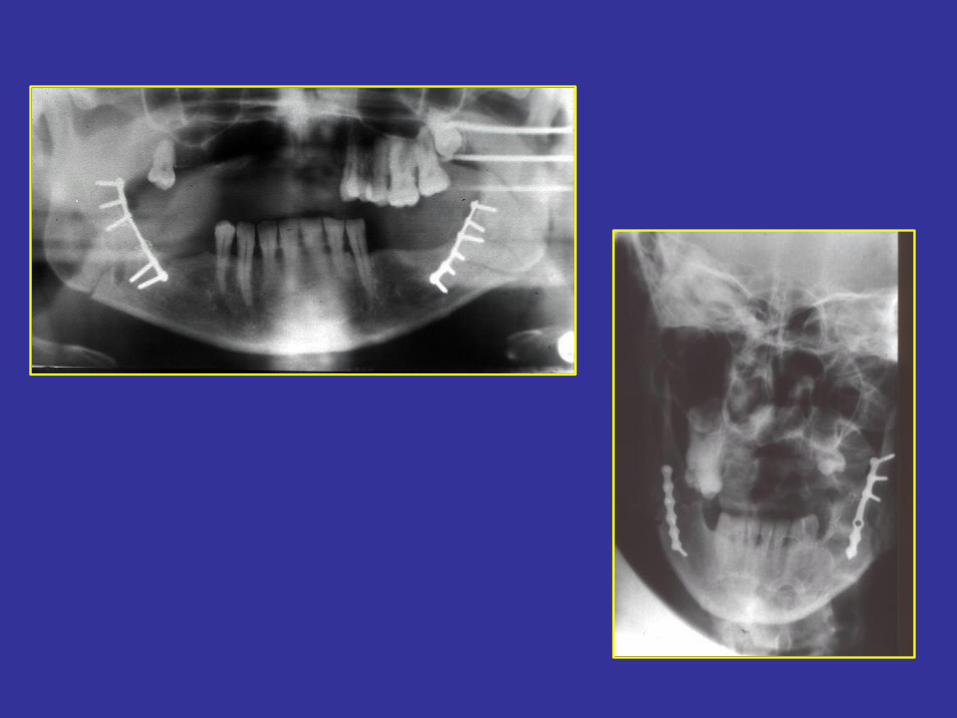
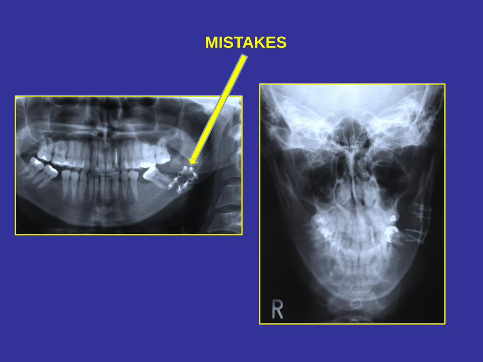
MISTAKES
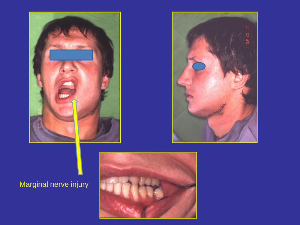
Marginal nerve injury
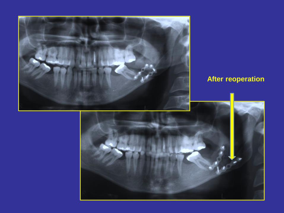
After reoperation
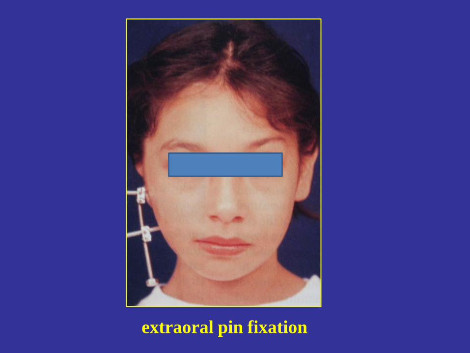
extraoral pin fixation
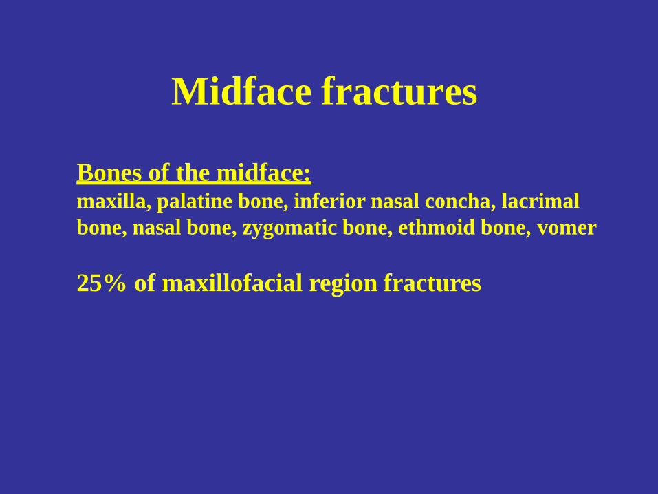
Midface fractures
Bones of the midface: maxilla, palatine bone, inferior nasal concha, lacrimal
bone, nasal bone, zygomatic bone, ethmoid bone, vomer
25% of maxillofacial region fractures
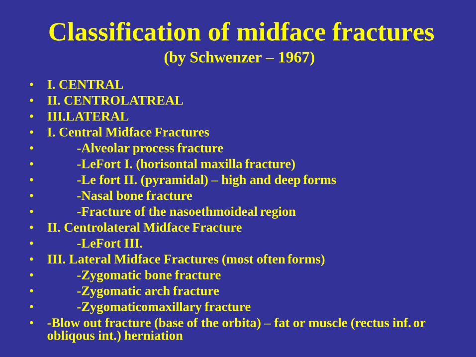
Classification of midface fractures (by Schwenzer – 1967)
• I. CENTRAL
• II. CENTROLATREAL
• III.LATERAL
• I. Central Midface Fractures
• -Alveolar process fracture
• -LeFort I. (horisontal maxilla fracture)
• -Le fort II. (pyramidal) – high and deep forms
• -Nasal bone fracture
• -Fracture of the nasoethmoideal region
• II. Centrolateral Midface Fracture
• -LeFort III.
• III. Lateral Midface Fractures (most often forms)
• -Zygomatic bone fracture
• -Zygomatic arch fracture
• -Zygomaticomaxillary fracture
• -Blow out fracture (base of the orbita) – fat or muscle (rectus inf. or obliqous int.) herniation
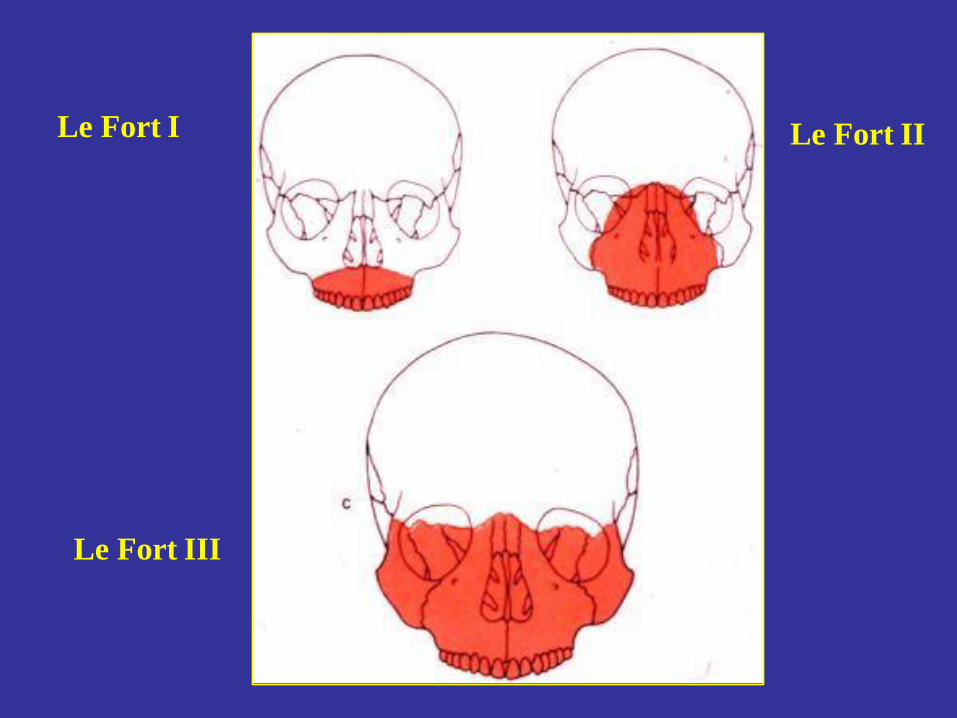
Le Fort I Le Fort II
Le Fort III
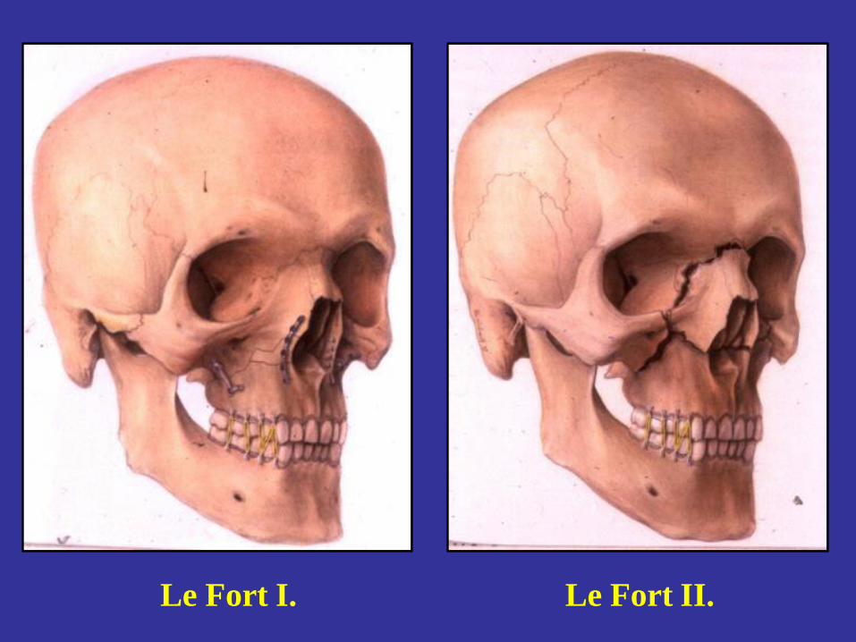
Le Fort II. Le Fort I.
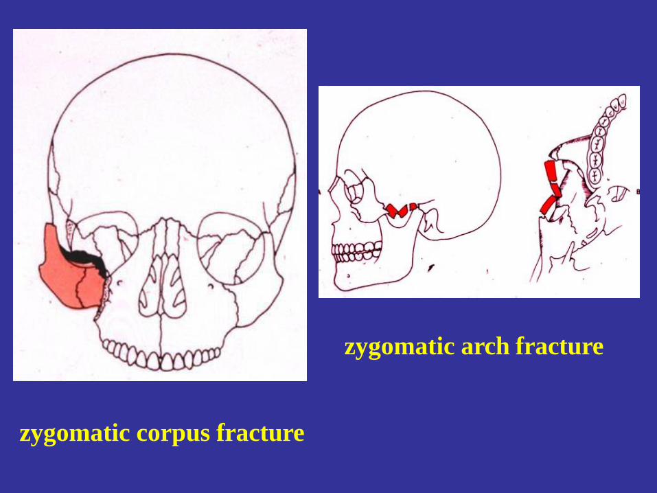
zygomatic arch fracture
zygomatic corpus fracture
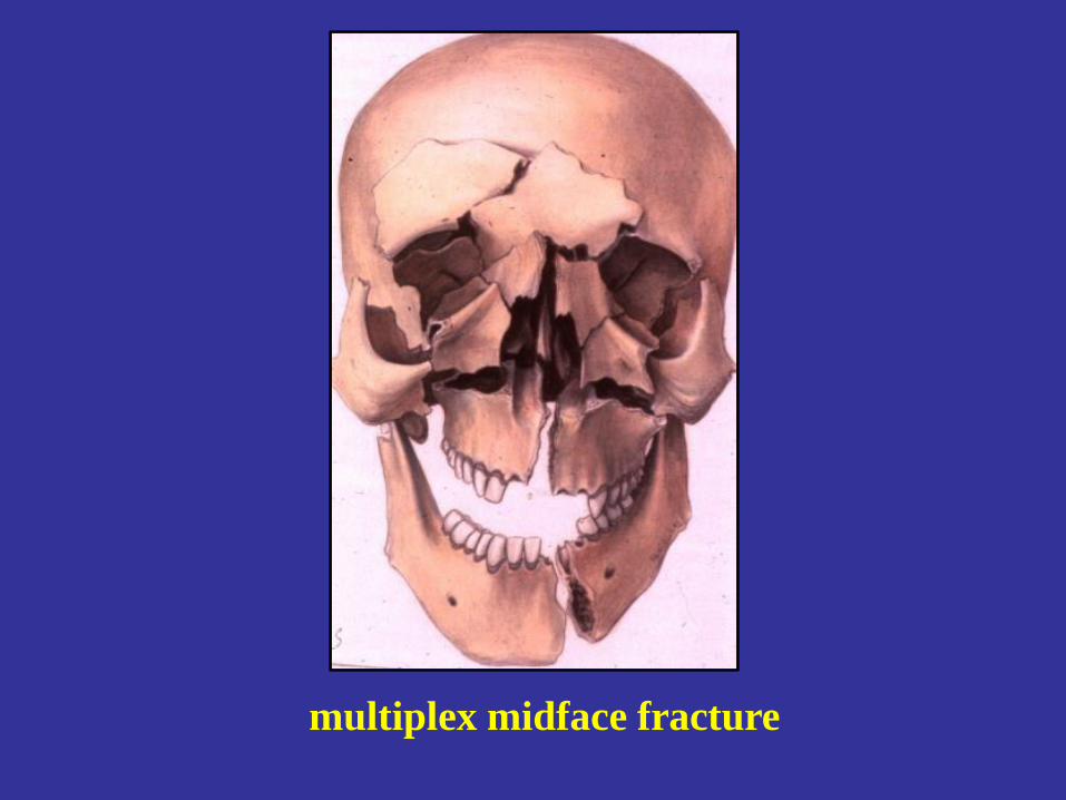
multiplex midface fracture
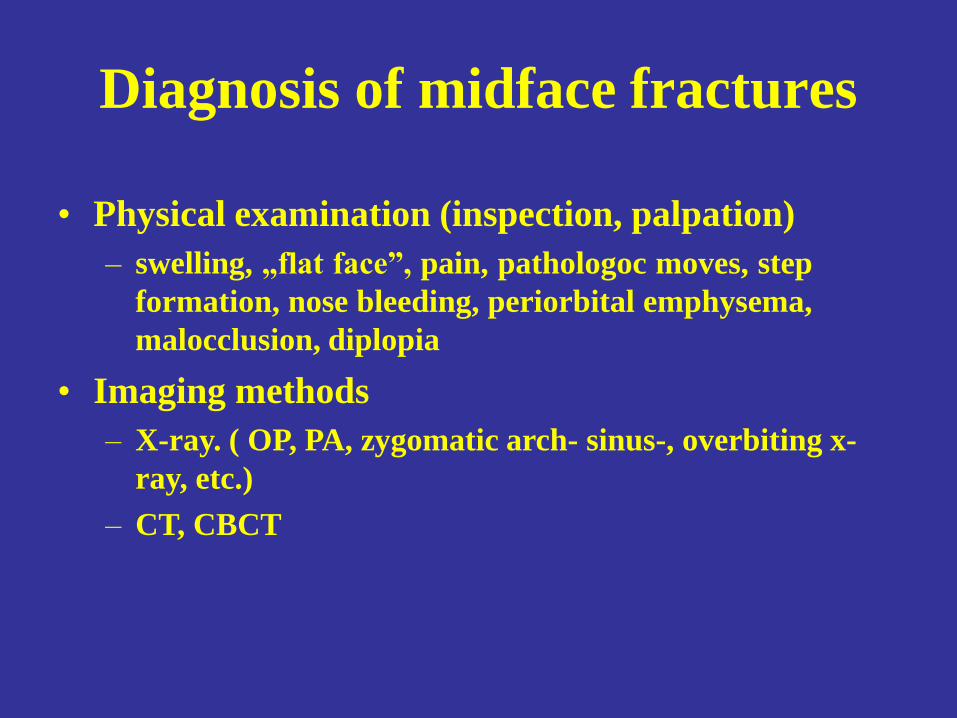
Diagnosis of midface fractures
• Physical examination (inspection, palpation)
– swelling, „flat face”, pain, pathologoc moves, step
formation, nose bleeding, periorbital emphysema,
malocclusion, diplopia
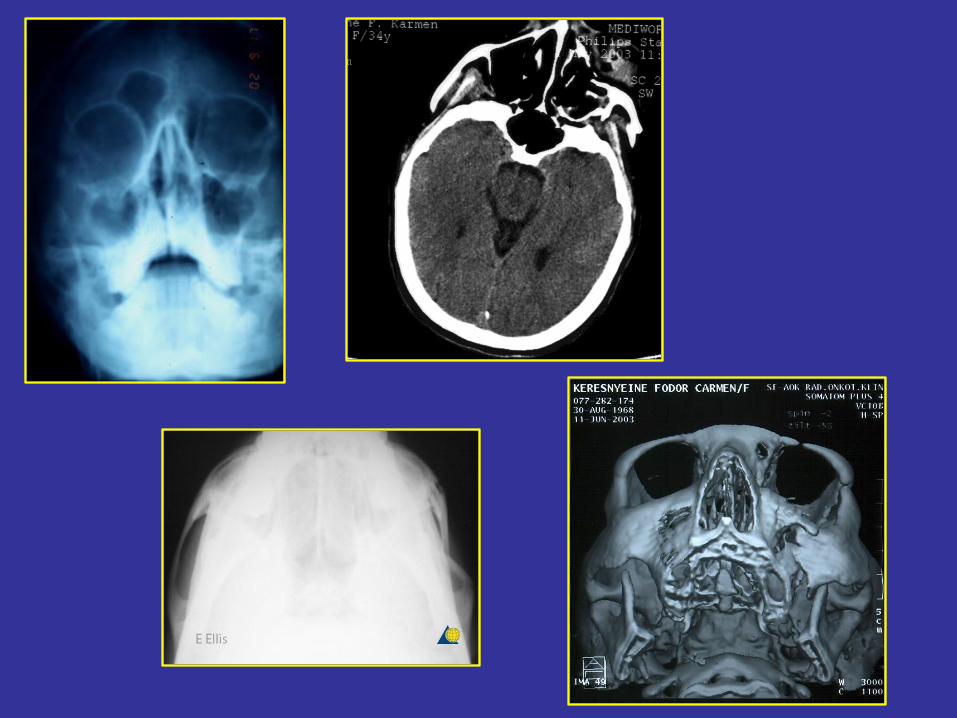
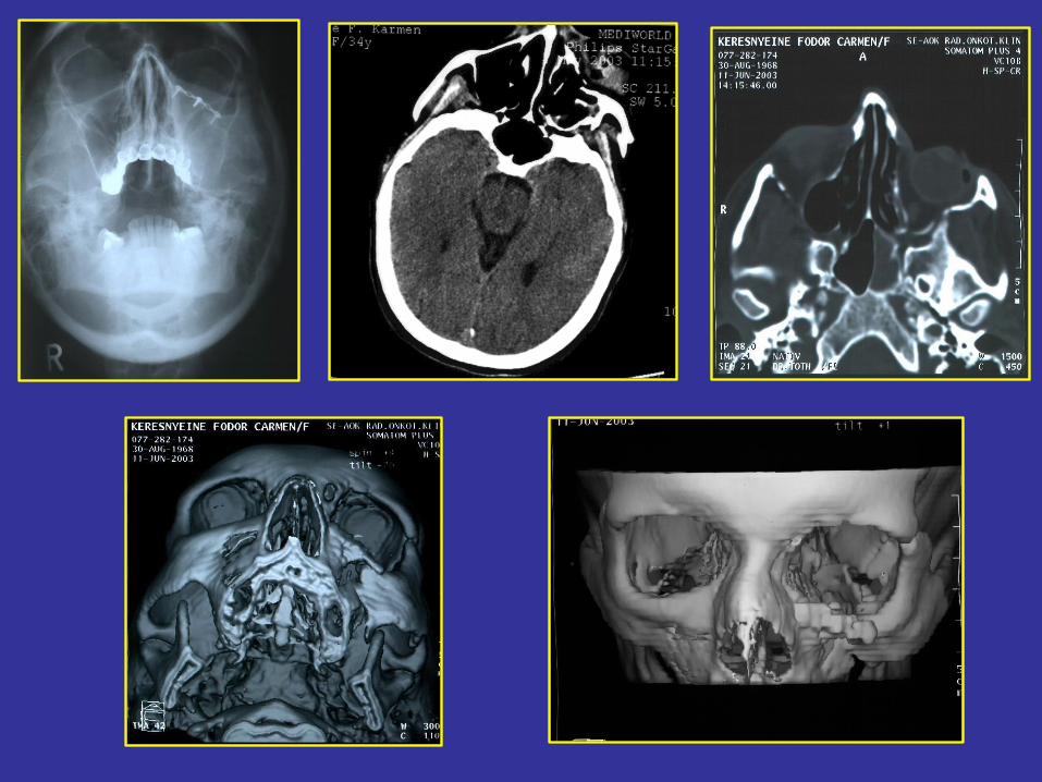
• Imaging methods
– X-ray. ( OP, PA, zygomatic arch- sinus-, overbiting x-
ray, etc.)
– CT, CBCT
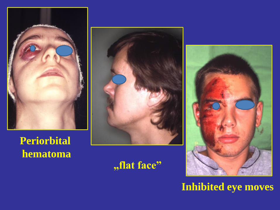
Periorbital
hematoma „flat face”
Inhibited eye moves
Therapy of midface fractures I.
Aim:
• Reconstruion of occlusion, functions and esthetics
Steps:
• reposition
• immobilisation (fixation)
• rehabilitation
Therapy of midface fractures II.
• conservative (rare)
• surgical
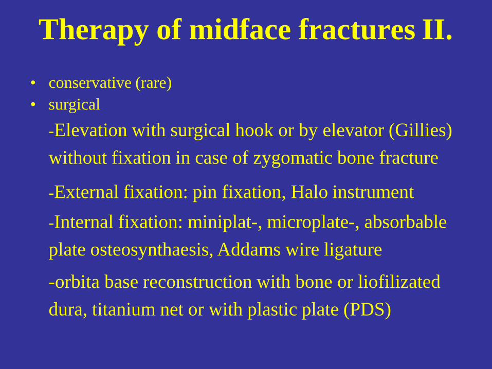
-Elevation with surgical hook or by elevator (Gillies)
without fixation in case of zygomatic bone fracture
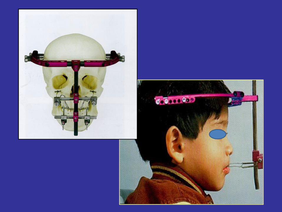
-External fixation: pin fixation, Halo instrument
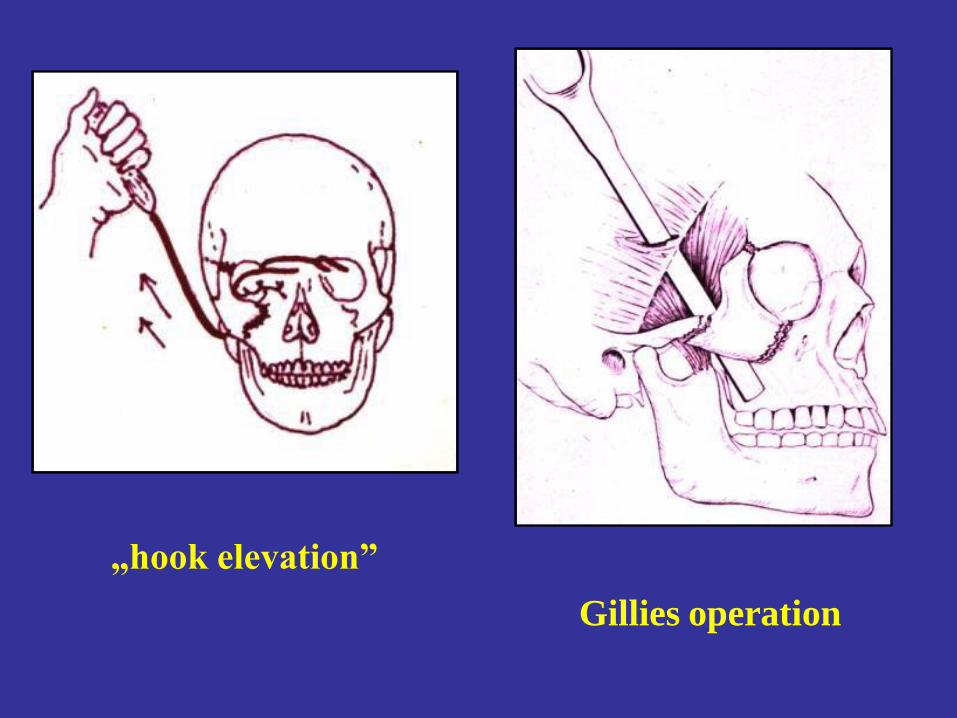
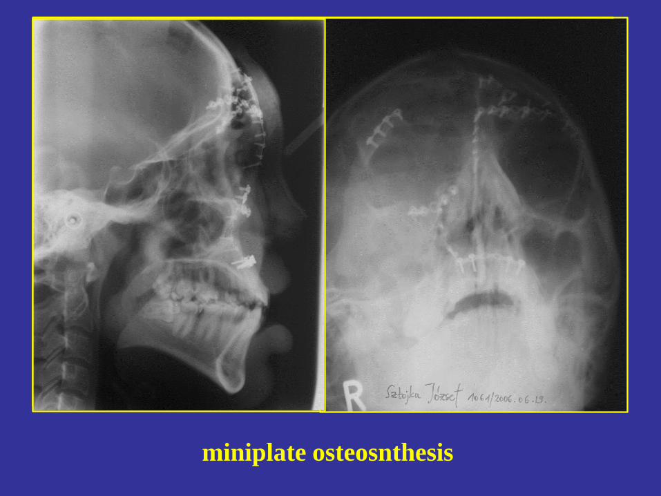
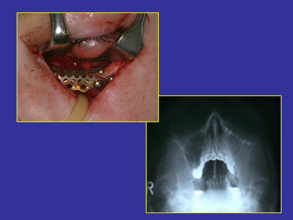
-Internal fixation: miniplat-, microplate-, absorbable
plate osteosynthaesis, Addams wire ligature
-orbita base reconstruction with bone or liofilizated
dura, titanium net or with plastic plate (PDS)
„hook elevation”
Gillies operation
miniplate osteosnthesis
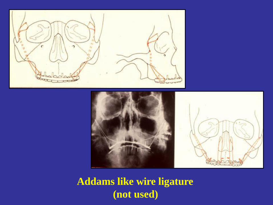
Addams like wire ligature
(not used)
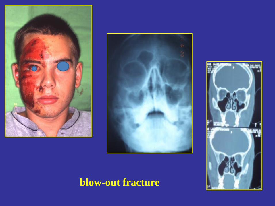
Blow- out fracture
Content of the orbita (fat or muscle
/rectus inf. or obliqous int./ herniation through
the orbital base impressional fracture into the
sinus cavity due to sudden increase of orbital
content pressure
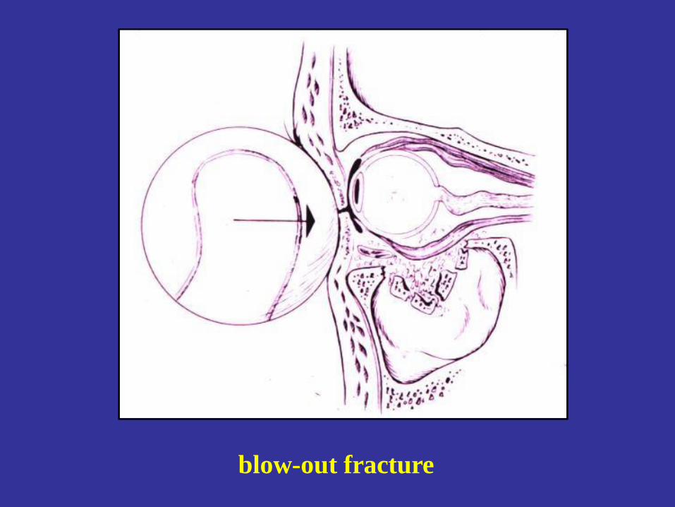
blow-out fracture
Symptoms
• decreased eye moves
• dyplopia
• enophtalmus
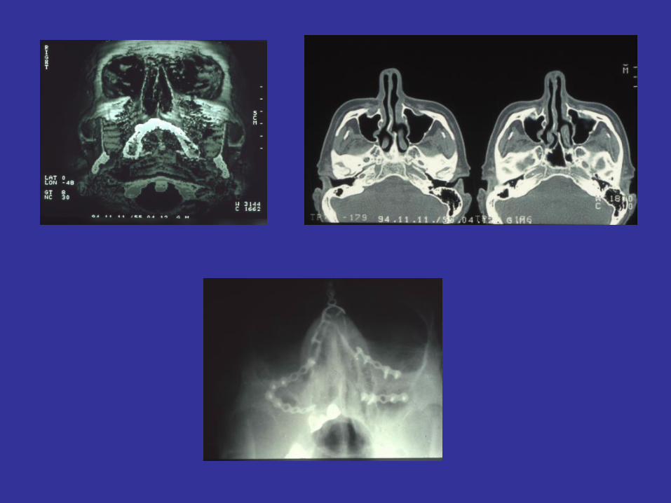
Diagnosis
• Physical examination
• Imaging methods
– PA skull x-ray, CT (coronal) !!!
blow-out fracture
Therapy of blow-out fracture
• exploration of orbital floor
• reposition
• fixation
– Reconstruction of orbital floor (titanium net,
Lyodura, PDS membrane, autologous bone etc.
– Support of the orbital floor through the
maxillary sinus ( Folley cateter)
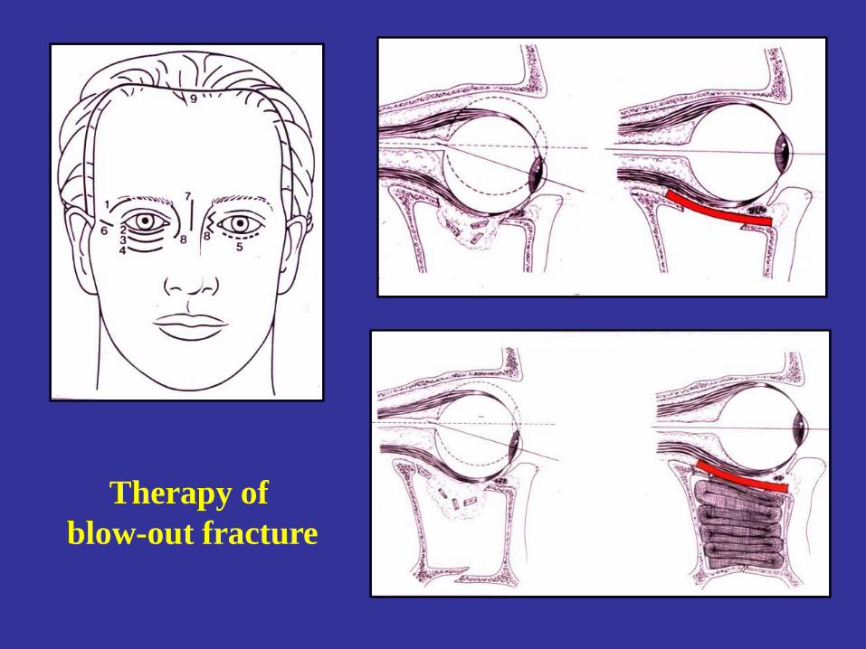
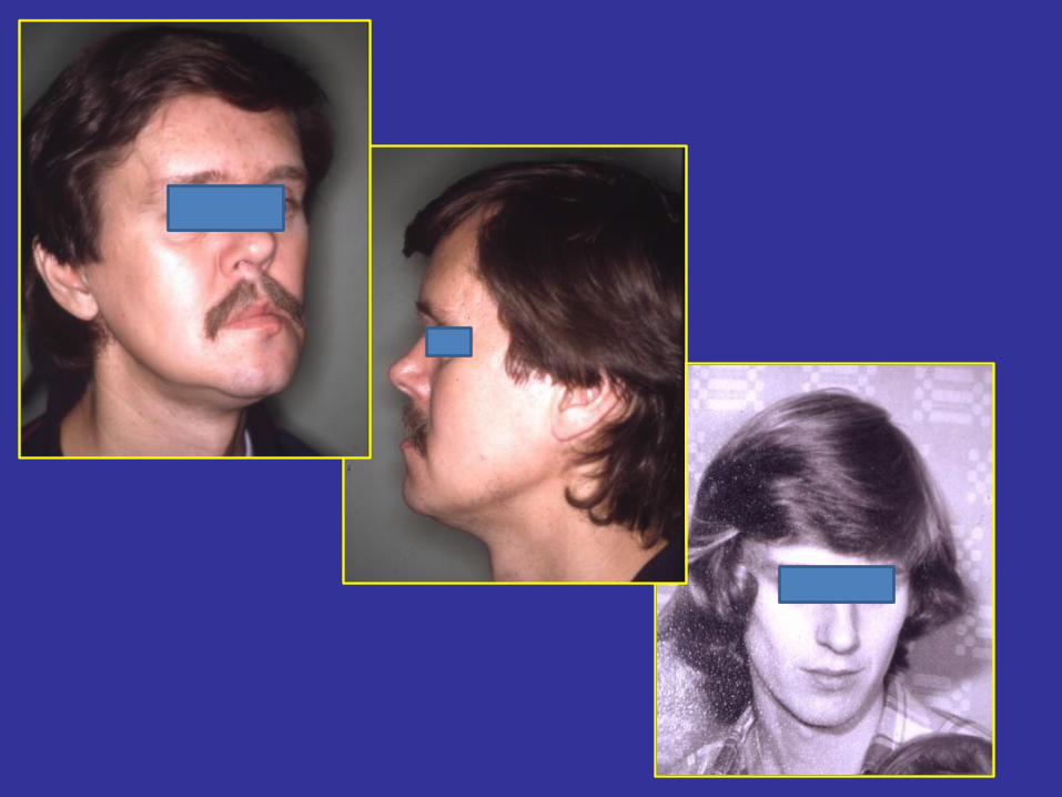
Therapy of
blow-out fracture
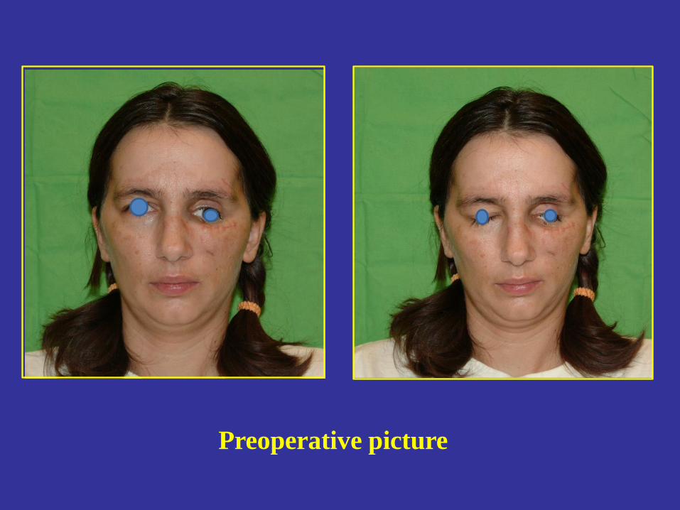
Preoperative picture
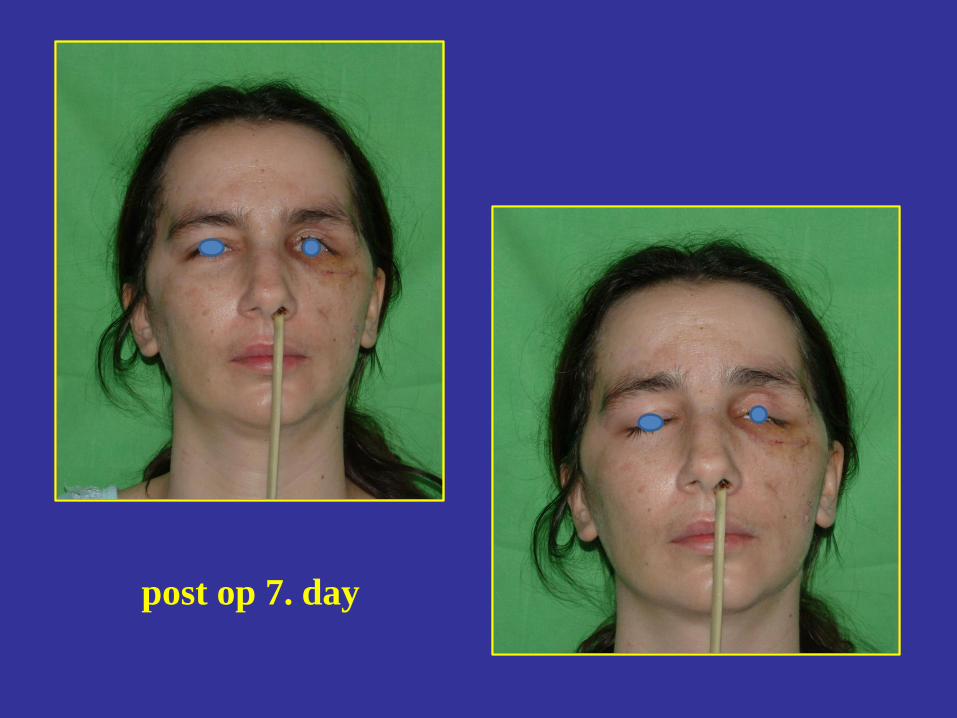
post op 7. day
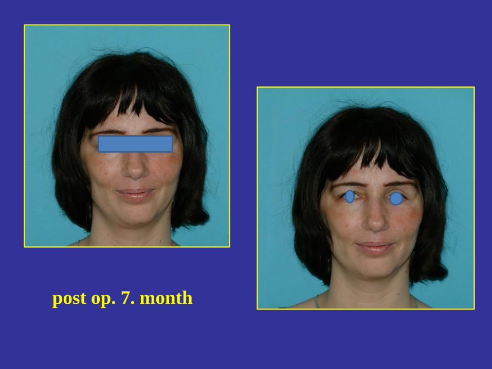
post op. 7. month
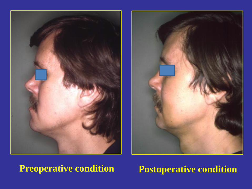
Postoperative condition Preoperative condition

THANK YOU!
Top Related