


Languages
Pages
Legal

Load Distributing Band
Mechanical Chest
Compression Device for
Treatment of Cardiac Arrest A/Prof Marcus Ong
Senior Consultant, Clinician Scientist
& Director of Research
Department of Emergency Medicine
Singapore General Hospital
Associate Professor
Duke-NUS Graduate Medical School
Health Services and Systems Research

Traditional CPR
• The problem with standard CPR (STD-
CPR): provides only 1/3 of normal blood
supply to the brain and 10-20% to the heart
• Problem of rescuer fatigue, CPR not
consistent, and need to stop CPR during
rescuer changes and patient transfers

With permission from Dr Sang Do Shin, Seoul National University
Load Distributing Band CPR
The AutoPulse™ (Revivant Corporation, Sunnyvale,
CA) is a non-invasive Load Distributing Band
device.
Distributing force over the entire chest improves the
effectiveness of chest compressions.
Less harm, elimination of rescuer fatigue, more
consistent, and eliminating the need to stop CPR
during rescuer changes and patient transfers

Use of an Automated, Load-Distributing
Band Chest Compression Device for Out-of-
Hospital Cardiac Arrest Resuscitation
JAMA 2006 June 295(22): 2629-2637
Ong MEH, Ornato JP, Edwards DP, Best AM,
Ines CS, Hickey S, Williams D, Clark B, Powell R, Overton J,
Peberdy MA.
Methods
Phased, non-randomized, interventional trial
Before and after replacement of standard CPR with the LDB-
CPR device in adult OHCA victims treated by paramedics
in Richmond, Virginia
Richmond metropolitan area: population of approximately 200
000, representative of a mid-size North American city
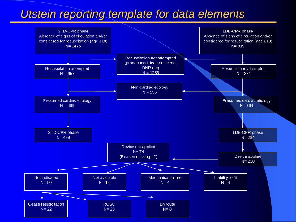
Resuscitation attempted
N = 381
Resuscitation not attempted
(pronounced dead on scene,
DNR etc)
N = 1256
Presumed cardiac etiology
N =284
Non-cardiac etiology
N = 255
LDB-CPR phase
N= 284
STD-CPR phase
N= 499
STD-CPR phase
Absence of signs of circulation and/or
considered for resuscitation (age 18)
N= 1475
LDB-CPR phase
Absence of signs of circulation and/or
considered for resuscitation (age 18)
N= 819
Resuscitation attempted
N = 657
Presumed cardiac etiology
N = 499
Device applied
N= 210
Device not applied
N= 74
(Reason missing =2)
Not indicated
N= 50
Not available
N= 14
Mechanical failure
N= 4
Inability to fit
N= 4
Cease resuscitation
N= 22
ROSC
N= 20
En route
N= 8
Utstein reporting template for data elements
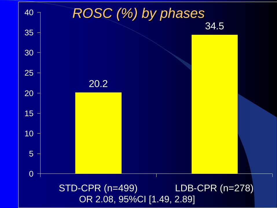
20.2
34.5
0
5
10
15
20
25
30
35
40
STD-CPR (n=499) LDB-CPR (n=278)
ROSC (%) by phases
OR 2.08, 95%CI [1.49, 2.89]
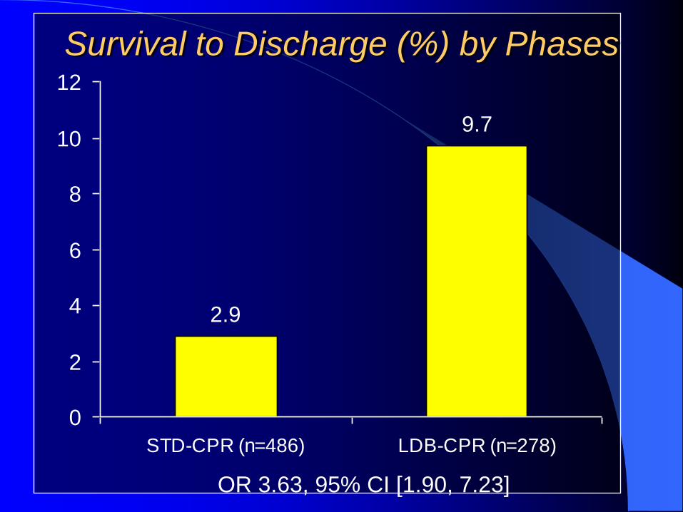
2.9
9.7
0
2
4
6
8
10
12
STD-CPR (n=486) LDB-CPR (n=278)
Survival to Discharge (%) by Phases
OR 3.63, 95% CI [1.90, 7.23]
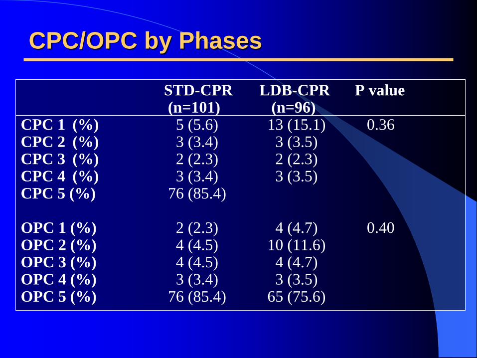
CPC/OPC by Phases
STD-CPR LDB-CPR P value (n=101) (n=96) CPC 1 (%) 5 (5.6) 13 (15.1) 0.36 CPC 2 (%) 3 (3.4) 3 (3.5) CPC 3 (%) 2 (2.3) 2 (2.3) CPC 4 (%) 3 (3.4) 3 (3.5) CPC 5 (%) 76 (85.4) OPC 1 (%) 2 (2.3) 4 (4.7) 0.40 OPC 2 (%) 4 (4.5) 10 (11.6) OPC 3 (%) 4 (4.5) 4 (4.7) OPC 4 (%) 3 (3.4) 3 (3.5) OPC 5 (%) 76 (85.4) 65 (75.6)
Manual chest compression vs use of an automated
chest compression device during resuscitation
following out-of-hospital cardiac arrest: a
randomized trial.
Jama. Jun 14 2006;295(22):2620-2628.
Hallstrom A, Rea TD, Sayre MR, et al.

Device should not be seen as the ‘miracle’ solution to
cardiac arrest
Multiple factors will affect cardiac arrest outcomes
The AutoPulseTM should be seen as a possible new
component of an overall resuscitation strategy
Challenge is to incorporate this in current treatment
protocols seamlessly
Reconciling the results

Interruptions
to CPR during
device
deployment
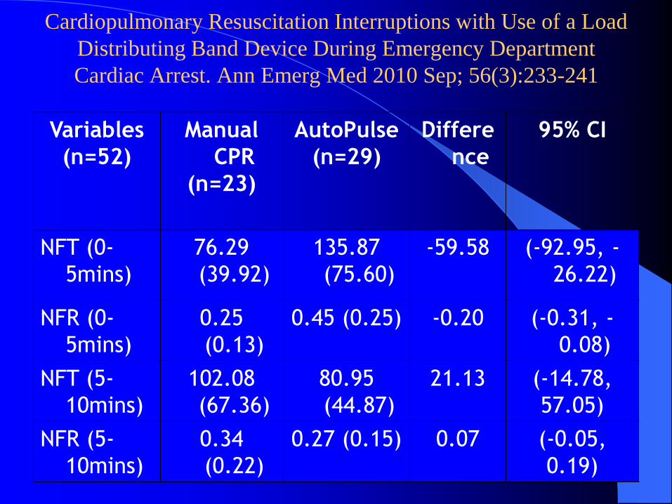
Cardiopulmonary Resuscitation Interruptions with Use of a Load
Distributing Band Device During Emergency Department
Cardiac Arrest. Ann Emerg Med 2010 Sep; 56(3):233-241
Variables
(n=52)
Manual
CPR
(n=23)
AutoPulse
(n=29)
Differe
nce
95% CI
NFT (0-
5mins)
76.29
(39.92)
135.87
(75.60)
-59.58 (-92.95, -
26.22)
NFR (0-
5mins)
0.25
(0.13)
0.45 (0.25) -0.20 (-0.31, -
0.08)
NFT (5-
10mins)
102.08
(67.36)
80.95
(44.87)
21.13 (-14.78,
57.05)
NFR (5-
10mins)
0.34
(0.22)
0.27 (0.15) 0.07 (-0.05,
0.19)

Error bar of No Flow Time for 1st 5
mins of resuscitation over LDB
phase of the study
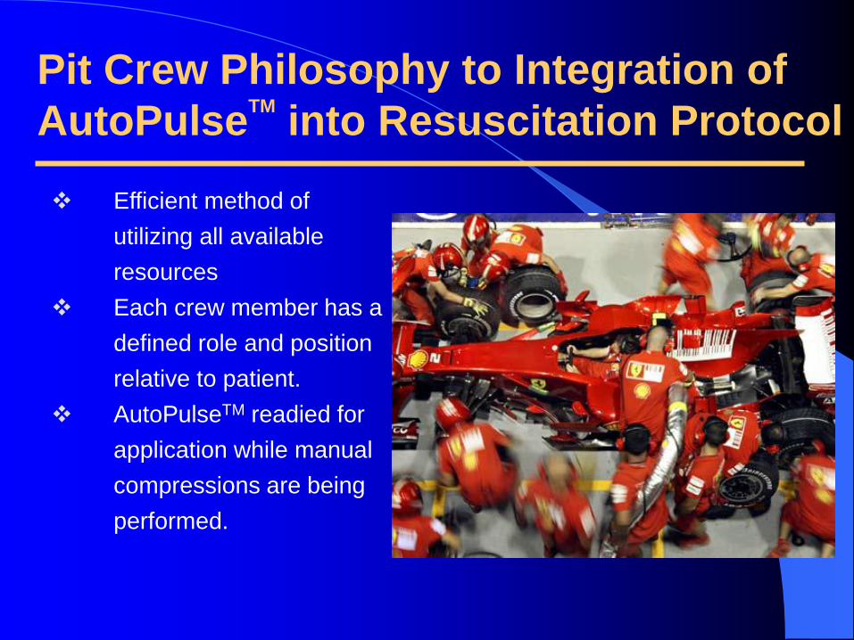
Pit Crew Philosophy to Integration of
AutoPulseTM
into Resuscitation Protocol
Efficient method of
utilizing all available
resources
Each crew member has a
defined role and position
relative to patient.
AutoPulseTM readied for
application while manual
compressions are being
performed.
Use of an Automated, Load-
Distributing Band Chest
Compression Device for
Cardiac Arrest Resuscitation :
A Multi-Centre Clinical Trial
A/Prof Marcus Ong Eng Hock MBBS, FRCS (A&E) Edin, MPH (VCU), FAMS
Senior Consultant, Director of Research and Clinician Scientist
Department of Emergency Medicine, Singapore General Hospital
Office of Clinical Sciences, Duke-NUS GMS
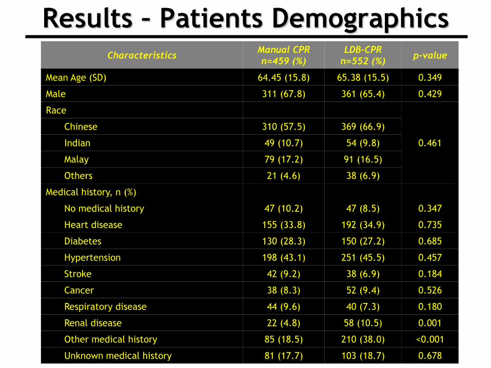
Results – Patients Demographics Characteristics
Manual CPR
n=459 (%)
LDB-CPR
n=552 (%) p-value
Mean Age (SD) 64.45 (15.8) 65.38 (15.5) 0.349
Male 311 (67.8) 361 (65.4) 0.429
Race
0.461
Chinese 310 (57.5) 369 (66.9)
Indian 49 (10.7) 54 (9.8)
Malay 79 (17.2) 91 (16.5)
Others 21 (4.6) 38 (6.9)
Medical history, n (%)
No medical history 47 (10.2) 47 (8.5) 0.347
Heart disease 155 (33.8) 192 (34.9) 0.735
Diabetes 130 (28.3) 150 (27.2) 0.685
Hypertension 198 (43.1) 251 (45.5) 0.457
Stroke 42 (9.2) 38 (6.9) 0.184
Cancer 38 (8.3) 52 (9.4) 0.526
Respiratory disease 44 (9.6) 40 (7.3) 0.180
Renal disease 22 (4.8) 58 (10.5) 0.001
Other medical history 85 (18.5) 210 (38.0) <0.001
Unknown medical history 81 (17.7) 103 (18.7) 0.678

Results – Patients Demographics
Characteristics Manual CPR
n=459 (%)
LDB-CPR
n=552 (%) p-value
Hospital
<0.001 Hospital A 186 (40.5) 293 (53.1)
Hospital B 273 (59.5) 259 (46.9)
Arrest location
<0.001 Prehospital 437 (95.2) 463 (83.9)
ED 22 (4.8) 89 (16.1)
Bystander witnessed 292 (63.6) 233 (42.2) <0.001
EMS witnessed 41 (8.9) 23 (4.2) 0.002
Bystander CPR 110 (24.0) 50 (9.1) <0.001
Initial rhythm
<0.001
Ventricular fibrillation 23 (5.0) 40 (7.3)
Ventricular tachycardia 0 (0.0) 10 (1.8)
Asystole 340 (74.0) 336 (60.9)
Pulseless electrical activity 80 (17.4) 119 (21.6)
Pre-hospital defibrillation 100 (21.8) 84 (15.2) 0.007
Defibrillated at ED 124 (27.0) 154 (27.9) 0.754
AutoPulse applied - 454 (82.3) <0.001
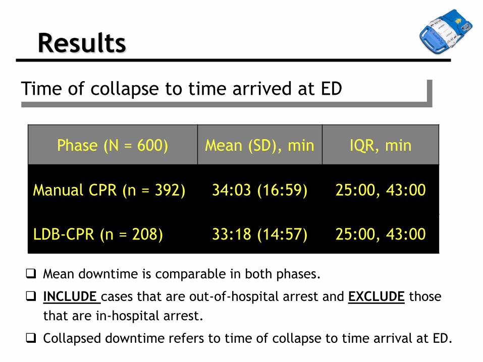
Results
Phase (N = 600) Mean (SD), min IQR, min
Manual CPR (n = 392) 34:03 (16:59) 25:00, 43:00
LDB-CPR (n = 208) 33:18 (14:57) 25:00, 43:00
Time of collapse to time arrived at ED
Mean downtime is comparable in both phases.
INCLUDE cases that are out-of-hospital arrest and EXCLUDE those
that are in-hospital arrest.
Collapsed downtime refers to time of collapse to time arrival at ED.

Manual CPR n=459(%)
LDB-CPR n=552(%)
Crude OR (95% CI)
Adjusted† OR (95% CI)
Return of spontaneous circulation
103 (22.4) 195 (35.3) 1.89
(1.43,2.50) 1.60
(1.16, 2.22)
Survival to hospital
admission 65 (14.2) 109 (19.8)
1.49 (1.07, 2.09)
1.23 (0.84, 1.81)
Survival to hospital
discharge 6 (1.3) 18 (3.3)
2.55 (1.00, 6.47)
1.42 (0.47, 4.29)
Results - Comparison of Clinical Outcomes
† The model was adjusted for hospital, arrest location, bystander witnessed, EMS witnessed, initial rhythm, prehospital defibrillation and LDB-CPR applied.

Results – CPC/OPC of Survivors
Performance categories Manual CPR
n=6 (%)
LDB-CPR
n=16 (%) p-value*
CPC 1 1 (16.7%) 12 (75%)
0.01 CPC 2 1 (16.7%) 1 (6.3%)
CPC 3 4 (66.7%) 1 (6.3%)
CPC 4 0 (0.00) 2 (12.5%)
OPC 1 1 (16.7%) 10 (62.5%)
0.06 OPC 2 1 (16.7%) 2 (12.5%)
OPC 3 4 (66.7%) 2 (12.5%)
OPC 4 0 (0.00) 2 (12.5%)
* Fisher’s exact test was used to compare percentages
CPC= Cerebral Performance Category; OPC= Overall Performance Category
Conclusion
A resuscitation strategy using LDB-CPR
in an ED environment was associated
with improved neurologically intact
survival to discharge in adults with non-
traumatic cardiac arrest, in a setting
with long arrest times.
A Multicenter Randomised Controlled Trial
Comparing Shock Success With Synchronized
Defibrillation (Compression Upstroke Versus
Precompression) During Ongoing Mechanical
Cardiopulmonary Resuscitation In The
Emergency Department
A/Prof Marcus Ong Senior Consultant, Senior Medical Scientist
& Director of Research
Department of Emergency Medicine
Singapore General Hospital
Overview
Objective •To compare shock success during defibrillation synchronized with the
upstroke of chest compression (peak upstroke), and precompression
(control)
Primary Outcomes:
•Shock success defined as the termination of VF or pulseless VT and
establishment of organised rhythm within 60 sec and requires at least 2
QRS complexes separated by no more than 5 sec
Second Outcomes:
•Termination of VF for at least 5 sec after the shock, regardless of the
resulting rhythm
•ROSC
•Survival to hospital admission
•Survival to hospital discharge •Glasgow Outcomes Score (CPC/OPC)
•European Quality of Life in 5 Dimensions (EQ-5D)

Synchronized Shocking Phases
Li Y YT, Ristagno G, Chung SP, Bisera J, Quan W, Freeman G, Weil MH, Tang W. The optimal phasic relationship
between synchronized shock and mechanical chest compressions. Resuscitation. 2010;81(6):724-729.

Synchronized Shock Phases
Li Y YT, Ristagno G, Chung SP, Bisera J, Quan W, Freeman G, Weil MH, Tang W. The optimal phasic relationship
between synchronized shock and mechanical chest compressions. Resuscitation. 2010;81(6):724-729.
Learning points
• Clinical trials in emergency situations
• Importance of device training and
quality of implementation
• Co-ordination of multi-site trials (eg
battery maintenance issues)
• PI initiated, industry supported trials
• Intellectual property and clinical trials
agreements
Top Related