


Languages
Pages
Legal

Leukemia
Leukemia vs lymphoma The major types of leukemia are based on whether the
disease is: Acute or chronic Lymphocytic or myeloid

Acute vs chronic leukemia
rapid increase in immature blood cells.
rapid progression and accumulation of the malignant cells over weeks or months.
Immediate treatment is required in acute leukemia
most common forms of leukemia in children.
excessive build up of relatively mature, but still abnormal, white blood cells.
Typically taking months or years to progress,
chronic forms are sometimes monitored for some time before treatment to ensure maximum effectiveness of therapy.
Chronic leukemia mostly occurs in older people.

Myeloid vs lymphoid

Acute myeloid leukemia
Incidence ~ 3.5/100000 people/yearM/F = 4.3 : 2.9Incidence increases with age 1.7<65yrs
15.9>65yrsEtiology Hereditary down syndrome (trisomy 21chromosome), fanconi
anemia, bloom syndrome, ataxia telangiectasia & kostmann syndrome
Radiation survivors of atomic bomb explosion Chemicals benzene exposure used as a solvent in chemical, plastic,
rubber & pharmaceutical industries. exposure to petroleum products, paints, embalming fluids, ethylene oxide, herbicides & pesticides
Drugs alkylating agents, topoisomerase II inhibitors, chloramphenicol, phenylbutazone

Classification(morphology,cytochemistry)
French-American-British (FAB) Classification IncidenceM0: Minimally differentiated leukemia 5%M1: Myeloblastic leukemia without maturation 20%M2: Myeloblastic leukemia with maturation 30%M3: Hypergranular promyelocytic leukemia 10%M4: Myelomonocytic leukemia 20%M4Eo: Variant: Increase in abnormal marrow eosinophilsM5: Monocytic leukemia 10%M6: Erythroleukemia (DiGuglielmo's disease) 4%M7: Megakaryoblastic leukemia 1%

WHO Classification (clinical,cytogenetics,molecular)
I. AML with recurrent genetic abnormalities AML with t(8;21)(q22;q22) Acute promyelocytic leukemia [AML with t(15;17)(q22;q12) AML with 11q23 (MLL) abnormalities
II. AML with myelodysplasia related changes Following a myelodysplastic syndrome or myelodysplastic syndrome/myeloproliferative disorder Without antecedent myelodysplastic syndrome
III. Therapy related myeloid neoplasms, therapy-related Alkylating agent–related Topoisomerase type II inhibitor–related
IV. AML not otherwise categorized
V.Myeloid sarcoma
VI.Myeloid proliferations related to Down syndrome
VII.Blastic plasmacytoid dendritic cell neoplasm
VIII.Acute leukemia of ambiguous lineage

Clinical feature
Anemia fatigue, weakness, anorexia,
Leucopenia/leucocytosis/leucocyte dysfn fever, recurrent infection
Thrombocytopenia bleeding episodes Nearly half have symptoms >3 months before leukemia was
diagnosed.Mass lesion –located in soft tissue ( granulocytic sarcoma,
chloroma ) Sign –fever, splenomegaly, hepatomegaly,
lymphadenopathy,sternal tenderness, evidence of infection, hemorrhage, bleeding tendency(M3),infiltration of gingiva, skin,soft tissue,meninges(M4&M5)

Investigation
Anemia NN,RC low Leucocyte ↓ / ↑ 25-40% <5000/ul
20% >1lakh/ul Platelet 75% <1lakh/ul
25%<25,000/ul Biochemistry : electrolyte, creatinine, calcium,phosphate,
LFT,LDH Coagulation profile Marrow aspirate & biopsy HLA typing
Diagnosis ≥ 20%myeloblasts –blood/marrow, cytoplasmic granules, auer rods, A+ MPO reaction >3%blast
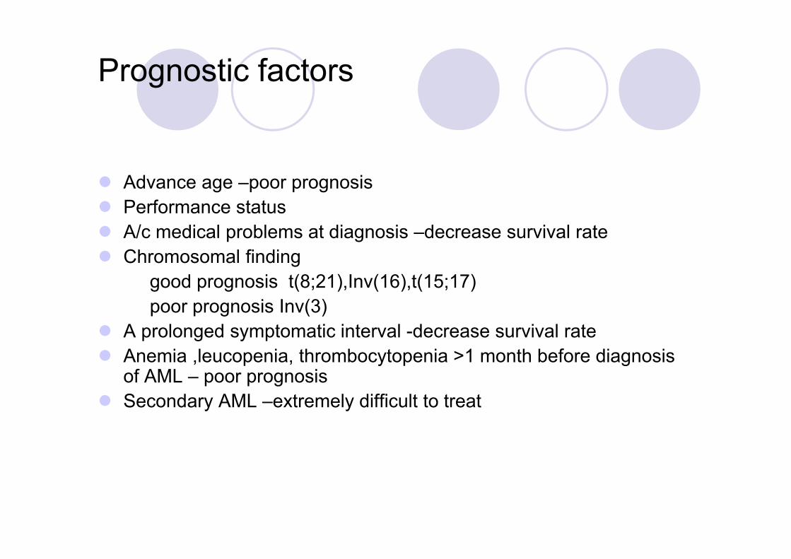
Prognostic factors
Advance age –poor prognosis Performance status A/c medical problems at diagnosis –decrease survival rate Chromosomal finding
good prognosis t(8;21),Inv(16),t(15;17)poor prognosis Inv(3)
A prolonged symptomatic interval -decrease survival rate Anemia ,leucopenia, thrombocytopenia >1 month before diagnosis
of AML – poor prognosis Secondary AML –extremely difficult to treat
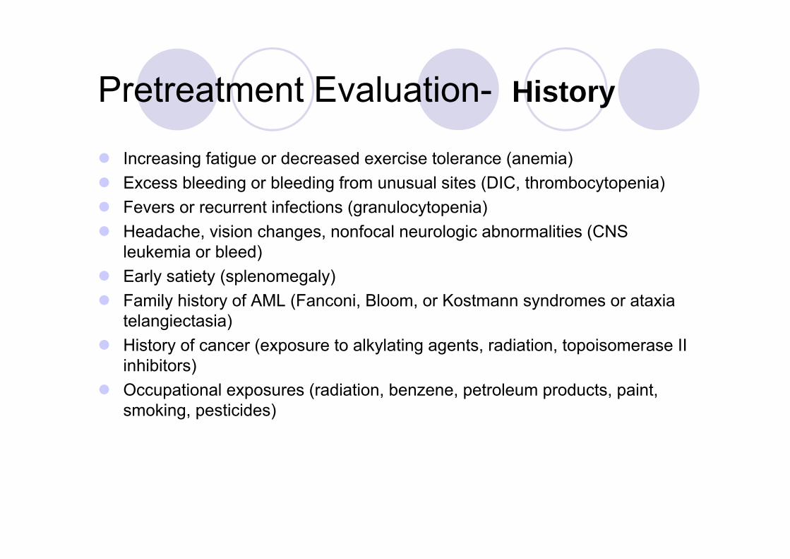
Pretreatment Evaluation- History
Increasing fatigue or decreased exercise tolerance (anemia) Excess bleeding or bleeding from unusual sites (DIC, thrombocytopenia) Fevers or recurrent infections (granulocytopenia) Headache, vision changes, nonfocal neurologic abnormalities (CNS
leukemia or bleed) Early satiety (splenomegaly) Family history of AML (Fanconi, Bloom, or Kostmann syndromes or ataxia
telangiectasia) History of cancer (exposure to alkylating agents, radiation, topoisomerase II
inhibitors) Occupational exposures (radiation, benzene, petroleum products, paint,
smoking, pesticides)

Physical Examination
Performance status (prognostic factor) Ecchymosis and oozing from IV sites (DIC, possible acute promyelocytic
leukemia) Fever and tachycardia (signs of infection) Papilledema, retinal infiltrates, cranial nerve abnormalities (CNS leukemia) Poor dentition, dental abscesses Gum hypertrophy (leukemic infiltration, most common in monocytic
leukemia) Skin infiltration or nodules (leukemia infiltration, most common in monocytic
leukemia) Lymphadenopathy, splenomegaly, hepatomegaly Back pain, lower extremity weakness [spinal granulocytic sarcoma, most
likely in t(8;21) patients]

Laboratory and Radiologic Studies
CBC with manual differential cell count Chemistry tests (electrolytes, creatinine, BUN, calcium, phosphorus, uric
acid, hepatic enzymes, bilirubin, LDH, amylase, lipase) Clotting studies (prothrombin time, partial thromboplastin time, fibrinogen,
D-dimer) Viral serologies (CMV, HSV-1, varicella zoster) HLA typing of patient, siblings, and parents for potential allogeneic SCT Bone marrow aspirate and biopsy (morphology, cytogenetics, flow
cytometry, molecular studies) Cryopreservation of viable leukemia cells Echocardiogram or heart scan PA and lateral chest radiograph Placement of central venous access device

Treatment Symptomatic therapy Cytopenia – G-CSF, GM-CSF Multilumen R atrial catheter insertion Thromboctopenia –thrombopoietin, platelet transfusion Anemia –erythropoietin, packed cell transfusion Oral nystatin or clotrimazole is recommended to prevent localized
candidiasis Infection –prompt & early initiation of empirical broad spectrum
antibiotics & antifungal
Allogenic SCT – treatment of choice
Chemothreapy - Induction therapy (aim – complete remission)- Post remission therapy- Relapse
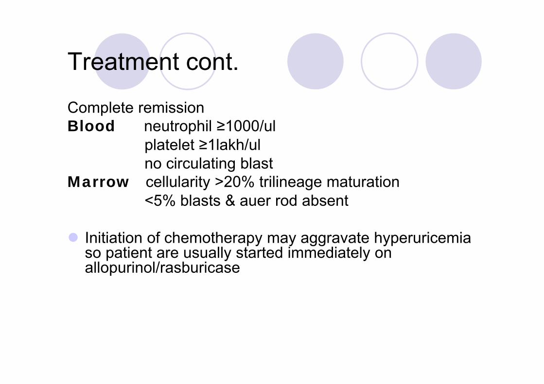
Treatment cont.
Complete remission Blood neutrophil ≥1000/ul
platelet ≥1lakh/ulno circulating blast
Marrow cellularity >20% trilineage maturation<5% blasts & auer rod absent
Initiation of chemotherapy may aggravate hyperuricemia so patient are usually started immediately on allopurinol/rasburicase

Induction therapy
Cytarabin i/v infusion 100-200mg/m2 x7d+anthracycline daunonorubicin i/v 45-60mg/m2 x1,2,3d
idarubicin i/v 12-13mg/m2 x1,2,3 d±etoposide
Complete CR
Post remission therapy
cure relapse
2/3
Incomplete CR
2 More inductioncourse
Incomplete CR3-4 cyclesof highDose cytarabin
Allogenic SCT
1/3

Post remission therapyDesigned to eradicate any Residual leukemic cells
age
<55yrs >55yrs
Intensive chemotherapyHigh dose cytarabin& allogenic/autologusSCT
Attenuated intensive therapyChemotherapy/non myeloablativeAllogenic SCT

Relapse
Once relapse occurs, patients are rarely cured with further standard-dose chemotherapy. Patients eligible for allogeneic SCT should receive transplants at the first sign of relapse.
Treatment with novel approaches should be considered if SCT is not possible. One promising therapy is decitabine, a nucleoside analog that inhibits DNA methyltransferase and subsequently reverses aberrant methylation in AML cells.
For elderly patients (age >60) for whom clinical trials are not available, gemtuzumab ozogamicin (Mylotarg) is another alternative

Treatment of Promyelocytic Leukemia
Tretinoin is an oral drug that induces the differentiation of leukemic cells bearing the t(15;17).
Tretinoin (45 mg/m2 per day orally until remission is documented) plus concurrent anthracycline chemotherapy appears to be among the safest and most effective treatments for APL.
Arsenic trioxide produces meaningful responses in up to 85% of patients refractory to tretinoin.
The detection of minimal residual disease by RT-PCR amplification of the t(15;17) chimeric gene product appears to predict relapse.
Disappearance of the signal is associated with long-term disease-free survival;

Chronic myeloid leukemia
chronic granulocytic leukemia (CGL)
clonal bone marrow stem cell disorder in which proliferation of mature granulocyte (neutrophils, eosinophils, and basophils) and their precursors

Chronic myeloid leukemia
Incidence 1.5/100000 people/yearM/F=1.9:1.1increase with age
Pathophysiology Clonal expansion of a hematopoietic stem cell possessing a
reciprocal translocation between chromosome 9 &22 t(9;22). untreated disease is characterized by inevitable transition from a
chronic phase to an accelerated phase and on to blast crisis. The product of fusion gene result from the t(9;22) plays a central
role in development of CML

Clinical feature
Insidious onset Anemia Recurrent infection Bleeding tendency & thrombosis (vasoocclusive disease) Hyper metabolic state (wt. loss, night sweats) Splenomegaly (early satiety, LUQ pain) Pruritis ,diarrhea, flushing (histamine production
secondary to basophilia)

Investigation
Anemia Leucocytosis with varying degree of immaturity Platelet N /low Marrow ↑M/E ratio Cytogenetics - Philadelphia chromosome t(9;22)
- 90-95% cases Cytochemistry neutrophil alkaline phosphatase (NAP)
- lowOthers serum B12 elevated & B12 binding capacity hyperuricimia

Classification(clinical/laboratory)
chronic phase(Approximately 85%/usually asymptomatic
accelerated phase blast crisis(terminal phase of CML/acute
leukemia)

Disease acceleration(WHO)
10–19% myeloblasts (blood/bone marrow) >20% basophils (blood/bone marrow) Platelet count <100,000, unrelated to therapy Platelet count >1,000,000, unresponsive to therapy Cytogenetic evolution with new abnormalities in addition
to the Philadelphia chromosome Increasing splenomegaly or white blood cell count,
unresponsive to therapy
The patient is considered to be in the accelerated phase if any of the above are present. The accelerated phase is significant because it signals that the disease is progressing and transformation to blast crisis is imminent. Drug treatment often becomes less effective in the advanced stages

Blast crisis(WHO)
Blast crisis is the final phase in the evolution of CML, and behaves like an acute leukemia, with rapid progression and short survival.
Blast crisis is diagnosed if any of the following are present in a patient with CML.
>20% myeloblasts/lymphoblasts in the blood or bone marrow
Large clusters of blasts in the bone marrow on biopsy Development of a chloroma (solid focus of leukemia
outside the bone marrow)

Response criteria in CML
Hematologic Complete response ; WBC <10,000/ul N. morphology
N Hb & platelet countIncomplete response ; WBC ≥10,000/ul
Cytogenetic - % of bone marrow metaphasis with t(9;22)Complete response 0Partial ≤35Minor 36-85No response 85-100
Molecular- presence of BCR/ABL transcript by RT-PCRComplete response -noneIncomplete response -any

Treatment
Allogenic SCT – currently only curative therapy &when feasible is treatment of choice. outcome of SCT depends on multiple factors
Patient (age, phase of disease)<65yrs,should have acceptable end organ function.
Donor – transplant from a family donor who is either fully matched or mismatched at only one HLA locus should be considered.7yrs disease free survival 55%relapse rate 30%
Preparative regimen - cyclophosphamide + total body irradiationcyclophosphamide + busulphan
Development & type of GVHDHigher degree of GVHD – higher risk of relapseDepletion of T lymphocytes from donor marrow can prevent GVHD but results in an increase risk of relapse
Post transplant treatment - ( imatinib, interferon α)

Imatinib mesylate (STI571)
Competitive inhibition at adenosine triphosphate (ATP) binding site of Abl kinase which leads to inhibition of tyrosine phosphorylation of proteins involved in Bcr/Abl signal transduction.
Administered orally dose -400mg/d Common side effects- fluid retention, nausea, muscle cramps,
diarrhoea & skin rashes. rarer –myleosuppression First line therapy for newly diagnosed CML patient. Patients in accelerated/blast phase of disease are less sensitive to
imatinib. Most patients with CML in chronic phase have a rapid hematological
response Dasatinib, Nilotinib,bosutinib

Treatment
Interferon ; when allogenic SCT not feasible. interferon α is second choice to imatinib
Side effects ; flu like symptoms, fatigue, lethargy, depression, wt.loss, myalgia, arthralgia,
Chemothrapy hydroxyureabusulphanhomoharringtonin (HHT) plant alkaloid
derived from a treearsenic trioxide
Autologous SCT Leukapharesis and splenectomy (for leukostasis related
complication)
Top Related