


Languages
Pages
Legal

1
Calcaneal Malunion
Approach & Management
Dr.Rajiv ShahFoot & Ankle Surgeon‘Foot and ankle orthopaedics’Vadodara, Surat, Gujarat
2
Diagnosis H/o
trauma Difficulty
on walking on uneven ground
Pain sites
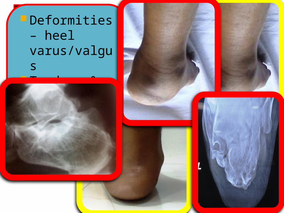
Deformities – heel varus/valgus
Tendons & ligaments

3
Joints Heel fat pad Gait Footwear RSD &
compartment syndrome
4
Radiology
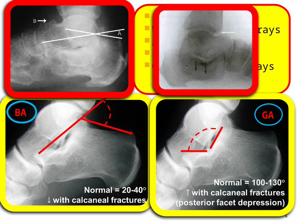
Plain X-rays Weight bearing x-rays Axial view Broden view Opposite side x-rays
BA
GA

Normal Xray Parameters: Lateral View
• A)Talocalcaneal height• B)Talar declination
angle
• C)Lateral talocalcaneal angle
• D)Calcaneal pitch
All parameters distorted!! Axial view Lateral wall exostosis Hindfoot malalignment Calcaneal shortening
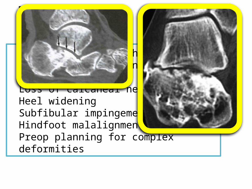
CT Scan
Degree of joint arthrosisPresence of nonunion Cystic lesionsLoss of calcaneal heightHeel wideningSubfibular impingementHindfoot malalignmentPreop planning for complex
deformities
7
Treatment
Conventional methodology is to treat Calcaneus Malunion based on classification!!
1996: Stephens & Sanders
CT based –axial images
Three types
Exosectomy & peroneal tenolysis
Addition of subtalar fusion
Addition of osteotomy
Dwyer or MCO
Classification is not complete!!Classification does not cover… ProtrusionsCC arthritisNerve issuesTendon issuesDeformities

8
How should one treat calcaneus malunion?
There could be many reasons of pain!Reasons very from case to case!!
Like in case of back pain find out every pain generator
Diagnostic injections in joints & nerve blocks help to identify pain generators
Beyond 3 weeks every calcaneus fracture should be treated like a case of Calcaneus Malunion!
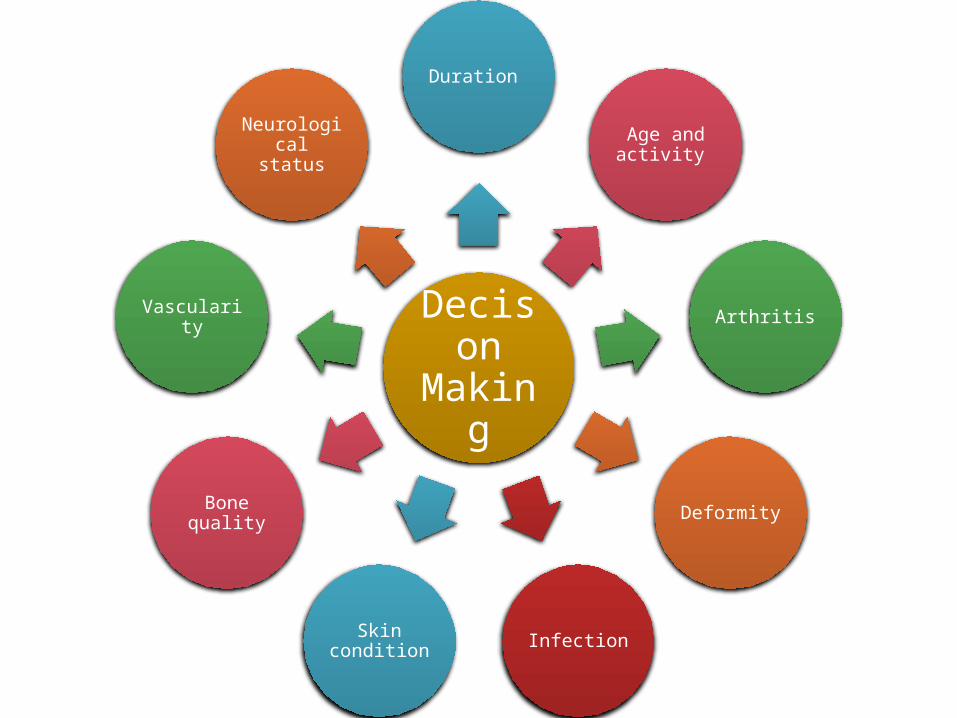
Decison
Making
Duration
Age and activity
Arthritis
Deformity
InfectionSkin condition
Bone quality
Vascularity
Neurological status

10
Treatment options available…Conservative treatment
NSAIDS Local
injections Braces & shoe
modifications
Activity modification

Calcaneal malunion: surgical
procedures
Joint salvage procedures
Revision fixation
Realignment with
osteotomy & fixation
Joint sacrificing
procedures Arthrodesis + osteotomy
Arthrodesis

Revision fixation
Calcaneus
13
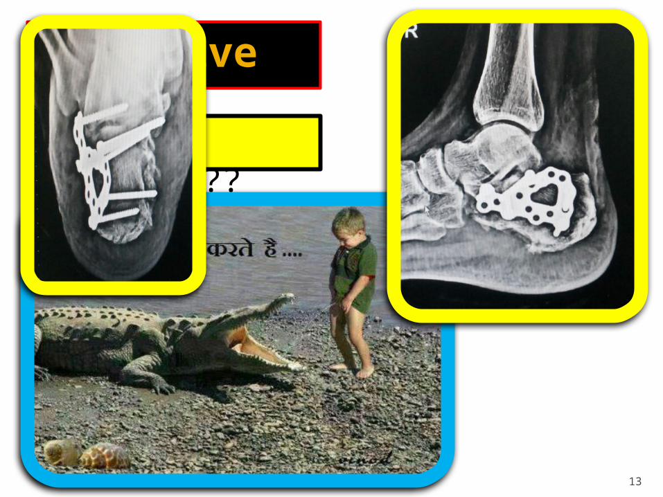
Operative careRevision fixation??
Pressure effects
Calcaneus

15
Excision of bony prominences Decompression of tendons, ligaments & nerves

Realignment with osteotomy
Calcaneus
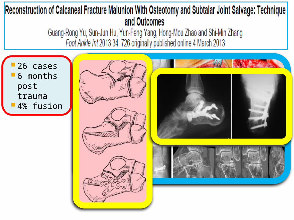
5 cases 3 months
post trauma
No fusion
26 cases 6 months
post trauma
4% fusion

Arthrodesis
Calcaneus
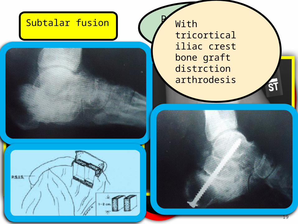
19
Subtalar fusion In situPost fixation arthrodesis
With tricortical iliac crest bone graft distrction arthrodesis
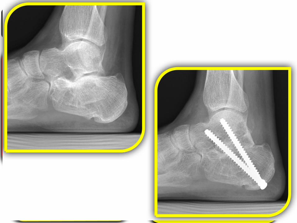
20
Subtalar fusion after delayed presentationDistraction subtalar fusion

Realignment with osteotomy + Fusion
Calcaneus

Corrective Osteotomy & Subtalar Arthrodesis
• For sanders type III
• Varus Dwyer: Lateral closing wedge
• Valgus Medializing calcaneal slide osteotomy

23
Vertical slide osteotomy + Subtalar fusion
Case courtesy: Dr.Selene Parekh
Romash’s methodosteotomy through old fracture & distraction & arthrodesis
Case courtesy: Dr.Selene Parekh
Final fixation
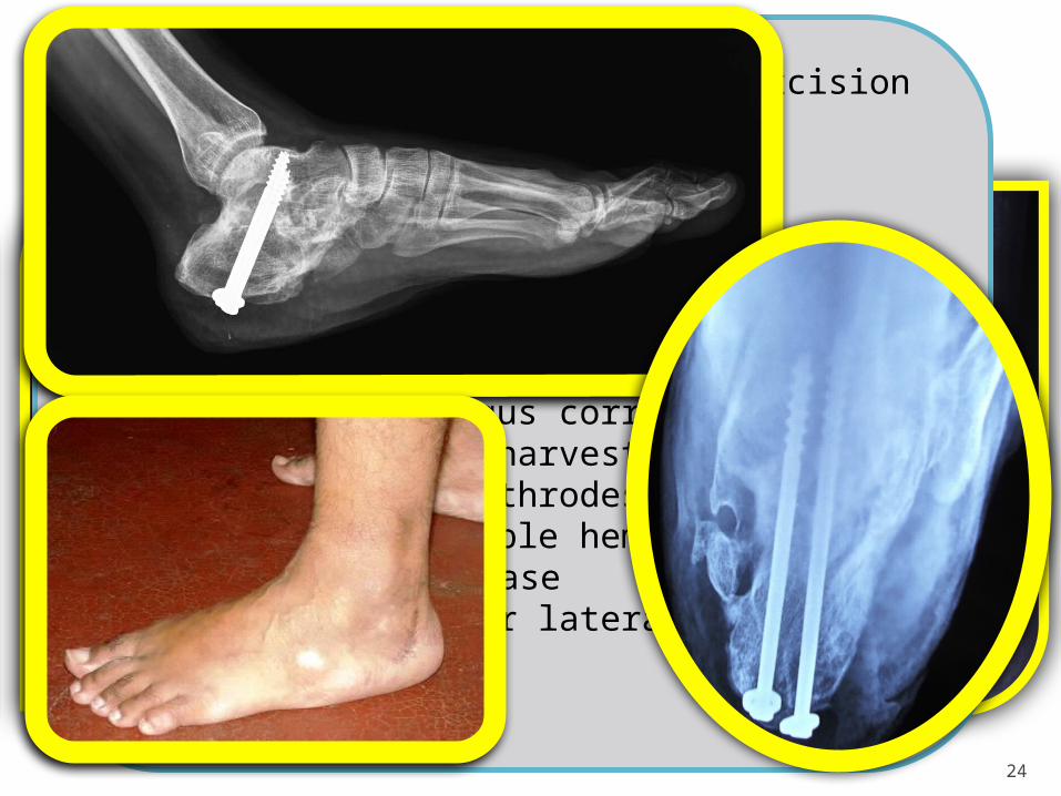
24
My worst case?!
Medial side: FHL impingement Lateral side– peroneal
impingement Plantar – painful plantar bump Sural & posterior tibial nerve
neuralgia Forefoot abduction on weight
bearing Hindfoot valgus with secondary
flat arch Arthritis of Subtalar & CC joint Tendoachiless tightness Claw toe deformities
1) Peroneal tenolysis with bump excision 2) Plantar bump excision 3) Medial bump excision 4) TT release5) Subtalar arthrodesis6) Calcaneal distraction for gaining height
with use of excised bony prominences7) MCO for heel valgus correction8) Iliac crest bone harvesting9) CC distraction arthrodesis10) Percutaneous triple hemisection
tendoachilles release11,12,13,14) All four lateral claw toe release

25
“ Try to correct as much damage as possible in the acute phase with surgery as late salvage procedures are quite difficult with absolutely uncertain results !! ”
Sander
26
That’s allThank you…
Top Related