


Languages
Pages
Legal


Journal of Proteomics & Bioinformatics - Open Access Research Article JPB/Vol.1/November 2008
J Proteomics Bioinform Volume 1(8) : 401-407 (2008) - 401
ISSN:0974-276X JPB, an open access journal
Comparative Modeling and Analysis of 3-D Structure of EMV2, a Late
Embryogenesis Abundant Protein of Vigna Radiata (Wilczek)1,2Subramanian Rajesh *, 1Muthurajan Raveendran and 1Ayyanar Manickam
1Centre for Plant Molecular Biology, Tamil Nadu Agricultural University, Coimbatore 641003, India2National Research Centre for Soybean, Indore 452001, Madhya Pradesh, India
*Corresponding authors: Dr. S. Rajesh, National Research Centre for Soybean, Indore-452 001,Madhya Pradesh, India, E-mail: [email protected];
Tel: +91-731-2362835; Fax: +91-731-2470520
Dr. A. Manickam, Centre for Plant Molecular Biology TamilNadu Agricultural University,
Coimbatore-641 003, India, E-mail: [email protected]
Received September 28, 2008; Accepted November 07, 2008; Published November 11, 2008
Citation: Subramanian R, Muthurajan R, Ayyanar M (2008) Comparative Modeling and Analysis of 3-D Structure of EMV2, a
Late Embryogenesis Abundant Protein of Vigna Radiata (Wilczek). J Proteomics Bioinform 1: 401-407. doi:10.4172/jpb.1000049
Copyright: © 2008 Subramanian R, et al. This is an open-access article distributed under the terms of the Creative Commons
Attribution License, which permits unrestricted use, distribution, and reproduction in any medium, provided the original author
and source are credited.
Abstract
LEA proteins are ubiquitous among photosynthetic organisms and have been reported in mono- and dicot
plants as well as in nematodes, yeast, bacteria and cyanobacteria. EMV2 is a Group 1 LEA protein isolated from
Vigna radiata, which is speculated to impart desiccation tolerance in plants. The homology model of this protein
was generated by using the LOOPP software based on available structural homologues in protein databases. The
final model obtained by molecular mechanics and dynamics method was assessed by PROCHECK that showed
that the final refined model is reliable. The model could prove useful in further functional characterization of this
protein.
Keywords: LEA proteins; Vigna radiate; Desiccation; Homology modeling; Validation
Abbreviations: SCOP: Structural Classification Of Proteins; PDB: Protein Data Bank; LOOPP: Learning Observing and
Outputting Protein Patterns
Introduction
Late embryogenesis abundant (LEA) protein genes are
highly expressed during late stages of seed development at
normal growth condition, but many of the LEA class genes
are also frequently expressed in vegetative tissues when
plants are exposed to environmental stress (Bray et al., 2000).
Several groups of LEA protein genes have been demon-
strated to confer water-deficit and salt-stress tolerance.
On the basis of sequence similarities, LEA proteins have
been classified in six groups (Dure, 1993; Bray, 1993). Group
2 LEA proteins or dehydrins are by far the most frequently
described LEA protein family and have been classified in
distinct groups (Close, 1997) that differ in the arrangement
and number of conserved motifs: the lysine-rich repeat
(KIKEKLPG) or K segment, the stretch of serine or S seg-
ment and the V/T DEYGNP motif or Y segment. Some of
these structural motifs are predicted to form amphipathic
alpha helices, which may be important for their function in
protecting plant cells against dehydration. Evidence of func-
tional links between LEA protein accumulation and improved
stress tolerance of transgenic yeast and plants support this
hypothesis (Imai, 1996; Xu et al., 1996; Sivamani et al.,
2000). It was therefore proposed that most LEA and
dehydrin proteins exist as largely unfolded structures in their
native state, although a few members exist as dimers or
tetramers (Ceccardi et al., 1994; Kazuoka and Oeda, 1994).
Hydrophilicity is a common characteristic of LEA-type and
other osmotic stress-responsive proteins. Our earlier work
Journal of Proteomics & Bioinformatics - Open Access Research Article JPB/Vol.1/November 2008
J Proteomics Bioinform Volume 1(8) : 401-407 (2008) - 402
ISSN:0974-276X JPB, an open access journal
on the occurrence of LEA proteins in the embryonic axes
of Vigna radiata (L.) Wilczek referred as EMV proteins,
the first ever report in the Fabaceae family (Manickam and
Carlier, 1980). cDNA encoding these proteins were isolated,
characterized (Manickam et al., 1996). In silico analysis of
the 20-mer motif of this EMV2 categorize this protein to
Group1 LEA and hypothesize to function as DNA/RNA
binding proteins in stabilizing membranes/macromolecules
at the time of dehydration process (Rajesh and Manickam,
2006; Gillies et al., 2007).
In the present study, effort was made to generate the
three-dimensional (3D) structure of the EMV2 protein based
on the available template structural homologues from Pro-
tein Data Bank and SCOP databases and the model vali-
dated with standard parameters. This study could prove
useful in further functional characterization of this impor-
tant group of proteins.
Materials and Methods
Datasets
The peptide sequences of Vigna radiata, EMV2 (NCBI
GenBank accession number U31211; UniProt acc. Nos.
Q41685 ) and other sequences examined in this study were
retrieved from the public databases, http://
www.ncbi.nlm.nih.gov and http://www.ebi.ac.uk. Structur-
ally homologous subsets of the experimentally determined
3D structures of the EMV proteins were retrieved from
PDB and SCOP databases. The template used for com-
parative modeling of EMV2 is a DNA Binding Protein from
Homo sapiens (1IG6_A.pdb) and a bifunctional inhibitor/
lipid-transfer protein/seed storage 2S albumin (1RZL.pdb)
with the sequence similarity of 56.44 and 56.67 % at the
loop/coiled coil regions. Similarity at these regions was con-
sidered because LEA proteins are generally loosely struc-
tured with predominantly random coiled regions. However
structure was further refined with sequence of 1RZL.pdb
as it showed overall 40 % sequence identity in the sequence
length of 89% to a bifunctional inhibitor/lipid transfer pro-
tein/seed storage 2S albumin of rice
Comparative modeling of EMV2 protein
Tertiary structure of the Vigna radiata LEA protein, EMV2
was modeled by submitting the deduced amino acid se-
quences to the Computational Biology Service Unit, Cornell
Theory Center, Cornell University. The Atomic coordinates
for the protein models were generated by aligning to the
structural homologues in the fold recognition program of
LOOPP v3.0 server (Teoderescu et al., 2004).
Validation of EMV2 protein model
PROCHECK, a versatile protein structure analysis pro-
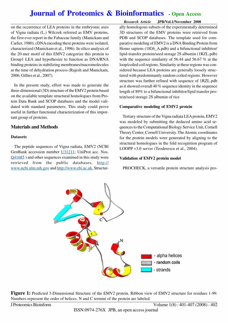
Figure 1: Predicted 3-Dimensional Structure of the EMV2 protein. Ribbon view of EMV2 structure for residues 1-99.
Numbers represent the order of helices. N and C termini of the protein are labeled.

-180 -135 -90 -45 0 45 90 135 180Phi (degrees)
180
135
90
45
0
-45
-90
-135
(Most favoured regions - A, B, L; Additional allowed regions - a, b, l, p; Genorously allowed region - ~a, ~b, ~l, ~p)
Psi(degrees)
B
L
A
b
a
-b
-b
-b
-b
b
-p
-a
p
b
-l
l
ASN 94
LYS 63
Journal of Proteomics & Bioinformatics - Open Access Research Article JPB/Vol.1/November 2008
J Proteomics Bioinform Volume 1(8) : 401-407 (2008) - 403
ISSN:0974-276X JPB, an open access journal
gram (Laskowski et al., 1993) available at the Joint Centre
for Structural Genomics, Bioinformatics core, University of
California, San Diego was used in validation of protein struc-
ture and models by verifying the parameters like
Ramachandran plot quality, peptide bond planarity, Bad non-
bonded interactions, main chain hydrogen bond energy, C-
alpha chirality and over-all G factor and the side chain pa-
rameters like standard deviations of chi1 gauche minus, trans
and plus, pooled standard deviations of chi1 with respect to
refined structures( Morris et al., 1992).
Results
Comparative modeling of EMV2 protein
Tertiary structure of a protein is build by packing of its
secondary structure elements to form discrete domains or
autonomous folding units. Comparative modeling to build
3D structure of the EMV2 protein was made based on the
experimentally solved structural homologues. The amino acid
sequences of EMV2 were submitted to LOOPP server,
Cornell Bioinformatics Structural Unit (CBSU) and atomic
coordinates for the proteins were generated based on Hid-
den Markov Model. The hypothetical protein models cre-
ated were stored as PDB output file. The hypothetical pro-
teins were visualized and computed by Swiss PDB Viewer
and Rastop. The 3D structure of the proteins were repre-
sented by cartoon display and colored based on the second-
ary structure (Figure 1).
Validation of protein structures of EMV2
The hypothetical protein models generated were analyzed
online by submitting to Joint Center for Structural Genomics
(JCSG), Bioinformatics core, University of California, San
Diego. Accuracy of the protein model generated was judged
by validity report generated by PROCHECK. Parameter
comparisons of these proteins were made with well-refined
structures that have similar resolution.
The main chain parameters plotted are Ramachandran
plot quality, peptide bond planarity, Bad non-bonded inter-
actions, main chain hydrogen bond energy, C-alpha chirality
and over-all G factor. In the Ramamchandran plot analysis,
the residues were classified according to its regions in the
quadrangle. The Ramachandran map for EMV2 (Figure 2)
and the plot statistics (Table 1) is represented. Non-bonded
interactions check revealed the value of 30 bad contacts
Figure 2: Ramachandran map of EMV2 protein. The Plot calculation was done with PROCHECK program.
Journal of Proteomics & Bioinformatics - Open Access Research Article JPB/Vol.1/November 2008
J Proteomics Bioinform Volume 1(8) : 401-407 (2008) - 404
ISSN:0974-276X JPB, an open access journal
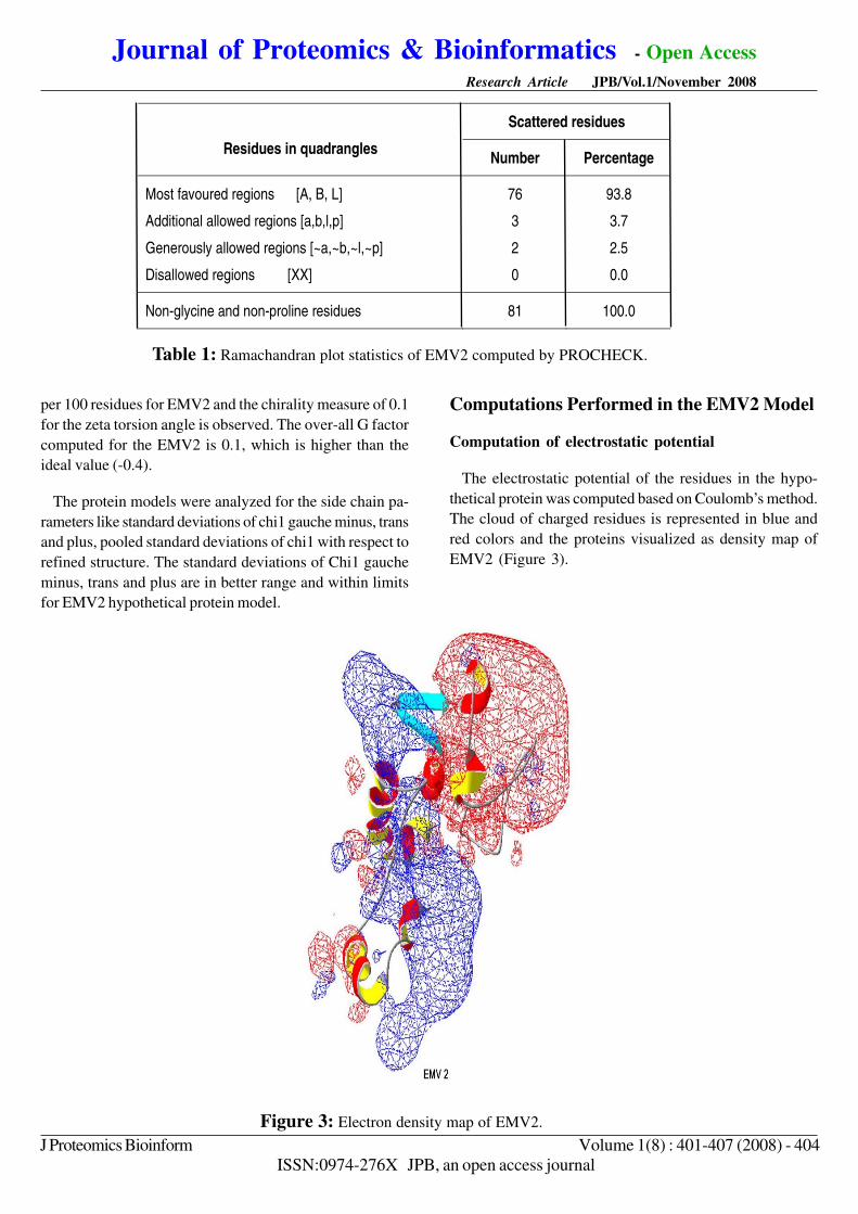
per 100 residues for EMV2 and the chirality measure of 0.1
for the zeta torsion angle is observed. The over-all G factor
computed for the EMV2 is 0.1, which is higher than the
ideal value (-0.4).
The protein models were analyzed for the side chain pa-
rameters like standard deviations of chi1 gauche minus, trans
and plus, pooled standard deviations of chi1 with respect to
refined structure. The standard deviations of Chi1 gauche
minus, trans and plus are in better range and within limits
for EMV2 hypothetical protein model.
Computations Performed in the EMV2 Model
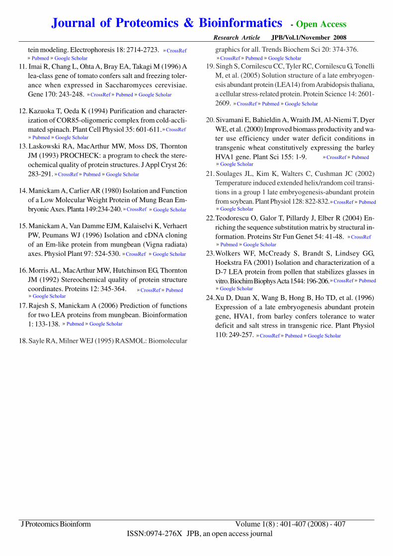
Computation of electrostatic potential
The electrostatic potential of the residues in the hypo-
thetical protein was computed based on Coulomb’s method.
The cloud of charged residues is represented in blue and
red colors and the proteins visualized as density map of
EMV2 (Figure 3).
Table 1: Ramachandran plot statistics of EMV2 computed by PROCHECK.
Figure 3: Electron density map of EMV2.
Scattered residues
Residues in quadrangles Number Percentage
Most favoured regions [A, B, L]
Additional allowed regions [a,b,l,p]
Generously allowed regions [~a,~b,~l,~p]
Disallowed regions [XX]
76
3
2
0
93.8
3.7
2.5
0.0
Non-glycine and non-proline residues 81 100.0

Journal of Proteomics & Bioinformatics - Open Access Research Article JPB/Vol.1/November 2008
J Proteomics Bioinform Volume 1(8) : 401-407 (2008) - 405
ISSN:0974-276X JPB, an open access journal
Computation of force field energy
Force field energy was computed for EMV2 protein model.
Positive values were observed (64x108 forEMV2). Model
refinement was done by energy minimization. Energy mini-
mization was carried out to reduce clashing amino acids,
using GROMOS96 force field algorithm. Decrease in the
force field energy was observed for both the protein mod-
els after successive energy minimization (-1718 for EMV2).
The energy minimized models will however needs further
refinement in order to reduce the non-bonded interactions
for the model to be judged as a good homology model.
Discussion
3D Modeling of EMV2, LEA protein from vigna ra-
diata
Prediction of tertiary structure of a protein molecule sig-
nifies an important step towards understanding the struc-
ture –function relationships in the concerned protein family.
Recently, the first solution structure of a LEA protein, LEA14
from Arabidopsis thaliana has been reported (Singh et al.,
2005). In the present study, model of EMV2 LEA protein of
Vigna radiata was generated from the LOOPP server, based
on the structural homologues derived from the SCOP and
protein data banks.
There exists biological sequence-structure deficit with
more than 3 lakhs protein sequences and millions of partial
nucleotide sequences, available in the public non-redundant
databases (Boguski et al., 1994); and by contrast, the num-
ber of unique 3D structures in the protein data bank is still
less than 1500 (Attwood and Parry-Smith, 2005). The dif-
ference of scale in sequence and structural information is
an important factor to be considered when assigning func-
tions to hypothetical proteins. Structure based functional
implications of such proteins have always been speculative.
Generally, under stress situations the plants may induce
formation of coiled coil / folding of the natively unfolded
proteins into more rigid structures upon binding to the part-
ner molecules. Since all natively unfolded proteins have
defined partner molecules that can be as small as nucle-
otide or cations or a macromolecule, LEA proteins being
natively unfolded is believed to have such binding partners
to attain a rigid structures.
The LEA proteins from Vigna radiata are being classified
as Group 1 LEA protein because of its extreme hydrophilic-
ity and adoption of helical conformation as revealed by ab
initio secondary structure predictions, in combination with
the predominant random-coiled arrangement of the residues
of Vigna LEA protein, is hypothesized to function as water
replacement molecule. Such a property may facilitate hy-
drogen bonding of this EMV proteins with essentially any
macromolecular or membrane surface. However, additional
experiments on physico-chemical analyses including exami-
nation of hydration properties of these proteins need to be
done to determine if EMV proteins can adopt certain struc-
tures upon interaction with other macromolecules.
Structure homologues identified for these EMV proteins
show closest structural homology to proteins with helical
bundles of small proteins and DNA/RNA binding proteins.
These observations are contradictory to the earlier findings
from our group that the low molecular weight protein iso-
lated from Vigna was believed to be located in the cyto-
plasm The structural motifs of these proteins are predicted
to form amphipathic α-helices which may be important for
their function in protecting cells against dehydration. How-
ever, not all LEA proteins are folded and structured. Group
1 LEA proteins are reported to be very hydrophilic, loosely
structured with predominantly random-coiled structures.
These proteins are reported to form regular α-helical struc-
ture when subjected to altered physiological conditions.
Observations from ab initio predictions of these Em pro-
teins of Vigna indicate 32.32% of EMV2 proteins attains
helical conformation as represented by helical blocks in the
3D hypothetical models (Manickam and Carlier, 1980).
Temperature-induced extended helix/random coil transi-
tion was reported for a Group 1 LEA protein from soybean.
These proteins are by native, largely unstructured but at-
tained 6- 14% helical conformation under temperature stress
or at high salt concentrations (Souglaes et al., 2002). Simi-
lar reports from Goyal et al., (2003) for AavLEA1, a Group
3 LEA protein from the nematode, Aphelenchus avenae in-
dicate oligomerization of these proteins in immunoblotting
and cross-linking experiments, however majority of these
proteins was found to be monomeric in analytical ultracen-
trifugation and gel filtration studies. Also, formation of
α-helical structures on drying was reported in partially char-
acterized protein from Typha latifolia, probably a Group 3
LEA protein based on Fourier transform-Infra red (FT-IR)
spectroscopy studies (Wolkers et al., 2001).
The LEA proteins from Vigna radiata are being classified
as Group 1 LEA protein because of its extreme hydrophilic-
ity and adoption of helical conformation as revealed by ab
initio secondary structure predictions, in combination with
the predominant random-coiled arrangement of the residues
of Vigna LEA protein, is hypothesized to function as water-

Journal of Proteomics & Bioinformatics - Open Access Research Article JPB/Vol.1/November 2008
J Proteomics Bioinform Volume 1(8) : 401-407 (2008) - 406
ISSN:0974-276X JPB, an open access journal
replacement molecule. Such a property may facilitate hy-
drogen bonding of this EMV proteins with essentially any
macromolecular or membrane surface. However, additional
experiments on physico-chemical analyses including exami-
nation of hydration properties of these proteins need to be
done to determine if EMV proteins can adopt certain struc-
tures upon interaction with other macromolecules.
Validation of the Model
The hypothetical protein model generated was subjected
to structure validation, for testing the accuracy of the model.
The quality of the final ensemble of conformers was as-
sessed using PROCHECK, a protein structure validation
program. The visual displays of the models were performed
with either the Swiss PDB viewer (Guex and Peitsch, 1997)
or RasTop (Sayler and Milner-white, 1995).
Stereochemical parameters of the proteins like main-and
side chains data of EMV2 was considered for determining
the quality of the model. The main chain parameters like
Ramachandran plot quality; peptide bond planarity, C-alpha
chirality and over-all G factor are found to be within the
limits for the model. However, the bad contacts per 100
residues are high. The side chain parameters are in better
range and within the limits for EMV2. These parameters
are compared to essentially satisfy the generated models
with well-refined structures at similar resolution as described
by Morris et al., (1992).
The validation reports for the protein models are analyzed,
and energy minimization of the models was made after
checking the force field energy of the models. For a model
to be validated based on quality, a good quality protein model
should have 90% or more residues in the most favored re-
gions of quadrangle in the Ramachandran plot. In the gen-
erated model of EMV2, the distribution of residues in the
most favored regions is 87.6 and 93 %, respectively. This
infers that EMV2 as good hypothetical protein model. Also,
a tertiary structure of a protein can be worth considering
only from its solution structures, obtained from the experi-
mentations using either NMR or crystallographic studies.
The homology model of mungbean LEA proteins, thus gen-
erated in this study, could some extent stimulate investiga-
tions at determining the mechanistic function of this stress
associated proteins.
Future Perspectives
Functional implications based on the structural homologues
hypothesize EMV2 to act as a water replacement molecule.
LEA proteins generally are reported to posses multiple func-
tions like salt, drought, heat and cold tolerance. Hence, the
present study will be useful in further in vitro studies by
over expression in model systems like E.coli or yeast cells
and the recombinant protein can be subjected to Salinity,
Cold shock, thermal stability analysis and the stress induced
structural changes can be monitored to ascertain the pos-
sible functions of this important class of proteins.
Acknowledgements
S.Rajesh is grateful to the Council of Scientific and In-
dustrial Research, New Delhi for grant of Research Fel-
lowship. We are grateful to the crews of NCBI, EBI, MRC
Lab-UK and SIB for making computational biology data/
tools publicly available.
References
1. Attwood TK, Parry SDJ (2005) In. Introduction to
Bioinformatics. Ed Wood (ed.). Pearson Education Asia
ltd India p3.
2. Boguski MS, Tolstoshev CM, Bassett DE (1994) Gene
discovery in DBEST. Science 265: 194-199.
3. Bray EA (1993) Molecular responses to water deficit.
Plant Physiol 103:1035-1040.
4. Bray EAJ, Bailey SE, Weretilnyk E (2000) Responses
to abiotic stresses, In: Biochemistry and Molecular Biol-
ogy of Plants. Buchanan W Gruissem R Jones (Eds.)
American Society of Plant Physiologists pp1158-1176.
5. Ceccardi TL, Meyer NC, Close TJ (1994) Purification
of a maize dehydrin. Protein Express Purif 5: 266-269 .
6. Close TJ (1997) Dehydrins: A commonality in the re-
sponse of plants to dehydration and low temperature.
Physiol Plant 100: 291-296.
7. Dure L (1993) A repeating 11-mer amino acid motif and
plant desiccation. Plant J 3: 363-369.
8. Gilles GJ, Hines KM, Manfre AJ, Marcotte WR (2007)
A predicted N-terminal helical domain of a Group 1 LEA
protein is required for protection of enzyme activity from
drying. Plant Physiol Biochem 45: 389-399.
9. Goyal K, Tisi L, Basran A, Browne J, Burnell A, et al.
(2003) Transition from natively unfolded to folded state
induced by desiccation in an anhydrobiotic nematode
protein. J Biol Chem 278: 129770-12984.
10. Guex N, Peitsch MC (1997) SWISS-MODEL and the
Swiss-PdbViewer: An environment for comparative pro-
» CrossRef » Pubmed » Google Scholar
» CrossRef » Pubmed » Google Scholar
» CrossRef » Pubmed » Google Scholar
» CrossRef » Google Scholar
» CrossRef » Pubmed » Google Scholar
» CrossRef » Pubmed » Google Scholar
» CrossRef » Pubmed » Google Scholar
Journal of Proteomics & Bioinformatics - Open Access Research Article JPB/Vol.1/November 2008
J Proteomics Bioinform Volume 1(8) : 401-407 (2008) - 407
ISSN:0974-276X JPB, an open access journal
tein modeling. Electrophoresis 18: 2714-2723.
11. Imai R, Chang L, Ohta A, Bray EA, Takagi M (1996) A
lea-class gene of tomato confers salt and freezing toler-
ance when expressed in Saccharomyces cerevisiae.
Gene 170: 243-248.
12. Kazuoka T, Oeda K (1994) Purification and character-
ization of COR85-oligomeric complex from cold-accli-
mated spinach. Plant Cell Physiol 35: 601-611.
13. Laskowski RA, MacArthur MW, Moss DS, Thornton
JM (1993) PROCHECK: a program to check the stere-
ochemical quality of protein structures. J Appl Cryst 26:
283-291.
14. Manickam A, Carlier AR (1980) Isolation and Function
of a Low Molecular Weight Protein of Mung Bean Em-
bryonic Axes. Planta 149:234-240.
15. Manickam A, Van Damme EJM, Kalaiselvi K, Verhaert
PW, Peumans WJ (1996) Isolation and cDNA cloning
of an Em-like protein from mungbean (Vigna radiata)
axes. Physiol Plant 97: 524-530.
16. Morris AL, MacArthur MW, Hutchinson EG, Thornton
JM (1992) Stereochemical quality of protein structure
coordinates. Proteins 12: 345-364.
17. Rajesh S, Manickam A (2006) Prediction of functions
for two LEA proteins from mungbean. Bioinformation
1: 133-138.
18. Sayle RA, Milner WEJ (1995) RASMOL: Biomolecular
graphics for all. Trends Biochem Sci 20: 374-376.
19. Singh S, Cornilescu CC, Tyler RC, Cornilescu G, Tonelli
M, et al. (2005) Solution structure of a late embryogen-
esis abundant protein (LEA14) from Arabidopsis thaliana,
a cellular stress-related protein. Protein Science 14: 2601-
2609.
20. Sivamani E, Bahieldin A, Wraith JM, Al-Niemi T, Dyer
WE, et al. (2000) Improved biomass productivity and wa-
ter use efficiency under water deficit conditions in
transgenic wheat constitutively expressing the barley
HVA1 gene. Plant Sci 155: 1-9.
21. Soulages JL, Kim K, Walters C, Cushman JC (2002)
Temperature induced extended helix/random coil transi-
tions in a group 1 late embryogenesis-abundant protein
from soybean. Plant Physiol 128: 822-832.
22.Teodorescu O, Galor T, Pillardy J, Elber R (2004) En-
riching the sequence substitution matrix by structural in-
formation. Proteins Str Fun Genet 54: 41-48.
23.Wolkers WF, McCready S, Brandt S, Lindsey GG,
Hoekstra FA (2001) Isolation and characterization of a
D-7 LEA protein from pollen that stabilizes glasses in
vitro. Biochim Biophys Acta 1544: 196-206.
24.Xu D, Duan X, Wang B, Hong B, Ho TD, et al. (1996)
Expression of a late embryogenesis abundant protein
gene, HVA1, from barley confers tolerance to water
deficit and salt stress in transgenic rice. Plant Physiol
110: 249-257.
» CrossRef » Pubmed » Google Scholar
» CrossRef » Pubmed » Google Scholar
» CrossRef » Pubmed » Google Scholar
» CrossRef » Pubmed » Google Scholar
» CrossRef » Google Scholar
» CrossRef » Google Scholar
» CrossRef » Pubmed» Google Scholar
» Pubmed » Google Scholar
» CrossRef » Pubmed » Google Scholar
» CrossRef » Pubmed » Google Scholar
» CrossRef » Pubmed» Google Scholar
» CrossRef » Pubmed» Google Scholar
» CrossRef » Pubmed » Google Scholar
» CrossRef » Pubmed» Google Scholar
» CrossRef » Pubmed » Google Scholar
Top Related