


Languages
Pages
Legal


Joints of upper limb
By Dr. Eman AbdelGhany

Joints of Upper Extremity
• 1-Sternoclavicular– Synovial-saddle
• 2- Acromioclavicular– Synovial-plane
• 3- Glenohumeral joint(shoulder)– Synovial-ball&socket

1- Sternoclavicular joint
• Articular surfaces:
- sternal end of the clavicle.
- clavicular notch of manubrium sterni.
- first costal cartilage.• Ligaments:
- costoclavicular ligament.
- interclavicular ligament.
- ant. & post. Sternoclavicular ligaments.• Blood supply: suprascapular artery & internal thoracic artery.• Nerve supply: medial supraclavicular nerve & nerve to subclavius.• NB: It is divided by articular disc into two cavities.

2- Acromioclavicular joint
• Articular surfaces :
- acromial end of the clavicle
- lateral side of acromion process.• Ligaments:• Acromioclavicular ligament.
- coracoclavicular ligament ( conoid & trapezoid parts) .• Blood supply : suprascapular & thoracoacromial arteries.• Nerve supply : suprascapular & lateral supraclavicular nerves.
NB: Incompletely divided by articular disc into two compartments.
Movements of both joints allow scapular rotation up to 60 degrees.
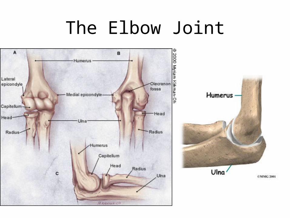
The Elbow Joint

Elbow joint• Elbow Joint type:
– Synovial – hinge• Articular surfaces:
– Humeroulnar ( trochla of humerus+ trochlear notch of ulna )– Humeroradial (capitulum of humerus + upper surface of head of
radius)
Capsule and synovial membrane are common for elbow & SRUJ
Capsule is attached to humerus, ulna & unular ligament. It is not attached to radius.
It includes the radial, coronoid & olecranon fossae of the humerus.

Ligaments of the Elbow
1- Ulnar Collateral Ligament
medial side – connects humerus to ulna
(3 bands ant. , post. And oblique bands)
2- Radial Collateral Ligament
Lateral side – connects humerus to radius
3- Annular Ligament
Surrounds radial head/holds it tight
to ulna

Movement
• Muscles Affecting the Elbow:
- Elbow Flexors
1. Brachialis
2. Biceps brachii
3. Brachioradialis
- Elbow Extensors
1. Triceps
2. Anconeus

Frolich, Human Anatomy,UpprLimb
Relations of elbow joint 1-Anterior: cubital fossa;
Contents• Median Cubital Vein+epitrochlear lymph node• Brachial & radial + ulnar arteries• Median & radial nerves• Biceps tendon
– Boundaries:Medial = Pronator teres
Lateral = Brachioradialis
Superior = Line between
Epicodyles
Roof = skin, fascia
+bicipital aponeurosis
Floor = supinator + brachialis muscles
Relations of elbow joint ( cont.)
2- Posterior relation : triceps and anconeus muscles.
3- Medial relation : ulnar nerve & common flexor origin.
4- Lateral relation : supinator & common extensor origin.
Blood supply : anastomosis around the elbow joint.
Nerve supply : musculocutanious + radial + ulnar nerves.

Radius and Ulna

Manual of Structural Kinesiology
The Elbow and Radioulnar Joints 6-12
Joints
• Radioulnar joint – Radial head rotates around at proximal ulna– Distal radius rotates around distal ulna– Annular ligament maintains radial head in its
joint– Joint between shafts of radius & ulna held
tightly together between proximal & distal articulations by an interosseus membrane (syndesmosis)• substantial rotary motion between the bones
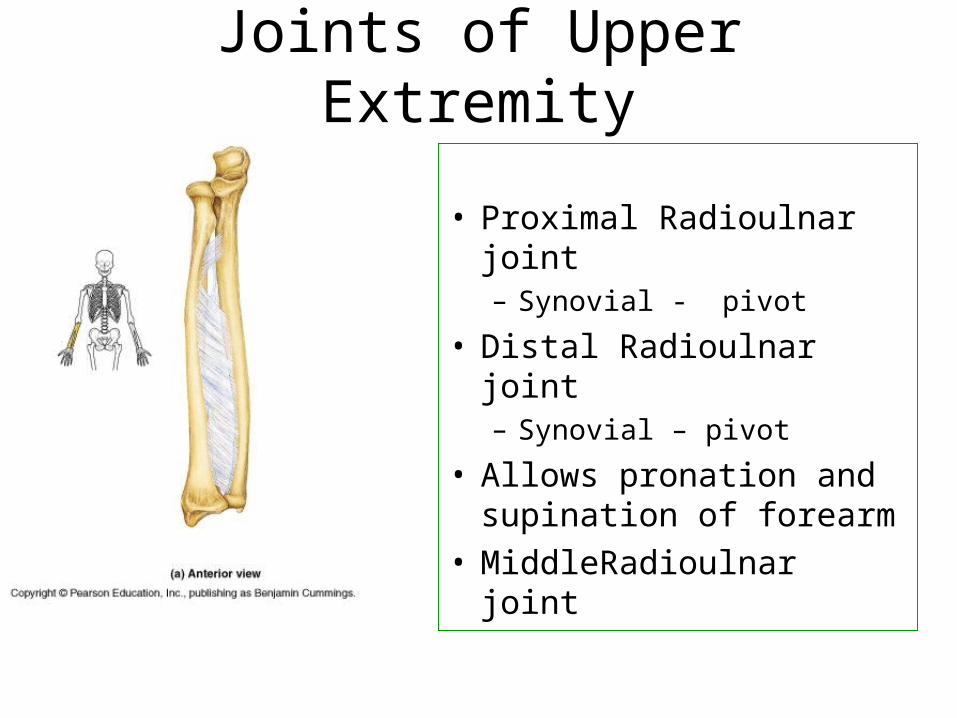
Joints of Upper Extremity
• Proximal Radioulnar joint– Synovial - pivot
• Distal Radioulnar joint– Synovial – pivot
• Allows pronation and supination of forearm
• MiddleRadioulnar joint
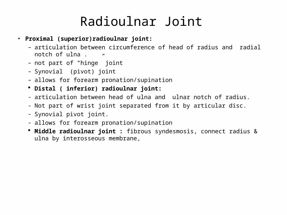
Radioulnar Joint• Proximal (superior)radioulnar joint:
– articulation between circumference of head of radius and radial notch of ulna .– not part of “hinge” joint– Synovial (pivot) joint– allows for forearm pronation/supination Distal ( inferior) radioulnar joint:- articulation between head of ulna and ulnar notch of radius.- Not part of wrist joint separated from it by articular disc.- Synovial pivot joint.- allows for forearm pronation/supination Middle radioulnar joint : fibrous syndesmosis, connect radius & ulna by
interosseous membrane,

6-15
Muscles of radioulnar joints
• Radioulnar pronators – Pronator teres– Pronator quadratus– Brachioradialis( begin movement )
• Radioulnar supinators– Biceps brachii(powerful supination for
flexed elbow)– Supinator muscle– Brachioradialis( begin movement)

1. Radial styloid
2. Scaphoid
3. Lunate
4. Triquetral
5. Pisiform
6. Trapezium
7. Trapezioid
8. Capitate
9. Hamate
10. Metacarpal
11. Proximal phalanx
12. Middle phalanx
13. Distal phalanx
14. ulna styloid
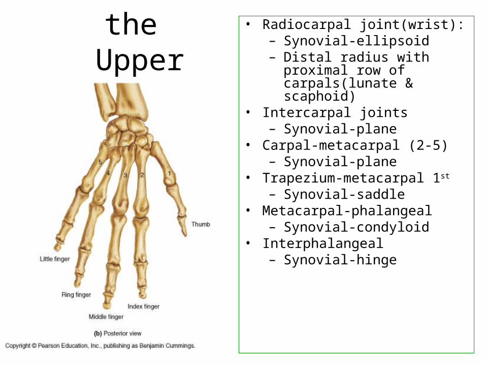
Joints of the Upper
Extremity
• Radiocarpal joint(wrist):– Synovial-ellipsoid– Distal radius with proximal row
of carpals(lunate & scaphoid)• Intercarpal joints
– Synovial-plane• Carpal-metacarpal (2-5)
– Synovial-plane• Trapezium-metacarpal 1st
– Synovial-saddle• Metacarpal-phalangeal
– Synovial-condyloid• Interphalangeal
– Synovial-hinge
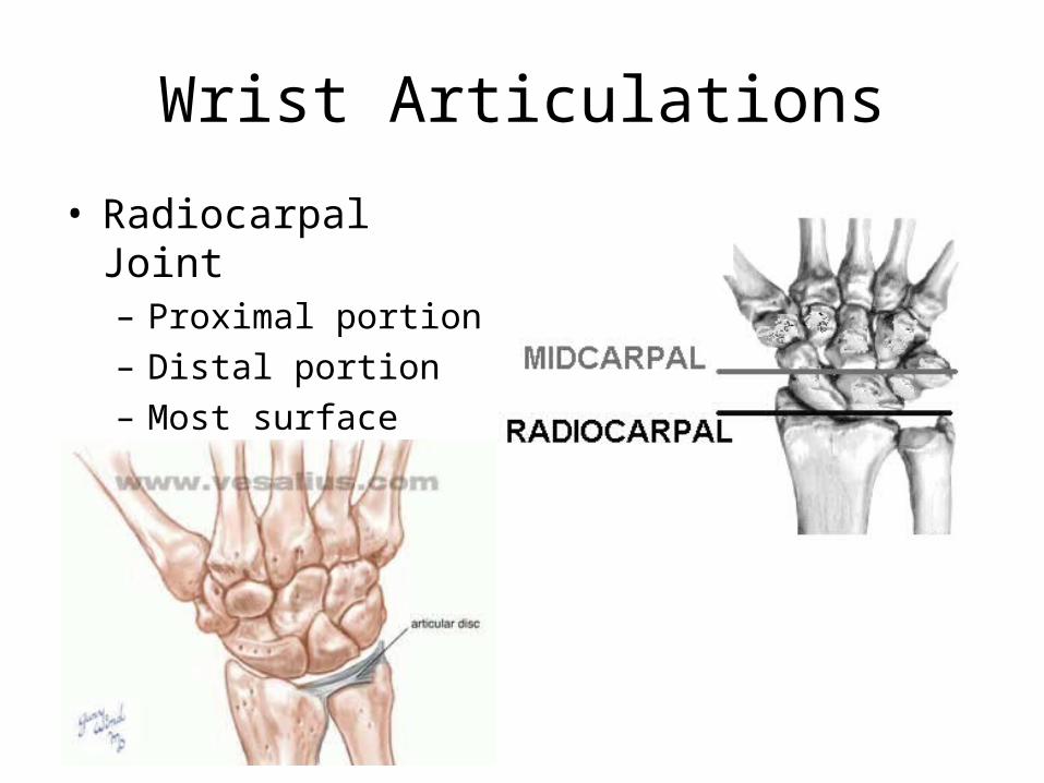
Wrist Articulations
• Radiocarpal Joint– Proximal portion– Distal portion– Most surface contact
found

Wrist (radiocarpal ) joint
• Articulation between (lower end of radius + scaphoid & lunate ), (triquetral +articular disc)=== the ulna not share in the wrist joint .
• Ligaments :
1- Ulnar collateral (styloid process of ulna to triquetral & pisiform);
2- Radial collateral ligament (styloid process of radius to scaphoid bone)
3- Ant. & post. Radiocarpal ligaments= thickened capsule.
Relations:
Ant. : carpal tunnel ?
Post. : Extensor compartments?
Lat.: Anatomical snuff box?
Med.: Dorsal branch of ulnar nerve.

Extensor Compartments• Anatomic
snuffbox:– EPL and
EPB – Scaphoid on
floor– Radial a.
inside
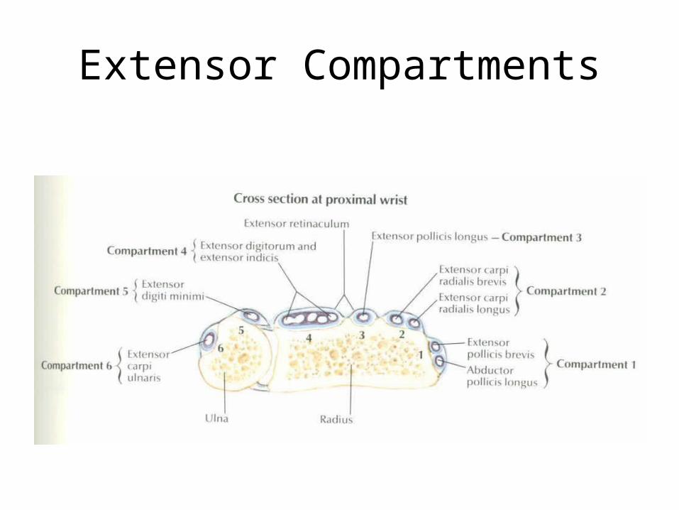
Extensor Compartments

Movement of the wrist joint
• Wrist Extensors (innervated by radial n.)• Superficial
– Extensor carpi radialis brevis/longus– Extensor carpi ulnaris– Extensor digitorium– brachioradialis
• Deep compartment– Extensor pollicus longus/brevis– Abductor pollicus longus– Extensor indices– supinator
• Secured by extensor retinaculum

• Wrist flexors (median n.)– Superficial
• Flexor carpi radialis• Palmaris longus• Flexor carpi ulnaris• Flexor digitorium superficialis• Pronator teres
– Deep• Flexor digitorium profundus• Flexor pollicis longus• Pronator quadratus

Cross Section just proximal to Carpal Tunnel
Carpal Tunnel
• Fibro-osseous structure– Floor is proximal carpal bones– Roof is transverse carpal ligament
• Tunnel contains 10 structures– Median n., flexor pollicis longus tendon, 4 slips of
flexor digitorium superficialis, flexor digitorium profundus
• Compression results in paresthesia 2-4 fingers and decrease grip

Bones of Wrist (palmar)

Ligaments of Wrist (dorsal)

(palmar)

Hand Anatomy
Tendon Sheaths
• Palmer Aspect

Carpal Tunnel

Wrist Anatomy
Tendon Sheaths• Dorsal Aspect
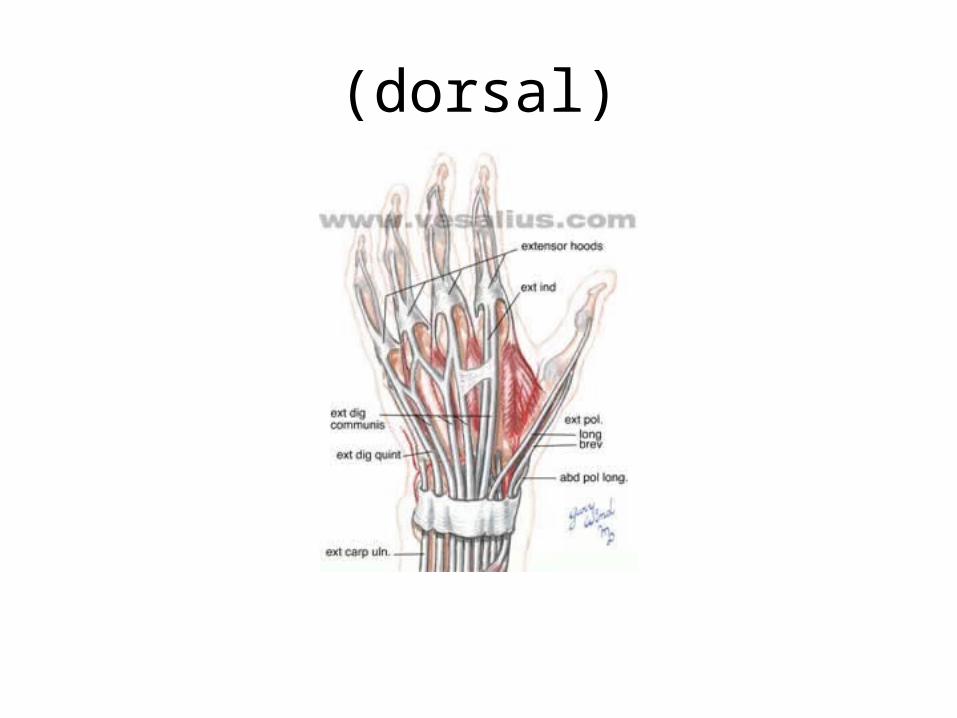
(dorsal)

Movement of wrist joint
• Flexion: flexor carpiradialis & ulnaris + palmaris longus helped by; FDS,FDP,FPL& ABD. P.L.
• Extension: extensor carpiradialis longus+ brevis & extensor carpiulnaris, helped by extensors of fingures.
• Abduction: flexor carpiradialis+extensor carpiradialis longus & brevis, helped by Abd. P.L. + Ext.P.L.& brevis
• Adduction: flexor & extensor carpiulnaris.• Circumduction : combination of flexion , abduction, extension &
adduction. • Blood supply : palmar & dorsal carpal branches of radial and ulnar
arteries & palmar arches.• Nerve supply : ant. & post. Interosseus nerves.
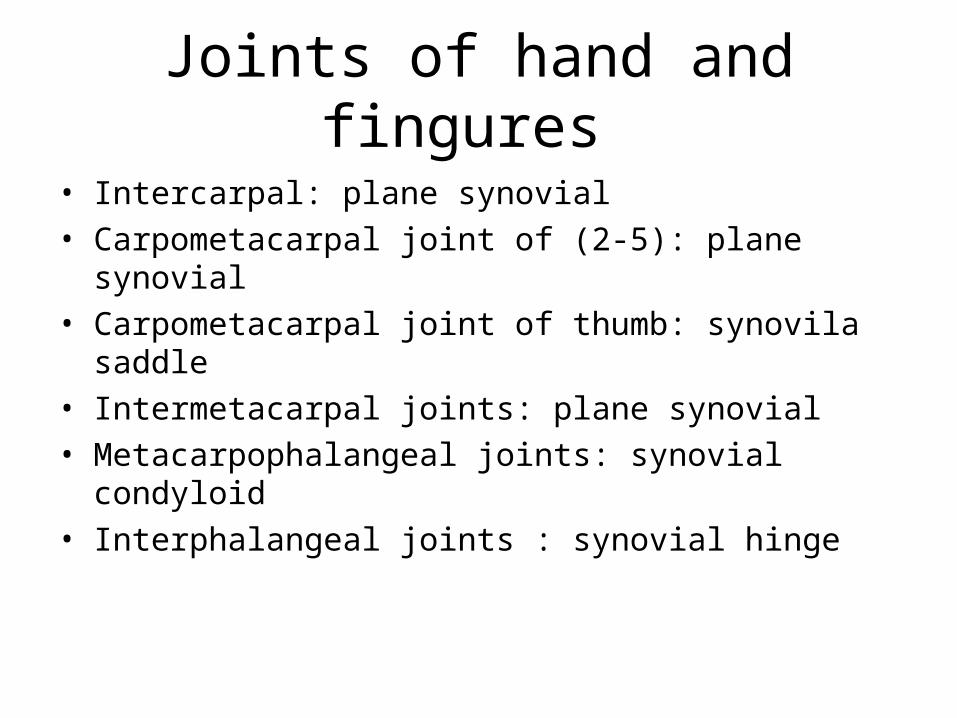
Joints of hand and fingures
• Intercarpal: plane synovial• Carpometacarpal joint of (2-5): plane synovial• Carpometacarpal joint of thumb: synovila saddle• Intermetacarpal joints: plane synovial• Metacarpophalangeal joints: synovial condyloid• Interphalangeal joints : synovial hinge

Interphalangeal Joints of 2-5 Fingers
• Hinge Joints• Motions

Ligaments of the Hand
• Palmar carpometacarpal ligaments
• Palmar metacarpal ligaments
• Deep transverse metacarpal lig
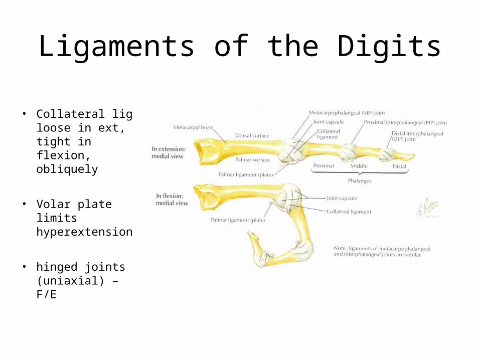
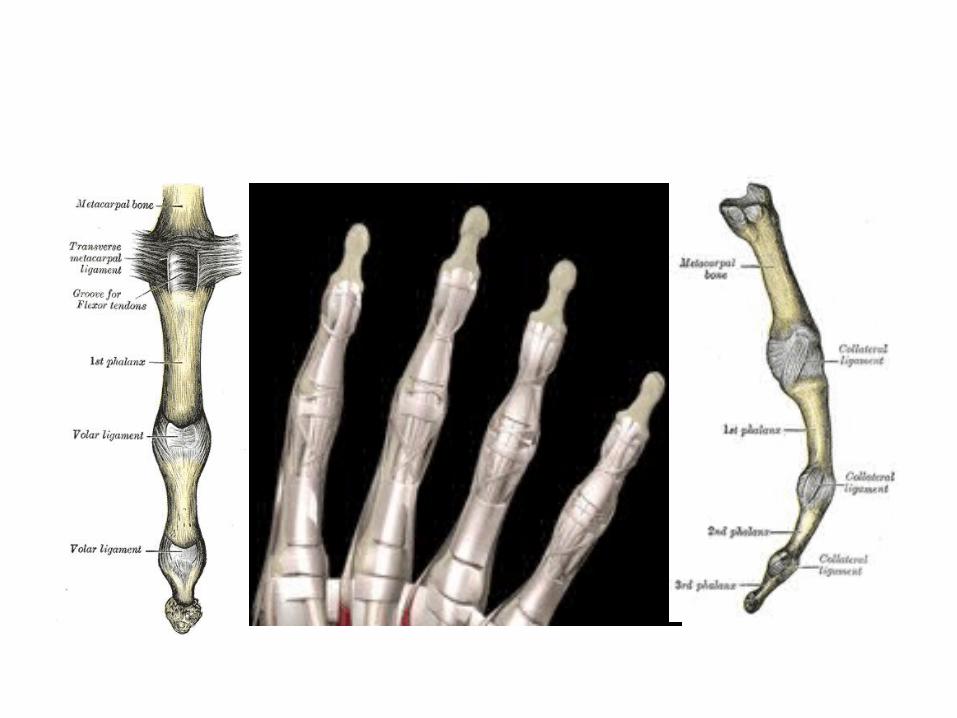
Ligaments of the Digits
• Collateral lig loose in ext, tight in flexion, obliquely
• Volar plate limits hyperextension
• hinged joints (uniaxial) – F/E
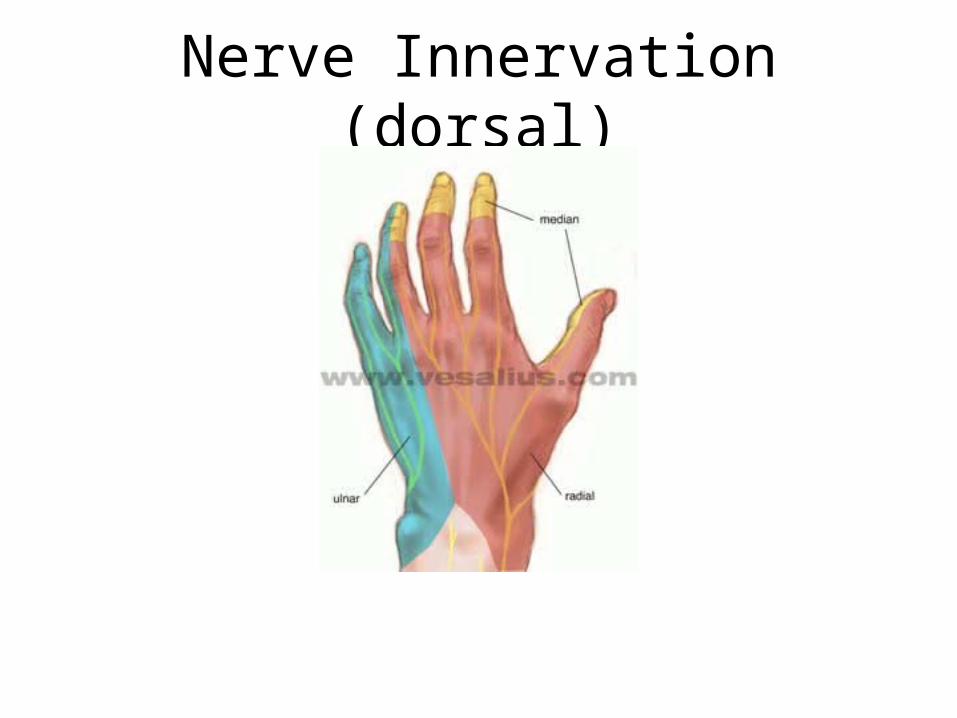
Nerve Innervation (dorsal)
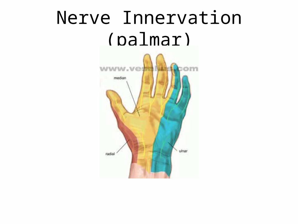
Nerve Innervation (palmar)
Stability of radius to ulna
• Interosseus membrane.
• Annular ligament.
• Disc of inferior radioulnar joint.
• Oblique cord.
• Supinator & pronator quadratus muscles.

Forces transmitted from the upper limb to axial skeleton pass through:
• Glenoid cavity.• Coracoid process.• Clavicle.• Costoclavicular ligament.• 1st rib & sternum.
And neither through acromioclavicular joint nor through sternoclavicular joint.
Top Related