


![Bacteriophage [Compatibility Mode] (2)](https://static.fdocuments.in/doc/165x107/577cd7461a28ab9e789e8922/bacteriophage-compatibility-mode-2.jpg)


Languages
Pages
Legal

International Journal of Scientific & Engineering Research, Volume 6, Issue 11, November-2015 539 ISSN 2229-5518
IJSER © 2015 http://www.ijser.org
Isolation and Characterization of Bacteriophage Against Methicillin Resistant Staphylococcus
aureus Mariem N. Mohammed-Ali and Nidham M. Jamalludeen
ABSTRACT- Methicillin resistant Staphylococcus aureus (MRSA) is a major human pathogen responsible for several life threatening conditions. MRSA have the ability to acquire resistance to several antimicrobial agents and phage therapy is one potential option to treat this pathogen. The aim of the study was to isolate and characterize bacteriophages effective against a wide range of methicillin-resistant Staphylococcus aureus (MRSA). A mixture of ten MRSA isolates was used for the isolation of phage from wastewater treatment plants. Three phages were selected for further characterization. All three phages belong to the Siphoviridae family and have long non-contractile flexible tails. The three phages showed a wide host range against S. aureus. Phages ɸSA1 and ɸSA2 were resistant to a pH range from 4-10 while ɸSA3 has a pH range from 3-11. DNA from all three phages were resistant to digestion by endonuclease enzymes such as EcoRI and AccI. There was a high degree of mosaicism among the three virulent phages and with their ancestor phages of Siphoviridae due to their non-uniform access to the common genetic pool by horizontal gene transfer and recombination. Since some of the staphylococcal toxins are phage encoded, the presence of genes for such toxins was tested by performing polymerase chain reaction and all three phages lacked genes for any of the staphylococcal toxins, including staphylococcal enterotoxins (sea, seb, sec and see), exfoliating toxins (eta and etb) and the toxic shock syndrome toxin (tst), therefore these bacteriophage are suitable candidates for future use in phage therapy against MRSA.
KEY WORDS- Staphylococcus aureus, phage therapy, MRSA phage, Siphoviridae, Anti-restriction mechanism, Mosaicism
1. INTRODUCTION
Methicillin-resistant Staphylococcus aureus (MRSA) is a worldwide pathogen that is responsible for a variety of diseases ranging from soft tissue and skin infections to life threatening conditions such as pneumonia, bacteremia and sepsis [1]. MRSA is one of the major human pathogens that may cause community and hospital acquired infections [2]. These organisms are frequently resistant to most of the commonly used antimicrobial agents, including β-lactam antibiotics [3]. The emergence and spread of strains resistant to oxacillin, methicillin and even vancomycin has made therapy of these multi drug resistant bacteria a global challenge [4]. MRSA has a wide variety of virulence factors that include structural and secreted factors [5]. These factors include superantigens, cytolytic toxins, exoenzymes and miscellaneous proteins [6]. Superantigens are a group of powerful immuno-stimulatory proteins implicated in a variety of human diseases including gastroenteritis and toxic shock syndrome [7].
One possible approach to treatment of methicillin resistant S. aureus is phage therapy, defined as the application of phage to selectively reduce or eliminate susceptible pathogens from specific environments [8]. Phage therapy may be a suitable alternative to antibiotic treatment due to the high specificity of and effectiveness against multi drug resistant bacteria [9, 10]. The use of bacteriophages in clinical medicine was first introduced by Félix d’Herelle [11]. Many therapeutic phage have been isolated against MRSA, most of which belong to the Myoviridae family such as the well-known phage K and MR-10 [12, 13]. Since all tailed phages are believed to share common ancestors, a high frequency of chimeric and mosaic
structures can be observed among different tailed phage families due to their access to a common genetic pool [14, 15]. Such mosaicism has resulted in a high degree of similarity among phages in both nucleic acids and proteins [16]. The main aims of this study were to isolate a set of bacteriophages effective against a wide range of MRSA isolates and characterize these phages according to their morphological features, host range, endonuclease enzyme digestion pattern, molecular identification, the presence of undesirable toxin encoding genes.
2. MATERIALS AND METHODS CULTURE MEDIA AND CHEMICALS
The following media and chemicals were used for the study: brain heart infusion agar (Salucea, Netherlands), brain heart infusion broth (HIMEDIA, India), blood agar (HIMEDIA, India), mannitol salt agar (OXOID, England), Mueller Hinton agar (HIMEDIA, India), Agar agar (HIMEDIA, India), agarose (Bio Basic, Canada), peptone (DIFCO, USA), beef extract (OXOID, England), yeast extract (Sigma-Aldrich, Switzerland), sodium chloride (Sigma-Aldrich, Switzerland), sodium hydroxide (Sigma-Aldrich, Switzerland), potassium dihydrogen phosphate (Sigma-Aldrich, Switzerland), Gelatin (BDH, England), barium chloride (Hopkins & Williams Limited, England), Gram stain (HIMEDIA, India), catalase test reagent (HIMEDIA, India), coagulase plasma (HIMEDIA, India), API Staph (bioMérieux, France), EcoRI (promega, USA), AccI (BioLabs, New England), sulfuric acid (BDH, England), hydrochloric acid (BDH, England), tris-(hydroxymethyl)-aminomethane (pH 7.5) (Riedel-deHaën, Germany), Chloroform (Sigma-Aldrich, Switzerland), glycerol (Chem-supply, Australia), absolute
IJSER
International Journal of Scientific & Engineering Research, Volume 6, Issue 11, November-2015 540 ISSN 2229-5518
IJSER © 2015 http://www.ijser.org
ethanol (BDH, England), nuclease free water (Promega, USA), ethidium bromide (Fisher, USA), bromophenol blue (Fisher, USA), TBE buffer (Promega, USA), TE buffer (Promega, USA), (Bioanalyse, USA), cefoxitin disc (30µg) (Bioanalyse, USA), DNA ladder marker (100-1000 bp) (Promega, USA) and DNA ladder marker (100-10000 bp (KAPA, USA).
SAMPLE COLLECTION A total of 100 isolates of staphylococci were obtained from
various clinical sources such as skin, anterior nares, ear wound, sputum, blood and urine all according to standard methods of sample collection [17]. The isolates were randomly collected from healthcare workers, hospital patients and patient’s escorts at Al-Sadar hospital, Al-Basra General Hospital, Ibn Ghazwan Hospital and day care centers in Basra.
MRSA IDENTIFICATION Staphylococcus aureus isolates were identified by standard
methods described [17]. Staphylococcus aureus isolates isolates were characterized as methicillin resistant (MRSA) using the cefoxtin disc (30 µg) diffusion method [18]. The diameter of the zone of inhibition was measured and the results were interpreted according to CLSI criteria [19].
ISOLATION OF BACTERIOPHAGE Bacteriophages were isolated from raw sewage samples
obtained from Al-Sadar hospital raw sewage in Basra according to previously described methods [20, 21]. A 200 ml volume of fresh sewage was mixed with 20 ml of bacteriophage broth [peptone (100 g/L), beef extract (30 g/L), yeast extract (50 g/L), sodium chloride (25 g/L), potassium dihydrogen phosphate (80 g/L)], 20 ml of a mixture of 2 MRSA strains (ATCC 25923 and ATCC 29213) in broth culture (optical density at 600 nm (OD600 = 1.4) and 20 ml of BHI broth were aseptically added to a 1 L flask and incubated at 37 ºC for 24 h with shaking (55 rpm). After incubation, the mixture was centrifuged at 4500 xg for 15 min and the supernatant was transferred into a clean flask and then filtered through a sterile 0.45 µm membrane filter (chm, Spain). The phage titre was determined by serial dilution in which 100 µl volumes of the filtrate was mixed with 100 µl of broth culture containing MRSA in a test tube and incubated at 37 ºC for 20 min then 3 ml of top agarose (7.0 g/L) was added, the tube contents were then mixed and poured onto the surface of a BHI agarose plate and allowed to harden for a few minutes and then incubated at 37 ºC for 16 h. Next day, the plates were examined for the presence of plaques. A control tube containing bacteria and 3 ml of top agarose without filtrate was also cultured on a BHI agarose plate.
A sterile Pasteur pipette with a rubber bulb was used to gently suction a well-isolated plaque. The pipette contents were transferred into a tube containing 1 ml of SM buffer (5.8 g/L NaCl, 2 g/L MgSO4 7H2O, 50 ml/L of 1 M Tris pH 7.5, 5 ml/L of 2% gelatin) and 1 drop of chloroform was added to each tube. The tubes were held at room temperature for 1–2 h
to allow the bacteriophage particles to diffuse out of the agar. The phage titre was determined by the soft agarose overlay method and finally the phages were stored at 4 ºC until stocks were prepared.
BACTERIOPHAGE STOCK PREPARATION Bacteriophage stock was prepared according to [21]. A 100-
µl volume of bacteriophage suspension was incubated with 100 µl of the selected host bacterium for 20 min at 37 ºC, then 3 ml of top agarose was added, mixed and poured onto a BHI agarose plate which was incubated for 6-8 h at 37 ºC. A 2 ml volume of SM buffer was then added to each plate in order to harvest the phages and then was transferred into tubes containing 0.2 ml of chloroform. The mixture was kept at 4 ºC for 2-3 h and gently vortexed and centrifuged at 4500 x g for 15 min. The supernatant was recovered and a drop of chloroform was added. The supernatant was filtered through a sterile 0.45 µm membrane filter (chm, Spain) and the filtrate was stored at 4 ºC and the titres of the phage stocks were determined by plaquing 10-fold dilutions using the soft agarose overlay method. The phage stock was purified for further studies by layering 4 ml of 5% glycerol solution in SM buffer over 40% glycerol and adding the sample on the top, followed by ultracentrifugation at 25000 x g, for 2 h at 4 ºC. The phage-containing pellet was resuspended in 1 ml SM buffer.
ELECTRON MICROSCOPY Phage particles were negatively stained with 2% uranyl
acetate (Sigma-Aldrich, Switzerland) on carbon-coated copper grid using standard procedures. TEM images were captured in Zeiss EM10C (Zeiss, Germany) at Khajeh Nasir Toosi University of Technology (IRAN). Three phages that were isolated were classified according to the International Committee on Taxonomy of Viruses ICTV [22].
HOST RANGE
The host range of each of the isolated bacteriophages was determined against a number of MRSA isolates as described by [20]. A lawn of a single MRSA isolate was inoculated on a BHI agar plate and the plate was divided into four squares by marking the surface of the plate. The plate was left to dry for a few minutes and then 10 µl of each phage suspension (109 pfu/ml) was dropped in the center of each square except for one square which was left as control. Following incubation at 37 ºC for 24 h, these plates were examined for lysis. A clear zone in the bacterial lawn was recorded as complete lysis.
RESISTANCE OF PHAGES TO ACIDITY AND ALKALINITY
Resistance of phages to acidity and alkalinity was done according to [20]. A 100 µl volume of phage suspension (109 pfu/ml) was added to 900 µl of saline adjusted to a specific pH. The mixture was incubated at 37 ºC for 16 h. A control sample (phage suspension and normal saline, pH 7.2) was also incubated at 37 ºC for 16 h. The titre of the surviving phages was determined by plaquing 10-fold dilutions by the soft
IJSER
International Journal of Scientific & Engineering Research, Volume 6, Issue 11, November-2015 541 ISSN 2229-5518
IJSER © 2015 http://www.ijser.org
agarose overlay method.
EXTRACTION OF BACTERIOPHAGE DNA The bacteriophage DNA was extracted using the QIAprep
Spin M13 kit (QIAGEN, Germany) and according to the manufacturer’s instruction. DNA was detected with the Nano drop (Optizen, Korea) and was visualized by agarose gel electrophoresis [21]. RESTRICTION ENDONUCLEASE ENZYME DIGESTION PATTERNS
A (5-10) µl volume of DNA was digested with EcoRI and AccI enzymes according to the manufacturer’s instructions. MOLECULAR IDENTIFICATION OF BACTERIOPHAGE
In an attempt for molecular identification of the three phages, conserved genomic sequences of Staphylococcus aureus phage type 3A, 11, 77, 187 and Twort like phages representing serogroups A, B, F, L and D were used. Each conserved sequence encodes a certain protein such as head protein, major capsid protein, packaging protein, tail protein or tail fiber protein [23]. Taq PCR master mix used and was supplied by Bioneer (Korea); the primers were designed by Macrogen (Korea). The primer sequences and their lengths are shown in Table 1. Table 1 Staphylococcal phage type, sero-group, primer sequence, PCR product length (bp) and type of protein.
DETECTION OF POSSIBLE TOXIN GENES BY POLYMERASE CHAIN REACTION (PCR)
Undesirable genes including staphylococcal enterotoxins and exfoliating toxins as well as toxic shock syndrome toxin of the isolated phages were searched by using QIAGEN multiplex PCR kit (QIAGEN, Germany) using a thermo cycler from Eppendorf (mastercycler, personal 5332, Germany) and according to the manufacturer’s instructions. The primers were designed for this study by Eurogentec (Belgium). A 100
µM (100 pmol/µl) stock of each primer was prepared according to the technical data sheet for each primer and kept in TE buffer at -70 °C. Staphylococcus aureus specific genes, primers and their exact sequence as well as the size of the amplified product (bp) are listed in Table 2 [24]. Table 2 Staphylococcus aureus specific genes, primers and their exact sequence as well as the predicted size of the amplified product (bp).
3. RESULTS BACTERIOPHAGE ISOLATION
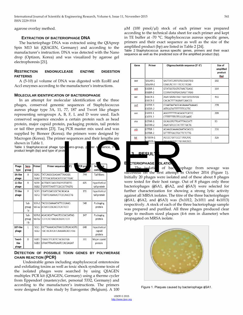
The isolation of bacteriophage from sewage was successful on the first attempt in October 2014 (Figure 1). Initially 20 phages were isolated and of these about 8 phages were tested for their host range. Out of 8 phages only three bacteriophages (ɸSA1, ɸSA2, and ɸSA3) were selected for further characterization for showing a strong lytic activity against all MRSA isolates. The titre of the three bacteriophages (ɸSA1, ɸSA2, and ɸSA3) was (5x10¹², 2x10¹¹ and 4x10¹³) respectively. A stock of each of the three bacteriophage sample was prepared and purified. All three phages produced clear large to medium sized plaques (4-6 mm in diameter) when propagated on MRSA isolate.
Figure 1. Plaques caused by bacteriophage ɸSA1.
IJSER
International Journal of Scientific & Engineering Research, Volume 6, Issue 11, November-2015 542 ISSN 2229-5518
IJSER © 2015 http://www.ijser.org
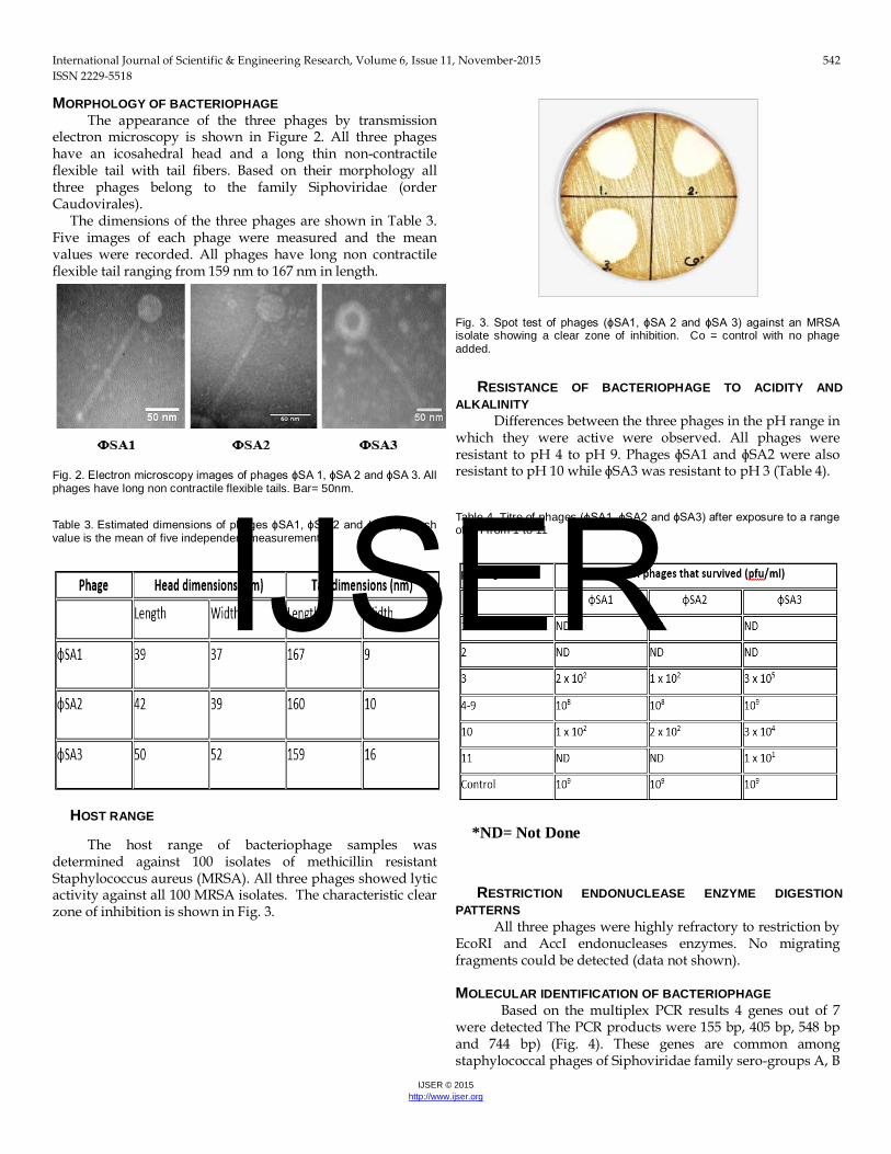
MORPHOLOGY OF BACTERIOPHAGE The appearance of the three phages by transmission
electron microscopy is shown in Figure 2. All three phages have an icosahedral head and a long thin non-contractile flexible tail with tail fibers. Based on their morphology all three phages belong to the family Siphoviridae (order Caudovirales).
The dimensions of the three phages are shown in Table 3. Five images of each phage were measured and the mean values were recorded. All phages have long non contractile flexible tail ranging from 159 nm to 167 nm in length.
Fig. 2. Electron microscopy images of phages ɸSA 1, ɸSA 2 and ɸSA 3. All phages have long non contractile flexible tails. Bar= 50nm.
Table 3. Estimated dimensions of phages ɸSA1, ɸSA 2 and ɸSA 3). Each value is the mean of five independent measurements.
HOST RANGE
The host range of bacteriophage samples was determined against 100 isolates of methicillin resistant Staphylococcus aureus (MRSA). All three phages showed lytic activity against all 100 MRSA isolates. The characteristic clear zone of inhibition is shown in Fig. 3.
Fig. 3. Spot test of phages (ɸSA1, ɸSA 2 and ɸSA 3) against an MRSA isolate showing a clear zone of inhibition. Co = control with no phage added.
RESISTANCE OF BACTERIOPHAGE TO ACIDITY AND ALKALINITY
Differences between the three phages in the pH range in which they were active were observed. All phages were resistant to pH 4 to pH 9. Phages ɸSA1 and ɸSA2 were also resistant to pH 10 while ɸSA3 was resistant to pH 3 (Table 4).
Table 4. Titre of phages (ɸSA1, ɸSA2 and ɸSA3) after exposure to a range of pH from 1 to 11
*ND= Not Done
RESTRICTION ENDONUCLEASE ENZYME DIGESTION PATTERNS
All three phages were highly refractory to restriction by EcoRI and AccI endonucleases enzymes. No migrating fragments could be detected (data not shown).
MOLECULAR IDENTIFICATION OF BACTERIOPHAGE
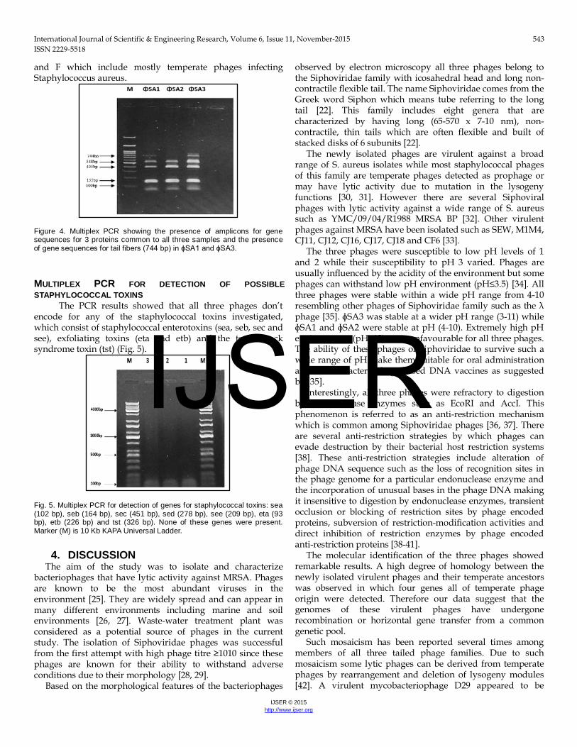
Based on the multiplex PCR results 4 genes out of 7 were detected The PCR products were 155 bp, 405 bp, 548 bp and 744 bp) (Fig. 4). These genes are common among staphylococcal phages of Siphoviridae family sero-groups A, B
IJSER
International Journal of Scientific & Engineering Research, Volume 6, Issue 11, November-2015 543 ISSN 2229-5518
IJSER © 2015 http://www.ijser.org
and F which include mostly temperate phages infecting Staphylococcus aureus.
Figure 4. Multiplex PCR showing the presence of amplicons for gene sequences for 3 proteins common to all three samples and the presence of gene sequences for tail fibers (744 bp) in ɸSA1 and ɸSA3.
MULTIPLEX PCR FOR DETECTION OF POSSIBLE STAPHYLOCOCCAL TOXINS
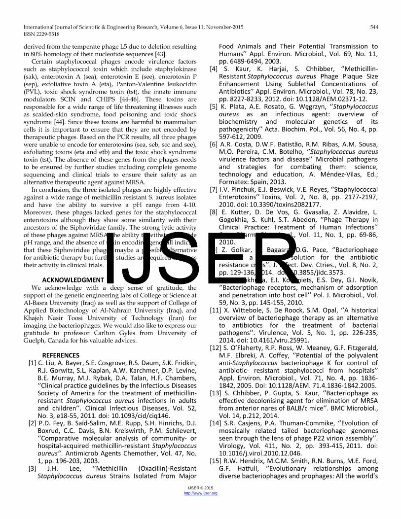
The PCR results showed that all three phages don’t encode for any of the staphylococcal toxins investigated, which consist of staphylococcal enterotoxins (sea, seb, sec and see), exfoliating toxins (eta and etb) and the toxic shock syndrome toxin (tst) (Fig. 5).
Fig. 5. Multiplex PCR for detection of genes for staphylococcal toxins: sea (102 bp), seb (164 bp), sec (451 bp), sed (278 bp), see (209 bp), eta (93 bp), etb (226 bp) and tst (326 bp). None of these genes were present. Marker (M) is 10 Kb KAPA Universal Ladder.
4. DISCUSSION The aim of the study was to isolate and characterize
bacteriophages that have lytic activity against MRSA. Phages are known to be the most abundant viruses in the environment [25]. They are widely spread and can appear in many different environments including marine and soil environments [26, 27]. Waste-water treatment plant was considered as a potential source of phages in the current study. The isolation of Siphoviridae phages was successful from the first attempt with high phage titre ≥1010 since these phages are known for their ability to withstand adverse conditions due to their morphology [28, 29].
Based on the morphological features of the bacteriophages
observed by electron microscopy all three phages belong to the Siphoviridae family with icosahedral head and long non-contractile flexible tail. The name Siphoviridae comes from the Greek word Siphon which means tube referring to the long tail [22]. This family includes eight genera that are characterized by having long (65-570 x 7-10 nm), non-contractile, thin tails which are often flexible and built of stacked disks of 6 subunits [22].
The newly isolated phages are virulent against a broad range of S. aureus isolates while most staphylococcal phages of this family are temperate phages detected as prophage or may have lytic activity due to mutation in the lysogeny functions [30, 31]. However there are several Siphoviral phages with lytic activity against a wide range of S. aureus such as YMC/09/04/R1988 MRSA BP [32]. Other virulent phages against MRSA have been isolated such as SEW, M1M4, CJ11, CJ12, CJ16, CJ17, CJ18 and CF6 [33].
The three phages were susceptible to low pH levels of 1 and 2 while their susceptibility to pH 3 varied. Phages are usually influenced by the acidity of the environment but some phages can withstand low pH environment (pH≤3.5) [34]. All three phages were stable within a wide pH range from 4-10 resembling other phages of Siphoviridae family such as the λ phage [35]. ɸSA3 was stable at a wider pH range (3-11) while ɸSA1 and ɸSA2 were stable at pH (4-10). Extremely high pH environments (pH≥12) were unfavourable for all three phages. The ability of these phages of Siphoviridae to survive such a wide range of pH make them suitable for oral administration as possible bacteriophage-based DNA vaccines as suggested by [35].
Interestingly, all three phages were refractory to digestion by endonuclease enzymes such as EcoRI and AccI. This phenomenon is referred to as an anti-restriction mechanism which is common among Siphoviridae phages [36, 37]. There are several anti-restriction strategies by which phages can evade destruction by their bacterial host restriction systems [38]. These anti-restriction strategies include alteration of phage DNA sequence such as the loss of recognition sites in the phage genome for a particular endonuclease enzyme and the incorporation of unusual bases in the phage DNA making it insensitive to digestion by endonuclease enzymes, transient occlusion or blocking of restriction sites by phage encoded proteins, subversion of restriction-modification activities and direct inhibition of restriction enzymes by phage encoded anti-restriction proteins [38-41].
The molecular identification of the three phages showed remarkable results. A high degree of homology between the newly isolated virulent phages and their temperate ancestors was observed in which four genes all of temperate phage origin were detected. Therefore our data suggest that the genomes of these virulent phages have undergone recombination or horizontal gene transfer from a common genetic pool.
Such mosaicism has been reported several times among members of all three tailed phage families. Due to such mosaicism some lytic phages can be derived from temperate phages by rearrangement and deletion of lysogeny modules [42]. A virulent mycobacteriophage D29 appeared to be
IJSER
International Journal of Scientific & Engineering Research, Volume 6, Issue 11, November-2015 544 ISSN 2229-5518
IJSER © 2015 http://www.ijser.org
derived from the temperate phage L5 due to deletion resulting in 80% homology of their nucleotide sequences [43].
Certain staphylococcal phages encode virulence factors such as staphylococcal toxin which include staphylokinase (sak), enterotoxin A (sea), enterotoxin E (see), enterotoxin P (sep), exfoliative toxin A (eta), Panton-Valentine leukocidin (PVL), toxic shock syndrome toxin (tst), the innate immune modulators SCIN and CHIPS [44-46]. These toxins are responsible for a wide range of life threatening illnesses such as scalded-skin syndrome, food poisoning and toxic shock syndrome [44]. Since these toxins are harmful to mammalian cells it is important to ensure that they are not encoded by therapeutic phages. Based on the PCR results, all three phages were unable to encode for enterotoxins (sea, seb, sec and see), exfoliating toxins (eta and etb) and the toxic shock syndrome toxin (tst). The absence of these genes from the phages needs to be ensured by further studies including complete genome sequencing and clinical trials to ensure their safety as an alternative therapeutic agent against MRSA.
In conclusion, the three isolated phages are highly effective against a wide range of methicillin resistant S. aureus isolates and have the ability to survive a pH range from 4-10. Moreover, these phages lacked genes for the staphylococcal enterotoxins although they show some similarity with their ancestors of the Siphoviridae family. The strong lytic activity of these phages against MRSA, the ability to withstand a wide pH range, and the absence of toxin encoding genes all indicate that these Siphoviridae phages maybe a possible alternative for antibiotic therapy but further studies are required to assess their activity in clinical trials.
ACKNOWLEDGMENT
We acknowledge with a deep sense of gratitude, the support of the genetic engineering labs of College of Science at Al-Basra University (Iraq) as well as the support of College of Applied Biotechnology of Al-Nahrain University (Iraq), and Khajeh Nasir Toosi University of Technology (Iran) for imaging the bacteriophages. We would also like to express our gratitude to professor Carlton Gyles from University of Guelph, Canada for his valuable advices.
REFERENCES
[1] C. Liu, A. Bayer, S.E. Cosgrove, R.S. Daum, S.K. Fridkin, R.J. Gorwitz, S.L. Kaplan, A.W. Karchmer, D.P. Levine, B.E. Murray, M.J. Rybak, D.A. Talan, H.F. Chambers, ‘’Clinical practice guidelines by the Infectious Diseases Society of America for the treatment of methicillin-resistant Staphylococcus aureus infections in adults and children’’. Clinical Infectious Diseases, Vol. 52, No. 3, e18-55, 2011. doi: 10.1093/cid/ciq146.
[2] P.D. Fey, B. Saïd-Salim, M.E. Rupp, S.H. Hinrichs, D.J. Boxrud, C.C. Davis, B.N. Kreiswirth, P.M. Schlievert, ‘’Comparative molecular analysis of community- or hospital-acquired methicillin-resistant Staphylococcus aureus’’. Antimicrob Agents Chemother, Vol. 47, No. 1, pp. 196-203, 2003.
[3] J.H. Lee, ‘’Methicillin (Oxacillin)-Resistant Staphylococcus aureus Strains Isolated from Major
Food Animals and Their Potential Transmission to Humans’’ Appl. Environ. Microbiol., Vol. 69, No. 11, pp. 6489-6494, 2003.
[4] S. Kaur, K. Harjai, S. Chhibber, ‘’Methicillin-Resistant Staphylococcus aureus Phage Plaque Size Enhancement Using Sublethal Concentrations of Antibiotics’’ Appl. Environ. Microbiol., Vol. 78, No. 23, pp. 8227-8233, 2012. doi: 10.1128/AEM.02371-12.
[5] K. Plata, A.E. Rosato, G. Węgrzyn, ‘’Staphylococcus aureus as an infectious agent: overview of biochemistry and molecular genetics of its pathogenicity’’ Acta. Biochim. Pol., Vol. 56, No. 4, pp. 597-612, 2009.
[6] A.R. Costa, D.W.F. Batistão, R.M. Ribas, A.M. Sousa, M.O. Pereira, C.M. Botelho, ‘’Staphylococcus aureus virulence factors and disease’’ Microbial pathogens and strategies for combating them: science, technology and education, A. Méndez-Vilas, Ed.; Formatex: Spain, 2013.
[7] I.V. Pinchuk, E.J. Beswick, V.E. Reyes, ‘’Staphylococcal Enterotoxins’’ Toxins, Vol. 2, No. 8, pp. 2177-2197, 2010. doi: 10.3390/toxins2082177.
[8] E. Kutter, D. De Vos, G. Gvasalia, Z. Alavidze, L. Gogokhia, S. Kuhl, S.T. Abedon, ‘’Phage Therapy in Clinical Practice: Treatment of Human Infections’’ Curr. Pharm. Biotechnol., Vol. 11, No. 1, pp. 69-86, 2010.
[9] Z. Golkar, O. Bagasra, D.G. Pace, ‘’Bacteriophage therapy: a potential solution for the antibiotic resistance crisis’’. J. Infect. Dev. Ctries., Vol. 8, No. 2, pp. 129-136, 2014. doi: 10.3855/jidc.3573.
[10] D.V. Rakhuba, E.I. Kolomiets, E.S. Dey, G.I. Novik, ‘’Bacteriophage receptors, mechanism of adsorption and penetration into host cell’’ Pol. J. Microbiol., Vol. 59, No. 3, pp. 145-155, 2010.
[11] X. Wittebole, S. De Roock, S.M. Opal, ‘’A historical overview of bacteriophage therapy as an alternative to antibiotics for the treatment of bacterial pathogens’’. Virulence, Vol. 5, No. 1, pp. 226-235, 2014. doi: 10.4161/viru.25991.
[12] S. O’Flaherty, R.P. Ross, W. Meaney, G.F. Fitzgerald, M.F. Elbreki, A. Coffey, ‘’Potential of the polyvalent anti-Staphylococcus bacteriophage K for control of antibiotic- resistant staphylococci from hospitals’’ Appl. Environ. Microbiol., Vol. 71, No. 4, pp. 1836-1842, 2005. Doi: 10.1128/AEM. 71.4.1836-1842.2005.
[13] S. Chhibber, P. Gupta, S. Kaur, ‘’Bacteriophage as effective decolonising agent for elimination of MRSA from anterior nares of BALB/c mice’’. BMC Microbiol., Vol. 14, p.212, 2014.
[14] S.R. Casjens, P.A. Thuman-Commike, ‘’Evolution of mosaically related tailed bacteriophage genomes seen through the lens of phage P22 virion assembly’’. Virology, Vol. 411, No. 2, pp. 393-415, 2011. doi: 10.1016/j.virol.2010.12.046.
[15] R.W. Hendrix, M.C.M. Smith, R.N. Burns, M.E. Ford, G.F. Hatfull, ‘’Evolutionary relationships among diverse bacteriophages and prophages: All the world’s
IJSER
International Journal of Scientific & Engineering Research, Volume 6, Issue 11, November-2015 545 ISSN 2229-5518
IJSER © 2015 http://www.ijser.org
a phage’’. Proc. Natl. Acad. Sci. USA., Vol. 96, No. 5, pp. 2192-2197, 1999.
[16] G.F. Hatfull, R.W. Hendrix, ‘’Bacteriophages and their Genomes’’. Curr. Opin. Virol., Vol. 1, No. 4, pp. 298-303, 2011. doi:10.1016/j.coviro.2011.06.009.
[17] B.A. Forbes, D.A. Sahm, A.S. Weissfeld, ‘’Bailey & Scott's Diagnostic Microbiology’’, 12th ed.; Mosby Elsevier Press: St. Louis, Missouri, 2007.
[18] N.M. Broekema, T.T. Van, T.A. Monson, S.A. Marshall, D.M. Warshauer, ‘’Comparison of Cefoxitin and Oxacillin Disk Diffusion Methods for Detection of mecA-Mediated Resistance in Staphylococcus aureus in a Large-Scale Study’’. J. Clin. Microbiol., Vol. 47, No. 1, pp. 217-219, 2009. doi: 10.1128/JCM.01506-08.
[19] Clinical and Laboratory Standards Institute. ‘’Performance Standards for Antimicrobial Susceptibility Testing; Twenty-Fourth Informational Supplement’’. CLSI document M100-S24, Vol. 34, No. 1, pp. 219, 2014.
[20] N. Jamalludeen, R.P. Johnson, R. Friendship, A.M. Kropinski, E.J. Lingohr, C.L. Gyles, ‘’Isolation and characterization of nine bacteriophages that lyse O149 enterotoxigenic Escherichia coli’’. Vet. Microbiol., Vol. 124, No. 1-2, pp. 47-57, 2007.
[21] J. Sambrook, and D.W. Russell, ‘’Molecular cloning: a laboratory manual’’, 3rd ed.; Cold Spring Harbor Laboratory Press: NY, 2001.
[22] ICTV-International Committee on Taxonomy of Viruses. ‘’Virus taxonomy; classification and nomenclature of viruses’’. Eighth report of the International Committee on Taxonomy of Viruses. Springer-Verlag/Wien, Austria, 2005, 57-70.
[23] R. Pantůček, J. Doškař, V. Růžičková, P. Kašpárek, E. Oráčová, V. Kvardová, S. Rosypal, ‘’Identification of bacteriophage types and their carriage in Staphylococcus aureus’’ Arch. Virol., Vol. 149, pp. 1689-1703, 2004. doi: 10.1007/s00705-004-0335-6.
[24] M. Mehrotra, G. Wang, W.M. Johnson, ‘’Multiplex PCR for Detection of Genes for Staphylococcus aureus Enterotoxins, Exfoliative Toxins, Toxic Shock Syndrome Toxin 1, and Methicillin Resistance’’ J. Clin. Microbiol., Vol. 38, No. 3, pp. 1032-1035, 2000.
[25] R.A. Sandaa, ‘’Environmental viral pool’’. In: Schmidt T. M. and Schaechter M., editors. Topics in Ecological and Environmental Microbiology. USA. Elsevier Inc, 2012.
[26] P. Mathur, ‘’Hand hygiene: Back to the basics of infection control’’. Indian J. Med. Res., Vol. 134, No. 5, pp. 611-620, 2011. doi:10.4103/0971-5916.90985.
[27] R. Międzybrodzki, W. Fortuna, B.W. Dąbrowska, A. Górski, ‘’Phage therapy of staphylococcal infections (including MRSA) may be less expensive than antibiotic treatment’’. Postepy. Hig. Med. Dosw., Vol. 61, pp. 461-465, 2007.
[28] J. Lasobras, M. Muniesa, F. Lucena, J. Jofre, ‘’Relationship between the morphology of bacteriophages and their persistence in the environment’’ Water. Sci. Tech., Vol. 35, No. 11-12,
pp. 129-132, 1997. doi: 10.1016/S0273-1223(97)00247-3.
[29] M. Muniesa, F. Lucena, J. Jofre, ‘’Study of the potential relationship between the morphology of infectious somatic coliphages and their persistence in the environment’’ J. Appl. Microbiol., Vol. 87, No. 3, pp. 402-409, 1999. doi: 10.1046/j.1365-2672.1999.00833.
[30] M. Deghorain, L. Van Melderen, ‘’The Staphylococci Phages Family: An Overview’’ Viruses, Vol. 4, No. 12, pp. 3316-3335, 2012. doi: 10.3390/v4123316.
[31] G. Xia and C. Wolz, ‘’Phage of Staphylococcus aureus and their impact on host evolution’’ Infect. Gene Evol., Vol. 21, pp. 593-601, 2014. doi. 10.1016/j.04.022.
[32] J. Jeon, R. D’Souza, S.K. Hong, Y. Lee, D. Yong, J. Choi, K. Lee, Y. Chong, ‘’Complete genome sequence of the Siphoviral bacteriophage YMC/09/04/R1988 MRSA BP: A lytic phage from a methicillin-resistant Staphylococcus aureus isolate’’ FEMS Microbiol Lett., 2014. doi: 10.1111/1574-6968.12580.
[33] K.C. Jensen, B.B. Hair, T.M. Wienclaw, M.H. Murdock, J.B. Hatch, A.T. Trent, T.D white K.J. Haskell B.K. Berges, ‘’Isolation and Host Range of Bacteriophage with Lytic Activity against Methicillin-Resistant Staphylococcus aureus and Potential Use as a Fomite Decontaminant’’ PLoS ONE, Vol. 10, No. 7, e0131714, 2015. Doi: org/10.1371/journal.pone.0131714.
[34] Z. Lu, F. Breidt, V. Plengvidhya, H.P. Fleming, ‘’Bacteriophage Ecology in Commercial Sauerkraut Fermentations’’. Appl. Environ. Microbiol., Vol. 69, No.6, pp. 3192-3202, 2003. doi: 10.1128/AEM.69.6.3192-3202.2003.
[35] C.D. Jepson, and J.B. March, ‘’Bacteriophage lambda is highly stable DNA vaccine delivery vehicle’’ Vaccine, Vol. 22, No. 19, pp. 2413-2419, 2004. doi: 10.1016/j.vaccine.2003.11.065.
[36] R. Barrangou, S. Yoon, F. Breidt, H. Fleming, T. Klaenhammer, ‘’Characterization of six Leuconostoc fallax bacteriophages isolated from an industrial Sauerkraut Fermentation’’ Appl. Environ. Microbial., Vol. 68, No. 11, pp. 5452-5458, 2002. Doi: 10.1128/AEM.68.11.5452-5458.2002.
[37] A. Kęsik-Szeloch, Z. Drulis-Kawa, B. Weber-Dąbrowska, J. Kassner, G. Majkowska-Skrobek, D. Augustyniak, M. Lusiak-Szelachowska, M. Zaczek, M.; Górski, A.M. Kropinski, ‘’Characterising the biology of novel lytic bacteriophages infecting multidrug resistant Klebsiella pneumoniae’’ Virol. J., Vol. 10, 100, 2013. doi: 10.1186/1743-422X-10-100.
[38] M.R. Tock, and D.T. Dryden, ‘’The biology of restriction and anti-restriction’’ Curr. Opin. Microbiol., Vol. 8, No. 4, pp. 466-472, 2005. Doi: 10.1016/j.mib.2005.06.003.
[39] I. Kovalchuk, and O. Kovalchuk, ‘’Epigenetics in health and disease’’ Pearson Education, Inc. FT press, 2012, 402.
IJSER
International Journal of Scientific & Engineering Research, Volume 6, Issue 11, November-2015 546 ISSN 2229-5518
IJSER © 2015 http://www.ijser.org
[40] P. Pristas, A. Vandzurova, P. Javorsky, ‘’Interplay between bacteriophages and restriction-modification systems in enterococci’’ Nova Biotechnologica et Chimica., Vol. 13, No. 1, pp. 13-20, 2014. doi: 10.2478/nbec-2014-0002.
[41] K. Vasu, and V. Nagaraja, ‘’Divers functions of restriction-modification systems in addition to cellular defense’’ Microbiol. Mol. Boil. Rev., Vol. 77, No. 1, pp. 53-72, 2013. doi: 10.1128/MMBR.00044-12.
[42] H. Brüssow, and F. Desiere, ‘’Comparative phage genomics and the evolution of Siphoviridae: insights from dairy phages’’ Mol. Microbial., Vol. 39, No. 2, pp. 213-222, 2001.
[43] E.J. Summer, C.F. Gonzalez, M. Bomer, T. Carlile, A. Embry, A.M. Kucherka, J. Lee, L. Mebane, W.C. Morrison, L. Mark, M.D. King, J.J. LiPuma, A.K. Vidaver, R.Y. Young, ‘’Divergence and Mosaicism among Virulent Soil Phages of the Burkholderia cepacia Complex’’ J. Bacteriol., Vol. 188, No. 1, pp. 255-268, 2006. doi:10.1128/JB.188.1.255-268.2006.
[44] E.F. Boyed, ‘’Bacteriophage-encoded virulence factors and phage-pathogenicity island interactions’’ Adv. virus Res., Vol. 82, pp. 91-118, 2012.
[45] H. Brüssow, C. Canchaya, W-D. Hardt, ‘’Phages and the Evolution of Bacterial Pathogens: from Genomic Rearrangements to Lysogenic Conversion’’ Microbiol. Mol. Biol. Rev., Vol. 68, No. 3, pp. 560-602, 2004. doi:10.1128/MMBR.68.3.560-602.2004.
[46] G.E. Christie, A.M. Matthews, D.G. King, K.D. Lane, N.P. Olivarez, S.M. Tallent, S.R. Gill, R.P. Novik, ‘’The complete genomes of Staphylococcus aureus bacteriophages 80 and 80α– implications for the specificity of SaPI mobilization’’ Virol., Vol. 407, No. 2, pp. 381-390, 2010. doi: 10.1016/j.virol.2010.08.036.
IJSER
Top Related