


Languages
Pages
Legal


Int.J.Curr.Microbiol.App.Sci (2018) 7(5): 3206-3218
3206
Original Research Article https://doi.org/10.20546/ijcmas.2018.705.375
Isolation, Molecular and Electron Microscopic Detection of PPR Virus from
Suspected Outbreaks of PPR in Goats of North – Eastern Karnataka, India
K.C. Mallinath1*
, A. Basavaraj1, Amitha R. Gomes
2, B.H. Jagadish
3,
M. Shivaraj2, R. Bhoyar
4, S.M. Byregowda
2, N.A. Patil
4,
C. Jagannathrao4, P. Ubhale
1 and Phani Kashyap
3
1Department of Veterinary Microbiology, Veterinary College, Bidar-585401,
Karnataka, India 2IAH &VB- Bengaluru, Karnataka, India
3ICAR-NIVEDI- Bengaluru, Karnataka, India
4Veterinary College, Bidar, Karnataka, India
*Corresponding author
A B S T R A C T
Introduction
Peste des Petits Ruminants (PPR) is an
economically important disease of sheep and
goats. World Organization for Animal Health
(OIE-previous name) identified as notifiable
infectious disease of sheep and goat caused by
virus belonging to Morbilli virus genus of
family Paramyxoviridae (Gibbs et al., 1979)
threatening food security and livelihood of
small and marginal farmers across the world.
Recent estimate of Food and Agriculture
Organisation (FAO), indicated the economic
loss due to PPR in India alone, is $2569.00
International Journal of Current Microbiology and Applied Sciences ISSN: 2319-7706 Volume 7 Number 05 (2018) Journal homepage: http://www.ijcmas.com
Region wise monitoring of the PPR virus by active surveillance is essential as it aids in
strategic planning of measures of disease control and prevention. The North Eastern
Karnataka with sizable sheep and goat population experiences the outbreaks of PPR but
the attempts to isolate and characterize the prevailing PPR virus is limited. Hence, the
present research work was carried out with the objective of isolation and characterization
of PPR virus isolates circulating in North Eastern Karnataka by microscopic (TEM) and
molecular (RT-PCR) methods. A total of 15 suspected PPR outbreaks were attended and
different types of clinical samples like nasal, oral, ocular, rectal swabs and blood from
ailing kids and goats were collected. The samples were initially screened for the presence
of PPR virus by observing the infected Vero-cells for development of characteristic CPE.
The isolates showing CPE were further confirmed for the virus by RT-PCR using N and F
genes of PPR virus. The PCR confirmed isolates were subjected to transmission electron
microscopy (TEM). A total of six PPR isolates were obtained representing five districts of
North Eastern Karnataka which were confirmed by RT-PCR. The TEM images showed
pleomorphic virion particles with varying sizes indicative of Morbilli-virus. These findings
provide the first documentation of PPR presence by microscopic and molecular
characterization in the region.
K e y w o r d s
PPR, TEM,
Morbilli-virus, N, F
gene etc.
Accepted:
22 April 2018
Available Online: 10 May 2018
Article Info

Int.J.Curr.Microbiol.App.Sci (2018) 7(5): 3206-3218
3207
million/year during 2012-2017. Realising the
economic importance of the PPR disease and
successful eradication of antigenically related
Rinder pest virus from globe in June 2011, the
International organizations (OIE and FAO)
have set target of global elimination of the
disease by 2030. Though disease is endemic in
India, caused by single serotype involving
lineage-IV (Asian), recent outbreaks of
lineage (IV) PPR virus in Africa and European
part of Turkey where outbreaks due to lineage
I, II, III were common, had necessitated the
continuous surveillance of the disease in an
endemic region (Banyard et al., 2010). Hence
continuous monitoring of the type of virus
circulating in the region by active surveillance
is necessary in understanding the
epidemiology of disease and planning the
measures of disease eradication. Considering
the above facts and figures, present study
reports about isolation, molecular and electron
microscopic detection of PPR virus from
suspected outbreaks of the disease recorded in
goats of North eastern region of Karnataka
comprising five districts.
Materials and Methods
Sample collection
Different clinical samples like nasal, oral,
ocular, rectal swabs and blood in viral
transport medium (VTM, Hi-media) from
ailing kids and adult goats were collected from
15 different outbreaks recorded in North
eastern region of Karnataka during the period
from Jan-2017 to Dec-2017. Using the clinical
score card the clinical signs of the disease
were classified in to mild, moderate and
severe form (Balamurugan et al., 2014).
A total of 85 nasal swabs, 85 ocular swabs, 85
oral swabs, 50 rectal swabs and 78 anti-
coagulated blood samples were collected
aseptically in VTM and brought to laboratory
in ice packs.
Virus isolation
Swab samples collected in VTM were initially
freeze thawed thrice and used directly for
isolation. From the whole blood, buffy coat is
extracted and eluted in 1X PBS and used as
inoculum for infection (OIE manual). Swabs
from same place and species of outbreaks
were pooled and syringe filtered using 0.45µ
pore sized membrane filter (Gomes et al.,
2016). One to one and half ml of filtrate was
used as inoculum to infect the monolayer
(80% confluency) of Vero cells (African green
monkey kidney cells at passage 140-160)
grown in T-25cm2culture flask by pre-
adsorption method (Nanda et al., 1996) with
minor modifications. Inoculated cells were
incubated at 370C for one hour with
intermittent shaking to allow adsorption of
virus. Decant the virus inoculum followed by
washing of infected cells with Minimum
Essential Medium (serum free DMEM) and
then added with maintenance medium
(DMEM with 2% serum) for further
incubation at 37oC for 6-8 days with change of
maintenance medium on alternate days. The
cells were monitored daily under inverted
microscope for cytopathic effects due to viral
replication. The samples were processed up to
6th
passage. The specificity of the isolated
virus was determined by RT-PCR. Total RNA
isolated from cell culture-adapted isolates was
subjected to reverse transcription to synthesize
complementary DNA followed by its PCR
amplification of N and F gene as described
below.
Reverse transcription polymerase chain
reaction (RT-PCR)
It was performed as per the protocol given in
OIE Manual of Diagnostic Tests and Vaccines
for Terrestrial Animals 2017. The total RNA
was extracted from clinical materials and
Vero-cell adopted isolates using RNA easy kit
(RNeasy®Minikit Qiagen Inc, Valencia, CA,
Int.J.Curr.Microbiol.App.Sci (2018) 7(5): 3206-3218
3208
USA). RT–PCR was performed using Thermo
Scientific Revert Aid First Strand cDNA
Synthesis Kit for c-DNA synthesis followed
by PCR amplification using multiple sets of
PPR virus genes (N and F) specific primers
(Table 1), master mix reagents (Qiagen Inc,
Valencia, CA, USA) and PCR conditions
(Table 2).
Detection of virus by Transmission
Electron Microscopy (TEM)
The concentrated and purified virus pellet of
each of the isolate was reconstituted in PBS
and 3 µl of each one of them were pipetted out
on a paraffin wax strip fixed on a glass slide.
The samples were then deposited onto glow-
discharger carbon coated copper grids (200
mesh Sigma Aldrich, India) by floating it on
the respective samples present on wax strip
and incubating it for 1 minute at room
temperature. Excess sample was blotted using
filter paper after incubation and the grid was
washed twice with Milli-Q water. Samples
were negatively stained with 2% uranyl
acetate for 1-2 minutes (Brenner and Horne,
1959), excess stain was blotted away with
filter paper and the grids were allowed to dry.
The grids were finally examined on
Transmission Electron Microscope HT7700
series at Indian Institute of Horticulture
Research (IIHR), Bengaluru operated at
80KVand images were captured at different
magnifications (5k, 10k, 15k, 20k, 25k and
30k)
Results and Discussion
Epidemiological observations
A total of 15 outbreaks were attended in the
region during the period under study. The
clinical signs like elevated temperature,
mucous/muco-purulent nasal and ocular
discharge, congested nasal and ocular mucous
membrane, crusts around nostrils, respiratory
distress, severe mucosal erosion, salivation,
ulcers on tongue, watery diarrhoea with soiled
hind quarters were observed in the outbreaks
and clinical cases attended. Based on the
clinical score card the disease was classified
into mild, moderate and severe form
(Balamurugan et al., 2014). The mild form of
disease was characterized by serous nasal and
ocular discharge, ulcers in the mouth,
respiratory distress (Fig.1A-C) and mild
diarrhea, while moderate form showed clinical
signs like moderate pyrexia, mucous nasal and
ocular discharge, ulcers and bran like depots
in the mouth, and watery diarrhoea (Fig.2D-
F). The severe form showed signs
characterized by hyperaemia, mucopurulant
nasal and ocular discharge, sloughing of oral
mucosa, formation of crust in and around
nostril and eyes, severe watery diarrhoea with
soiled hind quarters (Fig.3G-I). Severe form of
the disease was recorded more in the kids than
adults which experienced mild to moderate
forms of the disease.
Virus isolation
A total of six isolates were obtained from the
samples subjected for isolation among which
five isolates were recovered from nasal swabs,
one from ocular and none from blood and
rectal swabs. The isolates obtained showed
two patterns of characteristic cytopathic
effects like rounding and detachment from
surface, later on aggregation of cells followed
by fusion mass and in other type, CPE was
characterized by cells of increased refractivity,
rounding and ballooning of cells, clumping
into grape like clusters and appearance of fine
spindle cells with elongated processes [Fig.4
(A-D)] (Hamby and Dardiri, 1976).
The recovery of virus isolates was observed at
different passage levels in which one isolate
was recovered on 3rd
day of post infection
(dpi) in the first passage, two isolates on 2nd
and 4th
dpi in 2nd
passage, two isolates on 3rd
Int.J.Curr.Microbiol.App.Sci (2018) 7(5): 3206-3218
3209
and 5th
dpi in 3rd
passage and one isolate on
2nd
dpi in 5th
passage indicating specifically the
load of the initial virus in the clinical samples.
The analysis of the isolation results indicates
that the relative recovery of virus using nasal
swabs and ocular swab was better than faecal
and blood samples suggesting to consider
nasal swab and ocular swab be better choice of
sample for disease diagnosis in ailing animals
by isolation (Forsyth and Barrett, 1995; Diop
et al., 2005; Harshad et al., 2014). The failure
of isolation from blood and rectal swab could
be attributed to presence of certain inhibitory
or cytotoxic factors present in blood or rectum
and labile character of the virus, initial
concentration of virus in the clinical material,
sampling and delivery time of the sample to
laboratory (Harshad et al., 2014). Though
isolation of virus is laborious and time
consuming, yet is considered to be gold
standard test for confirming any aetiologcal
agent in an outbreak which is a mandatory
method for declaring disease free status in any
region.
RT-PCR Detection
RNA viruses are known to be subjected to
high nucleotide substitution error frequencies
(Steinhauer et al., 1989). Therefore for routine
detection of the virus from different origins,
there is a risk of a false negative result if the
PCR is carried out with two primers that were
designed according to the gene sequences of
only one virus strain which could be
minimized if two sets of primers used to
amplify two different fragments or if a set of
primers is synthesized after taking into
consideration of the sequences from several
strains of different origins (Couacy-Hymann
et al., 2002; Kerur et al., 2008 and Ularamu et
al., 2012).
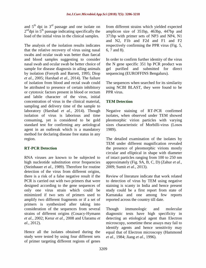
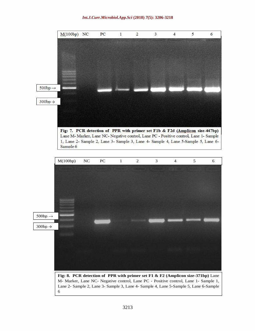
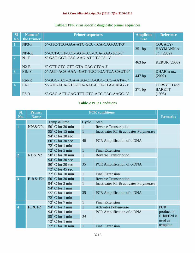
Hence all the isolates obtained during the
study were tested by using four different sets
of primer targeting different regions of genes
from different strains which yielded expected
amplicon size of 351bp, 463bp, 447bp and
371bp with primer sets of NP3 and NP4, N1
and N2, F1b and F2d and F1 and F2
respectively confirming the PPR virus (Fig. 5,
6, 7 and 8).
In order to confirm further identity of the virus
the N gene specific 351 bp PCR product was
gel purified and submitted for direct
sequencing (EUROFFINS Bengaluru).
The sequences when searched for its similarity
using NCBI BLAST, they were found to be
PPR virus.
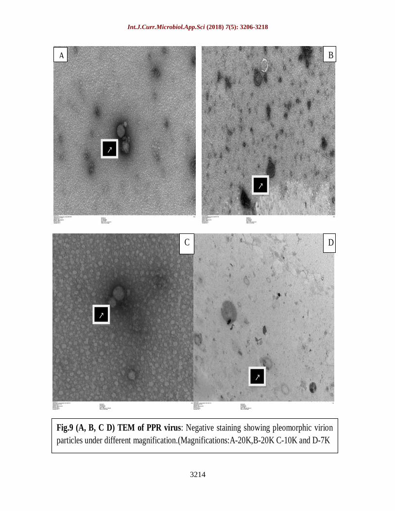
TEM Detection
Negative staining of RT-PCR confirmed
isolates, when observed under TEM showed
pleomorphic virion particles with varying
sizes characteristic of Morbilli-virus (Losos
1989).
The detailed examination of the isolates by
TEM under different magnification revealed
the presence of pleomorphic virions mostly
circular and elliptical in shape with diameter
of intact particles ranging from 100 to 250 nm
approximately (Fig. 9A, B, C, D) (Zahur et al.,
2009; Sumit et al., 2013).
Review of literature indicate that work related
to detection of virus by TEM using negative
staining is scanty in India and hence present
study could be a first report from state of
Karnataka and one among few reports
reported across the country till date.
Though immunologic and molecular
diagnostic tests have high specificity in
detecting an etiological agent than Electron
microscopy, sometime these assays may fail to
identify agents and hence sensitivity may
equal that of Electron microscopy (Hammond
et al., 1984; Jiang et al., 1996).

Int.J.Curr.Microbiol.App.Sci (2018) 7(5): 3206-3218
3210
A B C
D E F
G I H
Fig.3. Severe form of PPR: Mucopurulant nasal and oral discharge (G), sloughing of
oral mucosa (H), formation of crust in and around nostril and eyes (I)
Fig 2.Moderate form of PPR:Mucous nasal and ocular discharge (D & E),congestion
of mucus membrane, ulcers and bran like depots in the mouth (F)
Fig 1.Mild form of PPR: Serous nasal ocular and oral discharge, respiratory distress
(A,B,C)

Int.J.Curr.Microbiol.App.Sci (2018) 7(5): 3206-3218
3211
Fig.4.Pattern of CPE produced by field isolates A. Healthy Vero Cells (10X) B.
Rounding and ballooning of cells detachment from surface, later on aggregation of
cells followed by fusion mass (10X) C. Rounding and ballooning, clumping into
grape like clusters increased refractivity and appearance of fine spindle cells with
elongated processes (10X) D. Similar to C but of different isolate under 15X
magnification
A B
C D

Int.J.Curr.Microbiol.App.Sci (2018) 7(5): 3206-3218
3212
M(100bp) NC PC 1 2 3 4 5 6
Fig: 5. PCR detection of PPR with primer set NP3&NP4(Amplicon size-351bp)
Lane M- Marker, Lane NC- Negative control, Lane PC - Positive control, Lane 1- Sample
1, Lane 2- Sample 2, Lane 3- Sample 3, Lane 4- Sample 4, Lane 5-Sample 5, Lane 6-
Sample 6
500bp →
300bp →
M(100bp) NC PC 1 2 3 4 5 6
Fig: 6. PCR detection of PPR with primer set N1 & N2 (Amplicon size-463bp) Lane
M- Marker, Lane NC- Negative control, Lane PC - Positive control, Lane 1- Sample 1,
Lane 2- Sample 2, Lane 3- Sample 3, Lane 4- Sample 4, Lane 5-Sample 5, Lane 6-Sample
6
500bp →
300bp →
Int.J.Curr.Microbiol.App.Sci (2018) 7(5): 3206-3218
3213
Fig: 8. PCR detection of PPR with primer set F1 & F2 (Amplicon size-371bp) Lane
M- Marker, Lane NC- Negative control, Lane PC - Positive control, Lane 1- Sample 1,
Lane 2- Sample 2, Lane 3- Sample 3, Lane 4- Sample 4, Lane 5-Sample 5, Lane 6-Sample
6
M(100bp) NC PC 1 2 3 4 5 6
500bp →
300bp →
Int.J.Curr.Microbiol.App.Sci (2018) 7(5): 3206-3218
3214
Fig.9 (A, B, C D) TEM of PPR virus: Negative staining showing pleomorphic virion
particles under different magnification.(Magnifications:A-20K,B-20K C-10K and D-7K
↗
↗
↗
↗
A
D C
B
Int.J.Curr.Microbiol.App.Sci (2018) 7(5): 3206-3218
3215
Table.1 PPR virus specific diagnostic primer sequences
Table.2 PCR Conditions
Sl
No
Name of
the Primer
Primer sequences Amplicon
Size
Reference
1 NP3-F
NP4-R
5’-GTC-TCG-GAA-ATC-GCC-TCA-CAG-ACT-3’
5’-CCT-CCT-CCT-GGT-CCT-CCA-GAA-TCT-3’
351 bp
COUACY-
HAYMANN et
al., (2002)
2 N1-F
N2-R
5’-GAT-GGT-CAG-AAG-ATC-TGCA- 3’
5’-CTT-GTC-GTT-GTA-GAC-CTGA 3’
463 bp
KERUR (2008)
3 F1b-F
F2d-R
5’-AGT-ACA-AAA –GAT-TGC-TGA-TCA-CAGT-3’
5’-GGG-TCT-CGA-AGG-CTA-GGC-CCG-AATA-3’
447 bp
DHAR et al.,
(2002)
4 F1-F
F2-R
5’-ATC-ACA-GTG-TTA-AAG-CCT-GTA-GAGG -3’
5’-GAG-ACT-GAG-TTT-GTG-ACC-TAC-AAGC- 3’
371 bp
FORSYTH and
BARETT
(1995)
Sl.
No.
Primer
Name
PCR conditions
Remarks
Temp &Time Cycle Step
1 NP3&NP4 500 C for 30 min 1 Reverse Transcription
950 C for 15 min 1 Inactivates RT & activates Polymerase
940 C for 30 sec
40
PCR Amplification of c-DNA 600 C for 30 sec
720 C for 1 min
720 C for 5 min 1 Final Extension
2 N1 & N2 500 C for 30 min 1 Reverse Transcription
940 C for 30 sec
35
PCR Amplification of c-DNA 500 C for 30 sec
720 C for 45 sec
720 C for 10 min 1 Final Extension
3 F1b & F2d 500 C for 30 min 1 Reverse Transcription
940 C for 2 min 1 Inactivates RT & activates Polymerase
940 C for 1 min
35
PCR Amplification of c-DNA 550 C for 1 min
720 C for 1 min
720 C for 7 min 1 Final Extension
4 F1 & F2 940 C for 3 min 1 Activates Polymerase PCR
product of
F1b&F2d is
used as
template
940 C for 1 min
34
PCR Amplification of c-DNA
550 C for 1 min
720 C for 1 min
720 C for 10 min 1 Final Extension

Int.J.Curr.Microbiol.App.Sci (2018) 7(5): 3206-3218
3216
Change in the sequences of primer target
region on a viral genome due to mutations
may sometime decrease the sensitivity and
effectiveness of primer set and thus the ability
of nucleic acid amplifications tests like PCR.
The use of such tests may not able to identify
certain sub-viral components such as empty
virions which may be seen late in an infection
and which could be identified by Electron
microscopy.
Hence practical level of sensitivity of
molecular and immunological tests cannot
always exceed that of Electron microscopy
(Ando et al., 1995; Vinje et al., 2000; Green
et al., 2002). The TEM results of our study
had endorsed the confirmation of PPR virus
by PCR. Hence TEM technique could also be
used as a frontline diagnostic method in
parallel with other diagnostic tests for
diagnosis of viral infection if facility of TEM
is available.
Confirmation of outbreak
Based on above results the aetiology of
outbreaks were confirmed to be PPR virus
indicating its circulation in the region in-spite
of having a very good vaccine which many
investigators have justified with introduction
of new animals into flocks and movement of
animals from one place to other for food,
playing a key role in the transmission and
maintenance of PPRV in the nature.
Global target of PPR eradication can only be
achieved by prompt reporting of the outbreak,
rapid and accurate diagnosis and
implementation of prompt control measures.
Monitoring the clinical prevalence of the
virus in different geographical areas of the
country with varying agro-climatic conditions
may be helpful for establishing disease
control strategies and helps to the know the
actual infection rate (Balamurugan et al.,
2012).
Acknowledgment
I sincerely thank Dean, Veterinary College
Bidar, Director IAH&VB and Director
NIVEDI for their generous help provided in
terms of research facilities, technical guidance
and motivation in carrying out the present
work. I cannot forget to extend my sincere
thanks to the field veterinarians for their
invaluable help provided in collection of
samples, my thanks are due for senior and
junior friends for moral support in carrying
out present work.
References
Ando, T., Monroe, S.S., Gentsch, J.R., Jin, Q.,
Lewis, D.C. and Glass, R.I., 1995.
Detection and differentiation of
antigenically distinct small round-
structured viruses (Norwalk-like
viruses) by reverse transcription-PCR
and southern hybridization. J. Clin.
Microbiol, 33: 64-71.
Balamurugan, V., Govindaraj, G.,
Nagalingam, M., Hemadri, D., and
Gajendragad, M.R. 2014. Peste Des
Petits Ruminants (PPR), Clinical score
card for assessing the disease pattern
during PPR outbreaks in the field.
NIVEDI Tech. Bulletin., 31.
Balamurugan, V., Saravanan, P., Sen, A.,
Rajak, K.K., Venkateshan, G.,
Krishnamoorthy, G., Bhanuprakash, V.,
Singh, R.K. 2012.Prevalence of Peste
des Petits Ruminants among sheep and
goats in India. J. Vet. Sci., 13: 279–85.
Baynard, A.C., Parida, S., Batten, C., Oura,
C., Kwiatek, O., Libeau, G.
2010.Global Distribution of Peste Des
Petits Ruminants Virus and Prospects
for Improved Diagnosis and Control.
J.Gen.Virol., 91: 2885-2897.
Brenner, S. and Horne, R.W.1959. A negative
staining method for high resolution
electron microscopy of viruses.

Int.J.Curr.Microbiol.App.Sci (2018) 7(5): 3206-3218
3217
Biochimica et Biophysica Acta., 34:
103–110.
Couacy-Hymann, E., Roger, F., Hurard, C.,
Guillou, J. P., Libeau, G. and Diallo, A.
2002. Rapid and sensitive detection of
peste des petits ruminants virus by a
polymerase chain reaction assay. J.
Virol. Methods, 100:17-25.
Dhar, P., Sreenivasa, B.P., Barrett, T.,
Corteyn, M., Singh, R.P. and
Bandyopadhyay, S. K.2002. Recent
epidemiology of Peste des petits
ruminants virus (PPRV). Vet.
Microbiol, 88: 153-159.
Diop, M., Sarr, J., and Libeau, G. 2005
Evaluation of novel diagnostic tools for
Peste Des Petits Ruminants virus in
naturally infected goats. Epidemiol.
Infect, 133: 711-717
FAO, OIE, 2015. Global Strategy for the
Control and Eradication of Peste Des
Petits Ruminants. FAO, Rome.
http://www.fao.org/3/a-i4460e.pdf.
Accessed on 1/3/2016.
Felix, N. 2013. Current scenario and control
initiatives for PPR at global, regional
and country level according to the risk
factors and socioeconomic impact. In
Proceedings of the Second Regional
Conference on Progressive Control of
Peste Des Petits Ruminants in South
Asia, Kathmandu, Nepal, 19–20
December 2013.
Forsyth, A and Barret T 1995b. Isolation and
Identification of Peste Des Petits
Ruminants virus. J.Vet.Med.Series B,
42: 61-69
Forsyth, M., and Barrett, T. 1995a. Detection
and differentiation of rinderpest and
peste despetits ruminants viruses in
diagnostic and experimental samples by
polymerase chain reaction using P and F
gene-specific primers. Virus Res. 39:
151-163.
George, A. A. 2002. Comparative evaluation
of different gene targets for PCR
diagnosis of PPR. M. V. Sc. Thesis
submitted to Indian Veterinary Research
Institute, Deemed University, Izatnagar,
Bareilly India.
Gibbs, E. P., Taylor, W.P., Lawmann, M.J.
and Bryant, J. 1979. Classification of
Peste Des Petits Ruminants Virus as the
fourth member of the genus
Morbillivirus. Intervirology. 11: 268-
274
Gomes, A.R., Byregowda, S.M., Veeregowda,
B.M., Rathnamma, D., Chandranaik,
B.M., Shivashankar, B.P., Mallinath,
K.C., and Balamurugan, V. 2016.
Epidemiological investigation of the
Peste des Petits Ruminants outbreaks in
Karnataka, India. Adv. Anim. Vet. Sci.
4(2s): 27-33
Green, K.Y., Belliot, G., Taylor, J.L.,
Valdesuso, J., Lew, J.F., Kapikian, A.
Z. and Lin, F.Y. 2002. A predominant
role for Norwalk-like viruses as agents
of epidemic gastroenteritis in Maryland
nursing homes for the elderly. J. Infect.
Dis.185: 133-146.
Hamby, and Dardiri, A. H. 1976. Response of
white tailed deer to infection with Peste
des petits Ruminants.
J.wildl.dis.12:516-522.
Hammond, G.W., Ahluwalia, G.S., Klisko, B.
and Hazelton, P.R. 1984. Human
rotavirus detection by counter immune
electrophoresis versus enzyme
immunoassay and electron microscopy
after direct ultracentrifugation. J. Clin.
Microbiol.19: 439-441.
Harshad, C.C., Hemendra singh, K., Kaushal,
K.K., Arnab, S., Abidali, I.D., and
Bharat Singh, C. 2014. Epidemiology
and diagnosis of Peste des petits
Ruminants in sheep and goats by
Serological, Molecular and Isolation
Methods in Gujarat, India. Advances in
Animal and Veterinary Sciences.
2(4):192-198.
Int.J.Curr.Microbiol.App.Sci (2018) 7(5): 3206-3218
3218
Jiang, X., Turf, E., Hu, J., Barrett, E., Dai,
X.M., Monroe, S., Humphrey, C.,
Pickering, L.K. and Matson, D.O.
1996.Outbreaks of gastroenteritis in
elderly nursing homes and retirement
facilities associated with human
caliciviruses. J. Med. Virol.50: 335-341.
Kerur, N., Jhala, M.K. and Joshi, C.G., 2008.
Genetic characterization of Indian Peste
Des Petits Ruminants virus (PPRV) by
sequencing and phylogenetic analysis of
fusion and nucleoprotein gene
segments. Res.Vet.Sci.85:176-183
Losos, G.J., 1989. Infectious Tropical
diseases of domestic animals.
Longmann Scientific Technical Canada
12:549-556
Nanda, Y. P., Chatterjee, A.K., Purohit, A.,
Diallo, K., Innui, A., Sharma, K.R
Libeau, N., Thevasagayam, G.J.,
Bruning, A., Kitching, A., Anderson, P.,
T Barrett, J. and Taylor, T.W. P. 1996.
Isolation of Peste des petits ruminants
virus from Northern India. Vet
Microbiol. 51: 207-2016
Steinhauer, D.A., De La Torre, J. C and
Holland, J.J. 1989. High nucleotide
substitution error frequencies in clonal
pools of vesicular stomatitis virus. J.
Virol. 63:2063-2071.
Sumit, M., Rajesh, A., Mahesh, K., Anand,
M., Akhilesh, K., Nishe, P. and Raj, N.
T. 2013. Electron microscopy based
detection of PPR virus in goat and its
confirmation by sandwich-ELISA and
RT-PCR. Indian J.Vet Med. 33(2):92-
95
Ularamu, H.G., Owolodun, O.A., Woma,
T.Y., Audu, B.J., Aaron. G.B.,
Chollum, S.C., and Shamaki, D., 2012.
Molecular diagnosis of recent suspected
outbreaks of Peste Des Petits Ruminants
(PPR) in Yola, Adamawa state, Nigeria.
African Journal of Biotechnology.
11(5): 1158-1162
Vinje, J., Deijl, H., Van der Heide, R., Lewis,
D., Hedlund, K.O., Svensson, L. and
Koopmans, M.P. 2000. Molecular
detection and epidemiology of Sapporo-
like viruses. J. Clin. Microbiol.38: 530-
6.
Zahur, A.B., Ullah, A., Irshad, H., Farooq,
M.S., Hussain. M. And Jahangir, M.
2009. Epidemiological investigations of
a Peste-des petits ruminants (PPR)
outbreak in afghan sheep in Pakistan.
Pakistan Vet. J. 29(4): 174-178.
How to cite this article:
Mallinath, K.C., A. Basavaraj, Amitha R. Gomes, B.H. Jagadish, M. Shivaraj, R. Bhoyar, S.M.
Byregowda, N.A. Patil, C. Jagannathrao, P. Ubhale and Phani Kashyap. 2018. Isolation,
Molecular and Electron Microscopic Detection of PPR Virus from Suspected Outbreaks of
PPR in Goats of North – Eastern Karnataka, India. Int.J.Curr.Microbiol.App.Sci. 7(05): 3206-
3218. doi: https://doi.org/10.20546/ijcmas.2018.705.375
Top Related