


Languages
Pages
Legal


INJECTABLE HYDROGEL-MICRO GEL COMPOSITES
FOR DRUG DELIVERY

INJECTABLE IN SITU GELLABLE HYDRO GEL-MICRO GEL COMPOSITES
FOR DRUG DELIVERY
By
DARYL N. SIV AKUMARAN, B. Eng.
A Thesis
Submitted to the School of Graduate Studies
In Partial Fulfillment of Requirements
For the Degree
Master of Applied Science
McMaster University
©Copyright by Daryl N. Sivakumaran, September 2010

MASTER OF APPLIED SCIENCE (2010)
(Chemical Engineering)
McMaster University
Hamilton, Ontario
TITLE: Injectable In Situ Gellable Hydrogel-Microgel Composites For Drug Delivery
AUTHOR: Daryl N. Sivakumaran, B. Eng. (McMaster University)
SUPERVISOR: Dr. Todd Hoare
NUMBER OF PAGES: xiv, 120
ii

ABSTRACT
Hydrogels are water soluble polymer networks that are similar to the extra-cellular
matrix of cells. Drug delivery systems based on hydrogels are of interest given their high
biocompatibility. Obstacles with their use include their macroscopic dimensions
(requiring surgical implantation) and quick elution of drugs from the swollen hydrogel
matrix.
These challenges can be addressed through the use of microgels, hydrogel particles
with nanoscale dimensions. Microgels made from poly(N-isopropylacrylamide)
(PNIPAM)) are of particular interest given that the effective diameter and water content
of these microgels decreases at ~32°C. The degree of des welling and drug release rates
can be tuned by controlling distributions of comonomers inside micro gels. Microgels can
be immobilized within an injectable hydrogel network which is a liquid outside the body
but quickly gels upon injection inside the body.
The bulk, entrapping hydro gels were fabricated from carboxymethyl
cellulose (CMC) and dextran modified with hydrazide (CMC A) and aldehyde (Dex B)
functional groups. When mixed via co-injection through a needle at concentrations of 2
wt%, a hydrazone-crosslinked hydrogel network was formed. AA-NIPAM micro gels
were synthesized via mixed precipitation-emulsion free radical polymerization in a dilute
(~1 wt% monomer) aqueous solution and were co-injected with the B polymer for
encapsulation inside the hydrogel.
iii

Current results show that the release of bupivacaine, a cationic local anesthetic,
can be sustained over a period of up to 30 days using these composite hydrogel systems.
Release rates scaled directly with the anionic functional group content of the micro gel.
Release rates from the composite microgels appear to be driven by ion exchange between
the microgel and drug as opposed to simple diffusion.
The composite hydro gels, hydrogel pre-polymers, and microgels all showed no
significant cytotoxicity to fibroblasts or myoblasts at concentrations up to 2mg/mL
according to the MTT assay, suggesting their utility as effective in vivo drug delivery
vehicles.
iv

ACKNOWLEDGE:MENTS
Firstly, I would like to thank God and my savior Jesus Christ for giving me the
ability and desire to complete my studies. Secondly, I would like to thank my supervisor
Dr. Todd Hoare. He was willing to take me on as his first graduate student and has
allowed me to grow academically, learn independently and solve research problems on
my own. At the same time, I appreciate his openness to discuss any research problems
and lend his endless support. Thank you so much for everything you have given me.
Thank you for the support I have received in the Chemical Engineering
department and at McMaster University. Thank you Dr. Robert Pelton and Dr. Heather
Sheardown for allowing me to use your equipment in the fulfillment of my academic
requirements. Thank you Marta Prinz and Laura Wells for being my lab mothers and
helping me with almost everything. To my groups mates, thank you for all the stimulating
discussions and research advances. I cannot wait for the next four years. Thanks to my
only summer student DanielleMailtland.Withoutherhardwork.this research would not
have been possible.
To my parents, without your love and support I would not be the man I am today.
Dad, you push me to be a better man everyday and I can only hope that I can surpass what
you have accomplished in your life. Mom, you are my rock and lowe you everything.
My sister Diane, you are an awesome person and thank you for all your artistic skills. To
my dear Michelle Molon, you are my best friend in the world and you make everything
better. I am so glad that you are here for the next two years. And lastly, to all my lunch
buddies, workout friends, and teammates, Lets do it all again!
v

TABLE OF CONTENTS
Title Page .......................•............................................................. .i
Descriptive Note ............................................................................. .ii
Abstract. ...................................................................................... .iii
Acknowledgements ........................................................................... v
Table of Contents ............................................................................. vi
List of Figures ................................................................................. ix
List of Tables ................................................................................... xii
Nomenclature ........................ , ......................................................... xiii
Chapter 1- Introduction ................................................................. ... 1
Chapter 2 - Literature Review ........................................................... .4
2.1 Drug Delivery ............................................................................. 4
2.2 Mechanisms for Release ................................................................. 8
2.3 Hydrogels ................................................................................. 12
2.4 Microgels .................................................................................. 18
2.4.1: N-isopropylacrylamide (NIPAM) ......................................... 21
2.5 Composite Hydrogels .................................................................... 27
2.6 Materials Used ............................................................................ 29
2.6.1 Carboxymethyl Cellulose ..................................................... 30
2.6.2 Dextran .......................................................................... 32
2.6.3 Gel Formation ................................................................... 34
2.6.4 Poly(NIPAM-co-Acrylic Acid) ............................................. 36
2.6.5 Bupivacaine Hydrochloride .................................................. 36
2.7 Objectives of Research ................................................................... 39
Chapter 3 - Materials and Methods ..................................................... .41
3.1: Reagents .................................................................................... 41
3.2 Double Bane! Syringe Apparatus ...................................................... .43
vi
3.3 Acrylic Acid Functionalized Poly(NIPAM) Preparation ................................ .45
3.4 Acrylic Acid Functionalized Poly(NIPAM) Polymerization ............................. .45
3.5 Hydrazide Functionalized Carboxymethyl Cellulose Modification (A Polymer) ..... .46
3.6 Hydrazide Functionalized AA-P(NIPAM) Microgels .................................... .47
3.7 Aldehyde Functionalized DextraniCarboxymethyl Cellulose (B Polymer) ............ .47
3.8 Hydrogel-Microgel Composite ......................... ; .................................... .48
3.9 Hydrogel Characterization .................................................................... .49
3.9.1 Potentiometric and Conductometric Titration of H ydrazide-Functionalized
CMC ......................................................................................... 49
3.9.2 Aldehyde Detection Test. .......................................................... 50
3.9.3 Rheology Measurement of Hydrogels and Microgel-Hydrogel
Composites ........................................................................... 51
3.10 Microgel Characterization ................................................................... 51
3.10.1 Dynamic Light Scattering ........................................................ 51
310.2 Electrophoretic Mobility .......................................................... 52
3.10.3 Potentiometric and Conductometric Titration of AA -P(NIP AM)
microgels .................................................................................... 52
3.11 Dmg Release Studies ........................................................................ 52
3.12 Gel Swelling ................................................................................... 54
3.13 Cell Viability Testing ......................................................................... 55
3.13.1 Sterilization ........................................................................ 56
3.13.2 MTT Assay ........................................................................ 56
Chapter 4 - Material Characteristics ......................................................... 58
4.1 Hydrazide Functionalized Carboxymethyl Cellulose ..................................... 58
4.1.1 Degree of Functionalization ...................................................... 58
4.2 Aldehyde Functionalized DextranlCMC ................................................... 60
4.2.1 Degree of Functionalization ...................................................... 60
4.3 Acrylic Acid-P(NIPAM) Microgel Characteristics ....................................... 61
4.3.1 Particle Size - Dynamic Light Scattering ....................................... 62
vii
4.3.2 Drug Loading Effects on Microgel Size .......................................... 63
4.3.3 Electrophoretic Mobility ............................................................ 67
4.4 Hydrogel Characteristics ....................................................................... 68
4.5 Rheological Measurements .................................................................... 68
4.5.1 Degree of Cross-Linking In Hydrazide Functionalized CMC ................. 69
4.5.2 Impact of Imbedded Microgels on Hydrogel Rheology ........................ 71
Chapter 5 - Cell Cytotoxicity ..................................................................... 74
5.1 Raw Materials .................................................................................... 74
5.2 Hydrogel-Microgel Composite ............................................................... 78
Chapter 6 - Drug Release Profiles .......................... ................................... . 81
6.1 Composite Release Versus Raw Hydrogel and Microgel Release ....................... 81
6.2 Effect of Micro gel Functionalization on Drug Release .................................... 83
6.3 Effect of Cross-Linking Degree due to Hydrazide Functionalized Polymers .......... 86
6.4 Effect ofB polymer modification on Drug Release ....................................... 89
6.5 Hydrazide Functionalized Microgels ......................................................... 93
Chapter 7 - Discussion ........................................................................... 99
Chapter 8 - Conclusions and Recommendations ....... ................................... . 102
8.1 Conclusions .................................................................................... 102
8.2 Recommendations ............................................................................. 104
References .. ...................................................................................... .. 106
Appendices ........... ............................................................................ .. 113
Appendix A Material Information ............................................................... 113
Appendix Al Reagents .................................................................. 113
Appendix A2 Cell Culture Supplies .................................................... 114
Appendix B Cell Culture Procedures ............................................................ 115
Appendix C Calibration Curves .................................................................. 117
Appendix D Additional Data ..................................................................... 118
viii

LIST OF FIGURES
Figure 2.1: Clinical Window of Drug Delivery
Figure 2.2: Cross-linking of Hydrogels
Figure 2.3: Microgel Particle
Figure 2.4: N-Isopropylacrylamide (NIP AM)
Figure 2.5: Microgels imbedded within Hydrogel Network
Figure 2.6: Carboxymethyl Cellulose
Figure 2.7: Dextran Molecule
Figure 2.8: Hydrogel Formation via Hydrazone Formation
Figure 2.9: Oxidation of Dextran for Aldehyde Formation
Figure 2.10: Bupivacaine Hydrocholride
7
16
19
22
28
31
33
34
35
37
Figure 3.1: Double Barrel Syringe for Hydrogel-Microgel Composite Formation 44
Figure 4.1.1: Potentiometric Titration of Hydrazide-Functionalized CMC (50mg)
with 0.1 NaOH 59
Figure 4.3.1: Particle Size of Bupivacaine-Loaded (w/w%) 6mol%
AA-P(NIPAM) Microgels 64
Figure 4.3.2: Particle Size of Bupivacaine-Loaded (w/w%) 20mol%
AA-P(NIP AM) Microgels 65
Figure 4.5.1: G' and G" values for different hydrazide functionalized CMC 69
Figure 4.5.2: Complex Viscosity of Hydrogels 70
Figure 4.5.3: G' ,G" Characteristics of Hydrogels and Hydrogel-Microgel
Composites 72
ix

Figure 4.5.4: G'/G" Characteristics of Hydrogels and Hydrogel-Microgel
Composites 73
Figure 5.1.1: 3T3 Cell Viability versus Hydrogel/Microgel Precursors using
MTT Assay 75
Figure 5.1.2: C2C12 Cell Viability versus Hydrogel/Microgel Precursors
using MTT Assay 76
Figure 5.1.3: 3T3 Cell Viability Versus B-type Polymers using MTT Assay 77
Figure 5.1.4: C2C12 Cell Viability Versus B-type Polymers using MTT Assay 78
Figure 6.1.1: Comparison of Release Profiles from Microgel and Hydrogel
and that of the Hydrogel-Microgel Composite 83
Figure 6.2.1: Varying Microgel Effects on Dmg Release 84
Figure 6.2.2: Mixed Microgel Composite Dmg Release 85
Figure 6.3.1: Varying Degree of Hydrazide-Functionalized CMC Effects
on Dmg Release 88
Figure 6.3.2: Swelling Characteristics of Hydrazide-Functionalized CMC
with Dextran 88
Figure 6.4.1: Varying Aldehyde Functionalized Polymer Effects on Dmg Release 90
Figure 6.4.2: Swelling Differences in Aldehyde-Functionalized Polymer Type 91
Figure 6.5.1: Bupivacaine Release from Hydrazide-Functionalized
AA-P(NIPAM) Microgels
Figure 6.5.2: Potentiometric Titration of Hydrazide-Functionalized AA-P(NIPAM) Microgels with 0.1 NaOH
x
94
96

Figure 6.5.3: Swelling Characteristics of Hydrazide-Functionalized AA-P(NIPAM)
in comparison to Non-Functionalized (Entrapped) AA-P(NIPAM) 98
Figure B 1.1: Hemocytometer Grid 116
Figure C1: Absorbance - Bupivacaine Calibration Curve 117
Figure C2: Absorbance -Carbazate-TNBS Calibration Curve 117
Figure D 1: Reproducibility of Drug Release 118
Figure D2: Degree of Ionization of Adipic Acid Dihydrazide and CMC 119
Figure D3: pKa of Adipic Acid Dihydrazide and CMC 120
xi
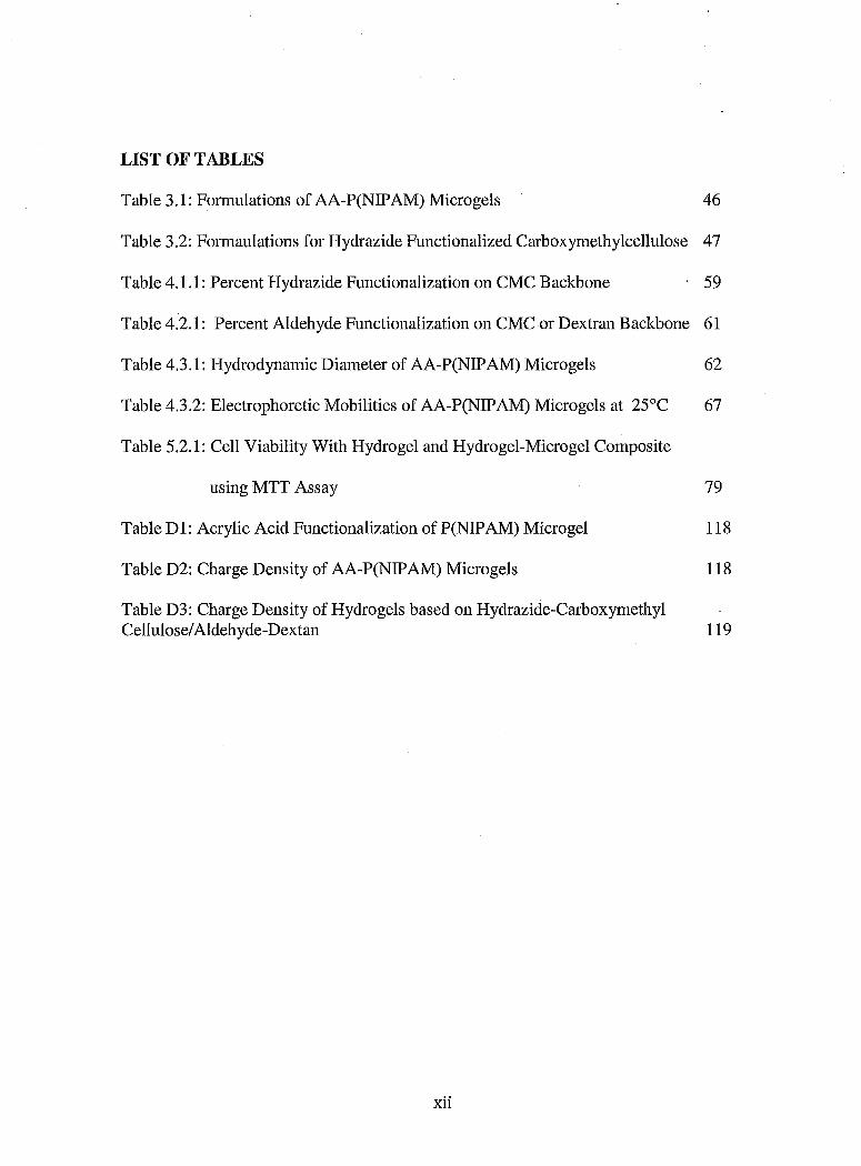
LIST OF TABLES
Table 3.1: Formulations of AA-P(NIPAM) Microgels 46
Table 3.2: Formaulations for Hydrazide Functionalized Carboxymethylcellulose 47
Table 4.1.1: Percent Hydrazide Functionalization on CMC Backbone 59
Table 4.2.1: Percent Aldehyde Functionalization on CMC or Dextran Backbone 61
Table 4.3.1: Hydrodynamic Diameter of AA-P(NIPAM) Microgels 62
Table 4.3.2: Electrophoretic Mobilities of AA-P(NIPAM) Microgels at 25°C 67
Table 5.2.1: Cell Viability With Hydrogel and Hydrogel-Microgel Composite
using MTT Assay 79
Table D 1: Acrylic Acid Functionalization of P(NIP AM) Microgel 118
Table D2: Charge Density of AA-P(NIPAM) Microgels 118
Table D3: Charge Density of Hydrogels based on Hydrazide-Carboxymethyl Cellulose/Aldehyde-Dextan 119
xii
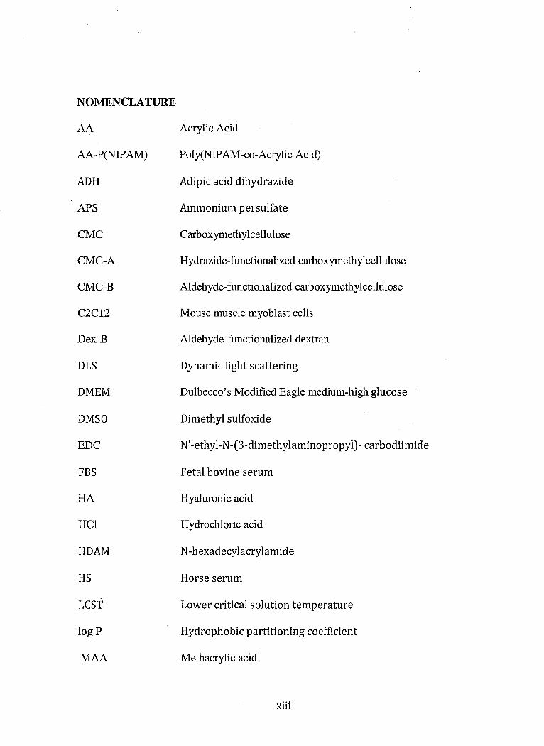
NOMENCLATURE
AA
AA-P(NIP AM)
ADH
APS
CMC
CMC-A
CMC-B
C2C12
Dex-B
DLS
DMEM
DMSO
EDC
FBS
HA
HCI
HDAM
HS
LeST
log P
MAA
Acrylic Acid
Pol y(NIP AM -co-Acrylic Acid)
Adipic acid dihydrazide
Ammonium persulfate
Cm"boxymethylcellulose
Hydrazide-functionalized carboxymethylcellulose
Aldehyde-functionalized carboxymeth ylcellulose
Mouse muscle myoblast cells
Aldehyde-functionalized dextran
Dynamic light scattering
Dulbecco's Modified Eagle medium-high glucose
Dimethyl sulfoxide
N' -ethyl-N -(3-dimethylaminopropyl)- carbodiimide
Fetal bovine serum
Hyaluronic acid
Hydrochloric acid
N -hexadecylacrylamide
Horse serum
Lower critical solution temperature
Hydrophobic partitioning coefficient
Methacrylic acid
xiii

MBA N,N'-methylene bisacrylamide
MTT Thiazolyl bluetetrazolium bromide
NaCl Sodium chloride
NaOH Sodium hydroxide
NHS N -Hydroxysuccinimide
NIPAM N -Isopropylacrylamide
PAA Poly(acrylic acid)
PBS Phosphate-buffered saline
pes Photon correlation spectroscopy
PEG Polyethylene glycol
PEO Poly(ethylene oxide)
PGLA Poly(lactic-co-glycolic acid)
P(NIPAM) Poly(NIP AM)
PPO Poly(propylene oxide)
PS Penicillin Streptomycin
RGD Arg-Gly-Asp peptide sequence
TNBS Trinitrobenzenesulfonic acid
VAA Vinyl acetic acid
VPTT Volume phase transition temperature
3T3 Mus musculus mouse cells
xiv
MASc Thesis - D. Sivakumaran McMaster University - Chemical Engineering
Chapter 1: Introduction
In recent years, there has been significant progress in the development of .
biotechnology tools for addressing human health challenges, including the development
of artificial organs, tissue engineering strategies, and drug delivery vehicles. Polymeric
materials have been key to many of these advances. In particular, the development of
novel biomaterials has become essential in the administration of drugs to patients.
Formulating drugs with biomaterials allows for long-term controlled release of drug,
significantly improving patient quality of life by affording more control over drug
administration. Of particular interest is the development of 'smart' materials that have
the potential to dynamically gauge the dose of drug delivered as a function of a physical
or biological stimulus (Hoare and McLean, 2006). Such materials would allow drug
delivery to be directly correlated with the patient's need for that drug. However, there
have been challenges in creating 'smart' materials that can alter the rate of drug release
based on environmental stimuli. The aim of this work is to investigate the use of 'smart'
hydrogels, microgels, and micro gel-hydrogel composites as potential biomaterials for
drug delivery.
The use of hydrogels as a drug delivery vehicle has been explored thoroughly.
Hydrogels are water soluble polymer networks that swell in an aqueous environment.
They are often physically and mechanically similar to the extra-cellular matrix of cells,
making them particularly favourable for use in the body. They can be made of a variety of
polymers such as dextran, hydroxyethylmethacrylate, and carboxymethycellulose
1

MASc Thesis - D. Sivakumaran McMaster University - Chemical Engineering
(Barbucci et al., 2000). Hydrogel delivery systems are advantageous due to their
biocompatibilityas well as their tunable affinity for dmgs. Obstacles with their use
include the quick elution of dmgs from the highly swollen hydrogel matrix and the
inability for macroscopic hydro gels to be injected into the body.
Materials at the micro or nanoscale offer an even wider range of potential
applications than traditional macro-scale biomaterials. In particular, microgels, hydrogel
particles with nanoscale dimensions, have great potential for dmg delivery. Microgels are
formed by water-soluble polymers and have a porous gel microstmcture consisting of a
cross-linked polymer network. Spherical microgels made from poly(N
isopropylacrylamide) (P(NIP AM)) are materials of particular interest given that the
effective diameter and water content of the micro gels decrease dramatically at ~32°C
(Pelton, 2000). When comonomers are polymerized into the gel network, the temperature
range of the phase transition can be altered according to the hydrophilic or hydrophobic
characteristics of the comonomer. This change in size due to temperature fluctuation can
lead to the development of smart materials. From previous research, it was shown that the
phase transition of acrylic acid functionalized PNIP AM micro gels would occur at around
35-40DC (Hoare, and Pelton 2004). This allows dmg loading to occur at room temperature
under swollen gel conditions, thus optimizing dmg entrapment by optimizing dmg
diffusion into the gel phase. Subsequently, when placed at body temperature, the
microgels would collapse, reducing the effective diffusion coefficient of dmg through the
microgel phase and slowing dmg release. However, the major disadvantage of microgels
is the nanoscale size of the gels results in relatively rapid dmg release and permits
2

MASc Thesis - D. Sivakumaran McMaster University - Chemical Engineering
consumption by the macrophages and rapid clearance from the injection site to the liver.
Thus, long-term local drug delivery is not possible using micro gels alone.
The aim of the research that follows is to optimize the features of both hydro gels
and micro gels to create an injectable in situ gellable long-term controlled release drug
delivery system. Microgels will be immobilized within an injectable hydrogel network
which is liquid outside the body but gels inside the body. By doing so, we can control
drug release according to the cross-link density, degree of swelling, and the drug affinity
of both the hydrogel and micro gel phases. Furthermore, studies will be made to determine
cytotoxicity as well as structural information regarding the hydrogels and microgels.
3

MASc Thesis - D. Sivakumaran McMaster University - Chemical Engineering
Chapter 2: Literature Review
2.1 Drug Delivery
Medication has become important in our daily lives. The effectiveness of any
given drug depends on several factors. One important factor is the dosage of the drug. An
effective drug would have maximum effects with a minimal amount of drug. However,
even highly potent drugs are not effective if they cannot be delivered to the required site
of action within the body. Thus, the main challenge of modem drug therapy is not
finding more potent drugs but rather finding improved ways to deliver those drugs to the
place required at the rate required inside the body.
There is a very important relationship between the chemical properties of the
medication and its subsequent movement through the body, that drug delivery technique
plays a vital role in the effective use of the drug (Saltzman). The method of drug
administration plays a large role in the pharmacokinetics of the drug in the body. There
are many routes and methods one can use in the administration of medication. The most
well-known are oral medicines such as pills or syrups. However, absorption of the drug
through the intestine may be hindered by the degradation of compounds in the
gastrointestinal tract. Thus, other methods such as injections, lotions, and suppositories
are popular (Saltzman). Injections are particularly effective, as they bypass many
degradation mechanisms and are delivered directly to the target site via intravenous,
4

MASc Thesis - D. Sivakumaran McMaster University - Chemical Engineering
intramuscular or subcutaneous routes. Encapsulation of the drug allows for protected and
timed release of the drug through the body.
Polymers have been primarily used in conjunction with drugs to alter the
limitations and pharmacokinetics of the drugs on their own. Polymer-based drug delivery
systems can allow for controlled and/or sustained release of the drug in the body which
differs than that of the characteristics of the naked agents. A controlled release system
would deliver drugs at a predetermined rate for a specified period of time, with little or no
effects from environmental conditions (Langer). Polymers can be utilized in a variety of
methods to effect controlled drug delivery. The first involves chemical modification of
the drug for attachment to the polymer delivery vehicle. This can be done for several
reasons, such as altering biodistribution or increasing agent solubility (Langer). With
attachment to a polymer, there is an increase in the effective size of the active agent,
changing its diffusion coefficient inside the body. Polymers like polyethylene glycol
(PEG) can also be attached to alter the immunogenic response of the host, impmting
"stealth" properties minimizing recognition of the drug by phagocytes or white blood
cells (Bazile et al.,1995). Polymers can also be used to increase the solubility of poorly
soluble drugs; for example, PEG has been used extensively in the distribution of proteins
and other drugs (Hoare and Kohane, 2008). For polymer conjugates to be effective, the
polymer-drug conjugate must be connected using a reliable linker than can be degraded in
the body either via hydrolysis or specific enzymatic degradation at the target site. This is
5

MASc Thesis - D. Sivakumaran McMaster University - Chemical Engineering
important as in many cases the dmg cannot be absorbed into cells with the delivery
vehicle (Saltzman).
The creation of particles/vesicles made of polymer materials can also be used to
improve dmg delivery. Much like dmg-macromolecule conjugates, factors such as
biodistribution can be altered by encapsulating the dmg relative to a naked dmg agent
alone (Saltzman). However, with particles, there is the added benefit of potentially having
a significantly higher dmg loading capacity. Vesicles can be composed of lipids,
carbohydrates, or other types of natural block copolymers. By using these materials, one
can help ensure maximum biocompatibility, since these materials are routinely found in
the body.
Vesicles or particles can be designed to be targeted by cells for uptake by
controlling their particle size, hydrophobic interactions, or ligand-receptor or antigen-
antibody interactions (Mohanraj and Chen, 2006). This will increase the efficacy of the
dmg while minimizing side-effects. Particle size is important in the absorption of
particles into cells. It has been found that 100nm nanoparticles had a 2.5 times greater
uptake than that of their 111m counterpalts using certain cell lines (Desai et al,1996).
The surface chemistry of the palticles can be used to enhance uptake of the dmg
palticles by cells as well as protect the dmgs from rapid degradation away from the target
site. Eilhanced or specific uptake can be achieved by altering the surface chemistry of the
vesicle or palticle. Direct cell communication can also be achieved with the addition of
certain ligands or antibodies that will interact with cell receptors or antigens to promote
6

MAScThesis - D. Sivakumaran McMaster University - Chemical Engineering
cell uptake. It has been seen that cell interactions can be enhanced with the incorporation
of the peptide RGD (ARg-Gly-Asp). This peptide has been known to bind with integrin
receptors on cell membranes, inducing cell uptake (Alsberg et al, 2001). The addition of
hydrophilic polyethylene glycol (PEG) has been observed to prevent phagocytosis
(Olivier, 2005).
Encapsulation or conjugation of drugs with polymers enables the engineering of
the release rate of that drug from the polymer matrix depending on the physical properties
of both the drug and the polymer. In general, the objective is to tune these properties to
control the therapeutic concentration of the drug in the body. Each drug has a therapeutic
concentration range above which it becomes toxic to the user and below which it is
clinically ineffective (Langer). The drug concentration in the blood stream between these
two extremes is known as the therapeutic window. This is represented graphically in
Figure 2.1.
Time
-Drug-onlyinjections -spikes in concentration
./ Range of safe clinical efficacy
Controtled release formulationconstant release VS. time
Figure 2.1: Clinical Window of Drug Delivery (Reproduced with Permission of Dr. Todd Hoare,
McMaster University)
7
MASc Thesis - D. Sivakumaran McMaster University - Chemical Engineering
In standard dosage systems (such as injections), the drug concentration in the blood
stream increases rapidly, peaks, and is followed by a decline out of the therapeutic
window. Thus, constant re-administration is needed for the patient to receive adequate
treatment. With constant or controlled release systems, the same increase and peak would
occur at point of administration. However, the subsequent decrease would last longer
within the therapeutic window, thus reducing the frequency of re-administration of the
drug (Saltzman). Ideally, therapy could be achieved by a single administration of the drug
delivery vehicle. Other advantages of a controlled delivery system include the ability to
delivery drugs locally to specific body parts or organs, thus lowering the systematic drug
concentration in the body. Local delivery could also lead to preservation of drug
bioactivity, as drugs would no longer be subjected to defense mechanisms in the blood
stream. Added benefits include the reduced need for follow-up care as well as improved
comfort and compliance from patients (Langer).
2.2 Mechanisms for Release
Depending on the type of drug, polymer, or delivery site, there are many different
mechanisms by which drug release can occur. Although many polymers can be used for
drug delivery systems, hydrophobic, non-degradable polymers have been chosen most
frequently. These polymers present the best features for retention of drug and
biocompatibility. They have been used extensively in the creation of drug reservoirs,
trans dermal systems, and matrix delivery systems. The most common mode of drug
release from drug reservoirs and other polymeric controlled release systems is via
8

MASc Thesis - D. Sivakumaran McMaster University - Chemical Engineering
molecular diffusion of the drug into the external media (Saltzman). In each of these
systems, the diffusive release of the drug occurs over the course of three steps. The first
involves the dissolution of the drug into the polymer matrix or slab prior to implantation
in or on the body. Once implanted in the patient, diffusion across the polymer membrane,
out the pores of the matrix, or through the matrix material itself occurs. Lastly, effective
delivery occurs with the dissolution of the drug into the external phase (i.e. bodily fluids
surrounding the drug delivery vehicle) (Saltzman).
Since diffusion is the main mechanism of release, the encapsulating polymer can
be engineered to alter the effective diffusion coefficient of drug through the polymeric
matrix to control drug release. Diffusion can be engineered in part by creating degradable
polymer systems. Degradable polymers may degrade via a bulk erosion or surface erosion
method. In bulk erosion, water infuses through the polymer matrix, uniformly attacking
cross-links until mass degradation occurs (Edlund and Albertsson, 2002). This typically
occurs when water penetration is faster than that of the erosion rate. It is important in
drug release systems that the entire drug content is released prior to the device collapsing
as to prevent a burst in concentration of the plasma drug concentration. Most currently
available degradable drug delivery systems degrade via a bulk mechanism (e.g. PLGA).
Surface erosion, however, is in many ways more favourable, as it results in the gradual
release of polymers at the surface due to low water flow into the hydrogel and the slow
and controlled release of drug. Ideal surface erosion would occur when the erosion rate is
proportional to the external surface area of the device (Edlund and Albertsson, 2002). For
9
MASc Thesis - D. Sivakumaran McMaster University - Chemical Engineering
optimal sustained release, surface erosion should be faster than that of water penetration
into the bulk phase. To achieve this, the polymer should be hydrophobic but have bonds
that are easily hydrolysable. Additionally, for added biocompatibility, the products of
hydrolysis should be easily processed by cells for removal from the body. For surface
eroding systems, the release would be a function of the polymer erosion rate as well as
the diffusion coefficient of drug through the polymer matrix. (Saltzman).
Another factor influencing dmg release from polymeric dmg delivery vehicles is
drug-polymer interactions. Electrostatics can playa large role in rate of release of the
drug from the polymer system. For example, cationic polysaccharides such as cellulose
ethers or guar gums can interact with anionic ibuprofen to regulate drug release
(Rodriguez et. ai., 2003). Similarly, anionic carboxylic groups in hyaluronic acid can
interact with cationic dmgs such as lidocaine (Doherty et aI., 1995). In either case, the
ionic binding of the drug to the polymer will slow the release of the drug from the dmg
polymer system. Covalent bonding between the drug and polymer side groups may take
place, leading to slower release times depending on the increase in the effective dmg
molecular weight andlor the degradation time of the drug-polymer bond. For example,
chitosan and salicylic acid have been reported to react to create salicylate
(Puttipipatkhachorn et aI., 2001). This results in diffusion driven release initially,
followed by controlled release as the salicylate reverts back to salicylic acid for release
into the external environment.
10
MASc Thesis - D. Sivakumaran McMaster University - Chemical Engineering
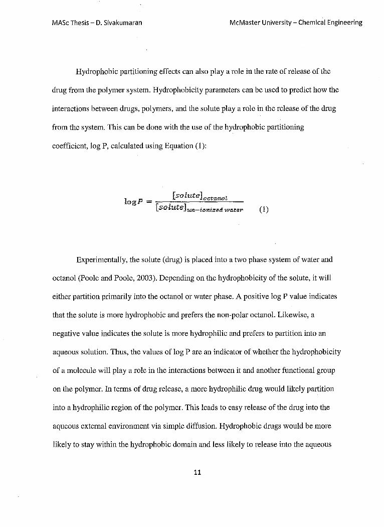
Hydrophobic partitioning effects can also playa role in the rate of release of the
drug from the polymer system. Hydrophobicity parameters can be used to predict how the
interactions between drugs, polymers, and the solute playa role in the release of the drug
from the system. This can be done with the use of the hydrophobic partitioning
coefficient, log P, calculated using Equation (1):
Experimentally, the solute (drug) is placed into a two phase system of water and
octanol (Poole and Poole, 2003). Depending on the hydrophobicity of the solute, it will
either partition primal'il y into the octanol or water phase. A positive log P value indicates
that the solute is more hydrophobic and prefers the non-polar octanol. Likewise, a
negative value indicates the solute is more hydrophilic and prefers to partition into an
aqueous solution. Thus, the values of log P are an indicator of whether the hydrophobicity
of a molecule will playa role in the interactions between it and another functional group
on the polymer. In terms of drug release, a more hydrophilic drug would likely partition
into a hydrophilic region of the polymer. This leads to easy release of the drug into the
aqueous external environment via simple diffusion. Hydrophobic drugs would be more
likely to stay within the hydrophobic domain and less likely to release into the aqueous
11

MASc Thesis - D. Sivakumaran McMaster University - Chemical Engineering
environment (Kang, et ai, 2006). Initial release occurs due to diffusion, but some drug
will stay within the hydrophobic domains. A secondary controlled release may occur at
later stages in the lifetime of the release system via hydrolysis or other mechanisms of
degradation of the release system. Degradation would occur within the core which would
lead invariably to a lowering the number of end groups available for drug-polymer
interaction. With this increase in free volume, more degradation would occur and more
drugs would be released from the system. Thus, a two-stage release profile would ensue.
This partitioning effect can be used as a means of secondary control in the drug release
profile of polymeric drug release systems. (Jeong et al., 2000)
2.3 Hydrogels
There has been substantial progress in the development of controlled release
systems. Hydrogel systems have attracted particular interest. Hydrogels are three
dimensional, cross-linked networks made of water soluble polymers (Hoare and Kohane,
2008). These networks can swell and absorb water, but do not dissolve in water
themselves (Saltzman). They can literally be made from any type of water-soluble
polymer, so their creation can employ a large field of polymers depending on the
specified application. The polymers are largely hydrophilic in nature and can absorb from
10-20% up to a thousand times their dry weight in water (Hoffman, 2002). Depending on
the polymer chosen and the type of crosslinking used to prepare the gel, the hydro gels can
be largely stable or designed to disintegrate or degrade as needed. They can be found in
various morphologies and forms, such as slabs, microparticles, coatings, pressed power
12
MASc Thesis - D. Sivakumaran McMaster University - Chemical Engineering
matrices, and films (Hoffman and Hoare, 2008). Examples of everyday hydrogels include
contact lenses, Jello, and diaper linings. Due to their versatility, they have been used in
various medical applications, including diagnostics, cellular immobilization, and
regenerative medicine (Hoare and Kohane, 2008).
The advantages of using hydrogels in drug delivery systems are numerous. The
high water content allows the hydro gels to be physically, mechanically, and (as desired)
chemically similar to the extracellular matrix of the host cells (Hoare and Kohane, 2008),
leading to generally good biocompatibility. This has been demonstrated with their
successful use in the peritoneum and other in vivo locations (Sutton, 2005). Their soft
nature also allows the hydrogels to protect transported materials that otherwise may be
fragile. The polymers themselves can easily be chemically modified for specific
applications, thus allowing precise tuning of the swelling and interfacial properties of the
hydrogels. Hydrogels can be prepared using both natural and synthetic polymers.
Examples of natural polymers include chitosan, dextran, hyaluronic acid, and collagen
(Sinha et ai, 2003). Synthetic polymers that are commonly utilized include poly(hydroxy
ethyl methacrylate,), poly(vinyl alcohol) poly(propylene oxide)(PPO), or poly(n
Isopropylacylamide) (Dai et ai, 2005).
Hydrogels have a highly porous structure that can be tuned by controlling cross
link density as well as tuning the polymer-environment interactions. The cross-links
between polymer chains determine key hydrogel parameters such as pore size, pore size
distribution, and tortuosity (Hoffman, 2002). In tum, these factors will all influence how
13
MASc Thesis - D. Sivakumaran McMaster University - Chemical Engineering
much the network will swell when the hydrogel is exposed to water. Swelling will vary
the inter-chain separation, which regulates the average pore size of the hydrogel and thus
the rate of drug diffusion through the hydrogel matrix. By controlling the cross-link
extent, one can thus predict how fast the drug can escape the system (Saltzman).
Other factors besides crosslink density can also influence gel swelling equilibrium
and thus the rate of drug release from hydrogel systems. Charged hydrogel networks can
swell via Donnan counterion partitioning effects, increasing the average pore size and
thus the rate of drug release (Hoare and Pelton, 2008). Other factors that govern diffusion
include the drug size and charge interactions between the drug and hydrogel (Hoffman,
2002). Environmental effects can also change the degree of crosslinking andlor the
equilibrium water content within the hydrogel depending on the type of polymers used,
thus leading to tunable hydrogels. For example, for typical hydrogel systems, temperature
increases lead to increases in the mobility of chains, resulting in a greater degree of
swelling of the hydrogel (Bajpai and Mishra, 2004). Thus, the main benefit of using a
hydrogel system is the ability to manage pharmokinetics within the body.
Despite these advantages, one must be mindful of some disadvantages of
hydrogels. Hydrogel materials can be difficult to handle due to their low mechanical
strength. Hydrogels are often difficult to sterilize, which is of great impOltance for in vivo
applications (Hoffman, 2002). Hydrogels also typically exhibit low tensile strength,
which limits their use where load bearing occurs and/or the hydrogel is exposed to high
shear flows. The use of hydrophobic drugs is also limited with hydrogels, as the high
14

MASc Thesis - D. Sivakumaran McMaster University - Chemical Engineering
water content prevents partitioning of the drug into the system during the loading phase.
The macroscopic size of gels also generally requires that moderately invasive measures
be used for implantation in patients, such as subcutaneous implantation operations (Hoare
and Kohane, 2008). However, these limitations can be overcome depending on the nature
of the cross-links used to form the hydrogel network.
Cross-linking of hydro gels can occur via several mechanisms. These cross-linking
approaches are broadly separated into two distinct categories: physical cross-linking and
covalent cross-linking. Physical cross-linking includes hydrophobic interactions, charge
interactions, and hydrogen bonding.
Hydrophobic gelation occurs via the formation of aggregates of hydrophobic
polymers. When this occurs, the hydrophobic domains minimize the hydrophobic surface
area contacting the water, reducing the amount of structured water surrounding the
domains and maximizing the entropy of the system (Hoare and Kohane, 2008). The
formation of the gel is dependent on the polymer concentration, degree of hydrophobicity,
and the chemical structure of the polymer. Examples of such hydro gels include PEO
PPO-PEO tri-block polymer hydro gels (Hoare and Kohane), 2008 and PEO-PPO-PAA
(Cleary et ai, 2003.). Charge interactions can allow for polymer-polymer or polymer-drug
cross-linking to occur, in which different charge domains of polymers interact ionically.
Release would then occur when competing ionic species would degrade the cross-links,
resulting in drug release at the delivery site. Hydrogen bonding can occur between
polymers such as gelatine-agar and starch-carboxymethyl cellulose to enhance the
15

MASc Thesis - D. Sivakumaran McMaster University ~ Chemical Engineering
rheological properties of the mixtures relative to the properties of the polymers alone.
However, these types are hydrogels are typically short-lived in vivo, as influxes of water
will lead to a breakdown of the hydrogen bonds. Indeed, via any physical gelation
technique, the major limitation of physically cross-linked hydro gels is their penchant to
breaking down in water. In addition, physically-cross-linked hydro gels have generally
weaker mechanical strength and are difficult to characterize, given the dynamic nature of
the crosslinking.
8!ockCopolymefs Micen" J>a<:klo9
Colloidal Ctystals
Figure 2.2: Cross-linking of Hydrogels (Reproduced with Permission of Dr. Todd Hoare,
McMaster University)
The shOlt half-lives of most physically-cross-linked hydro gels in vivo can be
resolved with the creation of covalently cross-linked hydrogels. The benefit of covalently
cross-linked hydrogels is the prevention of dilution of the hydrogel matrix and diffusion
of the polymer away from the injection site (Hoare and Kohane, 2008). In addition, the
design of covalently bonded hydrogels is much more flexible than that of their physically-
cross-linked counterparts. This is due to the fact that the properties of physically cross-
16

MASc Thesis - D. Sivakumaran McMaster University - Chemical Engineering
linked hydro gels are directly related to the chemical properties of its gelators (Hoare and
Kohane, 2008).
Hydrogels can be formed via covalent cross-linking using small-molecule
crosslinkers or polymer-polymer cross-linking. In small-molecule cross-linking, reactive
small molecule crosslinkers are added to covalently crosslink polymer chains. This has
been demonstrated with molecules such as genipin (for amino-functionalized polymers)
and glutaraldehyde (for carbohydrate-based polymers), both of which facilitate rapid
gelation. The main problem with small molecule chemistry is that many of the small
molecule additives can prove to be cytotoxic, resulting in a non-biocompatible hydrogel
not amenable to in vivo use (Hoare and Kohane, 2008).
Polymer-polymer cross-linking occurs when reactive side chains are attached to
the polymer backbone. An example of polymer-polymer cross-linking for hydrogel
formation is the process of photopolymerization. Some examples of photopolymerizable
macromers include PEG acrylate derivatives and polyvinyl alcohol derivatives (Nguyen
and West, 2002). However, with photo-cross-linking can lead to a local increase in
temperature when done in vivo, which may damage neighbouring tissues in the body (Jin
et al., 2007). The features of polymer-polymer cross-linking are especially favourable for
injectable in situ gelling. Covalently in situ gellable hydro gels can overcome the toxicity,
injectability, and degradation issues that may arise from standard hydro gels created
outside the body or prepared in situ via the use of small molecules. Several non-toxic
chemistries have been demonstrated. Schiff base cross-links can be formed via the
17
MASc Thesis - D. Sivakumaran McMaster University - Chemical Engineering
reaction of aldehydes and amines. Another example involves the reaction between
aldehydes and hydrazides to create hydrazone bonds. This has been demonstrated with
hyaluronic acid hydrazone cross-linked polymers for anaesthesia delivery (Ito et al,
2007). Michael addition between nucleophiles and vinyl groups is another common
technique that has been used, typically resulting in rapid gelation in under 10 minutes
(Hiemstra et al, 2007).
In most cases, cross-linking chemistries are designed such that the hydro gels will
degrade over a desired time frame inside the body. Degradable hydrogels have many
advantages for drug delivery. The major advantage is the added control of drug release
kinetics as a function of polymer degradation. Furthermore, residual degradation products
from the hydrogels could be absorbed by the body, thus eliminating the need for retrieval
of the device once the drug has been fully released (Saltzman). In all, to create an
effective device, the polymers that are used must degrade in a controllable fashion and
degrade into naturally occurring or inert compounds (Saltzman). Degradation of the
hydrogel occurs when cleavage of the main bonds occurs, resulting in the production of
oligomers. This can be accomplished either chemically (hydrolysis, oxidation) or
biologically (bacteria, enzymes) (Edlund and Albeltsson, 2002).
2.4 Microgels
Microgels are micro/nanoscale polymeric materials that are spherical in nature and
have a cross-linked gel microstructure. Dimensionally, they cart be considered colloidal
particles with average diameters ranging between 50nm and 5/lm (Pelton, 2000). 18

MASc Thesis - D. Sivakumaran McMaster University - Chemical Engineering
However, microgels are not restricted to this size and have been reported with larger
sizes. The structure of micro gels can be typically described as porous gel particles that
can be homogenous in nature or consist of different copolymers (Pelton, 2000). A
schematic of the typical micro gel structure is found on Figure 2.3 .
• Monomer Units
•• I .• CrossUnker
AIIII Colloidal , Stabilizer
Figure 2.3: Microgel Particle (Reproduced with Permission of Dr. Todd Hoare, McMaster
University)
On the macroscopic level, micro gels behave much colloidal particles which are
colloidally stable but can aggregate depending on salt concentration. On the microscale,
microgels have the ability to swell in response to their solvent environment (Saunders and
Vincent, 1999). Microgel swelling is dependent on balances in polymer/polymer
interactions as well as polymer/environment interactions, with all the same factors
contributing to hydrogel swelling also influencing microgel swelling (Pelton, 2000). Key
micro gel properties impOltant for applications include the extent of swelling in response
to a stimulus, the cross-link density, and the characteristic time constants of swelling and
shrinking (Pelton, 2000). Typical analysis of microgels can be done with photon
correlation spectroscopy (peS), dynamic light scattering (DLS), or electrophoresis
19

MASc Thesis - D. Sivakumaran McMaster University - Chemical Engineering
(Saunders and Vincent, 1999). Applications involving micro gels have included drug
delivery, diagnostics (Hoare and Kohane, 2008), the surface coatings, printing, and other
industrial processes (Saunders and Vincent, 1999).
Recently, much attention has been brought to the development of "smart"
microgels. These microgels are able to respond to environmental stimuli, resulting in
large changes in physical properties such as swelling characteristics, cross-link density,
and time constants for swelling and shrinking as a function of a stimulus (Pelton, 2000).
In addition, the physical changes observed should be reversible as to be able to regulate
the swelling changes in both directions (Roy and Gupta, 2003). The degree of
reversibility is dependent on the polymer structure, polymer functionality, and the
microgel's ability to dynamically adjust its nanostructure depending on stimuli (Roy and
Gupta, 2003). Various polymers have been developed to respond to various types of
stimuli. Sensitivity to pH can be achieved with the used of poly(propyl acrylic acid) or
poly(N,N9 -diethylaminoethyl meth acrylate), in which ionization occurs on the polymer
backbone (Qiu and Park, 2001). Light sensitivity can be used as a stimulus when
polyacrylamide is crosslinked with 4-(methacryloylamino )azobenzene (Roy and Gupta,
2003). Modifications to swelling can be dictated with the use of electric fields and
electroporation with micro gels that contain lipid bilayers (Kiser et ai, 2000). Magnetic
field sensitive polymers can be achieved by the incorporation of ferromagnetic
nanopalticles within the soluble region of the micro gel network (Roy and Gupta, 2003).
Temperature sensitive microgels are also possible with the use of poly(N-
20
MASc Thesis - D. Sivakumaran McMaster University ~ Chemical Engineering
isopropylacrylamide) (P(NIPAM)) or other similar N-alkylacrylamide-based polymers
(Pelton, 2000).
In all these cases, the swelling (or uptake of water) have similar mechanisms;
specifically, the affinity of water for the polymer changes as a function of the applied
stimulus. In the case of pH sensitive microgels, the ionization of basic or acidic
functional groups (such as carboxylic acids) can lead to changes in the swelling
characteristics due to Donnan equilibrium. In tum, more water would be allowed to either
enter or exit the microgel stmcture. Temperature sensitivity can be achieved by allowing
conformational changes to occur below and above a volume phase transition temperature
(VPTT) (Shibayama).
2.4.1: N-isopropylacrylamide (NIPAM)
The thenllosensitive class of "smart" polymeric microgels is among the most
intriguing materials being researched today. In particular, much interest in the field has
been focused on the development and use of poly(N-isopropylacrylamide) microgels
(Pelton 1986). The main motivator to this interest is P(NIPAM)'s ability to demonstrate
inverse solubility upon heating, which is the opposite behaviour to how most polymers
react at standard temperature and pressure. The linear version of the polymer has a base
stmcture as seen in Figure 2.4. P(NIP AM) contains two primary domains; the hydrophilic
amide bonds and the hydrophobic isopropyl stmcture.
21

MASc Thesis - D. Sivakumaran McMaster University - Chemical Engineering
Figure 2.4: N -Isopropylacrylamide (NIP AM) (Reproduced with Permission of Dr. Todd Hoare,
McMaster University)
The behaviour of the polymer in a solvent is determined by the interactions of the
polymer with itself as well as with the solvent. In particular, aqueous solutions allow for
hydrogen bonding to occur and are excellent at the pmtial structuring of polymers in
solution. The ordering of P(NIP AM) is dictated by the specific orientation of water
around the hydrophobic domain of the isopropyl group. It is best described by the Gibbs
free energy of the system which is represented by equation 2 below:
I1G = I1H - TI1S (2)
Here, G is the Gibbs free energy, H is enthalpy, T is temperature and S is the
entropy of the system. The cage-like structuring of water around the hydrophobic
isopropyl group in the polymer, known as the hydrophobic effect, results in decreased
entropy (a negative I1S) upon mixing (Schild and Tirrell, 1991). The cage structure also
forbids polymer-polymer interactions from occurring. Upon heating, the temperature-
entropy term (T I1S) is increased until a positive term is obtained. This results in the
22

MASc Thesis - D. Sivakumaran McMaster University - Chemical Engineering
entropy term dominating the hydrogen bonding interactions, thus cancelling the enthalpy
interactions (~H) between water and the amide, in which dissolution occurs. Entropy
primarily drives the ordering of the isopropyl group with other isopropyl groups and
allows for hydrophobic interactions to occur at which point full polymer-polymer
interactions transpire resulting in precipitation (cloud point) of the polymer (Pelton,
2000). This temperature point of phase separation is known as the lower critical solution
temperature (LSCT); the polymer is soluble below the LCST and insoluble above the
LCST. For P(NIPAM), the LCST can occur at temperatures between 15°C and 50°C, but
typically occurs at 32°C. (Pelton, 2000).
The LCST of P(NIP AM) can be adjusted with the addition of additives,
copolymers, or via the functionalization of the base polymer. Depending on the
hydrophobic/hydrophilic nature of the additive, the LCST can be altered. For instance, the
addition of a hydrophobic monomer/molecule, such as N-hexadecylacrylamide (HDAM),
can lead to a lowering of the LCST (Schild and Tirrell, 1991). Likewise, an increase in
hydrophilic domains, for example via copolymerization of vinyl acetic acid leads to an
increase in the LCST (Hoare and Pelton, 2004). This ability to shift temperatures allows
for the design of specifically "smart" materials depending on the application.
The first microgels ofP(NIPAM) were discovered in 1978 and consisted of a
highly monodisperse colloid dispersion. This micro gel was found to have a LSCT in
water of approximately 32°C. However, with gels, there is no distinct cloud point of
precipitation. Gels and microgels exhibit volume change behaviour, (Shibayama and
23

MASc Thesis - D. Sivakumaran McMaster University - Chemical Engineering
Tanaka, 1993) over a range of temperatures at the VPTT. In most cases, the LSCT and
VPTT ofpoly(NIPAM) (linear and gel) differs by ~1-3°C, with the gel exhibiting a
higher temperature due to the presence of the cross-linker. This volume change is
represented physically by the swelling and des welling of micro gels. Like linear polymers,
P(NIP AM) microgels can have their VPTT altered based on copolymerization or
functionalization. Copolymerization can lead to shifting of the VPTT due to changes in
the relative hydrophilicity/hydrophobicity of the gel matrix. There has been much
literature regarding the copolymerization of NIP AM, with extensive research being
conducted with carboxylic acid containing monomers such as acrylic acid (AA),
methacrylic acid (MAA), or vinyl acetic acid (V AA) (Hoare and Pelton, 2008)
Carboxylic acids are ionisable, leading to an increase in VPTT upon ionization, and
provide an electrophilic reaction site for polymer modification. Thus, carboxyl groups can
allow for P(NIP AM) gels to be tunable as a function of both temperature and pH.
Research has shown that the microstructure of such microgels would predominately be
that of a random co-polymer with carboxylic acid groups dispersed throughout the
P(NIP AM) regions. There is very little evidence of many block regions of carboxylic acid
groups (Hoare and Mclean, 2006). However, with V AA, the microstructure would
resemble that of a core-shell particle, with an internal NIP AM core and carboxyl groups
on the outer shell. Aside from providing a tunable effect on the VPTT, comonomers or
functionalized groups can also provide a basis for attachment of small molecules to the
microgel via covalent bonding or further post-modification depending on the final
application of the micro gel.
24

MASc Thesis - D. Sivakumaran McMaster University - Chemical Engineering
P (NIP AM) microgels are typically synthesized via a mixed precipitation
emulsion polymerization reaction, as described by Pelton. Polymerization is conducted in
water until the polymer reaches a critical molecular weight, at which point it undergoes a
phase transition and deposits on to the growing micro gel particles. The gels are
electrostatically stabilized by a surfactant concentration below the critical micelle
concentration. Initiation is achieved using a persulfate initiator.
Applications for P(NIP AM) micro gels are numerous due to their ability to
reversibly respond to temperature changes. Interesting applications include the use of
P(NIPAM) micro gels as a flocculants in oil reservoirs above the VPTT. Microgels can
also be used as rheological modifiers, taking advantage of their dynamically-tunable
water content as well as their ability to aggregate and create substantially larger particles
that would slow the flow of the solution, leading to a higher viscosity (Bokias et al,
2000). The creation of colour-tunable colloidal crystals is another interesting application.
With temperature changes, the Bragg diffraction of light through P(NIP AM) colloidal
crystals can be changed. Thus, with changes to the structure and resulting size of the
microgel, the Bragg peak and thus the colour can be tuned (Das et al.,2006). The use of
microgels can be further extended to the use of micro-optical arrays. It has been shown
that P(NIPAM) microgels have good affinity for celtain substrates (such as gold) and thus
can be attached to a variety of surfaces. By applying light to a lens shaped microgel, the
focal length can be altered due to the radiation-induced heating, leading to better focusing
25

MASc Thesis - D. Sivakumaran- McMaster University - Chemical Engineering
for the array. Given the relatively fast response time of microgels through the VPTT, the
creation of dynamic tunable optical devices is very promising.
Another exciting application involves the use of P(NIP AM) micro gels as a
delivery vehicle for small molecules and macromolecules. Their small size allows for a
greater surface area thus fast uptake and release of molecules (Sanders). Rates of delivery
can be adjusted by the use of the VPTT, as this is a tunable feature depending on the
application. In particular, the use of PNIP AM) as a delivery vehicle is realistic with
applications for biotechnology and drug delivery. P (NIPAM)'s ability to react to changes
in temperature allow for temperature-triggered drug release. It has been shown that rates
of diffusion out of the gel will be higher below the VPTT since the increased water
content and open pore space between polymer chains allow for low resistance to molecule
movement. Above the VPTT, the micro gel would collapse, providing higher resistance to
molecules exiting the gel. This has been demonstrated with both fluorescein-labelled
dextran and salicylamide, among other drugs (Snowden, 1992). Microgels can also be
modified to have certain functional groups so that specific drug-polymer interactions can
occur. This has been shown with hydrophobic/hydrophilic interactions as well as charge
interactions (Hoare and Pelton, 2008). Likewise, controlled release can be obtained by
"turning off' these interactions through a modification to environmental pH or
temperature. Drug loading into the gels can also be enhanced because of the temperature
sensitivity of the gels. Drug loading can be conducted in the swollen state, leading to
facile drug diffusion into the micro gel and optimal entrapment. When placed in the body,
26

MASc Thesis - D. Sivakumaran McMaster University - Chemical Engineering
as long as the VPTT is below physiological temperature, drug release can be slowed in
the collapsed state of the gel (Pelton, 2000).
Cytotoxicity of P(NIP AM) has been observed as minimal at low concentrations
over a range of temperatures. It is predicted that this will result in low in vivo toxicity.
However, not much is known about bioaccumulation and physiological responses to
degradation products in the body. Approaches in which known hydrolysable bonds can be
used to fraction the polymer in known locations may be a suitable remedy for this issue.
2.5 Composite Hydrogels
The proven benefits of microgels and other pmticle-based drug delivery systems
offer a potential solution to many of the limitations of bulk hydrogels. However, the high
mobility of micro gels in vivo and the fast release rates from micro gels present challenges
that are not problematic in bulk hydrogel systems. By combining microgels and hydrogels
together to form soft nanocomposite hydrogels, also known as "plum pudding" hydrogels,
the limitations of both micro gels and hydro gels can both be mitigated (McGillicuddy et
al., 2006). In such systems, the drug-loaded micro gels would be entrapped within the
hydrogel matrix as seen in Figure 2.5.
27

MASc Thesis - D. Sivakumaran McMaster University - Chemical Engineering
Figure 2.5: Microgels imbedded within Hydrogel Network (Reproduced with Permission of Dr.
Todd Hoare, McMaster University)
Composite hydro gels offer several benefits from a biomedical engineering
perspective. The hydrogel can mask any issues regarding particle biocompatibility, as the
particles will be able to "hide" within the hydrogel system. The bulk hydrogel network
prevents migration of the microgels from target sites (Hoare and Kohane, 2008). The
hydrogel network also provides an additional diffusive barrier for drug release, offering
potential for the generation of novel drug release profiles. As an example, the burst effect
often seen in microgel-based drug release could be mitigated in a composite hydrogel
system. Composite hydro gels also offer the unique potential to independently engineer
both the hydrogel and micro gel phase to optimize the drug release profile. This will allow
for total control of how drug can be released and allow for tunable features to be used.
28

MASc Thesis - D. Sivakumaran McMaster University - Chemical Engineering
Microgels can also be functionalized to cross-link with the hydrogel to give added
stability and prevent any migration of micro gels out of the hydrogel matrix.
There are several examples of the use of composite hydro gels for dmg delivery
currently in literature. Liposomes have been entrapped within carbopol and
hydroxyethy1cellulose-based hydrogel and can control the release of ca1cein depending on
the liposome membrane characteristic. Micropartic1es have been embedded within same
polymer hydro gels to delivery dmgs. Microgels made of N-isopropylacrylamide-N-tert
butylacrylamide-acrylic acid entrapped in dimethylacrylamide hydro gels can release
rhodamineB at a controlled speed (Lynch et ai, 2005). Hydrogel films made of N
isopropylacrylamide-N-tert-butylacrylamide and incorporated with the same polymer
micro spheres have been observed to release controlled rates of fluvastatin (McGillicuddy
et al., 2006). However, significant oppOltunities still exist for intelligently engineering the
compositions, degradabilities, and swelling properties of each phase to achieve improved
dmg release profiles.
2.6 Materials Used
The materials chosen for use in designing soft composite hydrogels in this thesis
were carboxymethy1cellulose, dextran, acrylic acid functionalized (AA-P(NIPAM)), and
bupivacaine. The materials chosen had to be easily modifiable, biocompatible, and allow
for easy analysis of results. The materials chosen have all had previous literature
regarding their use as hydrogels (carboxymethy1cellulose, dextran), micro gels (AA-
29

MASc Thesis - D. Sivakumaran McMaster University - Chemical Engineering
P(NIPAM)), and drug targets (bupivacaine), indicating that their use will be relevant for
future drug delivery systems.
2.6.1 Carboxymethyl Cellulose
Carboxymethyl cellulose is a linear ionic ether derivative of traditional cellulose.
It consists of ~-linked glucopyranose residues (Li et at, 2007). It differs from cellulose
with the addition of carboxymethyl groups on some of the cellulose hydroxyl groups.
Substitution of the groups ranges in between 25-75% per disaccharide unit of cellulose
(Barbucci et aI, 2000). The structure is found below in Figure 2.6. Synthesis is commonly
done by the alkali-catalyzed reaction of cellulose with chi oro acetic acid. It can be
purchased in several different molecular weight ranges, with common sizes ranging
between 200 kDA and 700 kDa. Even at high molecular weight, it is a highly water
soluble anionic compound that is chemically stable. Its most common form is that of a
powered salt. This polysaccharide has been widely used as an emulsifying agent in
pharmaceuticals, cosmetics, and in the food industries (Barbucci et aI, 2000). In the
biomedical field, it has been used to prevent epidermal scarring and tissue adherences
(Barbucci et aI, 2000). However, its potential for use as a hydrogel is promising in the
realm of drug delivery.
30
MASc Thesis - D. Sivakumaran McMaster University - Chemical Engineering
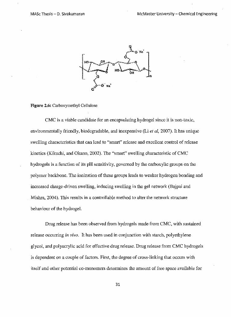
Figure 2.6: Carboxymethyl Cellulose
CMC is a viable candidate for an encapsulating hydrogel since it is non-toxic,
environmentally friendly, biodegradable, and inexpensive (Li et ai, 2007). It has unique
swelling characteristics that can lead to "smart" release and excellent control of release
kinetics (Kikuchi, and Okano, 2002). The "smart" swelling characteristic of CMC
hydrogels is a function of its pH sensitivity, governed by the carboxylic groups on the
polymer backbone. The ionization of these groups leads to weaker hydrogen bonding and
increased charge-driven swelling, inducing swelling in the gel network (Bajpai and
Mishra, 2004). This results in a controllable method to alter the network structure
behaviour of the hydrogel.
Drug release has been observed from hydro gels made from CMC, with sustained
release occUlTing in vivo. It has been used in conjunction with starch, polyethylene
glycol, and polyacrylic acid for effective drug release. Drug release from CMC hydro gels
is dependent on a couple of factors. First, the degree of cross-linking that occurs with
itself and other potential co-monomers determines the amount of free space available for
31

MASc Thesis - D. Sivakumaran McMaster University - Chemical Engineering
drug diffusion as well as the amount the gel may swell. Cross-linking can occur with
several agents such that modifications can be made on the hydroxyl or carboxyl groups of
the polymer (Bajpai and Mishra, 2004). Another factor is the dosage amount placed in the
polymer hydrogel. Various amounts will lead to differences in the release profile, with
higher concentrations resulting in faster release from the system due to diffusion effects
(Ikechukwu et at, 2000). Drug release has been observed from acrylamide-functionalized
carboxymethyl cellulose hydro gels, with swelling behaviour determining the rate of drug
release (Kulkarni and Sa, 2008).
2.6.2 Dextran
Dextran is a branched homopolysaccharide of glucose that contains mainly a-(1,6)
linkages of D-glucopyranose residues (Zhang et at, 2005). These types of linkages
constitute 50-97% of the total linkages (Naussens, et al. 2005). However, some chains
will also contain branched a-(1,3) and a-(I,4) linkages depending on production. The
structure of dextran is shown below in Figure 2.7. Molecular weights can vary but are
typically in the range of 10-150 kDa (Naussens et at, 2005). Production of dextrans is
routinely done via fermentation of sucrose by several different types of bacterial species.
These species include Leuconostoc, Streptococcus and Lactobacillis. All of these species
will synthesize dextran by excreting dextransucrase enzymes on sucrose media for
conversion (Nauss ens et at, 2005). Dextran has many applications in the food industry,
for photofilm manufacture, and as a soil conditioner (Shamala and Prasad, 1995). Its main
32

MASc Thesis - D. Sivakumaran McMaster University - Chemical Engineering
biomedical use is as a blood thinner since it is retained in the blood stream much better
than crystalloids which penetrate into vascular membranes .(Naussens et aI, 2005).
Figure 2.7: Dextran Molecule
'0 l
·.~ .. ~O
.. ~o OH I
.. ~/ ... L .•.. ~ ............ 'Q ... . HO t~ ... ,
H t
As a potential hydrogel for a dmg delivery device, dextran has great promise since
it has already been well-established as a highly biocompatible material in medicine (Chiu,
1999). Dextran hydrogels show low protein absorption and water soluble versions can
have molecular weights below 30kDa, which can allow for excretion through the kidneys
(Hiemstra et aI, 2007). Dextran exhibits low toxicity and high enzymatic degradability in
the body, which could lead to the development of a biodegradable polymer (Chiu, 1999).
Of great impOltance is the abundance of hydroxyl groups on the glucose resides, which
means side chains can easily be modified to make dextran a tunable polymer.
There have been several reported examples of dextran-based hydrogels. Many of
the developed hydro gels are homologous in nature with different crosslinking methods.
Michael addition chemistry has been used with vinyl sulfone-functionalized dextrans to
create degradable hydrogels (Heimstra et aI, 2007). Carboxymethyl functionalized
33
MASc Thesis - D. Sivakumaran McMaster University - Chemical Engineering
dextran has been created with N' -ethyl-N-(3-dimethylaminopropyl)- carbodiimidel N-
Hydroxysuccinimide (EDCINHS) chemistry to create "zero-length" ester cross-links
between carboxylic acid and hydroxyl groups (Zhang et al, 2005). Other dextran
hydro gels contain dextran in conjunction with other biocompatible materials to create
copolymers blends. Examples of dextran-based copolymer hydro gels include dextran-
HEMA, dextran-methacrylate copolymers (Van Dujk et al, 1997). In these cases, drug
release from the hydro gels has been demonstrated and release rate has been controlled.
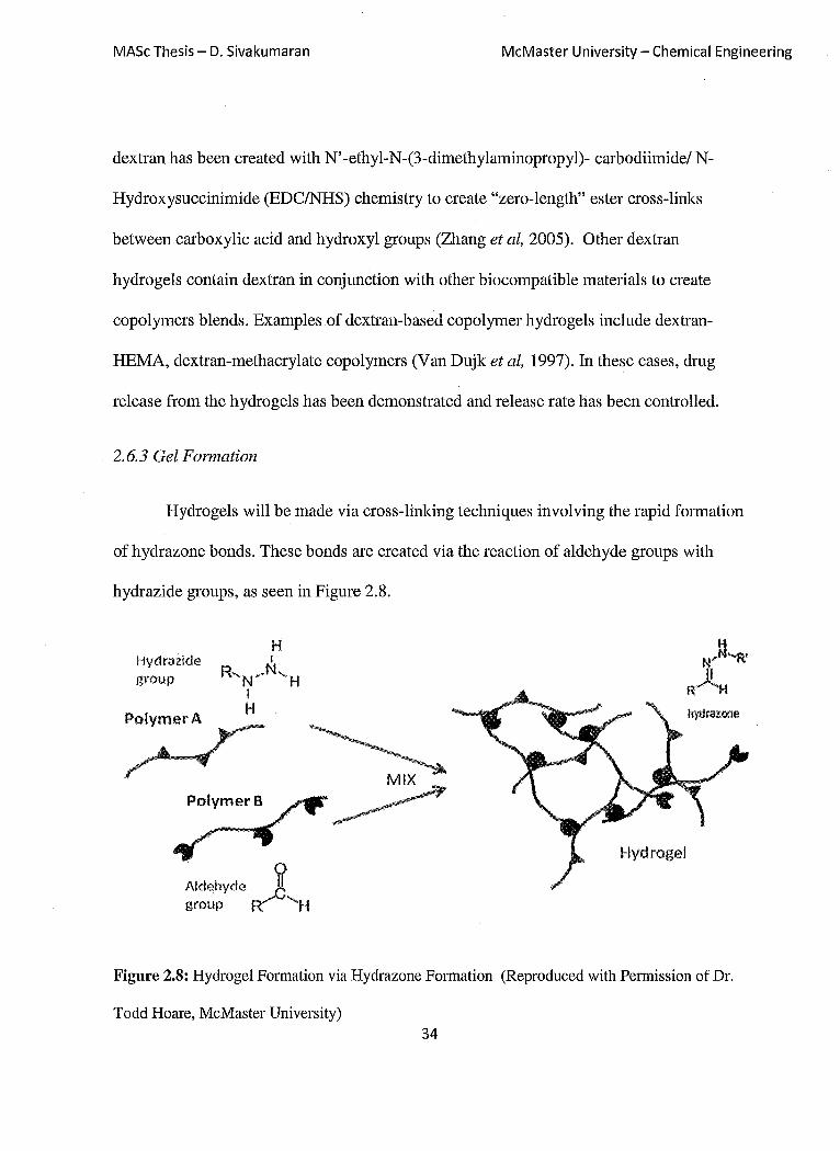
2.6.3 Gel Formation
Hydrogels will be made via cross-linking techniques involving the rapid formation
of hydrazone bonds. These bonds are created via the reaction of aldehyde groups with
hydrazide groups, as seen in Figure 2.8.
H Hydrazide I
R'N/N'H I
group
::::r PolymerB
Aldehyde 8' group R/ 'H
Hydrogel
NJJ"'R' R)lH hydrazone
Figure 2.8: Hydrogel Formation via Hydrazone Formation (Reproduced with Permission of Dr.
Todd Hoare, McMaster University) 34

MASc Thesis - D. Sivakumaran McMaster University - Chemical Engineering .
Hydrazones are created via a rapid condensation reaction between the aldehyde and
hydrazide groups, resulting in the release of water from the reaction. Carboxymethyl
cellulose will be functionalized with hydrazide groups via NHSIEDC chemistry using
adipic acid dihydrazide (pKa -3.4 from potentiometric titration, see Appendix D). The
associated aldehyde functionalized polymers will consist of either carboxymethyl
cellulose or dextran, modified to contain aldehydes via sodium periodate oxidation
(Figure 2.9). With either component, an injectable in-situ gelling hydrogel will be
prepared upon mixing the two functionalized polymers. It must be noted that hydrazone
bonds can undergo degradation.
Figure 2.9: Oxidation of Dextran for Aldehyde formation
However, the erosion of the bonds is a slow acid-labile reaction that results in hydrolysis
(Nathan et at., 1996). Hydrazone degradation has been used as a controllable method to
control the rate of degradation for hydro gels such as ones based on poly(aldehyde
guluronate) (Lee et ai., 2000). Degradation is mediated by a bulk erosion process and is
35
MASc Thesis - D. Sivakumaran McMaster University - Chemical Engineering
dependent on the cross-link density of the hydrogel. Low cross-linked hydro gels tend to
erode faster than that of hydrogels that are more cross-linked. In addition, during
degradation, it is possible for the hydrazide bonds to re-cross-link with other aldehyde
groups. The probability of this occurring increases for heavily cross-linked hydrazone
hydrogels, which explains the slow degradation kinetics of highly cross-linked gels (Lee
et al. 2000).
2.6.4 Poly(NIPAM-co-Acrylic Acid)
Poly(NIPAM-co-acrylic) microgels were used for several reasons in the
development of the hydrogel-micro gel composites. From previous research conducted by
Hoare, AA-P(NIPAM) can be tuned for specific applications with modifications to
functional monomer content, functional group distribution and changes in micro gel size.
Loading of drug has been proven to occur on the basis of the anionic micro gels being able
to bind onto cationic molecules. Furthermore, loading of drug into the micro gel matrix
can be differed based on temperature and functional group distributions. Lastly, AA
P(NIP AM) microgels can be further modified as to be able to cross-link with the hydrogel
portion of the composite if necessary.
2.6.5 Bupivacaine Hydrochloride
The management of pain in a patient after surgery is impOltant in the
improvement of quality of life. Post-operative pain is found to be significant and is one of
the primary concerns of patients admitted to the intensive care unit. Most analgesic
36

MASc Thesis - D. Sivakumaran McMaster University - Chemical Engineering
methods to alleviate pain center around intravenous injections that must be carried out by
the patient or by the nurse every few hours (White et al.). Thus, developing strategies for
reducing the frequency of drug administration and dosage for pain management via the
development of controlled release vehicles is an important step toward improving patient
care.
Bupivacaine is a cationic local anaesthetic that blocks the conduction of nerve
impulses by decreasing nerve membrane permeability to sodium (Lexi-Comp). It is used
mainly in pain management. In solution it behaves like a weak acid and has a pKa of 8.2.
Its solubility is ~40mg/mL in acidic conditions, with solubility dropping 1000 times in
basic conditions (Shah and Maniar, 1993). The structure is seen in Figure 2.10.
Figure 2.10: Bupivacaine Hydrocholride
Bupivacaine gives typically long-lasting anaesthesia effects, with recorded
therapeutic times being four to nine hours when injected in solution. It is generally used
to moderate post-surgical pain in both medicine and dentistry. It can be administered via
central routes such as spinal or epidurals (Le Cone et al, 1994). It can also be
administered via dextrose injection or in combination with epinephrine (Shah and Mania,
37
MASc Thesis - D. Sivakumaran McMaster University - Chemical Engineering
1993). Although considered long lasting, bupivacaine therapy only routinely uses
concentrations that are low in nature varying from 0.5-0.75% due to the myotoxicity of
bupivacaine at higher concentrations (Lexi-Comp). Thus repeated administration is
needed. There has been much research to find ways to deliver fixed amounts of the drug
for extended periods of time culminating in controlled release. Controlled release has
been achieved in a few polymer systems such as polylactide and PLGA microspheres (Le
COlTe, et ai, 1994). Very promising results have been found with the use of P(NIP AM)
microgels, in which the micro gels allow for a higher dose of bupivacaine to be
administered without adverse effects (Hoare and Kohane, unpublished).
Bupivacaine was chosen to due the presence of anionic acrylic groups in the AA
P(NIP AM) microgels. Bupivacaine is cationic at physiological pH, allowing for
electrostatic interactions to take place between the micro gels and the drug, thus
optimizing the load of drug into the hydrogel-micro gel system. In addition, at
physiological pH, there are hydrophobic partitioning driving forces that can act on the
drug. The octanol-water partition coefficient (log P) of NIP AM residues and bupivacaine
are similar. The log P of a NIPAM monomer sub-unit is approximately 0.06. For the
cationic bupivacaine, the log P value is 0.18 (Sudoh et ai, 2004). Since the log P values
are similar, bupivacaine would prefer to partition into the hydrophobic domains of the
micro gel phase at physiological conditions rather than any bodily fluid (Hoare and
Kohane, unpublished).
38

MASc Thesis ~ D. Sivakumaran McMaster University - Chemical Engineering
2.7 Objectives of Research
There has been recent progress in the development of composite hydrogel
networks that contained entrapped micro gel particles within the hydrogel matrix. These
systems have great potential for drug delivery applications for controlled release of drugs
(Yeo). These systems have several desirable features that utilize the benefits of both
hydrogels and microgels. Hydrogel delivery systems are advantageous due to their
biocompatibility as well as their tunable affinity for drugs. Obstacles with their use
include the quick elution of drugs from the highly swollen hydrogel matrix and the
inability for macroscopic hydro gels to be injected into the body. These hydrogels are
routinely gelled and fabricated outside of the body and subsequently implanted.
Microgels based on P(NIP AM) have great tunable responses that can maximize drug
loading and control release and are readily injectable. However, there is typically rapid
sequestering of the microgels into the liver or spleen. We can combat these problems by
creating microgels that can be immobilized within an injectable hydrogel network which
is liquid outside the body but gels inside the body. The goal of this work is to use acrylic
acid-functionalized PNIPAM micro gels combined with in situ-gelling hydrogel materials
to make an injectable long-term drug delivery system for the delivery of local
anaesthetics, aiming to address clinical needs in long-term pain management. The
hydrogels were made of carboxymethyl cellulose (CMC) and dextran (Dex) modified
with hydrazide (CMC-A) and aldehyde (Dex-B, CMC-B) functional groups. When
mixed via co-injection through a needle, these two polymers rapidly form a hydrogel. By
39
MASc Thesis - D. Sivakumaran McMaster University - Chemical Engineering
co-injecting micro gels together with one of the reactive polymers, composite, in situ
geUable hydrogel-microgel networks are formed. The impact of the crosslink density,
degree of swelling, and the ionic dmg affinity of both the hydrogel and micro gel phases
on the release kinetics of bupivacaine were investigated to determine how to engineer the
micro gel-hydrogel composites to achieve optimized dmg release profiles. The effect of
changes to the functional group content of either the micro gel or the hydrogel was also be
investigated to determine the impact of phase functionalization on dmg release. Cross
linking of the micro gels to the hydrogel matrix was also explored to see the effects of
mechanically restricting the micro gel phase transition on dmg release. Rheological
analysis was conducted to find the mechanical strength of these network polymers. Cell
viability tests were also conducted to ensure that these polymer networks and the reactive
gel precursors are not cytotoxic.
40
MASc Thesis - D. Sivakumaran
Chapter 3: Materials and Methods
3.1,' Reagents
Acrylic Acid Functionalized P(NIP AM) Microgels
McMaster University - Chemical Engineering
N-isopropylacylamide (NIPAM), acrylic acid (AA), ammonium persulfate (APS), N,N'
methylene bisacrylamide (MBA), and bupivacaine hydrochloride were all purchased
directly from Sigma Aldrich (Oakville, ON). Sodium Dodecylsulfate (SDS) was obtained
from Bioshop Canada (Burlington, ON).
Hydrazide Functionalized Carboxymethyl Cellulose
Sodium carboxymethyl cellulose (CMC) [MW 250,000] was purchased from Sigma
Aldrich (Oakville, ON). Adipic acid dihydrazide (ADH), N-Hydroxysuccinimide (NHS),
and N' -ethyl-N-(3-dimethylaminopropyl)- carbodiimide (EDC) were purchased from
Sigma Aldrich (Oakville, ON). Dimethyl sulphoxide (DMSO) was purchased from
Caledon Laboratory Chemicals (Georgetown, ON).
Aldehyde Functionalized Carboxymethyl Cellulose and Dextran
Sodium carboxymethyl cellulose (CMC) [MW 700,000], dextran from Leuconstroc spp
[Mr - 500,000], ethylene glycol, and sodium periodate were all purchased from Sigma
Aldrich (Oakville, ON).
41
MASc Thesis - D. Sivakumaran McMaster University - Chemical Engineering
Cell Culture
3T3 Mus musculus mouse cells and C2C12 mouse muscle myoblast cells were acquired
from ATCC: Cederlane Laboratories Ltd. (Burlington, ON). Media contents included
Dulbecco's Modified Eagle Medium-high glucose (DMEM), fetal bovine serum (FBS),
horse serum (HS), and Penicillin Streptomycin (PS), which were obtained from
Invitrogen Canada (Burlington, ON). Recovery cell culture freezing and trypsin-EDTA
were purchased from Invitrogen Canada (Burlington, ON). Thiazolyl Blue Tetrazolium
Bromide (MTT) was purchased from Sigma Aldrich (Oakville, ON).
Additional information regarding reagents is found in Appendix A.
42

MASc Thesis - D. Sivakumaran McMaster University - Chemical Engineering
3.1 Double Barrel Syringe Apparatus
Double barrel syringe supplies were purchased from Medmix Systems
(Switzerland). The double barrel syringes are comprised of four components as seen in
Figure 3.1. The first component 'is the double barrels (1) which have the capability to
house two separate solutions that are to be mixed. The mixer (2) is affixed to the ejection
side of the barrel and is screwed in. The mixer is responsible for the mixing of the two
solutions within the barrels. The mixer can also be affixed with a needle (20 gauge) to
provide further control of the resultant mixed solution/gel. Injection is facilitated by the
double plunger (3) and the o-rings (4).
43
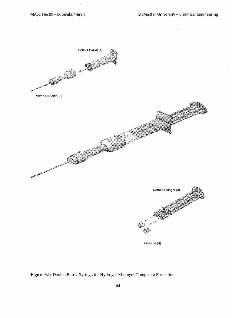
MASc Thesis -D. Sivakumaran McMaster University - Chemical Engineering
Double Barrel (1)
Mixer + Needle (2)
Double Plunger (3)
O-Rings (4)
Figure 3.1: Double Barrel Syringe for Hydrogel-Microgel Composite Formation
44

MASc Thesis - D. Sivakumaran McMaster University - Chemical Engineering
3.3 Acrylic Acid Functionalized Poly(NIPAM) Preparation
N-Isopropylacrylamide monomer was purified with a 60:40 toluene: hexane
mixture. The amount of NIP AM added was equivalent in 1: 1 w/v % ratio with toluene.
The mixture was placed in an oil bath and heated to 60°C. Once dissolved, the solution
was placed in ice bath for 2 hours until recrystallization occurred. The solution was then
filtered to isolate the NIP AM crystals and the recrystallized product was dried under
nitrogen for 12 hours prior to storage.
3.4 Acrylic Acid Functionalized Poly(NIPAM) Polymerization
Formulations for various degrees of micro gel functionalization are found in Table
3.1. Microgels were synthesized based on methods described by Hoare and Pelton.
Polymerizations were done in a three necked flask with magnetic stir bar and attached
condenser. Appropriate amounts of NIP AM monomer, acrylic acid, MBA, and SDS were
dissolved in 150mL Milli-Q water. Polymerization was done at a temperature of 70°C
under nitrogen purge at a mixing rate of 200rpm. After 30 minutes of heating, APS was
dissolved in lOmL of Milli-Q water and added to the mixture to initiate polymerization.
Polymerization was allowed to continue for 12 hours followed by dialysis for
purification. Dialysis was performed against MilliQ water with membrane tubing from
Spectmm Labs (132680) with a molecular weight cut-off of 12,000 - 14,000 Da. A
minimum of 6 dialysis cycles were conducted prior to the solutions being lyophilized and
stored dry for future use.
45

MASc Thesis - D. Sivakumaran McMaster University - Chemical Engineering
Table 3.1: Formulations of AA-P(NIPAM) Microgels
Code NIPAM Acrylic MBA SDS APS (g) + H2O
(g) Acid (g) (g) (g) lOmLH2O (mL)
AA1 (6mol % 1.4 0.058 0.1 0.2 0.1 150
AA)
AA2 (llmol% 1.4 0.116 0.1 0.2 0.1 150
AA)
AA4 (20mol% 1.4 0.232 0.1 0.2 0.1 150
AA)
AA8 (33mol% 1.4 0.464 0.1 0.2 0.1 150
AA)
3.5 Hydrazide Functionalized Carboxymethyl Cellulose Modification (A Polymer)
Formulations for various degrees of hydrazide attachment are found in Table 3.2.
Hydrazide attachment was conducted following methods described by Hudson.
Carboxymethyl cellulose (250kDa) was dissolved in a 200mL Milli-Q water in a 250mL
round bottom flask. A magnetic stir bar at 200rpm was used. Adipic acid dihydrazide was
added, resulting in a solution with a pH of ~ 7. N-hydroxysuccinimide was dissolved in
4mLDMSO:H20 (1:1) and added dropwise to the CMC solution. N'-ethyl-N-(3-
dimethylaminopropyl)- carbodiimide was dissolved in 1mL of 1: 1 DMSO:H20 and added
dropwise to CMC solution. The pH of the resulting mixture was continuously adjusted to
46

MASc Thesis - D. Sivakumaran McMaster University - Chemical Engineering
a pH of 6.8 with 0.1M NaOH. The reaction was permitted to react until the pH became
constant, which took approximately 1 hour. The resulting polymer was dialyzed with
membrane tubing from Spectrum Labs (S632724) with a molecular weight cut-off of
3,500 Da. A minimum of 6 dialysis cycles were conducted prior to the solutions being
lyophilized and stored dry for future use.
Table 3.2: Forrnaulations for Hydrazide Functionalized Carboxymethy1cellulose
Code CMC (g) ADH(g) NHS (g) EDC (g) H20 (mL)
CMC-AO.5 1 3 0.035 0.3 200
CMC-A1 1 3 0.07 0.3 200
CMC-A1.5 1 3 0.10 0.3 200
CMC-A2 1 3 0.13 0.3 200
3.6 Hydrazide FunctionalizedAA-P(NIPAM) Microgels
Recipes and methods are identical to hydrazide-functionalization of CMC.
However, 19 of AA-P(NIPAM) was used instead of 19 of CMC per recipe.
3.7 Aldehyde Functionalized Dextran/Carboxymethyl Cellulose (B Polymer)
Dextran or carboxymethyl cellulose (1.5g, 250kDa) was slowly dissolved in
150mL Mill-Q water in a 250mL round bottom flask. A magnetic stir bar was rotated at
200rpm. Sodium periodate (0.8g) was dissolved in lOmL water, added dropwise to
47

MASc Thesis - D. Sivakumaran McMaster University - Chemical Engineering
polymer solution, and allowed to stir for 2hours. Ethylene glycol (O.4mL) was then
injected into the solution and allowed to react for 1 hour. The resulting polymer was
dialyzed with membrane tubing from Spectmm Labs (S632724) with a molecular weight
cut-off of 3,500 Da. A minimum of 6 dialysis cycles were conducted prior to the solutions
being lyophilized and stored dry for future use.
3.8 Hydrogel-Microgel Composite
Polymer solutions to be used for hydrogel fabrication were prepared in 2mL
micro-centrifuge tubes. In the first tube, CMC-A polymer (40mg) was dissolved in 2mL
of 0.15M saline at a concentration of 2w/v%. In the second tube, 2w/v% of CMC-B or
Dex-B polymer (40mg), 20mg oftotal solution bupivacaine (5mg/mL), and 4w/v% of
AA-poly(NIPAM) micro gels (80mg) were dissolved in 2mL of 0.15M saline.
The micro gel-hydrogel composites were created using double barrel syringes
purchased from Medmix Systems (Switzerland). Each individual barrel contained the
contents of one of the premade solutions from the micro-centrifuge tubes described
above. A 20-gauge needle was affixed to the mixer end of the double barrel syringe to
allow for ease of injection. Upon mixing within the mixer and needle, the resulting
composite was injected into cylindrical moulds prepared from silicone lUbber obtained
from McMaster Carr (Illinois, USA). The moulds had a diameter of 0.95cm and height of
0.32cm, resulting in a disk shaped hydrogel-microgel composite.
48

MASc Thesis - D. Sivakumaran McMaster University - Chemical Engineering
Control hydro gels were made in identical fashion to the procedure described above.
However, bupivacaine was not included in the formulation. Special hydrogel controls
were also made that consisted of just the hydrogel components and contained no micro gel
fraction.
3.9 Hydrogel Characterization
3.9.1 Potentiometric and Conductometric Titration of Hydrazide-Functionalized CM C
Hydrazide attachment to the CMC backbone was primarily determined via
potentiometric titration, as described by Hoare and Pelton (Langmuir, 2004). Titrations
were performed using a Burivar-12 automatic buret (ManTech associates) with PC-Titrate
software (Version 2.0.0.79) for control. Samples of 50mg CMC-A polymers were
dissolved in 50mL of 1O-3M NaCI for titration. Titrations were run in clean 100mL beaker
and temperature controlled at 25°C. The beaker was fitted with overhead mixer,
thermometer, pH electrode and conductivity electrode from ManTech associates.
Nitrogen was purged into the system for 30 minutes to remove dissolved carbon dioxide
from the solution. The pH of the solution was controlled by 0.1M HCI and 0.1M NaOH
from Acculute standards. Prior to titration, the solution pH was lowered to 3.5. With a
gentle nitrogen purge onto the sample to keep an inert atmosphere, data was obtained
with a base into acid titration, using an end pH value of 11.5. Injections of NaOH were
performed at a rate of lOminiunit pH over the course of the titrations. Since the pKa of
hydrazide groups (-3.4) is approximately 2 units lower than the pKa of carboxylic acid
groups (-5.3), independent characterization of hydrazide and carboxylic acid groups is
49
MASc Thesis - D. Sivakumaran McMaster University - Chemical Engineering
possible in this system. Indeed, >SO% of hydrazide groups will be ionized before the
ionization of CMC-bound -COOH groups begins (see Appendix D).
3.9.2 Aldehyde Detection Test
The quantity of aldehyde functional groups derived from the oxidation of dextran
and CMC was determined by using a tert-butyl carbazate assay. Approximately Smg of
aldehyde functionalized polysaccharide was dissolved in ImL sodium acetate buffer
(lOmM, pH S.2, 0.2M NaCl) which contained tert-butyl carbazate (0.04g). This solution
was mixed with the polysaccharide for 24 hours at room temperature. O.OOS mL of the
reacted solution was extracted and added to 1.9SmL borate buffer (SOmM, pH S). A
further SOuL of S w/v% 2,4,6-tri-nitrobenesulfonic acid solution (TNBS) was added and
reacted with the borate-buffered solution at room temperature. The resulting red solution
was diluted 1: 1 with O.SM hydrochloric acid. This resulted in a yellow solution, of which
SOIlL was extracted and added to 1.9SmL of O.S M hydrochloric acid. The absorbance of
the solution was measured taken on a DU SOO UV Nisible Spectrophotometer (Beckman
Coulter) with associated Beckman Coulter DU SOO software (version 3.0) at a wavelength
of 334nm. Blanks were measured on the UV spectrophotometer to account for
background noise generated by the solvent. The same dilutions with solvent were
conducted above without reactive species (aldehyde, carbazate, or TNBS). The
absorbance was then subtracted from the measured absorbencies of the carbazate-TNBS
complex.
50
MASc Thesis - D. Sivakumaran McMaster University - Chemical Engineering
3.9.3 Rheology Measurements of Hydrogels and Microgel-Hydrogel Composites
Measurements on the hydrogel composite and hydrogel-micro gel composite were
done by following procedures set by Motlagh et. al (2007). Complex viscosity( 11 *) as
well as storage (G') and loss moduli (G") were measured using an ARES rheometer (TA
Instmments) using parallel-plate geometry. The parallel plates had diameters of 7mm
with a gel thickness adjusted to Imm. A strain sweep test was conducted with strains
between 0.1-100% and constant frequency of 1Hz to identify the linear viscoelastic
range. This was done by finding the linear region of G' and 11* as a function of strain. A
constant strain was then chosen within the linear viscoelastic region and a frequency
sweep test was performed using the characteristic strain as a control variable. A frequency
sweep range of 0.1-100 rad/s was determine the complex viscosity, G', and G" of the
composite. All rheological measurements were conducted at 21 DC.
3.10 Microgel Characterization
3.10.1 Dynamic Light Scattering
Particle sizing of the microgels was conducted with dynamic light scattering using
a Melles Griot HeNe laser with a 632.8 nm light source. Detection was done at a 900
angle with a Brookhaven Instmments Corporation (BIC) detector model BI-APD.
Correlation data was analysed with a BIC TurboCorr digital correlator and corresponding
software (Version 3.34). Dried microgels were suspended in a lOmM PBS solution at a
concentration of 4mglmL, producing an intensity count between 100-250 kilocounts per
second. Duration times for measurement were 5 minutes per sample. Readings were 51

MASc Thesis - D. Sivakumaran McMaster University - Chemical Engineering
conducted at 25°C and 37°C. At least five replicates were conducted for each sample; the
experimental uncertainties represent the standard deviation of the replicate measurements.
Dmg solutions with lOmM PBS were mixed with microgels to determine dmg-microgel
interactions. Dmg solutions were varied by weight percent with relation to the microgel
content that was in solution (4mglmL). The same measuring procedures as above were
followed.
310.2 Electrophoretic Mobility
Electrophoretic mobility was measured using a ZetaPlus zeta potential analyser
from Brookhaven Instmment Corporation operating in PALS (phase analysis light
scattering) mode with polystyrene cuvettes. Samples were prepared in lOmM PBS and
done in triplicates, with each run consisting of 15 cycles; the experimental uncertainties
represent the standard deviation of the replicate measurements.
3.10.3 Potentiometric and Conductometric Titration of AA-P(NIPAM) microgels
Methods were similar to hydrogel titration in Section 3.8.1. For micro gel
titrations, solutions contained 50mg of AA-P (NIPAM) dissolved in 50mL of 1O-3M
NaCl.
3.11 Drug Release Studies
Drug release studies for the AA-P(NIPAM) micro gels were conducted in ImL
lOOkDa float-a-Iyzer cellulose membranes. Dmg release for the hydrogels and
52

MASc Thesis - D. Sivakumaran McMaster University - Chemical Engineering
composites were conducted in 12 well plates from Falcon. Six hydrogel-microgel
composites were used for each test sequence and were placed cell culture inserts (2.5cm
diameter, 811m pore size), which were subsequently placed in the 12 well plates. Prior to
testing, the cell inselts were perforated 20 times using a 20 gauge needle to ensure facile
release medium flow in and out of the insert. A total of 2mL of lOmM PBS was injected
into each well for bupivacaine elution to occur. Incubation was allowed to occur in an
incubating orbital shaker (VWR) at 37°C at a stir rate of 100 rpm. Gels were switched
into fresh media every 30 minutes for the first 2 hours, then every hour for the next five
hours. This was followed by subsequent measurements over the course of the gel lifetime.
At all times, the dmg concentration measured was less than 10% of the dmg solubility in
the PBS buffer, ensuring infinite sink conditions at all points during the release
experiment.
Measurements of eluted dmg content were performed using a DU 800 UV Nisible
Spectrophotometer (Beckman Coulter) with associated Beckman Coulter DU 800
software (version 3.0). The dmg-PBS solution was placed in a freshly cleaned quartz
cuvette from VWR. Cuvettes were cleaned with Milli-Q water as well as acetone
followed by an air purge to ensure excess reside was removed before each measurement.
A wavelength scan was used with a UV light scale from 190nm to 400nm. A known peak
at 262nm was observed from bupivacaine in measurements. A calibration curve was
created with various known amounts of bupivacine with lOmM PBS buffer to quantify
the amount of dmg being released from the gels.
53
MASc Thesis - D. Sivakumaran McMaster University - Chemical Engineering
Blanks of the composites were created using the same hydrogel and micro gel
precursors of those used in dmg release. The blank composites were exposed to 2mL of
lOmm PBS for the same time periods as their dmg-loaded composite counterparts. This
was followed with UV measurements being made on the release media using the same
wavelength range as used for the bupivacaine release studies. This was done to account
for the dissolution of polymers into the release medium from the degradation of the
hydrogel, which gave a small concentration-dependent signal at the UV wavelength use
to quantify bupivacaine. The absorbance measured from the blank sample was
subtracted from the absorbance measured using bupivacaine release samples collected at
the same time with the same composite hydrogel to calculate the final release curves. At
least six replicates were conducted for each sample; the experimental uncertainties
represent the standard deviation of the replicate measurements.
3.12 Gel Swelling
Swelling of the micro gel-hydrogel composite was determined by comparing the
room temperature mass of the gel with its mass as a function of time at physiological
temperature. Percentage mass changes were then calculated according to the equation:
Mass370C
- Mass200C
Mass200C
(3)
The mass of the cell insert and the gel was measured every time the lOmM PBS media
was changed (i.e. at each time point for the release study), allowing continuous tracking
54

MASc Thesis - D. Sivakumaran McMaster University - Chemical Engineering
of hydrogel swellingl deswellingldegradation over the lifetime of the gel composite. At
least six replicates were conducted for each sample; the experimental uncertainties
represent the standard deviation of the replicate measurements.
3.13 Cell Viability Testing
Cell viability was tested with the use of 3T3 Mus musculus mouse cells and
C2C12 mouse muscle myoblast cells. Proliferation media consisted of 500mLDMEM,
50mL FBS, and 5mL PS. Differentiation media for the C2C12 cells (to facilitate
differentiation of myoblasts to myotubes) used lOmL HS instead of FBS. A detailed
procedure for cell splitting and culturing is available in Appendix A. Tests were
conducted in polystyrene 24 well plates (2cm2) containing either 10,000 3T3 cells or
50,000 C2C12 cells and containing 1mL HS containing media. A blank well containing
no cells and wells containing cells but no polymer solutions were used as controls. Cells
were exposed to various solutions of A polymer, B polymer, and poly(NIP AM-co-AA)
microgels at concentrations ranging from lOOllglmL to 10,000 IlgimL. In addition, cells
were exposed to the hydrogel-micro gel composite using two different approaches: (1)
placing the composite hydrogel on top of a cell layer and (2) casting the hydrogel in the
24 well plate and plating cells on top of the hydrogel layer. Each experiment was
replicated 4 times each to ensure consistent behaviour among the cell response, with the
error bars representing the standard deviation of the measurements.
55

MASc Thesis - D. Sivakumaran -McMaster University - Chemical Engineering
3.13.1 Sterilization
Polymer precursors were sterilized using UV radiation. Polymer precursors were
exposed to 2 hours ofUV radiation per side of polymer. The polymer was then rotated
and exposed for 2 hours again for a total of 4 hours of UV radiation to ensure
sterilization. Hydrogel solvent (0.15M saline) was filtered though 0.2mllm pre-sterilized
filters and exposed to UV light for 2 hours. Hydrogel composites were fabricated in a
biohood to minimize bacterial contamination.
3.13.2 MIT Assay
Cell viability was assessed via the thiazolyl blue tetrazolium bromide (MTT)
assay, modified from manufacturer's protocols as described by Pawlikowska (2006). The
MTT stock solution (3mL) was reconstituted in PBS and sterile filtered. The
concentration of the stock solution was 40mg/mL, prepared in lOmM PBS. When applied
to cells, the solution was diluted to O.4mg/mL with appropriate HS containing medium.
Mter 24 hour exposure to polymers, cells were exposed to 150llm of MTT solution and
incubated for 4 hours. After the incubation, 250llL DMSO was added to each well to
solubilise the formazan precipitate. Plates were shaken for 10-20 minutes or until
formazan was dissolved. The resulting solution was transfened to a 96 well plate (200IlL
per plate) and read in a microplate reader (Biorad, Model 550) at 540nm, with absorbance
due to the plate and medium itself used as a control. Viability was measured as a function
of formazan absorption at 540nm. Absorption was compared to a blank well, in which no
56

MASc Thesis - D. Sivakumaran McMaster University - Chemical Engineering
polymer was exposed to cells. Resultant cell viability was calculated using the following
equation:
Absorbance . Cell Viability(%) = PoiYlIlerSoilltlO1l (4)
Absorbance Bialik
57

MASc Thesis - D. Sivakumaran McMaster University - Chemical Engineering
Chapter 4: Material Characteristics
4.1 Hydrazide Functionalized Carboxymethyl Cellulose
Production of the hydrazide-functionalized CMC (CMC-A) was conducted at
room temperature, with the extent of reaction indicated by increasing solution viscosity
over time. After purification and lyophilisation, the solubility of the polymer varied
depending on the degree of functionalization. The polymers functionalized with fewer
hydrazide groups (CMC-AO.5) dissolved readily in O.15M saline solution while the
polymer with the highest degree of hydrazide functionalization (CMC-A1.5) required
three days of constant shaking to fully dissolve. For all polymer solutions, the viscosity of
the solution was much higher than that of water.
4.1.1 Degree of Functionalization
To assess the success of hydrazide functionalization onto the carboxymethyl
cellulose backbone via NHSIEDC chemistry, base-into-acid potentiometric and
conductometric titration was conducted with O.IM NaOH. Reaction of adipic acid
dihydrazide with CMC via EDCINHS chemistry consumes one titratable -COOH group
per hydrazide group conjugated to the polymer. Thus, the change in the number of
titratable functional groups before and after hydrazide functionalization permits
characterization of the hydrazide content of the CMC-A polymers. Figure 4.1.1 confirms
that increased hydrazide functionalization of CMC results in fewer titratable carboxylic
acid functional groups.
58

MASc Thesis - D. Sivakumaran
12
10
8
::z: 6 c..
4
2
o o 2
McMaster University - Chemical Engineering
~~~-CMC
..... CMCA1
4 NaOH Added
"-Bl-CMCAO.5
~CMCA1.5
6 8
Figure 4.1.1: Potentiometric Titration of Hydrazide-Functionalized CMC (50mg) with 0.1 NaOH
Using the conductometric data, we can calculate how many of the carboxyl groups were
converted using unmodified CMC as a reference point. Results are shown in Table 4.1.1.
Table 4.1.1: Percent Hydrazide Functionalization on CMC Backbone
Polymer Percent of Carboxyl Groups Converted
CMCAO.5 30.4%
CMCA1.0 43.9 %
CMCA1.5 49.4%
59

MASc Thesis - D. Sivakumaran McMaster University - Chemical Engineering
The polymer with the highest degree of functionalization (CMC-Al.S) has ~ 50%
conversion of carboxylic acids to hydrazides (average of ~700 hydrazide groups per
single CMC chain), while the polymer with the lowest degree of functionalization (CMC
AO.S) has ~30% hydrazide functionalization (average of ~400 hydrazide groups per
single CMC chain). Higher degrees of functionalization led to issues with polymer
solubility, while lower degrees of functionalization led to issues with slow gelation times.
The 20% range in hydrazide functionalization achieved offers potential control over the
cross-linking density of the resulting hydrogel, which we will assess later.
4.2 Aldehyde Functionalized Dextran/CMC
Production of the aldehyde-functionalized dextran and CMC was conducted at
room temperature and was followed by dialysis and lyophilisation. Solubility of the
aldehyde polymers was very high in O.ISM saline solution.
4.2.1 Degree of Functionalization
The percentage oxidation of hydroxyl groups into aldehydes on the CMC and
dextran backbones was determined using tert-butyl carbazate assay. Carbazates are well
known to react to form stable carbazones via a mechanism that is similar to the formation
of hydrazones (Bouhadir et ai, 1999). The carbazates will react with aldehydes to form
carbazones in a 1: 1 reaction. The amount of unreacted carbazate can then be determined
with the addition of trinitrobenzenesulfonic acid (TNBS). This results in a coloured
solution which can be assayed using UV/VIS spectrophotometery. After the
60

MASc Thesis - D. Sivakumaran McMaster University - Chemical Engineering
determination of residual carbazate, we can subtract that value from the total added
carbazate to determine the number of aldehydes in the original solution. Table 4.2.1
shows the results of this analysis. Overall, 30-40% of hydroxyl groups along the
carbohydrate backbones are converted to aldehydes via the oxidation reaction performed,
with dextran exhibiting a slightly higher degree of aldehyde functionalization.
Table 4.2.1: Percent Aldehyde Functionalization on CMC or Dextran Backbone
Polymer Hydroxyl Groups Converted
Dextran 37.0%
Carboxymethyl Cellulose 31.6%
4.3 Acrylic Acid-P(NIPAM) Microgel Characteristics
Production of the microgels occurred rapidly after injection of the initiator into the
monomer solution. The colour of the solution changed from transparent to a blue-white
colour while the micro gel was formed. The turbidity of the suspension increased with the
degree of acrylic acid functionalization, with the most functionalized microgels appearing
completely opaque. Once lyophilized, the microgels were easily re-suspended in saline.
To prepare the composite hydro gels , the micro gels were mixed with the aldehyde
polymer solution since the viscosity of that solution was much lower, offsetting the
moderate viscosity of the microgel suspension at the concentrations used to prepare the
61

MASc Thesis - D. Sivakumaran McMaster University - Chemical Engineering
hydrogels (4wt%). Degree of functionalization was verified via conductometric titration
and can be seen in Table D 1.
4.3.1 Particle Size - Dynamic Light Scattering
Microgel sizing was conducted using dynamic light scattering. The particle sizes
were measured at room temperature (25°C), at which the micro gels are in their swollen
state, and at physiological temperature (37°C), in which the microgels have at least
partially undergone a phase transition. Particle sizes for each micro gel were tested in
lOmM PBS, are displayed below in Table 4.3.1.
Table 4.3.1: Hydrodynamic Diameter of AA-P(NIPAM) Microgels
Microgel Type Particle Size at 2SoC Particle Size at 37°C
(nm) (nm)
6mol%AA 133.2 ±8.3 89.1 ± 0.4
Ilmol% AA 170.2 ± 6.4 161.8 ± 3.8
20mol %AA 270.8 ± 19.0 206.3 ±4.6
34mol%AA 324.5 ± 8.5 282.2 +7.2
The data suggests that with an increase in acrylic acid groups per micro gel, there is an
associated increase in particle diameter. The least functionalized micro gel had an average
diameter of 133nm at room temperature, while a five-fold increase in acrylic acid groups
results in an average diameter of 324nm. This can be attributed to charge-driven swelling
62
MASc Thesis - D. Sivakumaran McMaster University - Chemical Engineering
in microgels, in which micro gels with more acrylic acid groups have a higher overall
charge density and thus swell more due to Donnan equilibrium effects. The same trends
are observed at elevated temperatures. The particles shrink in diameter at physiological
temperatures but still increase in size with increased acrylic functionalization. It should be
noted that AA-6% experiences a significantly higher volume change upon heating to 37
°C (-70%) than the AA-34% micro gel (-34%), again owing to the increased
hydrophilicity of AA residues that shift the volume phase transition temperature of more
highly AA-functionalized microgels to higher temperatures.
4.3.2 Drug Loading Effects on Microgel Size
Dmg-microgel interactions were studied to determine if the addition of
bupivacaine would playa role in the swelling characteristics of the micro gel. Microgel
condensation has previously been observed when cationic dmgs are loaded into anionic
micro gels, inducing large size changes and significantly inhibiting dmg release at high
loading levels (Hoare and Pelton, 2008). Correspondingly, if the microgel size increases
as drug is exchanged out of the microgel phase, the effective diffusion coefficient of dmg
inside the micro gel phase will increase over time. This may in part compensate for the
lower concentration gradient present at longer release times as the dmg delivery vehicle is
depleted of dmg, resulting in a more constant release over an extended duration.
The microgel containing 6 mol% and 20 mol% acrylic acid was used in the study,
with measurements taken at both room temperature and physiological temperature. Dmg
loading was conducted at bupivacaine concentrations ranging from 0% to 12.5% on a
63

MASc Thesis - D. Sivakumaran McMaster University - Chemical Engineering
weight basis to the total micro gel added (i.e. mass of dmg:dry mass of microgel). In the
standard hydrogel-micro gel composite release studies, the micro gels were loaded using
bupivacaine solutions at 5mg/mL, which corresponds to a wt:wt ratio of approximately
12.5 wt%. The results of the dmg-microgel interactions are displayed below in Figure
4.3.1 and Figure 4.3.2.
180
160 -
140 -
-. E 120 I: -C1J 100 -N u; C1J 80 (3
t: 60 rtI -
Q.
40 -
20 -
0
0 5
.. 25 Degrees Celsius m37 Degrees Celsius
10
Bupivacine Load (%)
15
Figure 4.3.1: Particle Size of Bupivacaine-Loaded (w/w%) 6mol% AA-P(NIPAM) Microgels
64

MASc Thesis - D. Sivakumaran McMaster University - Chemical Engineering
140
120 -
- 100 -E c: -(J) 80
.!:::! (J)
(J) 60 13 ~ a-
--25 Degrees Celsius rcs c.. 40
-m-37 Degrees Celsius
20
0
0 5 10 15
Bupivacine Load (%)
Figure 4.3.2: Particle Size of Bupivacaine-Loaded (w/w%) 20mol% AA-P(NIPAM) Microgels
When the drug is loaded at room temperature, the micro gel diameter decreases with the
amount of drug loaded into the micro gel. The direct opposite effect is seen at
physiological temperatures. There is a rapid increase in size in relation to drug content.
The reduction in particle size as a function of bupivacaine binding at low
temperature is a result of the reduced charge density of the micro gel network as the
cationic bupivacaine is introduced, which neutralizes the acidic microgel sites, which in
tum reduces swelling. Additionally, increased hydrophobicity caused by bupivacaine
partitioning into the microgel can lead to the removal of water from the system (Hoare 65

MASc Thesis - D. Sivakumaran McMaster University - Chemical Engineering
and Pelton, 2008). Microgels reach their collapsed state at different drug concentrations
depending on acrylic acid functionalization. The 6mol% AA microgel collapses fully at
about 6wt% drug concentration where as the 20% AA micro gel collapses fully at
concentrations about 10 wt% drug. This suggests that the higher concentrations of acrylic
groups allow for more drug binding before the charge density of the micro gel is
completely neutralized and the micro gel reaches an equilibrium swelling.
At physiological temperatures (above the VPTT of the microgel), the micro gel is
already pmtially or completely collapsed at the high-salt conditions used in the test (and
in vivo). However, with drug present, NIPAM-NIPAM interactions driving the microgel
des welling may be disrupted by the presence of adsorbed bupivacaine, either through
direct blocking of isopropyl groups or steric restrictions on local chain mobility imposed
by the bound bupivacaine molecule. As a result, des welling is not as efficient as in the
absence of drug in the collapsed state and the average diameter increases as a function of
the bupivacaine loading. Microgel aggregation may also account for the increased
particle size, particularly at high concentrations of bupivacaine. Bupivacaine binding to
anionic functional groups on microgels consume the charges that are predominantly
responsible for stabilizing the micro gel above the VPTT, at which point the hair-like
surface chains on the micro gel which provide steric stabilization become insoluble. Thus,
although no evidence is observed for the formation of large aggregates, small micro gel
clusters may form that increase the measured average diameter.
66

MASc Thesis - D. Sivakumaran McMaster University - Chemical Engineering
4.3.3 Electrophoretic Mobility
Electrophoretic mobilities for each of the AA -P(NIP AM) micro gels tested, as
measured in m2N·s, are shown in Table 4.3.2.
Table 4.3.2: Electrophoretic Mobilities of AA-P(NIPAM) Microgels at 25°C
Microgel Type Electrophoretic Mobility
(xlO·8 m2/Vs)
6mol%AA -0.51 ± 0.09
llmol%AA -1.07 ± 0.14
20mol %AA -1.87 ± 0.18
34mol%AA -1.80 ± 0.18
As anticipated, an increase in acrylic acid incorporation into the microgelleads to a
higher mobility (i.e. surface charge). This data confirms that each type of microgel was
synthesized with different fractions of acrylic acid incorporation into the micro gel. The
only exception to this trend occurs at higher degrees of functionalization; the 20mol %
and 34mol% AA-NIPAM microgels have statistically identical electrophoretic mobilites.
This may be attributable to changes in micro gel aggregation kinetics as the AA:NIP AM
ratio increases. Alternately, charge-driven swelling due to the presence of high charge
densities in AA-34% may reduce the effective volumetric charge density, thereby
reducing the measured mobility value. Approximate values for the charge density of the
micro gel and hydrogel phases used can be seen in Appendix D. 67

MASc Thesis - D. Sivakumaran McMaster University - Chemical Engineering
4.4 Hydrogel Characteristics
The polymer solutions were loaded into the dual-barrel syringe and injected into
cylindrical moulds to create the hydrogel-microgel composites. The polymer solutions
were easy to extrude and flowed from the syringe at a consistent rate. Gelling times for
the hydrogel structure varied between 5-15 seconds, depending on the degree of
hydrazide functionalization on the CMC-A polymer used. Less hydrazide-functionalized
CMC polymers require longer gelation times. The hydro gels were optically clear. The
gels were elastic to the touch and were easily handled without inducing any mechanical
degradation.
4.5: Rheological Measurements
The mechanical properties of the hydrogel and hydrogel-microgel composite were
studied with the use of parallel-plate rheometry. Using the parallel-plate rheometer, the
G' (storage modulus), G" (loss modulus), and complex viscosity (11*) ofthe composites
were determined. Tests were conducted on gels prepared with aldehyde-functionalized
dextran (Dex-B) with hydrazide-functionalized CMC containing different fractions of
hydrazide functional groups, permitting detection of the differences in mechanical
properties of the hydrogels as a function of hydrogel cross-linking. Gels prepared using
CMC-AO.5 and/or CMC B polymers were extremely weak and did not possess sufficient
friction with the rotating rheometer plate to record accurate results.
68
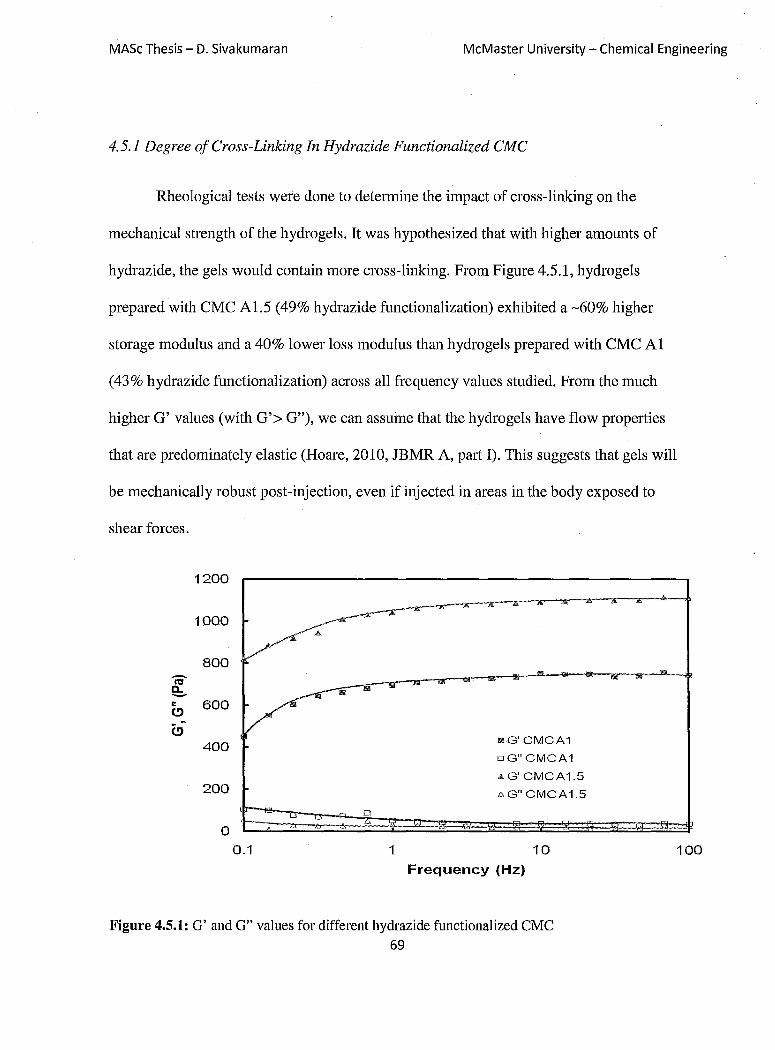
MASc Thesis - D. Sivakumaran McMaster University - Chemical Engineering
4.5.1 Degree of Cross-Linking In Hydrazide Functionalized CMC
Rheological tests were done to determine the impact of cross-linking on the
mechanical strength of the hydro gels. It was hypothesized that with higher amounts of
hydrazide, the gels would contain more cross-linking. From Figure 4.5.1, hydrogels
prepared with CMC Al.5 (49% hydrazide functionalization) exhibited a -60% higher
storage modulus and a 40% lower loss modulus than hydro gels prepared with CMC Al
(43% hydrazide functionalization) across all frequency values studied. From the much
higher G' values (with G'> G"), we can assume that the hydrogels have flow properties
that are predominately elastic (Hoare, 2010, JBMR A, pmt I). This suggests that gels will
be mechanically robust post-injection, even if injected in areas in the body exposed to
shear forces.
1200
1000
800
"' f!:.. = 600 (!) ~
C> 400
200
0 o
0.1 1
mG'CMCA1
oG"CMCA1
AG' CMCA1.5
i,G"CMCA1.5
10 Frequency (Hz)
Figure 4.5.1: G' and G" values for different hydrazide functionalized CMC 69
100

MASc Thesis - D. Sivakumaran McMaster University - Chemical Engineering
Figure 4.5.2 shows the complex viscosity of the hydrogels as a function of applied
frequency.
10000
9000
8000
7000
- 6000 II) • n:I 5000 a. -= 4000
3000
2000
1000
0 0.1 1
Frequency (Hz)
Figure 4.5.2: Complex Viscosity of Hydrogels
10
.... CMCA1
-'&-CMCA1.5
At low frequencies, there is a large difference in viscosities, with the higher
functionalized CMC having a viscosity nearly twice that of its lower functionalized
100
counterpalt. As the oscillation frequencies are increased, there is an associated decrease in
viscosity as the polymer chains of the hydrogel between cross-linking points are aligned
in the shear field. The reduction in complex viscosity with increased oscillation
frequencies indicates that the hydrogels exhibit shear thinning capabilities. Shear thinning
occurs in polymers due to the alignment and de-entanglement of polymer chains induced
70

MASc Thesis - D. Sivakumaran McMaster University - Chemical Engineering
by an applied shear field (Lapasin, 1995). In this case, for an in situ injectable hydrogel,
this kind of behaviour is desirable. This allows for maximum elasticity at low shear (ie. in
the body), while promoting flow at higher shears (during injection), enabling the injection
of high concentrations of polymer solutions and thus the formation of dense hydrogels in
vivo. (Hoare, 2010, JBMR A, part I).
4.5.2 Impact of Imbedded Microgels on Hydrogel Rheology
Within the composite, the bulk hydrogel component is responsible for the majority
of the mechanical strength in the system. The physical entrapment of micro gels within
this hydrogel phase may significantly alter the mechanical properties of the hydrogel.
Specifically, the lack of cross-linking between the embedded micro gel phase and the bulk
hydrogel has the potential to significantly weaken the hydrogel composite, reducing its
potential applicability in the body. The rheology of a CMC-A1/Dex-B hydrogel, with
and without a 50wt% (dry basis) fraction of embedded AA-6% microgel, is presented in
Figure 4.5.3.
71
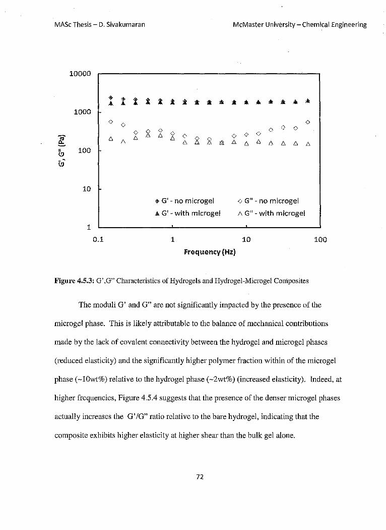
MASc Thesis - D. Sivakumaran McMaster University - Chemical Engineering
10000
• t t 1 A. t t :t * * * .. • At • • • At
1000
~
<> <> <>
~ <> (;, () <> <> 6. <> ¢ <> <> <>
6. t:, t:, g 2 t:, 6. ~ 6. t:, li 6. 6. 6. 6.
~ 100 ...
l!J
10
• G' - no microgel <> Gil - no microgel
&. G' - with microgel 6. Gil - with microgel
1
0.1 1 10 100
Frequency (Hz)
Figure 4.5.3: G' ,G" Characteristics of Hydrogels and Hydrogel-Microgel COniposites
The moduli G' and G" are not significantly impacted by the presence of the
microgel phase. This is likely attributable to the balance of mechanical contributions
made by the lack of covalent connectivity between the hydrogel and micro gel phases
(reduced elasticity) and the significantly higher polymer fraction within of the micro gel
phase (~lOwt%) relative to the hydrogel phase (~2wt%) (increased elasticity). Indeed, at
higher frequencies, Figure 4.5.4 suggests that the presence of the denser micro gel phases
actually increases the G' /G" ratio relative to the bare hydrogel, indicating that the
composite exhibits higher elasticity at higher shear than the bulk gel alone.
72
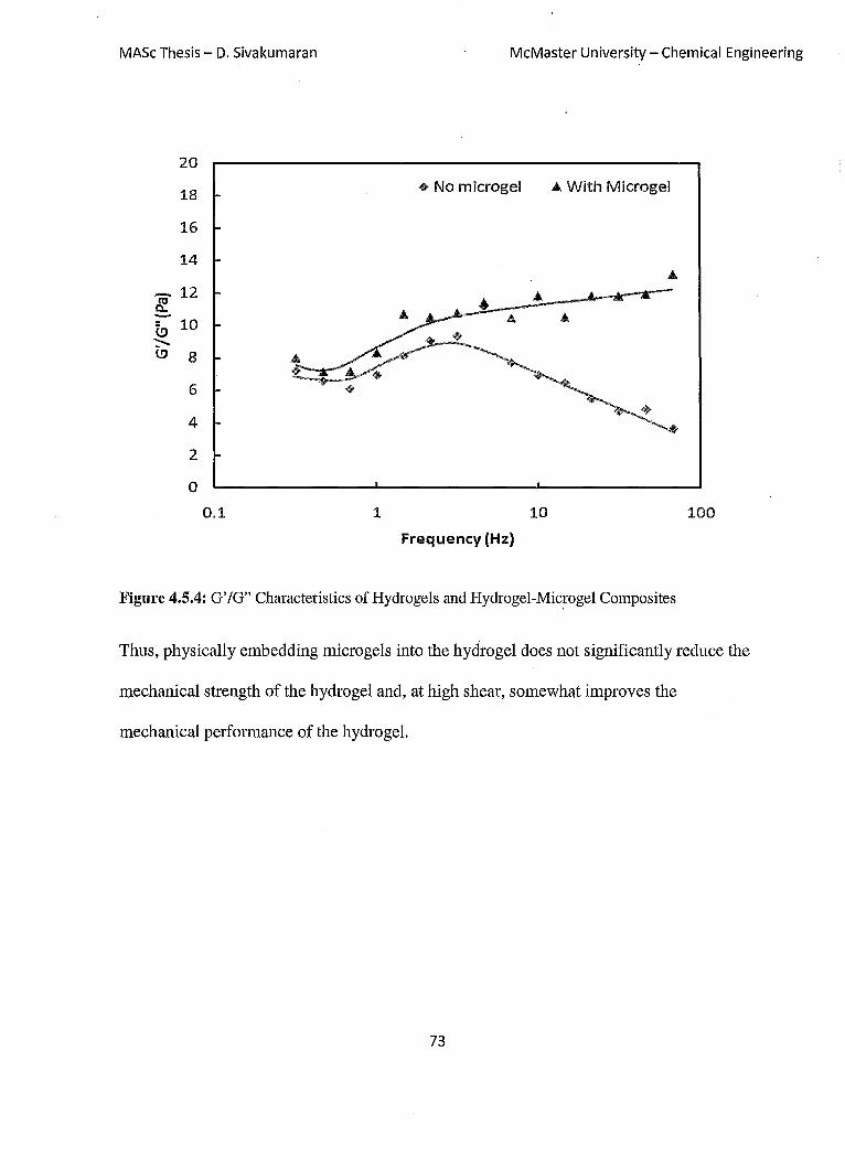
MASc Thesis - D. Sivakumaran McMaster University - Chemical Engineering
20
18 • No microgel ... With Microgel
16
14
- 12 IE ~ 10 ~ (.!) 8 A
+' 6
4
2
0
0.1 1 10 100
Frequency (Hz)
Figure 4.5.4: G' /G" Characteristics of Hydrogels and Hydrogel-Mic~ogel Composites
Thus, physically embedding microgels into the hydrogel does not significantly reduce the
mechanical strength of the hydrogel and, at high shear, somewhat improves the
mechanical performance of the hydrogel.
73

MASc Thesis - D. Sivakumaran McMaster University - Chemical Engineering
Chapter 5: Cell Cytotoxicity
5.1,' Raw Materials
For practical use in vivo, both the composite hydro gels as well as each of their
constituent components (A polymer, B polymer, and microgels) must not induce
significant cytotoxicity around the site of the injection. To determine the effects of the
polymers, microgels, and hydrogels on cellular bodies, an in vitro thiazolyl blue
tetrazolium bromide (MTT) assay was used. This assay is a metabolic process in which
the concentration of the purple metabolite of the MTT dye can be con-elated with the
overall level of cell metabolism and thus the total number of viable cells. For these
assays, tests were conducted with the raw materials as well as the hydrogel and hydrogel
microgel composites. Two cell types were used: 3T3 Mus musculus mouse cells and
C2C12 mouse muscle myoblast cells. These cells were chosen since they are
characteristic of the local cells the composite hydrogel would encounter when injected
intramuscularly. Figure 5.1.1 shows the cell viability (measured as a percentage relative
to a cell-only control) for the composite hydrogel precursors on 3T3 cells. No significant
cytotoxicity is noted for CMC-Al, Dex-B, CMC-B, or AA-6% microgels at
concentrations up to Img/mL, typically considered a threshold for cytotoxicity
assessment. Indeed, at low concentrations, the presence of CMC-Al and CMC-B slightly
up-regulate cell metabolism. Minor decreases in cell viability were observed at
concentrations> Img/mL, decreasing to -80% cell viability at concentrations of 2mglmL
74
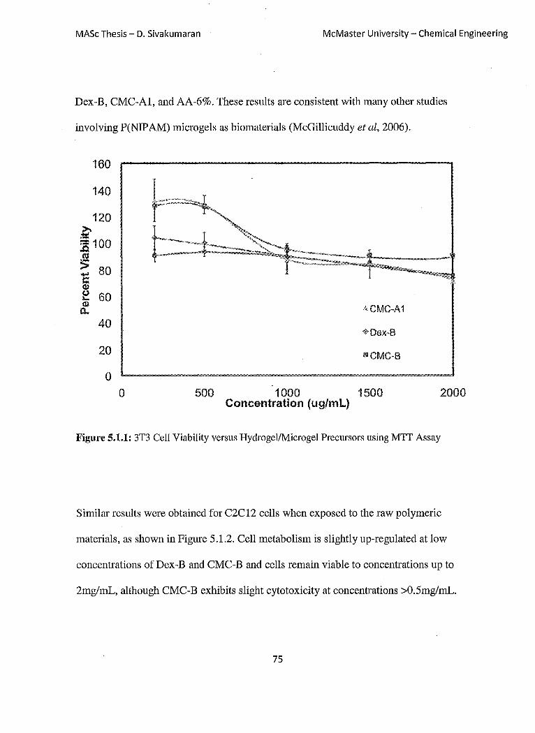
MASc Thesis - D. Sivakumaran McMaster University - Chemical Engineering
Dex-B, CMC-Al, and AA-6%. These results are consistent with many other studies
involving P(NIP AM) microgels as biomaterials (McGillicuddy et ai, 2006).
160
140
120 » ..... :g 100 «i
:;: 80 -c (I)
e 60 4)
0..
40
20
o
kCMC-A1
+ Dex-B
o 500 1000 1500 Concentration (ug/mL)
2000
Figure 5.1.1: 3T3 Cell Viability versus Hydrogel/Microgel Precursors using MTT Assay
Similar results were obtained for C2C12 cells when exposed to the raw polymeric
materials, as shown in Figure 5.1.2. Cell metabolism is slightly up-regulated at low
concentrations of Dex-B and CMC-B and cells remain viable to concentrations up to
2mglmL, although CMC-B exhibits slight cytotoxicity at concentrations >0.5mglmL.
75

MASc Thesis - D. Sivakumaran McMaster University - Chemical Engineering
160
140 9
120 >-:::: :s 100 !U
~ ~ ............. " T
"-----1 '<'''/'''''~"""",.~""""", _e~ """i '- -"'-,,- ,
'~ 1,
:; 80 - T
T
C <Ll (.) 60 '-
ACMC-A1 -<Ll
0... $Oex-B
40 mCMC-B
20 <l!I6mol% Acrylic Acid Microgel
0
o 500 1000 1500 2000
Concentration (ug/mL)
Figure 5.1.2: C2C12 Cell Viability v.ersus Hydroge1JMicrogel Precursors using MIT Assay
Many aldehyde-containing polymers have previously been shown to exhibit
cytotoxicity to cells, predominantly through their high reactivity to nucleophilic sites on
proteins (e.g. amines and thiols) (Deneer, 1988). Such reactions can result in protein
denaturation and subsequent problems with cell growth and development, specifically via
modification of membrane proteins and enzymatic systems. However, Figures 5.1.1 and
5.1.2 indicate that the aldehyde polymers used in this study maintain high cell viability at
concentrations up to 2mg/mL. To assess the cell toxicity threshold of CMC-B and Dex-
B, cytotoxicity to 3T3 cells (Figure 5.1.3) and C2C12 cells (Figure 5.1.4) was assessed up
to concentrations of lOmg/mL. From previous experiments, dextran has been seen to be
cell friendly and does not result in cell death (Cadee et ai, 2000). With aldehyde
76

MASc Thesis - D. Sivakumaran McMaster University - Chemical Engineering
functionalized dextran, cell viability drops to -50% at a concentration of 10,000 IlglmL
for both 3T3 and C2C12 cells. Aldehyde-functionalized CMC, however, maintains at
least -80% cell viability even when added at a concentration of 10mglmL. Thus, CMC-B
and Dex-B both indtlce only minimal to moderate cytotoxicity even at concentrations far
in excess of what would be present in vivo due to the presence of unreacted polymers
and/or the degradation of the composite hydrogel. This is similar to the results found by
previously, in which up to 100llglmL of hydro gels (carboxymethyl dextran) were found to
have promising cell viabilities. In addition, the same hydrogels were found to have low
toxicity in the peritoneum, which is one of the most demanding regions of the body (Ito et
ai, 2007). This result suggests that the composite hydro gels we have developed may have
practical applications in vivo.
140
120
100 :>..
= :.c 80 !'!l :> -s:: 60 (I.) <.J ... (I.) a..
40
20
0 0 2000 4000 6000
Concentration (ug/mL)
.. Oex-B mCMC-B
8000
Figure 5.1.3: 3T3 Cell Viability for B-type Polymers using MIT Assay
77
10000

MASc Thesis - D. Sivakumaran McMaster University - Chemical Engineering
140
120
100
.~ :c 80 ttl
:> .... s::
60 aJ 1.1 .... OJ 0-
40
20
0
it.
0 2000 4000 6000
1> Dex-B
.!ill CMC-B
Concentration (ugjml)
Figure 5.1.4: C2C12 Cell Viability for B-type Polymers using MIT Assay
5.2 Hydrogel-Microgel Composite
8000
The cell viability of 3T3 and C2C12 cells in the presence of a CMC-A1/Dex-B
10000
hydrogel and a composite CMC-Al/Dex-B/AA-6% hydrogel was also assessed using the
MTT assay. Two geometries were tested: (1) plating cells in the polystyrene well and
placing a hydrogel disk on top of the cells and (2) extruding the hydrogel to fill the
polystyrene well and plating cells on top of the hydrogel. These two geometries were
selected to mitigate potential complications in terms of nutrient accessibility (hydrogel on
top), cell-hydrogel contact (hydrogel on top) and cell adhesion (hydrogel on bottom) that
78
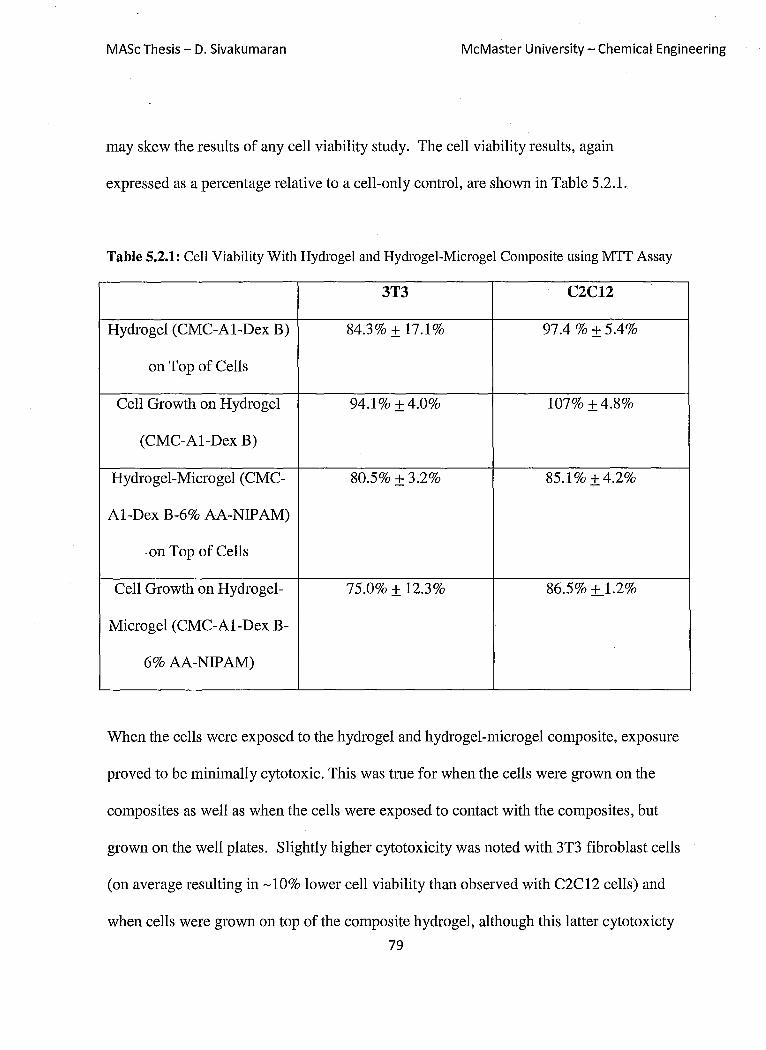
MASt Thesis - D. Sivakumaran McMaster University - Chemical Engineering
may skew the results of any cell viability study. The cell viability results, again
expressed as a percentage relative to a cell-only control, are shown in Table 5.2.1.
Table 5.2.1: Cell Viability With Hydrogel and Hydrogel-Microgel Composite using MIT Assay
3T3 C2C12
Hydrogel (CMC-A1-Dex B) 84.3% ± 17.1% 97.4 % ± 5.4%
on Top of Cells
Cell Growth on Hydrogel 94.1% ±4.0% 107% ±4.8%
(CMC-A1-Dex B)
Hydrogel-Microgel (CMC- 80.5% ±3.2% 85.1% ±4.2%
A1-Dex B-6% AA-NIPAM)
. on Top of Cells
Cell Growth on Hydrogel- 75.0% ± 12.3% 86.5% ±J.2%
Microgel (CMC-A1-Dex B-
6% AA-NIPAM)
When the cells were exposed to the hydrogel and hydrogel-microgel composite, exposure
proved to be minimally cytotoxic. This was tme for when the cells were grown on the
composites as well as when the cells were exposed to contact with the composites, but
grown on the well plates. Slightly higher cytotoxicity was noted with 3T3 fibroblast cells
(on average resulting in ~1O% lower cell viability than observed with C2C12 cells) and
when cells were grown on top of the composite hydrogel, although this latter cytotoxicty
79

MASc Thesis - D. Sivakumaran McMaster University - Chemical Engineering
may be in part attributable to the highly hydrophilic nature of these composite hydrogels
that may inhibit adhesion of the cells and thus normal metabolic function. The hydrogel
microgel composite appeared to be slightly more cytotoxic to the cells than the hydrogel
on its own, particularly toward C2C12 cells. This could be due to the cumulative nature
of exposing another component with a minor cytotoxic response (microgels) to the cells.
fudeed, the cell response to the hydro gels seem to mimic cumulatively what would
happen if all precursors (CMC, dextran, hydrazide, aldehyde) were combined. Overall,
however, the high apparent cytocompatibility of the hydro gels observed here is consistent
with other "plum-pudding" matrices reported in the literature (McGillicuddy et ai, 2006).
It should also be noted that any toxicities observed through in vitro experiments are
typically significantly lower in the infinitely diluting in vivo environment, in which fluids
around the hydrogel are rapidly exchanged (Hoare, 2010, JBMR A, part II).
80

MASc Thesis - D. Sivakumaran McMaster University - Chemical Engineering
Chapter 6: Drug Release Profiles
Drug release of bupivacaine was observed from the hydrogel-micro gel composites into
the external media, which consisted of lOmM PBS. In all cases, a large burst effect was
observed at the beginning of the composite exposure to external media. This is consistent
with many polymer type systems in which diffusion mechanisms playa role in delivery of
medication (Edlund and Albertsson, 2002). This was followed by varying release profiles
that were dependent on the type of cross-linking, polymer-drug interactions, and other
factors. In most cases, the burst effect follows a first-order release profile. This is
followed by the reduction of bupivacaine release until slow, near zero-order release is
obtained.
When media was changed, the hydrogel-micro gel composites retained their shape
and degree of swelling during the testing protocol. It can be stated that over the duration
of drug release the hydrogel-microgel composites displayed excellent structural strength,
which in tum resulted in controlled release of bupivacaine. Over the course of time, the
hydrogel-micro gel composites experienced bulk erosion. Any degradation of the
hydrogel matrix can most likely be attributed to the slow hydrolysis of the hydrazone
groups (Hiemstra et al, 2007).
6.1 Composite Release Versus Raw Hydrogel and Microgel Release
Prior to the discussion of tunable features of the hydrogel-micro gel composite, the
benefits of the composite were compared to that of the hydrogel and micro gel on its own.
81

MASc Thesis - D. Sivakumaran McMaster University - Chemical Engineering
Each type of microgel was loaded with 5mglmL bupivacaine to study the release
behaviour of the micro gel. The same experiment was done for the hydrogel without
embedded micro gels. The results of release from the CMC-dextran hydrogel and a 20%
AA-NIPAM microgel are displayed in Figure 6.1.1. These results are then compared to
the release observed from a composite hydrogel containing a 50:50 mass ratio of the
CMC-dextran hydrogel and the 20% AA-NIP AM microgel. The drug content had the
same mass to mass ratio of microgel:composite. Over the first two days of release, each
of the bulk hydrogel, the microgel, and the micro gel-hydrogel composite exhibits a burst
release profile followed by nearly first-order release characteristic of a diffusion-based
process. However, the amounts of drug released from the micro gel and hydrogel are
significantly higher than that of the composite hydrogel, with release from both phases
between 50-70% faster than from the composite hydrogel. The small size of the micro gel
(small diffusional path length) and the minimal affinity binding expected between
bupivacaine and the bulk hydrogel promotes rapid diffusion-based release from both the
microgel and the hydrogel alone. In comparison, drug loaded into the high-affinity
micro gel phase inside the composite hydrogel is not released to a great extent over this
time period. Instead, after the burst release (diffusion of drug contained in bulk hydrogel
phase out of the gel composite), slow and near zero-order release is observed from the
hydrogel-microgel composite. We expect this slow, near-constant release is attributable
to partitioning of drug from the micro gel to the hydrogel phase and finally into the release
medium, facilitating slow release of drug over time. Thus, it appears that microgel-
hydrogel composites have the potential to facilitate long-lasting controlled release.
82

MASc Thesis - D. Sivakumaran
1.2
- 1
E -
'(5
~ 0.4 '5. ::J
CO 0.2
o 1
McMaster University - Chemical Engineering
2 3 4 5 6 7
Time (days)
Figure 6.1.1: Comparison of Release Profiles from Microgel and Hydrogel and that of the
Hydrogel-Microgel Composite
6.2 Effect of Microgel Functionalization on Drug Release
Figure 6.2.1 shows the effect of increasing the number of acrylic acid functional
groups in the micro gel on the drug release kinetics of the hydrogel-micro gel composite.
As the number of acrylic acid groups in the micro gel is increased, the amount of drug
released and the rate of that release both decreased. This result is attributable to charge-
charge interactions between bupivacaine (cationic) and acrylic acid residues in the
micro gel (anionic), leading to tighter binding of the drug in the micro gel phase and a
lower driving force for partitioning of drug into the bulk phase for release. Indeed, for
83
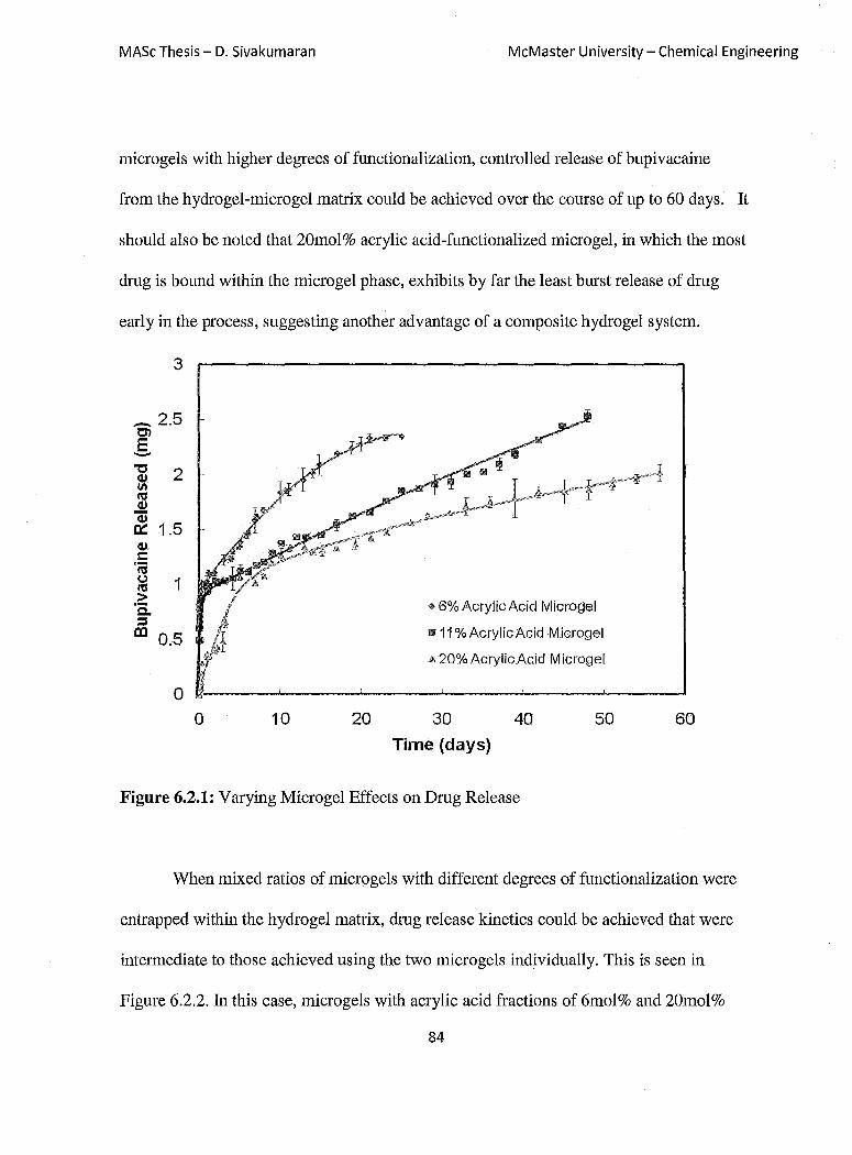
MASc Thesis - D. Sivakumaran McMaster University - Chemical Engineering
micro gels with higher degrees of functionalization, controlled release of bupivacaine
from the hydrogel-microgel matrix could be achieved over the course of up to 60 days. It
should also be noted that 20mol% acrylic acid-functionalized microgel, in which the most
dmg is bound within the micro gel phase, exhibits by far the least burst release of dmg
early in the process, suggesting another advantage of a composite hydrogel system.
3
_ 2.5 -en E -
<1>. s:: 'r; ~ 1 > 'Q. :::I
III 0.5
o o 10 20
.. 6% Acrylic Acid Microgel
m11 % Acrylic Acid M icrogel
A 20% AcrylicAcid M icrogel
30
Time (days)
40 50
Figure 6.2.1: Varying Microgel Effects on Dmg Release
60
When mixed ratios of micro gels with different degrees of functionalization were
entrapped within the hydrogel matrix, dmg release kinetics could be achieved that were
intermediate to those achieved using the two micro gels individually. This is seen in
Figure 6.2.2. In this case, microgels with acrylic acid fractions of 6mol% and 20mol%
84

MASc Thesis - D. Sivakumaran
-C')
E -(!J VI
2.5
2
m 1.5
~ <IJ ,5 n:t 1 u "' > 'Q.. :::7 III 0.5
o o 5 10 15
Time (Days)
McMaster University - Chemical Engineering
.6mol%AcrylicAcid Microgel
f1!120mol%AcrylicAcid Microgel
.1/2 6mol% AA Microgel; 1/2 20mol%AA Microgel
20 25 30
Figure 6.2.2: Mixed Microgel Composite Drug Release
were mixed in a 1:1 ratio and entrapped in a CMC-AIlDextran B hydrogel. The release
profile achieved fit directly between the release profiles achieved using the two microgels
individually in the same bulk hydrogel. Thus, any kinetic profile can be achieved using a
composite hydrogel simply by mixing nanophases with different affinities for the drug to
be delivered.
Together, Figures 6.1.1 and 6.2.1 suggest that the primary factor controlling drug
release from microgel-hydrogel composites is the degree of microgel functionalization
and, specifically, the electrostatic interactions present between the micro gel phase and the
drug. Bupivacaine is a cationic drug which binds ionic ally with the acrylic acid groups
85

MASc Thesis - D. Sivakumaran McMaster University - Chemical Engineering
within the micro gel, which are anionic at physiological pH. This electrostatic attraction
can explain why bupivacaine is loaded at higher fractions in microgels that contain higher
acrylic acid fractions (Hoare and Pelton, 2008). This also explains the additive behaviour
of mixing microgel fractions, as the degree of drug-micro gel interaction achieved is
dependent on the total number of acrylic groups present in the micro gel mixture.
6.3 Effect of Cross-Linking Degree due to Hydrazide Functionalized Polymers
The effect of degree of cross-linking in the bulk hydrogel phase on composite
hydrogel drug release was studied by varying the degree hydrazide attachment to the
CMC backbone of the A-type polymers. Increased hydrazide functionalization is
associated with an increased degree of cross-linking, as the presence of more hydrazide
groups results in a greater probability for reaction with aldehyde groups during the cross
linking process and thus a higher cross-linking density. Differences in cross-link density
between hydrogels prepared using polymers with different degrees of hydrazide
functionalization were previously confirmed based on rheological measurements on these
hydro gels in the absence of an embedded microgel phase (Figure 4.5.1). Figure 6.3.1
indicates that higher degrees of cross-linking in the bulk hydrogel phase resulted in
slower drug release. This is due to the increased cross-link density effectively reducing
the diffusion of the drug through the matrix. Thus, these hydrogels most effectively
reduced the rate of diffusion of drug out of the matrix and water into the hydrogel phase.
86

MASc Thesis - D. Sivakumaran McMaster University - Chemical Engineering
This result suggests that tuning the cross-link density of the bulk gel offers additional
control over the dmg release kinetics that can be achieved with composite systems.
The observed release behaviour of the low cross-linked composite (CMC-AO.5)
can be explained by considering the swelling characteristics as a function of time ·of the
various hydro gels tested, shown in Figure 6.3.2. In general, by increasing the cross-link
density, the potential for the hydrogel to swell or shrink is restricted (Huang et aI, 2004).
The CMC-AIDextran B polymers all deswelled over time, due to unfavourable polymer
solvent interactions generated upon cross-linking in this system due to consumption of the
hydrophilic functional groups upon cross-linking. As a result, increasing the cross-link
density reduced the degree of des welling that could be achieved, resulting in higher pore
sizes in the more highly cross-linked bulk hydrogels (CMC Al and CMC A1.5).
It should be noted from Figure 6.3.2 that increasing the degree of hydrazide
functionalization in the A polymer is effective at changing hydrogel swelling and thus
dmg diffusion only to a limit. Hydrogels prepared with CMC-AO.5 (30% hydrazide
functionalization) released dmg significantly more slowly than hydrogels prepared with
CMC-Al (44% hydrazide) due to des welling ofthe matrix. However, when the degree of
hydrazide functionalization was increased to 49% (CMC-A1.5), Figure 6.3.1 indicates
that no significant change in dmg release was observed and Figure 6.3.2 indicates that
there is no statistically significant
87

MASc Thesis - D. Sivakumaran
'En E -
"'0 Q)
2.5
~ 1.5 iU 0::: Q) c 'n; 1 (,J
~ 'Q.. ::s
CQ 0.5
o o 10 20
Time (days)
McMaster University - Chemical Engineering
+CMC-AO.5 it! CMC-A1 A CMC-A1.5
30 40
Figure 6.3.1: Varying Degree of Hydrazide-Functionalized CMC Effects on Drug Release
o
-10
Q) -20 OJ c rtI
...c: o -30 -c Q)
~ Q)
a. -40
-50
-60
10 20 30
Time (Days)
40
+CMC-AO.5
f!lCMC-A1
"'CMC-A1.5
Figure 6.3.2: Swelling Characteristics of Hydrazide-Functionalized CMC with Dextran
88

MASc Thesis - D. Sivakumaran McMaster University - Chemical Engineering
difference in the des welling response of two composite hydro gels. This observation is
likely attributable to steric limitations introduced during the cross-linking process.
Increasing the degree of cross-linking decreases the mobility of the polymer chains
present between the cross-linking points, thus restricting the capacity of additional
hydrazide groups to cross-link with excess aldehyde groups present in the system.
The degradation rates of the hydro gels varied significantly with the degree of
cross-linking. From Figure 6.3.2, it can be determined that degradation occurred within
thirteen days via hydrolytic cleavage of the hydrazone bonds between the CMC and
dextran polymers comprising the hydrogel for the low functionalized CMC backbone
(Hiemstra et al, 2007). Rapid degradation occurred since fewer hydrazone bonds need to
hydrolyze before the hydrogel loses structural integrity and becomes a viscous polymer
solution. While the higher cross-linked hydrogels also degraded over time, degradation
occurred 4-5 weeks after immersion into the lOmM PBS solution. In this case, more
hydrazone bonds must be hydrolyzed before the elastic nature of the hydrogel is
compromised, resulting in slower overall degradation.
6.4 Effect of B polymer modification on Drug Release
The composition of the aldehyde-containing polymer used to prepare the bulk
hydrogel was also varied to assess the impact of the composition of the bulk gel phase on
drug release from hydrogel-microgel composites. Figure 6.4.1 indicates that drug release
is not significantly changed with CMC-B is substituted for Dextran-B in preparing the
bulk hydro gels.
89
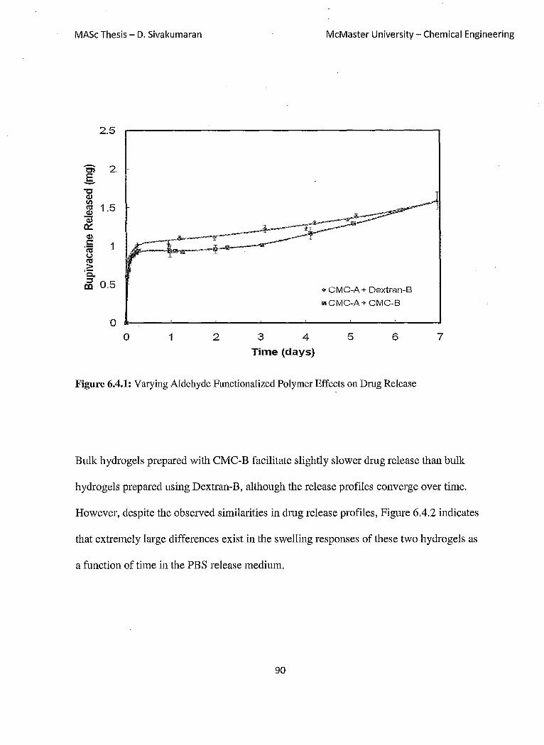
MASc Thesis - D. Sivakumaran
2.5
Ci 2 .s "C <I.)
~ 1.5 -<I.)
~ <1.) .
. ~ 1 (,,) I'tI > 'Q. ::::s
OJ 0.5
o o 1 2 3 4
Time (days)
McMaster University - Chemical Engineering
.. CMC-A + Dextran-B
IllICMC-A+ CMC-B
5 6 7
Figure 6.4.1: Varying Aldehyde Functionalized Polymer Effects on Drug Release
Bulk hydrogels prepared with CMC-B facilitate slightly slower drug release than bulk
hydro gels prepared using Dextran-B, although the release profiles converge over time.
However, despite the observed similarities in drug release profiles, Figure 6.4.2 indicates
that extremely large differences exist in the swelling responses of these two hydro gels as
a function of time in the PBS release medium.
90

MASc Thesis - D. Sivakumaran McMaster University - Chemical Engineering
290
240
~ 190 lij ..c (.) ... 140 I: <U U '-<U a. 90
40
,*,CMCB
II!II Dextran B
_F·····""'·-····"",·····-"""··"li!· .""' ...... r""'--m---"""T--_-m--_r-r--~.---.--... ---10 o 1 2 3 4 5 6 7
Time (Days)
Figure 6.4.2: Swelling Differences in Aldehyde-Functionalized Polymer Type
While CMC-A1IDextran-B hydro gels show little or no swelling as a function of
time, hydrogel-microgels composites made of CMC-A1 and CMC B swell by a factor of
up to 2.5 times of their initial weight (at formation) after just one day of exposure to the
PBS release solution. Water uptake in CMC hydro gels is govemed by the carboxylic
groups on the CMC. The ionization of the carboxylic acid groups (pKa =3.2) induces
Donnan equilibrium-driven swelling in the CMC-B-containing hydrogel (Barbucci et ai,
2000). It should be noted that the aldehyde content of the two polymers is essentially
equivalent (32% for CMC and 37% for dextran, see Table 4.1.1 and 4.2.1), such that this
difference in swelling cannot be attributed to different cross-link densities in the two
91
MASc Thesis - D. Sivakumaran McMaster University - Chemical Engineering
hydrogels. Thus, although the average pore size in CMC-AlICMC-B hydrogels is
significantly higher than that of CMC-AIlDextran B hydrogels, the bulk hydrogel
prepared with CMC-B releases drug more slowly. This is in contradiction to the result
shown in Figure 6.3.1, in which higher bulk hydrogel swelling facilitated faster release.
Electrostatic interactions between bupivacaine and the ionized carboxylic acid
groups in the CMC-B polymer, which are maintained during the oxidation process used to
convert CMC to CMC-B, can be used to rationalize this apparent contradiction. While
diffusion-based release will occur significantly faster in CMC-AlICMC-B hydrogel
systems, the presence of -COO- groups provides an electrostatic partitioning effect that
restricts the diffusion of bupivacaine through the highly swollen bulk gel matrix. As a
result, despite the significantly larger average pore size in CMC-AlICMC-B hydrogels,
slightly slower drug release is observed. This result is consistent with our observations
regarding the effect of the total charge density in the micro gel phase on bupivacaine
release (Figure 6.1.1) and suggests that release kinetics from composite hydrogels can be
sensitively tuned by altering both the cross-link density of the micro gel (i.e. average pore
size) as well as the affinity of either phase for the drug to be released.
Hydrophobic partitioning can also playa role. The log P of a NIP AM monomer
sub-unit is approximately 0.06. For cationic bupivacaine (the predominant form at
physiological pH), the log P value is 0.18 (Sudoh et aI, 2004). As a result, bupivacaine
has a similar hydrophobicity to the micro gel and would thus prefer to partition within the
microgel phase at physiological conditions rather than the water-based solvent (PBS).
92

MASc Thesis - D. Sivakumaran McMaster University - Chemical Engineering
Thus, even in the absence of charged groups, drug release should be slowed by the
presence ofthe NIPAM-based micro gel phase.
It should be noted that gel degradation via hydrolysis occurs rapidly with the very
swollen CMC-B based composites (7-10 days), owing to the large water fraction present
in these hydro gels. In comparison, the CMC-dextran hydro gels maintain their structure in
excess of 30 days depending on cross-link density.
6.5 Hydrazide Functionalized MicrogeZs
To investigate the effect of the micro gel phase transition on the drug release
achieved in hydrogel-microgel composites, cross-linkable microgels functionalized with
hydrazide groups were synthesized and incorporated into the CMC-dextran bulk
hydrogels. In this case, the hydrazide-modified microgels were mixed with CMC-Al in
the double barrel syringe and co-extruded with Dextran B to generate composite
hydro gels in which the micro gel phase is covalently cross-linked to the bulk hydrogel, not
just physically entrapped. Drug release results for composite hydro gels of the same
composition prepared with cross-linkable (hydrazide-functionalized) microgels and non
crosslinkable (acid-functionalized) microgels are shown in Figure 6.5.1.
93

MASc Thesis - D. Sivakumaran
2.S
"6i 2 E --g IJ? ~ 1.S cv Qi 0::: cv ,~ 1 u ~ > '5.. ::J al O.S
o o 10 20
McMaster University - Chemical Engineering
+ 6% Acrylic Acid Microgel t) 34%AcrylicAcid Microgel III 6% AA Microgel + Hydrazide A 34%AA-Mlcrogel + Hydrazide
30
Time (days)
40 so 60
Figure 6.5.1: Bupivacaine Release from Hydrazide-Functionalized AA-P(NIP AM) Microgels
In both cases, when micro gels are cross-linked to the bulk hydrogel matrix, the
dmg release rate from the hydrogel is slowed relative to hydro gels in which the micro gels
are only physically entrapped. This observation can be rationalized by two factors. First,
cross-linking between the microgel and hydrogel phase physically restricts the desweIling
of the microgels typically observed in the high salt environment at physiological
temperature. This avoids the potential formation of a macroporous hydrogel, in which
des welling of the entrapped micro gel within the swelling bulk hydrogel phase creates
large free volumes around the embedded micro gel phase, resulting in faster dmg release.
Second, cross-linking the microgel to the bulk hydrogel increases the total cross-link
94

MASc Thesis - D. Sivakumaran McMaster University - Chemical Engineering
density in the network, reducing the average pore size and thus the overall diffusion
coefficient of drug through the polymer network.
However, the increased cross-link density (increasing diffusive resistances to drug
release) is offset in part by the consumption of ionic -COO- groups via adipic acid
dihydrazide functionalization, reducing the number of available ionic binding sites for
bupivacaine. These competing effects are evidenced by the change in release kinetics
observed upon microgel crosslinking between the 6mol% and 34mol%-functionalized
microgels. ill the 6mol% functionalized micro gel , few carboxylic acid groups are
present in the microgel in either the hydrazide-functionalized micro gel or the unmodified
microgel; as a result, relatively few electrostatic drug-polymer interactions are present
even in the non-cross-linked micro gel and consuming -COOH groups via reaction with
adipic acid dihydrazide does not significantly change the electrostatic partitioning of drug
into the microgel. Correspondingly, a large decrease in drug release rate is observed
when the micro gel is cross-linked to the hydrogel matrix due to the increase in overall
cross-link density and mechanical prevention of a micro gel phase transition. ill
comparison, adipic acid dihydrazide functionalization of the 34mol % functionalized
micro gels results in the consumption of a large number of -COOH groups, reducing the
-COOH content of the micro gel from 34mol% to 20 mol%. This can be seen from the
titration of the microgels, shown in Figure 6.5.2.
95
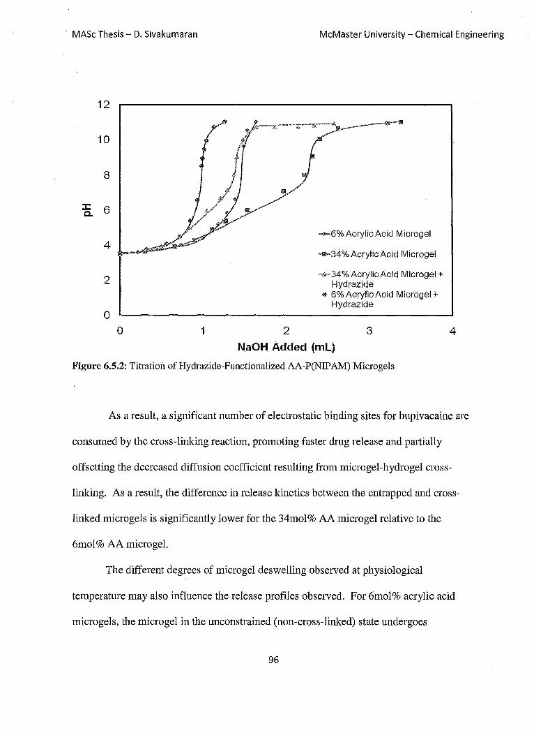
MASc Thesis - D. Sivakumaran McMaster University - Chemical Engineering
12
10
8
:z:: 6 c.
4
2
o o 1 2
--6% Aeryl ie Aeid M icrogel
-m-34%AcrylieAeid Microgel
-£r-34%AcrylieAcid Microgel + Hydrazide
(9 6%AerylieAeid Microgel + Hydrazide
3
NaOH Added (mL)
Figure 6.5.2: Titration of Hydrazide-Functionalized AA-P(NIPAM) Microgels
4
As a result, a significant number of electrostatic binding sites for bupivacaine are
consumed by the cross-linking reaction, promoting faster drug release and partially
offsetting the decreased diffusion coefficient resulting from microgel-hydrogel cross-
linking. As a result, the difference in release kinetics between the entrapped and cross-
linked microgels is significantly lower for the 34mol% AA micro gel relative to the
6mol% AA micro gel.
The different degrees of micro gel des welling observed at physiological
temperature may also influence the release profiles observed. For 6mol% acrylic acid
microgels, the microgel in the unconstrained (non-cross-linked) state undergoes
96

MASc Thesis - D. Sivakumaran McMaster University - Chemical Engineering
significant des welling at physiological temperature (-70% volume change, Table 4.3.1).
As a result, when the micro gel is simply entrapped in the bulk hydrogel phase, convective
mass transfer occurs to release bupivacaine from the micro gel, resulting in the fast, burst
release observed. In addition, larger free volumes are generated within the gel phase at
physiological temperature due to the collapse of the embedded micro gel phase, increasing
the average diffusion coefficient of drug from the composite. In comparison, when the
microgel is cross-linked to the bulk hydrogel, the phase transition is suppressed due to the
elastic constraints imposed by the covalent crosslinks, preventing the same degree of
burst release, minimizing or eliminating the formation of a macroporous hydrogel
structure, and significantly reducing the rate of diffusion-driven drug release. For the
34mol% acrylic acid functionalized microgels, the higher charge content of the micro gel
restricts des welling at physiological temperature, resulting in significantly less des welling
(-34% volume change, Table 4.3.1). Consequently, less deswelling is avoided by cross-
linking the micro gel to the hydrogel phase, less convective mass transfer occurs, and
smaller macropores are generated in the micro gel network at physiological temperature.
As a result, and less difference is observed between the release profiles of hydro gels
prepared using cross-linked or entrapped microgels.
While micro gel des welling is significantly affected by whether the microgel is
physically entrapped or covalently cross-linked to the bulk hydrogel matrix, the overall
swelling of the composite material is not affected by the presence of a cross-'linked
microgel phase, as shown in Figure 6.5.3. Thus, any differences in drug release are
attributable exclusively to changes in the microgel phase. This result demonstrates the
97

MASc Thesis - D. Sivakumaran McMaster University ~ Chemical Engineering
power of using composite hydrogel systems for engineering drug delivery systems, since
properties of both phases can be independently engineered to achieve new types of
release behaviours.
o
-10
.~ -20 (I,) C'l C rtf .c -30 (.) -c (I,)
~ (I,) -40
0...
-50
-60
10 20
~ 6% Acrylic Acid M icrogel + Hydrazide
A 34 % Acrylic Acid M icrogel + Hydrazide
1IiII 6% Acrylic Acid M icrogel
• 34%AcrylicAcid Microgel
30
Time (Days)
40 50
Figure 6.5.3: Swelling Characteristics of Hydrazide-Functionalized AA-P(NIP AM) in comparison to Non-Functionalized (Entrapped) AA-P(NIPAM)
98

MASc Thesis - D. Sivakumaran McMaster University - Chemical Engineering
Chapter 7: Discussion
The objective of this research project was to create a delivery vehicle for cationic,
hydrophobic drugs utilizing the benefits of two distinct types of gels. The macroscopic
hydrogel and the microscopic microgels have been shown to both be capable of
independently tuning the release mechanisms of the hydrogel-microgel composites. This
allows for the individual engineering of each component to optimize the release profiles
of drug from the system. The biocompatible hydrogels can dictate release rates of
bupivacaine depending on the degree of functionalization within the matrix, with release
being governed by a combination of diffusion and phase partitioning. The hydro gels have
also shown adequate mechanical strength to withhold the rigours of being in the body.
The composite hydrogels can control the release of bupivacaine via a combination
of three mechanisms: electrostatic binding, diffusion, and hydrophobic partitioning.
Electrostatic binding between cationic bupivacaine to the acrylic acid groups of the AA
P(NIP AM) micro gels appears to be the primary driving force for bupivacaine uptake into
the composite hydrogel and regulates the initial distribution of bupivacaine between the
bulk and micro gel phases, controlling the magnitude of the initial burst release achieved.
In addition, increased densities of anionic functional groups in either the bulk hydrogel
phase or the micro gel phase (i.e. increases in the affinity of hydrogel or microgel phase
for the drug) reduce the rate of drug release achieved. These ionic interactions appear to
be the most critical factor for predicting release kinetics. Diffusion is determined by the
cross-link density and thus the degree of swelling of the composite hydrogel as a function
99
MASc Thesis - D. Sivakumaran McMaster University - Chemical Engineering
of time. Composite hydro gels that swell more release drug faster due to the higher
average diffusion coefficient of bupivacaine within a more swollen network.
Hydrophobic partitioning of bupivacaine into the moderately hydrophobic domains of the
NIP AM residues also contributes to release control, although this effect appears to be
significantly less important than the ionic binding and cross-link effects.
By cross-liking the micro gel to the bulk hydrogel the release profile of
bupivacaine can be fmther tuned. The cross-linking of the micro gel will result in a
mechanical constriction on the phase transition of the microgel, reducing convective mass
transport out of the micro gel at short times via microgel des welling (thus reducing burst
drug release) and eliminating the potential for making macropores in the free volume
around the collapsed microgels. Increased overall cross-linking density within the
composite also contributes to slower drug release, although this effect is in part
counteracted by the consumption of micro gel charges upon crosslink formation.
The results of this work have significant relevance to the clinical challenge of pain
management. Bupivacaine is a local anaesthetic that is used short term for pain relief,
mainly post-surgery. Durations of release on the order of 8-10 hours are currently the
state-of-the-art technology for bupivacaine release, effective for relieving surgical
complications but ineffective for chronic pain relief. While controlled bupivacaine
release over a long period would effectively relieve pain, it would also impair local motor
function, a potential issue in the long-term use of the drug release systems studied
depending on the location of drug administration in the body. In such cases, structurally
similar drugs such as lidocaine-Q, an anaesthetic mainly used in chronic control of
100

MASc Thesis - D. Sivakumaran McMaster University - Chemical Engineering
posttherpetic neuralgia since it has no impact on motor functions, may be more relevant
toward achieving the goal of chronic pain relief. (Lexi-Comp).
From the observations collected in this research, it can be determined that the
creation of micro gel-hydrogel composites will result in drug release that can be tuned by
altering the crosslink density, chemistry, and charge density of different phases within the
soft composite. The ~60-day release observed for 20% acrylic acid microgel-embedded
composite hydrogels studied in this thesis, with a composition optimized for binding the
target drug bupivacaine, is significantly longer than release durations previously reported
from hydrogel-based materials. This duration of release is notable for a small molecule
through a hydrogel matrix and suggests the potential for using soft nanocomposite
hydrogels for long-term controlled local drug release. Similarly, we expect that the
partitioning and electrostatic properties of the micro gel can be tuned to design
customizable in situ gellable drug delivery systems with potential for achieving long-term
release of high doses of drugs from hydrogel-based materials. In theory, it should be
possible to independently tune the compositions of the bulk hydrogel and microgel phases
to optimize the affinity of those phases to any target drug and thus achieve the dual
partitioning-diffusion-based control over drug release kinetics demonstrated in this study.
The injectable nature of the hydro gels developed also contributes strongly to their
potential applicability in the clinic, as drugs could be administered via traditional,
minimally-invasive injection techniques rather than requiring minor surgical procedures
as with most bulk hydrogel systems with similar elastic properties.
101

MASc Thesis - D. Sivakumaran McMaster University - Chemical Engineering
Chapter 8: Conclusions and Recommendations:
8.1 Conclusions
As a result of the work performed in this thesis, the following conclusions can be made.
The charge content and particle size of acrylic acid-functionalized P(NIPAM)
microgels can be tuned by varying the amount of acrylic acid co-polymerized with the
NIP AM. The charge content of the micro gel primarily determined the amount of cationic
bupivacaine that could be entrapped within the anionic-functionalized microgels,
although hydrophobic partitioning also enhanced drug uptake.
The creation of a hydrogel based on hydrazone crosslinking is a suitable method for
creating an in situ gelling hydro gels. The functionalization of polymers (CMC and
dextran) with hydrazides and aldehydes produces polymers that facilitate easy flow in the
syringe and through the needle. Upon contact, gelation occurs within seconds for the
creation of a hydrogel matrix, creating a predominantly elastic hydrogel matrix even at
very high micro gel loadings (up to 50 dry weight percent) which is highly suitable for
many in vivo applications. Degradation times and swelling responses can both be altered
by changing the degree of cross-linking within the hydrogel. Degradation occurs
primarily via hydrolytic cleavage of the hydrazone cross-links.
The cytotoxicity of both the composite precursor materials and the composite
hydrogels was minimal at the concentrations at which the materials would be bioavailable
inside the body.
102
MASc Thesis - D. Sivakumaran McMaster University ~ Chemical Engineering
Dmg release from the hydrogel-micro gel composites consisted of two distinct release
regimes: fast burst or first-order release over the first 2-3 days (corresponding to diffusion
of dmg through the bulk hydrogel matrix) and slow, near zero-order release over the
remainder of the release period (corresponding to dmg partitioning from the micro gel
phase to the bulk hydrogel phase). The combining of the bulk hydrogel and the micro gel
significantly slowed dmg release versus either component used alone. Release durations
of up to 60 days were achieved for the small molecule dmg bupivacaine, significantly
longer than previously reported in other hydrogel dmg delivery studies
The release of bupivacaine can be altered by changing the degree of cross-linking of
the hydrogel. The higher the cross-link density, the slower the release ofbupivacaine
from the composite. However, significantly more control could be made via the
modification of the microgels. The electrostatic interactions between bupivacaine and the
acrylic acid groups and the hydrophobic partitioning behavior between bupivacaine and
NIPAM residues were the two most important rate-governing parameters. By increasing
the amount of acrylic groups in the microgels, the rate of release can be slowed.
Cross-linking the microgels directly to the bulk hydrogel matrix significantly changes
the release kinetics achieved in composite hydrogel-micro gel dmg delivery systems since
the phase transition of the thermoresponsive micro gel is mechanically inhibited by the
interphase cross-links. In general, release from composite hydro gels prepared with cross
linkable microgels is slower, attributable both to the lack of convective dmg transport
upon micro gel deswelling as well as the decreased average pore size at the microgel-
103

MASc Thesis - D. Sivakumaran McMaster University - Chemical Engineering
hydrogel interface. However, the consumption of -COO- groups in the micro gel via the
hydrazide functionalization reaction limits the effeCtiveness of this approach in terms of
further reducing the drug delivery rate achievable from composite hydro gels
8.2 Recommendations
More studies need to be conducted on how the degree of hydrogel cross-linking can
be controlled by varying the degree of aldehyde functionalization. This could be used to
further tune the rate of release of bupivacaine from the composite system.
The addition of a higher micro gel fraction should be studied to determine how an
increased fraction of micro gels alters release kinetics. Specifically, a maximum micro gel
loading can be identified at which a stable composite hydrogel can be fabricated.
Composite hydro gels with higher micro gel fractions should result in slower release as
there will be more acrylic acid locations for bupivacaine to interact.
Additional studies pertaining to the development of the hydrazide-functionalized AA
P(NIP AM) micro gels should be conducted to determine a more complete mechanism for
drug release from these type of systems.
To prevent aldehyde degradation after long periods of storage, the synthesis of
aldehyde-functionalized dextran should instead be pursued by synthesizing acetal
functionalized dextran and subsequently hydrolyzing the acetal group as the polymer is
104

MASc Thesis - D. Sivakumaran McMaster University - Chemical Engineering
required (Chen et al, 2002). This method has been observed to create much more stable
aldehyde-functionalized dextran polymers.
Additional bupivacaine detection tests should be conducted to determine the exact
concentration of drug being eluted without the need for blank (polymer-only) control
samples. It is suggested that HPLC be used in conjunction with UV spectrophotometer
readings in any future drug release studies.
Different types of drugs should be studied to see how the magnitude of drug-gel
interactions influence release kinetics. Examples of model drugs to be used are dopamine
(cationic), acetaminophen (neutral charge), or naproxen (anionic) (Hoare, 2008).
In vivo studies should be conducted with the hydrogel-microgel composites. This
should be done to determine cytotoxicity of the device in the body as well as to determine
if andlor where bioaccumulation occurs. In addition, drug release testing can be
completed in vivo to determine if localized drug delivery can be achieved with the
hydrogel-microgel devices.
105

MASc Thesis - D. Sivakumaran McMaster University - Chemical Engineering
References
Alsberg, E., Anderson, K., Albeituti, A, Franceschi, R, & Mooney, D. (2001). Cell interactive alginate hydro gels for bone tissue engineering. Journal of Dental Research, 80(11).
Anbergen, U., & Oppermann, W. (1990). Elasticity and swelling behaviour of chemically crosslinked cellulose ethers in aqueous systems. Polymer, 31(10), 1854-1858.
Bajpai, A K., & Mishra, A (2004). Ionizable interpenetrating polymer networks of carboxymethyl cellulose and polyacrylic acid: Evaluation of water uptake. Journal of Applied Polymer Science, 93(5),2054-2065
Barbucci, R, Magnani, A, & Consumi, M. (2000). Swelling behavior of carboxymethylcellulose hydro gels in relation to cross-linking, pH, and charge density. Macromolecules, 33(20), 7475-7480.
Bazile, D., Prud'homme, C., Bassoullet, M., Marlard, M., Spenlehauer, G., & Veillard, M. (1995). Stealth me.PEG-PLA nanoparticles avoid uptake by the mononuclear phagocytes system. J. Ph arm. Sci, 84,493.
Berger, J., Reist, M., Mayer, J. M., Felt, 0., Peppas, N. A, & Gumy, R (2004). Structure and interactions in covalently and ionically crosslinked chitosan hydrogels for biomedical applications. European Journal of Pharmaceutics and Bioph~rmaceutics, 57(1), 19-34.
Bokias, G., Hourdet, D., & Iliopoulos, I. (2000). Positively charged amphiphilic polymers based on poly(N-isopropylacrylamide): Phase behavior and shear-induced thickening in aqueous solution. Macromolecules, 33(8), 2929-2935.
Bouhadir, K. H., Hausman, D. S., & Mooney, D. J. (1999). Synthesis of cross-linked poly(aldehyde guluronate) hydrogels. Polymer, 40(12),3575-3584.
Bulpitt, P., & Aeschlimann, D. (1999). New strategy for chemical modification of hyaluronic acid: Preparation of functionalized derivatives and their use in the formation of novel biocompatible hydrogels. Journal of Biomedical Materials Research, 47(2), 152-169.
Cadee, J., Van Luyn, M., Brouwer, L., Plantinga, J., van Wachem, P., de Groot, C., et al. (2000). In vivo biocompatibility of dextran-based hydrogels. Journal of Biomedical Materials Research, 50(3), 397.
Chen, N., Hu, S., Pelton, R .. (2002). Mechanisms of aldehyde-containing paper wetstrength resins. Journal of Industrial and Engineering Chemistry Research. 41, 5366-5371
106
MASc Thesis - D. Sivakumaran McMaster University - Chemical Engineering
Chiu, H., Lin, Y., & Hsu, Y. (2002). Effects of acrylic acid on preparation and swelling properties of pH-sensitive dextran hydrogels. Biomaterials, 23(4),1103-1112.
Cleary, J., Bromberg, L. E., & Magner, E. (2003). Diffusion and release of solutes in pluronic-g-poly(acrylic acid) hydrogels. Langmuir, 19(22), 9162-9172.
Dai, C., Wang, B., & Zhao, H. (2005). Microencapsulation peptide and protein drugs delivery system. Colloids and Suifaces B: Biointeifaces, 41(2-3),117-120.
Das, M., Zhang, H., & Kumacheva, E. (2006). Microgels: Old materials with new applications. Annual Review of Materials Research, 36, 117.
Deneer, J. W., Seinen, W., & Hermens, J. L. M. (1988). The acute toxicity of aldehydes to the guppy. Aquatic Toxicology, 12(2), 185-192.
Desai, M. P., Labhasetwar, V., Amidon, G. L., & Levy, R J. (1996). Gastrointestinal uptake of biodegradable microparticles: Effect of particle size. Pharmaceutical Research, 13(12), 1838.
Doherty, M., Hughes, P., Korszniak, N., & Charman, W. (1995). Prolongation of lidocaine-induced epidural anesthesia by medium molecular weight hyaluronic acid formulations: Pharmacodynamic and pharmacokinetic studies in the rabbit. Anesthesia & Analgesia, 80(4), 740.
Edlund, U., & Albertsson, A. (2002). Degradable polymer microspheres for controlled drug delivery. Degradable aliphatic polyesters. Advances in Polymer Science,157, 67-112
Ghannam, M. T., & Esmail, M. N. (1997). Rheological properties of carboxymethyl cellulose. Journal of Applied Polymer Science, 64(2), 289.
Hiemstra, C., van, d. A, Zhong, Z., Dijkstra, P. J., & Feijen, J. (2007). Novel in situ forming, degradable dextran hydrogels by michael addition chemistry: synthesis, rheology, and degradation. Macromolecules, 40(4),1165-1173.
Hoare, T. R, & Kohane, D. S. (2008). Hydrogels in drug delivery: Progress and challenges. Polymer, 49(8), 1993-2007.
Hoare, T., Bellas, E., Zurakowski, D., & Kohane DS. (2010). Rheological blends for drug delivery. II. prolongation of nerve blockade, biocompatibility, and in vitro-in vivo correlations. Journal of Biomedical Materials Research A, 92(2), 586.
Hoare, T., & McLean, D. (2006). Kinetic prediction of functional group distributions in thermosensitive microgels. The Journal of Physical Chemistry B, 110(41),20327-20336.
Hoare, T., & Pelton, R (2004). Functional group distributions in carboxylic acid containing poly(N-isopropylacrylamide) microgels. Langmuir, 20(6), 2123-2133.
107

MASc Thesis - D. Sivakumaran McMaster University - Chemical Engineering
Hoare, T., & Pelton, R. (2004). Highly pH and temperature responsive micro gels functionalized with vinyl acetic acid. Macromolecules, 37(7), 2544-2550.
Hoare, T., & Pelton, R. (2008). Impact of micro gel morphology on functionalized Microgel-Drug interactions. Langmuir, 24(3), 1005-1012.
Hoare, T., Zurakowski, D., & Kohane, D. S. (2010). Rheological blends for drug delivery. I. characterization in vitro. Journal of Biomedical Materials Research Part A, 92A(2), 575.
Hoffman, A. S. (2002). Hydrogels for biomedical applications. Advanced Drug Delivery Reviews, 54(1), 3-12.
Huang, G., Gao, J., Hu, Z., St. John, J. V., Ponder, B. C., & Moro, D. (2004). Controlled drug release from hydrogel nanopartic1e networks. Journal of Controlled Release, 94(2-3),303-311.
Hudson, S. P., Langer, R., Fink, G. R., & Kohane, D. S. (2010). Injectable in situ crosslinking hydro gels for local antifungal therapy. Biomaterials, 31(6), 1444-1452.
Ikechukwu Ugwoke, M., Kaufmann, G., Verbeke, N., & Kinget, R. (2000). Intranasal bioavailability of apomorphine from carboxymethy1cellulose-based drug delivery systems. International Journal of Pharmaceutics, 202(1-2), 125-131.
Ito, T., Yeo, Y., Highley, C. B., Bellas, E.; & Kohane, D. S. (2007). Dextran-based in situ cross-linked injectable hydrogels to prevent peritoneal adhesions. Biomaterials, 28(23), 3418-3426.
Jeong, B., Bae, Y. H., & Kim, S. W. (2000). Drug release from biodegradable injectable thermo sensitive hydrogel ofPEG-PLGA-PEG triblock copolymers. Journal of Controlled Release, 63(1-2), 155-163.
Jia, X., Burdick, J. A., Kobler, J., Clifton, R. J., Rosowski, J. J., Zeitels, S. M., et al. (2004). Synthesis and characterization of in situ cross-linkable hyaluronic acid-based hydrogels with potential application for vocal fold regeneration. Macromolecules, 37(9), 3239-3248.
Jin, R., Hiemstra, C., Zhong, Z., & Feijen, J. (2007). Enzyme-mediated fast in situ formation of hydro gels from dextran-tyramine conjugates. Biomaterials, 28(18), 2791-2800.
Kang, G. D., Cheon, S. H., & Song, S. (2006). Controlled release of doxOlubicin from thermosensitive poly(organophosphazene) hydrogels. International Journal of Pharmaceutics, 319(1-2), 29-36.
108

MASc Thesis - D. Sivakumaran McMaster University - Chemical Engineering
Kikuchi, A., & Okano, T. (2002). Pulsatile drug release control using hydrogels. Advanced Drug Delivery Reviews, 54(1), 53-77.
Kiser, P. E, Wilson, G., & Needham, D. (2000). Lipid-coated microgels for the triggered release of doxorubicin. Journal of Controlled Release, 68(1), 9-22.
Kulkarni, R V., & Sa, B. (2008). Evaluation of pH-sensitivity and drug release characteristics of (polyacrylamide-grafted-xanthan)-Carboxymethyl cellulose-based pHsensitive interpenetrating network hydrogel beads. Drug Development and Industrial Pharmacy, 34(12),1406-1414.
Langer, R (1990). New methods of drug delivery. Science. 249, 1527-33
Lapasin, R, & Priel, S. (1995). Rheology of industrial polysaccharides: Theory and applications. New York: Aspen Publishers Inc.
Le Corre, P., Le Guevello, P., Gajan, V., Chevanne, F., Le Verge, R .. (1994), Preparation and characterization of bupivacaine-Ioaded polylactide and polylactide-co-glycolide micro spheres, International Journal of Pharmaceutics, 207, 41-49
Lee, K. Y., Bouhadir, K. H., & Mooney, D. J. (2000). Degradation behavior of covalently croSs-linked poly(aldehyde guluronate) hydrogels. Macromolecules, 33(1), 97-101.
Lexi-Comp, I., & American Pharmaceutical Association. (1994). Dmg information handbook (North American ed.); Washington, D.C.: American Pharmaceutical Association.
Li, J., Lewis, R B., & Dahn, J. R (2007). Sodium carboxymethyl cellulose. Electrochemical and Solid-State Letters, 10(2), AI7-A20.
Lopez, V. C., Hadgraft, J., & Snowden, M. J. (2005). The use of colloidal rnicrogels as a (trans)dermal dmg delivery system. International Journal of Pharmaceutics, 292(1-2), 137-147.
Lynch, I., de Gregorio, P., & Dawson, K. A (2005). Simultaneous release of hydrophobic and cationic solutes from thin-film plum-pUdding gels: A multifunctional platform for surface drug delivery? The Journal of Physical Chemistry B, 109(13),6257-6261.
McGillicuddy, E C., Lynch, I., Rochev, Y. A, Burke, M., Dawson, K. A, Gallagher, W. M., et al. (2006). Novel' 'plum pudding" gels as potential drug-eluting stent coatings: Controlled release of fluvastatin. Journal of Biomedical Materials Research Part A, 79A(4),923.
Mohanraj, V. J., & Chen, Y. (June, 2006). Nanopaltieles - A review. Tropical Journal of Pharmaceutical Research, 5(1), 561.
109

MASc Thesis - D. Sivakumaran McMaster University - Chemical Engineering
Motlagh, G. H., Hrymak, AN., & Thompson, M. R. (2007). Properties of a carbon filled cyclic olefin copolymer. Journal of Polymer Science Part B: Polymer Physics, 45(14), 1808-1820.
Nadagouda, M. N., & Varma, R. S. (2007). Synthesis of thermally stable carboxymethyl CelluloselMetal biodegradable nanocomposites for potential biological applications. Biomacromolecules, 8(9),2762-2767.
Naessens, M., Cerdobbel, A, Soetaert, & Vandamme, E. J. (2005). Leuconostoc dextransucrase and dextran: Production, properties and applications. Journal of Chemical Technology & Biotechnology, 80(8), 845.
Nathan, A, Zalipsky, S., & Kohn, J. (1996). PEG modified anticancer drugs: Synthesis and biological activity. Journal of Bioactive and Compatible Polymers, 11, 122.
Nguyen, K T., & West, J. L. (2002). Photopolymerizable hydrogels for tissue engineering applications. Biomaterials, 23(22), 4307-4314.
Olivier, J. (2005). Drug transport to brain with targeted nanoparticles. The Journal of the American Society for Experimental NeuroTherapeutics, 2(108)
Padula, C., Colombo, G., Nicoli, S., Catellani, P. L., Massimo, G., & Santi, P. (2003). Bioadhesive film for the trans dermal delivery of lidocaine: In vitro and in vivo behavior. Journal of Controlled Release, 88(2),277-285.
Park, H., Temenoff, J. S., Holland, T. A, Tabata, Y., & Mikos, A G. (2005). Delivery of TGF-pl and chondrocytes via injectable, biodegradable hydrogels for cartilage tissue engineering applications. Biomaterials, 26(34), 7095-7103.
Pawlikowska, P., Gajkowska, B., & Orzechowski, A (2007). Mitofusin 2 (Mfn2): A key player in insulin-dependent myogenesis in vitro. Cell and Tissue Research, 327(3), 571-58l.
Pelton, R. H., & Chibante, P. (1986). Preparation of aqueous latices with Nisopropylacrylamide. Colloids and Surfaces, 20(3), 247-256.
Pelton, R. (2000). Temperature-sensitive aqueous microgels. Advances in Colloid and Interface Science, 85(1), 1-33.
Poole, S. K, & Poole, C. F. (2003). Separation methods for estimating octanol-water partition coefficients. Journal of Chromatography B, 797(1-2),3-19.
Puttipipatkhachom, S., Nunthanid, J., Yamamoto, K, & Peck, G. E. (2001). Drug physical state and drug-polymer interaction on drug release from chitosan matrix films. Journal of Controlled Release, 75(1-2), 143-153
110

MASc Thesis - D. Sivakumaran McMaster University - Chemical Engineering
Qiu, Y., & Park, K. (2001). Environment-sensitive hydrogels for dmg delivery. Advanced Drug Delivery Reviews, 53(3), 321-339.
Rodriguez, R, Alvarez-Lorenzo, C., & Concheiro, A. (2003). Interactions of ibuprofen with cationic polysaccharides in aqueous dispersions and hydrogels: Rheological and diffusional implications. European Journal of Pharmaceutical Sciences, 20(4-5), 429-438.
Rosen, H., & Abribat, T. (2005). The rise and rise of dmg delivery. Nat Rev Drug Discovery, 4(5), 381-385.
Roy, I., & Gupta, M. N. (2003). Smart polymeric materials: Emerging biochemical applications. Chemistry & Biology, 10(12), 1161-1171.
Saunders, B. R, & Vincent, B. (1999). Microgel particles as model colloids: Theory, propel ties and applications. Advances in Colloid and Interface Science, 80(1), 1-25.
Schild, H. G. (1992). Poly(N-isopropylacrylamide): Experiment, theory and application. Progress in Polymer Science, 17(2), 163-249.
Schild, H. G., & TilTell, D. A. (1991). Microheterogeneous solutions of amphiphilic copolymers of N-isopropylacrylamide: An investigation via fluorescence methods. Langmuir, 7(7), 1319-1324.
Shah, J. c., & Maniar, M. (1993). pH-dependent solubility and dissolution ofbupivacaine and its relevance to the formulation of a controlled release system. Journal of Controlled Release, 23(3),261-270.
Shamala, T. R, & Prasad, M. S. (1995). Preliminary studies on the production of high and low viscosity dextran by leuconostoc spp. Process Biochemistry, 30(3), 237-241.
Shi, c., Morse, L., Ing, C., White, P. F., Rawal, S., Latham, P., et al. (2003). Use of a continuous local anesthetic infusion for pain management after median sternotomy. Anesthesiology, 99(4), 918.
Shibayama, M., & Tanaka, T. (1993). Volume phase transition and related phenomena of polymer gels, Advances inPolymerScience,109, 1-62
Sinha, V. R, & Trehan, A. (2003). Biodegradable micro spheres for protein delivery. Journal of Controlled Release, 90(3), 261-280.
Snowden, M. J. (1992). The use ofpoly(N-isopropylacrylamide) latices as novel release systems. J. Chem. Soc., Chem. Commun, (11), 803.
111

MASc Thesis - D. Sivakumaran McMaster University - Chemical Engineering
Sudoh, Y., Cahoon, E. E., De Girolami, u., & Wang, G. K. (2004). Local anesthetic properties of a novel derivative, N-methyl doxepin, versus doxepin and bupivacaine. Anesthesia & Analgesia, 98(3), 672-676.
Sutton, C. (2005). Adhesions and their prevention. The Obstetrician & Gynaecologist, 7(3), 168-176.
Van Dijk-Wolthuis, WNE., Hoogeboom, JAM., van Steentergen, MJ., Tsang, SKY., Hennink, WE .. (1997) Degredation and Release Behaviour of Dextran-Based Hydrogels. Macromolocules, 30, 4639-4645.
Vihola, H., Laukkanen, A., Valtola, L., Tenhu, H., & Hirvonen, J. (2005). Cytotoxicity of thermosensitive polymers poly(N-isopropylacrylamide), poly(N-vinylcaprolactam) and amphiphilically modified poly(N-vinylcaprolactam). Biomaterials, 26(16),3055-3064.
Yeo, Y., Ito, T., Bellas, E., Highley, C. B., Marini, R, & Kohane, D. S. (2007). In situ cross-linkable hyaluronan hydro gels containing polymeric nanoparticles for preventing postsurgical adhesions. Annals of Surgery, 245(5), 819.
Zhang, R, Tang, M., Bowyer, A., Eisenthal, R, & Hubble, J. (2005). A novel pH- and ionic-strength-sensitive carboxymethyl dextran hydrogel. Biomaterials, 26(22), 4677-4683.
112

MASc Thesis - D. Sivakumaran McMaster University - Chemical Engineering
Appendices
Appendix A: Material Information
Appendix Ai Reagents
Chemical Abbreviation Supplier Product ID Acrylic Acid, 99% AA Sigma Aldrich, Oakville, 147230
ON Adipic dihydrazide. 98% ADH Sigma Aldrich, Oakville, A0638
ON Ammonium Persulfate APS Sigma Aldrich, Oakville, 215589
ON Bupivicaine Hydrochloride Sigma Aldrich, Oakville, B5274
ON Dextran from Leuconstoc Sigma Aldrich, Oakville, 31392 spp. ON Dimethyl Sulphoxide DMSO Caledon Laboratory 4100-1
Chemicals, Georgetown, ON
Ethylene Glycol EG Sigma Aldrich, Oakville, 324558 ON
N' -ethyl-N-(3- EDC Sigma Aldrich, Oakville, E7750 dimethylaminopropyl)- ON carbodiimide, commercial grade N-hydroxysuccinimide, NHS Sigma Aldrich, Oakville, 130672 97% ON N -isopropylacrylamide, NIPAM Sigma Aldrich, Oakville, 415324 97% ON N'N' -Methylene MBA Sigma Aldrich, Oakville, 146072 Bisacrylamide, 99% ON Sodium Dodecyl Sulfate SDS Bioshop Canada, SDS001 (electrophoresis grade) Burlington, ON Sodium Periodate, Sigma Aldrich, Oakville, 311448 ACS Reagent> 99.8% ON
113

MASc Thesis - D. Sivakumaran McMaster University - Chemical Engineering
Appendix A2 Cell Culture Supplies
Product Abbreviation Supplier Product ID 3T3-Swiss albino Mus 3T3 ATCC: Cedarlane CCL-92 musculus Laboratories Ltd.,
Burlington, ON C1C12 Mouse Muscle C1C12 ATCC: Cedarlane CRL-1772 Myoblast Laboratories Ltd.,
Burlington, ON Dulbecco's Modified DMEM Invitrogen Canada, 11965 Eagle Medium (high Burlington, ON glucose) Fetal Bovine Serum FBS Invitrogen Canada, 10437
Burlington, ON Horse Serum HS Invitrogen Canada, 16050
Burlington, ON Penicillin Streptomycin Penn Strep Invitrogen Canada, 15140
Burlington, ON Recovery Cell Culture Invitrogen Canada, 12648 Freezing Burlington, ON Thiazolyl Blue MTT Sigma Aldrich, M5655 Tetrazolium Bromide Oakville, ON Trypsin-EDTA,0.25% Invitrogen Canada, 25200
Burlington, ON
Bright-Line Hemocytometer, Hausser Scientific, Horsham, PA, USA
114
MASc Thesis - D. Sivakumaran McMaster University - Chemical Engineering
Appendix B Cell Culture Procedures
Information from manufacturer procedures (ATCC)
Cell Thawing: 1. Thaw cryovial with gentle agitation in 37°C water bath for approximately 2
minutes. 2. Transfer vial contents into 15mL centrifuge tub~ and add lOmL proliferation
(FBS) media. 3. Centrifuge at 1000rpm for 5 minutes 4. Aspirate off media and add 15mL freshly warmed FBS containing media. 5. Pipette gently to disperse cells. 6. Add mixtures to 75cm3 (T75) flask. 7. Incubate at 37°C with 5% C02. 8. Passage every 2-4 days.
Changing Media: 1. Aspirate old media 2. Add 15mL freshly warmed FBS containing media. 3. Place back in incubator.
Passaging Cells: 1. Aspirate off old media. 2. Add 5mL trypsin-EDTA into flask and gently tilt back and forth to detach cells.
(5-10 minutes) 3. Add 6mL of media into flask and move contents to 15mL centrifuge tube. 4. Centrifuge at 1000rpm for 5 minutes. 5. Aspirate off media and add 15mL freshly warmed FBS containing media. 6. Pipette gently to disperse cells. 7. Add 100!J.L mixtures to 75cm3 (T75) flask with 15mL fresh FBS containing
media. 8. Incubate at 37°C with 5% C02. 9. Passage every 2-4 days.
Counting Cells: 1. Aspirate off old media. 2. Add 5mL trypsin-EDTA into flask and gently tilt back and forth to detach cells.
(5-10 minutes) 3. Add 6mL ofFBS containing media into flask and move contents to 15mL
centrifuge tube. 4. Centrifuge at 1000rpm for 5 minutes. 5. Aspirate off media and add 15mL freshly warmed HS containing media. 6. Remove lOO!J.L of mixture and place in hemocytometer.
115

MASc Thesis - D. Sivakumaran McMaster University - Chemical Engineering
l LOa """_
l 0.05 """_ =
i I LOa """_
LOa mDl_
r J 0_25 """
Figure BI.I: Hemocytometer Grid
7. Count number of cells in outer quadrants (single quadrant area = 0.lmm3) and calculate cells per quadrant. (See Figure A1.I)
8. Number of cells is calculated as follows:
Number of Cells = Cell Count (cellsI0.lmm3) x 10000 (0.lmm3 ImL)x15mL
116
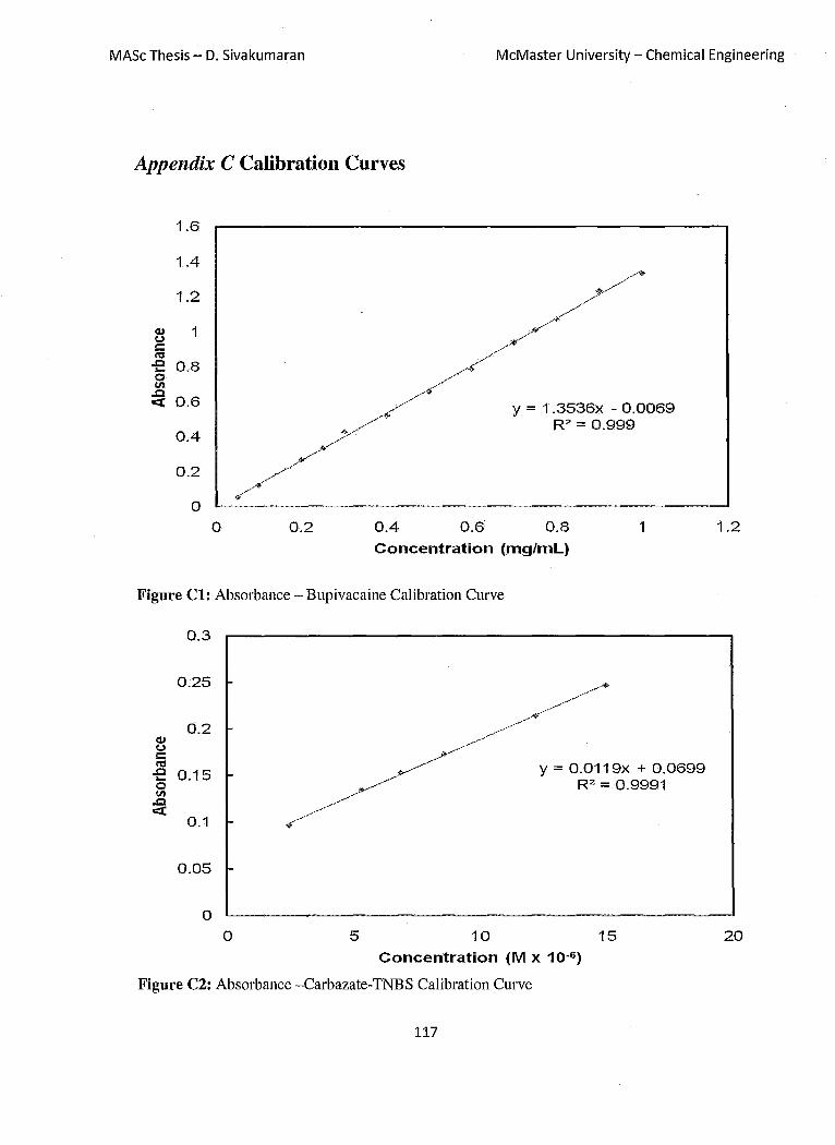
MASc Thesis - D. Sivakumaran McMaster University - Chemical Engineering
Appendix C Calibration Curves
1.6
1.4
1.2
<U 1 <oJ c: n:I .c 0.8 .... 0 II) .c
0.6 0:::(
0.4
0.2
0
0 0.2 0.4 0.6
y = 1.3536x - 0.0069 R2 = 0.999
0.8 1
Concentration (mg/mL)
Figure Cl: Absorbance - Bupivacaine Calibration Curve
0.3
0.25
0.2 <U <oJ c: n:I .c 0.15 .... 0 II) .c 0:::(
0.1
0.05
0
0 5
y = 0.0119x + 0.0699 R2 = 0.9991
10
Concentration (M x 10-6 )
15
Figure C2: Absorbance -Carbazate-TNBS Calibration Curve
117
1.2
20

MASc Thesis - D. Sivakumaran McMaster University - Chemical Engineering
Appendix D Additional Data
3
2.5 , I - I '" III
~ ~ Cl " i ~ .. E I~ ~
.. , y .. IIiII I!II .. - * 'f ~ .. :~
/. .. (1) 2
)I, ~ i U)
'" #1>. ~ .,+ (1)
~ A{j) ~ ~.;; at" 0:: "~ 'jJ<
1.5 I1l'~A, <0 " ! Ai , C ~t' on; I~t (J
'" 1 > 'a. :::I ... Ge11 IIiIIGel2 AGel3 co
0.5 xGel4 'KGeI5 egel6
a a 5 10 15 20 25 30
Time (Days)
Figure Dl: Reproducibility of Drug Release
Table Dl: Acrylic Acid Functionalization of P(NIP AM) Microgel
Polymer Mol COOH per gram of micro gel 6mol% Acrylic Acid Microgel 7.0 xlO-4
20mol % Acrylic Acid Microgel 1.3xlO-j
34mol% Acrylic Acid Microgel 2.4xlO-j
T bl D2 Ch a e arge D enslty 0 fAA P(NIPAM) M' - lcroge s Polymer Mol COOH per cm
j of AA-P(NIPAM)
Microgel 6mol% Acrylic Acid Microgel 5.6 xlO-4
20mol % Acrylic Acid Microgel 1.0 xlO-J
34mol% Acrylic Acid Microgel 1.8 xlO-J
118

MASc Thesis - D. Sivakumaran McMaster University - Chemical Engineering
Table D3: Charge Density of Hydrogels based on Hydrazide-Carboxymethyl Cellulose/ Aldehyde-Dextan
Polymer CMC-AO.5 CMC-A1.0 CMC-A1.5
1
0.9 -
0.8 c 0.7 0 -; ttl N 0.6 '2 .2 0.5 -0
Q> 0.4 Q> -en Q> 0.3 -Q
0.2
0.1
o -0 2 6
pH
Mol COOH per cmj Hydrogel 2.2 xlO-) 1.8 xlO-)
1.66 xlO-o
-+-AdipicAcid Dihydrazide -ilI-CMC
8 10
Figure D2: Degree of Ionization of Adipic Acid Dihydrazide and CMC
119
12

MASc Thesis - D. Sivakumaran McMaster University - Chemical Engineering
8
7 -~
6 ~
5 - ....
C"tS J ~4 c. -. ..........,.
3 -
2 -+-AdipicAcid Dihydrazide
1 - -m-CMC
o o 0.2 0.4 0.6 0.8 1 1.2
Degree of Ionization
Figure D3: pKa of Adipic Acid Dihydrazide and CMC
120
Top Related