


Languages
Pages
Legal


Article
Inhibitory Control in the C
ortico-Basal Ganglia-Thalamocortical Loop: Complex Regulation andInterplay with Memory and Decision ProcessesHighlights
d Modeling inhibitory control as a distributed process in the
cortico-subcortical system
d Long-distance recurrent dynamics of persistent activity
enhance inhibitory control
d Stop signal reaction time is insensitive to task difficulty in
perceptual decision
d Executive control differentially depends on various pathways
in the basal ganglia
Wei & Wang, 2016, Neuron 92, 1–13December 7, 2016 ª 2016 Elsevier Inc.http://dx.doi.org/10.1016/j.neuron.2016.10.031
Authors
Wei Wei, Xiao-Jing Wang
In Brief
Wei et al. propose a cortico-basal
ganglia-thalamocortical distributed
attractor network model for inhibitory
control. Besides reproducing recent
experimental findings and making
testable predictions, it provides a unified
framework to investigate inhibitory
control, perceptual decision making,
working memory, and their interactions.
Please cite this article in press as: Wei and Wang, Inhibitory Control in the Cortico-Basal Ganglia-Thalamocortical Loop: Complex Regulation andInterplay with Memory and Decision Processes, Neuron (2016), http://dx.doi.org/10.1016/j.neuron.2016.10.031
Neuron
Article
Inhibitory Control in the Cortico-BasalGanglia-Thalamocortical Loop: Complex Regulationand Interplay with Memory and Decision ProcessesWei Wei1 and Xiao-Jing Wang1,2,3,*1Center for Neural Science, New York University, New York, NY 10003, USA2NYU-ECNU Institute of Brain and Cognitive Science, NYU Shanghai, 200122 Shanghai, China3Lead Contact*Correspondence: [email protected]
http://dx.doi.org/10.1016/j.neuron.2016.10.031
SUMMARY
We developed a circuit model of spiking neuronsthat includes multiple pathways in the basal ganglia(BG) and is endowed with feedback mechanisms atthree levels: cortical microcircuit, corticothalamicloop, and cortico-BG-thalamocortical system. Wefocused on executive control in a stop signal task,which is known to depend on BG across species.Themodel reproduces a range of experimental obser-vations and shows that the newly discovered feed-back projection from external globus pallidus tostriatum is crucial for inhibitory control. Moreover,stopping process is enhanced by the cortico-subcor-tical reverberatory dynamics underlying persistentactivity, establishing interdependence betweenwork-ing memory and inhibitory control. Surprisingly, thestop signal reaction time (SSRT) can be adjusted byweights of certain connections but is insensitive toother connections in this complex circuit, suggestingnovel circuit-based intervention for inhibitory controldeficits associated with mental illness. Our modelprovides a unified framework for inhibitory control,decision making, and working memory.
INTRODUCTION
Across mammalian species, from rodent to monkey to human, a
conserved brain system is the prefrontal cortex-basal ganglia
(BG)-thalamic circuit. This cortico-subcortical system plays a
critical role in diverse cognitive functions, including perceptual
decision making (Ding and Gold, 2012; Forstmann et al., 2010;
Grinband et al., 2006), inhibitory control (Aron et al., 2003,
2007b; Aron and Poldrack, 2006; Jahanshahi et al., 2015), and
working memory (Floresco et al., 1999; Isseroff et al., 1982; Mills
et al., 2012; Parnaudeau et al., 2013;Wang, 2001;Watanabe and
Funahashi, 2012). A recent fMRI study revealed an overlapping
cortical representation of executive control and workingmemory
(Harding et al., 2016). It has also been found that degraded exec-
utive control function is usually associatedwithdeficits inworking
memory in schizophrenia (Gregoire et al., 2012; Zandbelt et al.,
2011). Intriguingly, in a task that engaged both perceptual deci-
sionmaking and inhibitory control, the two processeswere found
to be functionally independent of each other (Middlebrooks and
Schall, 2014), raising thequestionofwhether they could still share
a common underlying circuit. Understanding the circuit mecha-
nism for the interplay between these cognitive functions poses
a challenge for both experimental and modeling studies.
Inhibitory control, the ability to cancel a response when a
planned action becomes inappropriate, constitutes an essential
part of executive function (Schall and Godlove, 2012; Stuphorn,
2015; Verbruggen and Logan, 2009). Impaired inhibition function
has been found to be implicated in several neurological disorders
such as Parkinson’s disease (PD) (Gauggel et al., 2004; Mirabella
et al., 2012), attention-deficit hyperactivity disorder (Castellanos
et al., 2006; McAlonan et al., 2009), schizophrenia (Hughes et al.,
2012; Thakkar et al., 2011, 2015; Zandbelt et al., 2011), and also
normal aging (Andres et al., 2008; Coxon et al., 2012; Hu et al.,
2014). The stop signal task, in which a subject is required to sup-
press a planned action upon the occurrence of an unexpected
stop signal, provides a standard paradigm for investigating
inhibitory control (Verbruggen and Logan, 2008, 2009). It allows
for the estimation of the latency of a covert stop process, i.e.,
the stop signal reaction time (SSRT), according to the racemodel
(Boucher et al., 2007; Logan and Cowan, 1984; Logan et al.,
2014). SSRT measures the effectiveness of inhibitory control
(the shorter the SSRT, the better one’s ability to suppress prepo-
tent but inappropriate action), and it has been widely used to
assess impaired inhibitory control in psychiatric patients. A
recent experiment provides direct neurophysiological evidence
for the involvement of the BG in the stop process: a transient
surge of neural activity in the subthalamic nucleus (STN) upon a
stop signal presentation and the corresponding increase of activ-
ity in the substantia nigra pars reticulate (SNr) in successful stop
signal trials were well within the window of the SSRT (Schmidt
et al., 2013). In pro/anti-saccade tasks that involve response
inhibition, the higher-order thalamus (Th) also showed strong
modulation during response initiation (Tanaka and Kunimatsu,
2011). Meanwhile, higher-order Th receives topographic inputs
from the SNr (Gulcebi et al., 2012) and forms reciprocal connec-
tions with the frontal cortex (Alexander et al., 1986; Haber and
Calzavara, 2009; Parent and Hazrati, 1995). How inhibitory con-
trol depends on the multiple pathways through the BG and
Neuron 92, 1–13, December 7, 2016 ª 2016 Elsevier Inc. 1
A C
B
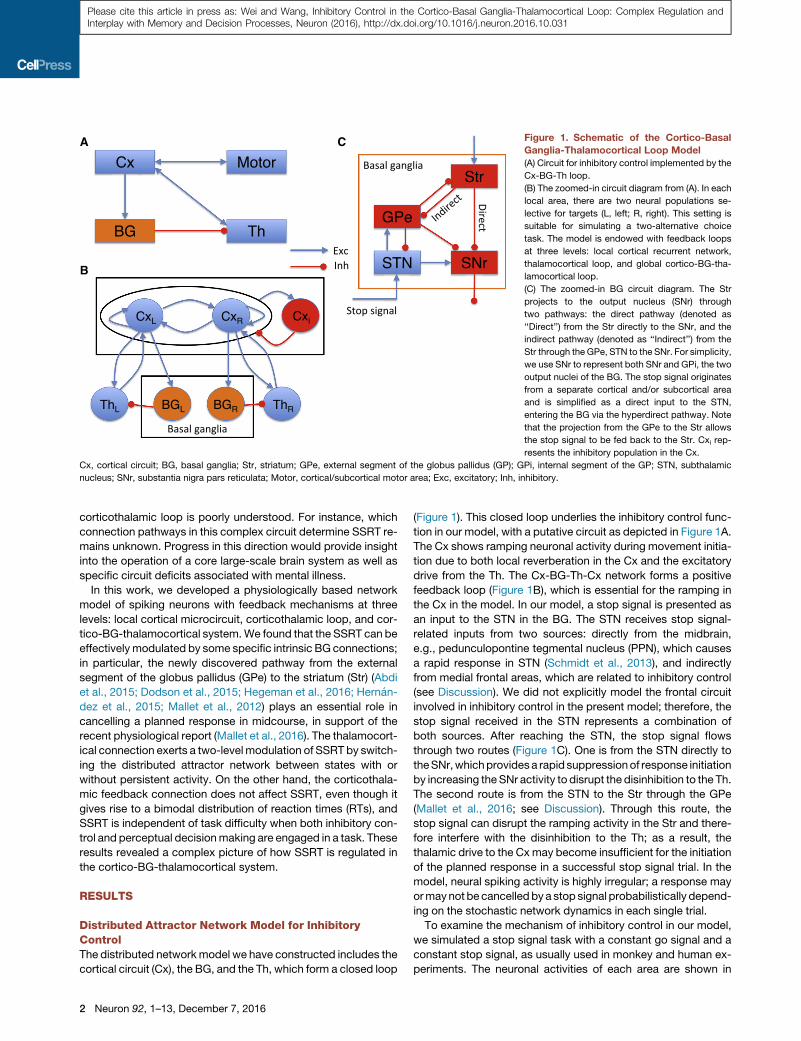
Figure 1. Schematic of the Cortico-Basal
Ganglia-Thalamocortical Loop Model
(A) Circuit for inhibitory control implemented by the
Cx-BG-Th loop.
(B) The zoomed-in circuit diagram from (A). In each
local area, there are two neural populations se-
lective for targets (L, left; R, right). This setting is
suitable for simulating a two-alternative choice
task. The model is endowed with feedback loops
at three levels: local cortical recurrent network,
thalamocortical loop, and global cortico-BG-tha-
lamocortical loop.
(C) The zoomed-in BG circuit diagram. The Str
projects to the output nucleus (SNr) through
two pathways: the direct pathway (denoted as
‘‘Direct’’) from the Str directly to the SNr, and the
indirect pathway (denoted as ‘‘Indirect’’) from the
Str through the GPe, STN to the SNr. For simplicity,
we use SNr to represent both SNr and GPi, the two
output nuclei of the BG. The stop signal originates
from a separate cortical and/or subcortical area
and is simplified as a direct input to the STN,
entering the BG via the hyperdirect pathway. Note
that the projection from the GPe to the Str allows
the stop signal to be fed back to the Str. Cxi rep-
resents the inhibitory population in the Cx.
Cx, cortical circuit; BG, basal ganglia; Str, striatum; GPe, external segment of the globus pallidus (GP); GPi, internal segment of the GP; STN, subthalamic
nucleus; SNr, substantia nigra pars reticulata; Motor, cortical/subcortical motor area; Exc, excitatory; Inh, inhibitory.
Please cite this article in press as: Wei and Wang, Inhibitory Control in the Cortico-Basal Ganglia-Thalamocortical Loop: Complex Regulation andInterplay with Memory and Decision Processes, Neuron (2016), http://dx.doi.org/10.1016/j.neuron.2016.10.031
corticothalamic loop is poorly understood. For instance, which
connection pathways in this complex circuit determine SSRT re-
mains unknown. Progress in this direction would provide insight
into the operation of a core large-scale brain system as well as
specific circuit deficits associated with mental illness.
In this work, we developed a physiologically based network
model of spiking neurons with feedback mechanisms at three
levels: local cortical microcircuit, corticothalamic loop, and cor-
tico-BG-thalamocortical system.We found that the SSRT can be
effectively modulated by some specific intrinsic BGconnections;
in particular, the newly discovered pathway from the external
segment of the globus pallidus (GPe) to the striatum (Str) (Abdi
et al., 2015; Dodson et al., 2015; Hegeman et al., 2016; Hernan-
dez et al., 2015; Mallet et al., 2012) plays an essential role in
cancelling a planned response in midcourse, in support of the
recent physiological report (Mallet et al., 2016). The thalamocort-
ical connection exerts a two-level modulation of SSRT by switch-
ing the distributed attractor network between states with or
without persistent activity. On the other hand, the corticothala-
mic feedback connection does not affect SSRT, even though it
gives rise to a bimodal distribution of reaction times (RTs), and
SSRT is independent of task difficulty when both inhibitory con-
trol and perceptual decisionmaking are engaged in a task. These
results revealed a complex picture of how SSRT is regulated in
the cortico-BG-thalamocortical system.
RESULTS
Distributed Attractor Network Model for InhibitoryControlThe distributed networkmodel we have constructed includes the
cortical circuit (Cx), the BG, and the Th, which form a closed loop
2 Neuron 92, 1–13, December 7, 2016
(Figure 1). This closed loop underlies the inhibitory control func-
tion in our model, with a putative circuit as depicted in Figure 1A.
The Cx shows ramping neuronal activity during movement initia-
tion due to both local reverberation in the Cx and the excitatory
drive from the Th. The Cx-BG-Th-Cx network forms a positive
feedback loop (Figure 1B), which is essential for the ramping in
the Cx in the model. In our model, a stop signal is presented as
an input to the STN in the BG. The STN receives stop signal-
related inputs from two sources: directly from the midbrain,
e.g., pedunculopontine tegmental nucleus (PPN), which causes
a rapid response in STN (Schmidt et al., 2013), and indirectly
from medial frontal areas, which are related to inhibitory control
(see Discussion). We did not explicitly model the frontal circuit
involved in inhibitory control in the present model; therefore, the
stop signal received in the STN represents a combination of
both sources. After reaching the STN, the stop signal flows
through two routes (Figure 1C). One is from the STN directly to
theSNr,whichprovidesa rapid suppressionof response initiation
by increasing the SNr activity to disrupt the disinhibition to the Th.
The second route is from the STN to the Str through the GPe
(Mallet et al., 2016; see Discussion). Through this route, the
stop signal can disrupt the ramping activity in the Str and there-
fore interfere with the disinhibition to the Th; as a result, the
thalamic drive to the Cxmay become insufficient for the initiation
of the planned response in a successful stop signal trial. In the
model, neural spiking activity is highly irregular; a response may
ormaynot be cancelled bya stop signal probabilistically depend-
ing on the stochastic network dynamics in each single trial.
To examine the mechanism of inhibitory control in our model,
we simulated a stop signal task with a constant go signal and a
constant stop signal, as usually used in monkey and human ex-
periments. The neuronal activities of each area are shown in

A B C
D E F
Figure 2. Single-Trial Simulations of the
Stop Signal Task
Spike trains (raster plots) are shown for a go trial
(black) and a successful stop signal trial (blue) in
each area (A–F; upper panels). Population firing
rates in each area (A–F; lower panels) are shown for
a go trial (black), and successful (blue) and failed
(green) stop signal trials. The schematic in (A) il-
lustrates a constant go stimulus that is turned on
at time 0 (black) and two SSDs for the stop signal
at 120 (blue) and 170 ms (green), respectively.
Please cite this article in press as: Wei and Wang, Inhibitory Control in the Cortico-Basal Ganglia-Thalamocortical Loop: Complex Regulation andInterplay with Memory and Decision Processes, Neuron (2016), http://dx.doi.org/10.1016/j.neuron.2016.10.031
Figure 2 for the go trial (black), successful (blue), and failed (non-
cancelled; green) stop signal trials, respectively. The raster plots
in each area represent spike trains of the population selective to
the go stimulus in the go trial (black) and successful stop signal
trial (blue). The lower panel of Figure 2A shows a schematic of the
go stimulus and two stop signal delays (SSDs) (blue, 120 ms;
green, 170 ms). In the go trial, the ramping activity in the Cx (Fig-
ure 2A) leads to an increase of Str activity (Figure 2D) and a
decrease of the SNr activity (Figure 2C), which releases the inhi-
bition to the Th (Figure 2F) and further facilitates the ramping ac-
tivity in the Cx. The stop signal elevates the STN activity (Fig-
ure 2E). In a successful stop signal trial (blue), at the time of
stop signal onset, the SNr activity is not suppressed too much
by the striatal input yet, and the elevated STN activity can in-
crease the SNr activity. This leads to decreased Th activity and
terminates the ramping in the Cx. The stop signal flowing through
STN-to-GPe-to-Str increases the GPe activity (Figure 2B) and
then reduces the Str activity, leading to a weakened go process.
In a non-cancelled stop signal trial (green), the SNr activity is
strongly suppressed by the striatal input at the time of the stop
signal arrival. In this case, the elevated STN activity is insufficient
to counteract the strong striatal input to the SNr and disrupt the
disinhibition to the Th, and the response is not cancelled.
Successful stopping happens only in a fraction of the trials for
a given SSD. In our model, the competition between two selec-
tive populations in the Cx results in varia-
bly ramping slopes of the winner popula-
tion, which provides the main drive for
the fluctuating neuronal activity in the Cx
and the downstream circuits from trial to
trial. In trials with faster cortical ramping,
the SNr also decreases faster and is less
likely to increase its activity at the occur-
rence of the stop signal due to the stron-
ger inhibitory drive from the Str. The frac-
tion of non-cancelled stop signal trials
(normalized by the performance in go tri-
als) gives the non-cancelled probability,
which can be used to estimate the SSRT
through the integration method (Logan
and Cowan, 1984). The idea is that SSRT
quantifies the time needed for a stop pro-
cess to ‘‘win the race’’ over the go pro-
cess; therefore, non-cancelled trials are
those with RTs shorter than the sum of SSD and SSRT; see Fig-
ure 3A and Supplemental Experimental Procedures, available
online, for details. This suggests that the large-scale spiking
network model of the cortico-BG-thalamocortical system we
have introduced is behaviorally consistent with the race model
(Boucher et al., 2007; Logan and Cowan, 1984). The non-
cancelled probability as a function of the SSD defines the inhibi-
tion function, as shown in Figure 3B. The non-cancelled proba-
bility increases with the SSD, from completely cancelling at small
SSD to totally failing to cancel at large SSD, as reported in
numerous experiments (e.g., Hanes and Schall, 1995; Logan
and Cowan, 1984). The mean neuronal activities for non-
cancelled and cancelled stop signal trials, and the correspond-
ing latency-matched fast and slow go trials, were shown in Fig-
ure S1. Here, latency-matched fast and slow go trials refer to
those go trials with RTs shorter and longer than SSD plus
SSRT, respectively. Consistent with recent experimental find-
ings (Schmidt et al., 2013), the variable timing of the striatal ac-
tivity for go trials determines whether the cancellation of
response by the stop signal is successful or not (Figures S1D
and S1G). Note that despite the similar activities between Cx
and Str in our model (Figure S1), the cortical activity itself does
not provide such a determinant since it only receives stop-
related activity through the BG-to-Th-to-Cx route, while as sug-
gested by both experiments and our modeling study (see next
Neuron 92, 1–13, December 7, 2016 3

A B Figure 3. SSRT and Inhibition Function from
the Model
(A) Upper panel: integration method for estimating
the SSRT. The black curve represents the cumulative
density function (cdf) for the reaction times (RTs) of
go trials, and the green curve represents the cdf for
the RTs of non-cancelled stop signal trials with
SSD = 270 ms, scaled by the overall non-cancelled
probability (fraction of non-cancelled stop signal tri-
als, blue dashed line). The vertical red dashed line
represents the intersection time of the blue dashed
line and the black curve. The solid red line indicates
the SSD. The interval between the vertical solid and
dashed red lines gives an estimate of the SSRT.
Lower panel: RT probability densities for the go trials
(black curve) and non-cancelled stop signal trials
(green curve; scaled by non-cancelled probability).
(B) The non-cancelled probability as a function of the
SSD (the inhibition function), which is fitted by the
Weibull function.
Please cite this article in press as: Wei and Wang, Inhibitory Control in the Cortico-Basal Ganglia-Thalamocortical Loop: Complex Regulation andInterplay with Memory and Decision Processes, Neuron (2016), http://dx.doi.org/10.1016/j.neuron.2016.10.031
section), the GPe-to-Str route for stop signal transmission is
essential for successful stopping.
SSRT Depends on Specific BG Circuit ConnectionsThe stop signal in our model is conveyed through the hyperdirect
pathway of the BG to the STN. To cancel a response, the activa-
tion of the stop signal on the SNr has to counterbalance the
direct pathway input to the SNr.We found that the non-cancelled
probability increases with direct pathway strength gStr�SNr (Fig-
ure 4A, left), since it is harder for the stop signal to counterbal-
ance the direct pathway input to the SNr and disrupt the disinhi-
bition to the Th when the strength of the direct pathway is
stronger. The SSRT increases with gStr�SNr (Figure 4A, right),
indicating a reduction of stopping function with the increase of
the direct pathway strength. At the neuronal level, the SNr has
a lower spontaneous firing rate and decreases faster during
response initiation for a larger gStr�SNr (Figure S2A), increasing
the chance that SNr activity drops to a critically low level by
the onset of the stop signal.
We further checked how the backpropagation of the stop
signal from the STN to the Str through the GPe might impact
inhibitory control. When the strength of the GPe-to-Str connec-
tion, gGPe�Str , is increased, we found that the non-cancelled
probability is reduced (Figure 4B, left) and the SSRT is decreased
(Figure 4B, right). On the other hand, in the absence of the GPe-
to-Str connection, the model failed completely in performing
inhibitory control, as indicated by the fact that the non-cancelled
probabilities are near 1 (black curve in the left panel of Figure 4B;
gGPe�Str = 0). Note that for fair comparison, we adjusted the
background input to the SNr and kept the spontaneous SNr firing
rate unchanged for different gGPe�Str , to distinguish from the
impact of direct pathway strength gStr�SNr , which influences
the stopping function by modulating the SNr activity.
4 Neuron 92, 1–13, December 7, 2016
With a stronger gGPe�Str , the stop signal
backpropagating to the Str can more easily
curtail the ramping activity there, which
makes the SNr activity decrease slower,
thereby facilitating the stopping function (Figure S2B). This result
resonates with the recent experimental finding of an important
role in the stopping of arkypallidal cells in the GPe that mediate
this feedback projection to the Str (Mallet et al., 2016). The
striatal activity therefore provides a determinant for stopping
behavior, where the two routes of stop-related activity transmis-
sion, from GPe to Str and from SNr to Cx through Th, converge
(Schmidt et al., 2013; see also Figures S1D and S1G). The inhibi-
tion functions for different gStr�SNr (Figures S3A and S3B) and
gGPe�Str (Figures S3C and S3D) are aligned with each other
when plotting against the z score of the relative finishing time
(ZRFT) (Logan and Cowan, 1984).
We have also performed simulations for an extended circuit, in
which the Str neurons are segregated into direct (D1-expressing)
and indirect (D2-expressing) pathway neurons, and the GPe
neurons are segregated into Ark and Pro types of neurons,
respectively (Figure 4C). With this extended circuit, we per-
formed simulations on the impact of Ark-to-Str back-projection
on inhibitory control, in which we assumed symmetric projection
from Ark neurons to D1- and D2-expressing striatal neurons
since no such data are available yet (Hegeman et al., 2016).
We found a similar impact as that shown in Figure 4B (Figure 4D).
Therefore, segregation of the Str and GPe into sub-populations
does not change the results we have obtained from the simplified
circuit. We also showed that choosing a different level of deci-
sion threshold at the Cx (Figures S4A–S4E) or scaling the sparse
GPe-STN connections to all-to-all connections (Figures S4F and
S4G) does not change the results.
Self-Sustained Persistent Activity Facilitates StoppingFunctionWhen excitatory feedback connections are sufficiently strong, a
recurrent neural circuit can generate self-sustained persistent

A C
B D
Figure 4. SSRT Depends on Weights of Specific Connections in the BG
(A) Left: dependence of inhibition functions on gStr�SNr . Right: the SSRT increases with a larger gStr�SNr .
(B) Left: dependence of inhibition functions on gGPe�Str . Right: the SSRT decreases with a larger gGPe�Str .
(C) Schematic of the extended BG circuit. The Str neurons are segregated into direct (D1-expressing) and indirect (D2-expressing) pathway neurons, and the GPe
neurons are segregated into Ark and Pro types of neurons. The Ark neurons project back to both types of Str neurons, and the Pro neurons project to the STN and
SNr. The indirect pathway striatal neurons and STN neurons project to both Ark and Pro neurons.
(D) Left: dependence of inhibition functions on the back-projection strength from the Ark neurons to the Str, gArk�Str . Right: the SSRT decreases with a larger
gArk�Str .
Error bars indicate SD of SSRT values estimated for each SSD. Note that in (B) and (D), for fair comparison when gGPe�Str or gArk�Str is varied, the spontaneous
firing rate of the SNr is fixed by adjusting the external input rate yext to it. The inhibition functions in (A), (B), and (D) are fitted by the Weibull function.
Please cite this article in press as: Wei and Wang, Inhibitory Control in the Cortico-Basal Ganglia-Thalamocortical Loop: Complex Regulation andInterplay with Memory and Decision Processes, Neuron (2016), http://dx.doi.org/10.1016/j.neuron.2016.10.031
activity (attractor states) (Wang, 2001). The existence of an at-
tractor state in our model is determined by both the local recur-
rent strength w+ in the Cx and the feedback strength of the cor-
tico-subcortical loop, for instance, gTh�Cx (see a schematic of the
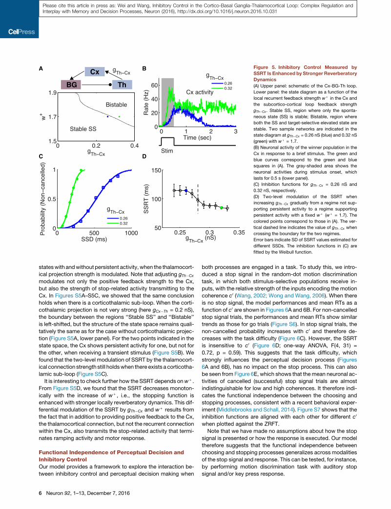
closed Cx-BG-Th loop in Figure 5A, upper panel). We assessed
self-sustained activity states of the model system (in the
absence of external input) as a function of w+ and gTh�Cx (Fig-
ure 5A, lower panel). In the left corner, the spontaneous state
(SS) is the only stable state, and the neuronal activity of the se-
lective population decays back to the SS after the stimulus is
offset. The upper right corner represents a bistable region, where
both the SS and the elevated state are stable. When gTh�Cx in-
creases further for a given w+ (region not shown), the SS be-
comes unstable and both stimulus-selective (for left and right,
L and R) populations can be found in that state. Note that the bi-
stable region exists only when w+ is larger than some critical
value, such that the competition between the two (L and R) pop-
ulations in theCx is strong enough to implement thewinner-take-
all mechanism (Wang, 2002). In Figure 5B, we showed the
neuronal activity of the winner population in the Cx for two repre-
sentative samples in the state space (blue and green squares in
Figure 5A, lower panel;w+ = 1.7) with gTh�Cx = 0.26 nS (blue) and
0.32 nS (green), respectively. When a transient stimulus is shown
for 500 ms (Figure 5B, lower panel), it triggers persistent activity
for gTh�Cx = 0.32 nS (green curve), but not for gTh�Cx = 0.26 nS
(blue curve). Therefore, by increasing the feedback strength of
the big loop, the circuit can be switched from a state without
persistent activity to a state that supports self-sustained persis-
tent activity and therefore working memory, under the assump-
tion that local reverberation in the Cx is insufficient to maintain
persistent activity. We further note that when gTh�Cx is above
some critical value (�0.2 nS here for w+ = 1.7), the Cx shows
ramping neuronal activity during the presentation of the stimulus
even though only the SS is stable after the stimulus offset. A local
circuit with ramping activity but without persistent activity has
been investigated in Wong and Wang (2006).
We compared inhibitory control function for these two cases.
When there is self-sustained persistent activity, the inhibition
function is left shifted (Figure 5C), indicating a faster go process
due to a stronger Cx-BG-Th-Cx feedback loop, as compared to
the case when there is no persistent activity. Meanwhile, the
SSRT is significantly reduced when there is persistent activity
(Figure 5D, colored points), indicating also a faster stop process.
This suggests an enhancement of stopping function due to self-
sustained persistent activity. To further elucidate the relationship
between persistent activity dynamics and SSRT, we increased
gTh�Cx gradually from the regime without persistent activity to
the regime supporting persistent activity with a fixed w+ (w+ =
1.7). We observed a two-level profile for the SSRT: when gTh�Cx
is varied but without crossing the phase boundary (vertical
dashed line in Figure 5D), the SSRT is insensitive to this change.
There are two different levels of the SSRT, corresponding to the
Neuron 92, 1–13, December 7, 2016 5
A B
C D
Figure 5. Inhibitory Control Measured by
SSRT Is Enhanced by Stronger Reverberatory
Dynamics
(A) Upper panel: schematic of the Cx-BG-Th loop.
Lower panel: the state diagram as a function of the
local recurrent feedback strength w+ in the Cx and
the subcortico-cortical loop feedback strength
gTh�Cx. Stable SS, region where only the sponta-
neous state (SS) is stable; Bistable, region where
both the SS and target-selective elevated state are
stable. Two sample networks are indicated in the
state diagram at gTh�Cx = 0.26 nS (blue) and 0.32 nS
(green) with w+ = 1.7.
(B) Neuronal activity of the winner population in the
Cx in response to a brief stimulus. The green and
blue curves correspond to the green and blue
squares in (A). The gray-shaded area shows the
neuronal activities during stimulus onset, which
lasts for 0.5 s (lower panel).
(C) Inhibition functions for gTh�Cx = 0.26 nS and
0.32 nS, respectively.
(D) Two-level modulation of the SSRT when
increasing gTh�Cx gradually from a regime not sup-
porting persistent activity to a regime supporting
persistent activity with a fixed w+ (w+ = 1.7). The
colored points correspond to those in (A). The ver-
tical dashed line indicates the value of gTh�Cx when
crossing the boundary for the two regimes.
Error bars indicate SD of SSRT values estimated for
different SSDs. The inhibition functions in (C) are
fitted by the Weibull function.
Please cite this article in press as: Wei and Wang, Inhibitory Control in the Cortico-Basal Ganglia-Thalamocortical Loop: Complex Regulation andInterplay with Memory and Decision Processes, Neuron (2016), http://dx.doi.org/10.1016/j.neuron.2016.10.031
stateswith andwithout persistent activity,when the thalamocort-
ical projection strength is modulated. Note that adjusting gTh�Cx
modulates not only the positive feedback strength to the Cx,
but also the strength of stop-related activity transmitting to the
Cx. In Figures S5A–S5C, we showed that the same conclusion
holds when there is a corticothalamic sub-loop. When the corti-
cothalamic projection is not very strong (here gCx�Th = 0.2 nS),
the boundary between the regions ‘‘Stable SS’’ and ‘‘Bistable’’
is left-shifted, but the structure of the state space remains quali-
tatively the same as for the case without corticothalamic projec-
tion (Figure S5A, lower panel). For the two points indicated in the
state space, the Cx shows persistent activity for one, but not for
the other, when receiving a transient stimulus (Figure S5B). We
found that the two-level modulation of SSRT by the thalamocort-
ical connection strength still holdswhen there exists a corticotha-
lamic sub-loop (Figure S5C).
It is interesting to check further how the SSRT depends onw+ .
From Figure S5D, we found that the SSRT decreases monoton-
ically with the increase of w+ , i.e., the stopping function is
enhanced with stronger locally reverberatory dynamics. This dif-
ferential modulation of the SSRT by gTh�Cx and w+ results from
the fact that in addition to providing positive feedback to the Cx,
the thalamocortical connection, but not the recurrent connection
within the Cx, also transmits the stop-related activity that termi-
nates ramping activity and motor response.
Functional Independence of Perceptual Decision andInhibitory ControlOur model provides a framework to explore the interaction be-
tween inhibitory control and perceptual decision making when
6 Neuron 92, 1–13, December 7, 2016
both processes are engaged in a task. To study this, we intro-
duced a stop signal in the random-dot motion discrimination
task, in which both stimulus-selective populations receive in-
puts, with the relative strength of the inputs encoding the motion
coherence c0 (Wang, 2002; Wong and Wang, 2006). When there
is no stop signal, the model performances and mean RTs as a
function of c0 are shown in Figures 6A and 6B. For non-cancelled
stop signal trials, the performances and mean RTs show similar
trends as those for go trials (Figure S6). In stop signal trials, the
non-cancelled probability increases with c0 and therefore de-
creases with the task difficulty (Figure 6C). However, the SSRT
is insensitive to c0 (Figure 6D; one-way ANOVA, F(4, 31) =
0.72, p = 0.59). This suggests that the task difficulty, which
strongly influences the perceptual decision process (Figures
6A and 6B), has no impact on the stop process. This can also
be seen from Figure 6E, which shows that the mean neuronal ac-
tivities of cancelled (successful) stop signal trials are almost
indistinguishable for low and high coherences. It therefore indi-
cates the functional independence between the choosing and
stopping processes, consistent with a recent behavioral exper-
iment (Middlebrooks and Schall, 2014). Figure S7 shows that the
inhibition functions are aligned with each other for different c0
when plotted against the ZRFT.
Note that we have made no assumptions about how the stop
signal is presented or how the response is executed. Our model
therefore suggests that the functional independence between
choosing and stopping processes generalizes across modalities
of the stop signal and response. This can be tested, for instance,
by performing motion discrimination task with auditory stop
signal and/or key press response.

A
C
E
B
D
Figure 6. SSRT Is Independent of Task Diffi-
culty in Perceptual Decision Making
(A and B) Performances (A) and mean RTs (B) from
model simulation of a random-dot motion direction
discrimination task.
(C) Inhibition functions for different task difficulty
represented by coherence of the random moving
dot input, c0. The values of c0 from bottom to top
are 0%, 3.2%, 6.4%, 12.8%, and 25.6%. The
inhibition functions are fitted by the Weibull
function.
(D) The SSRT as a function of c0, showing no sta-
tistically significant dependence. Error bars indi-
cate SD of SSRT values estimated for those SSDs
with corresponding non-cancelled probabilities
within the interval (0.1, 0.9).
(E) Mean neuronal activities for successful stop
signal trials (solid curves) and latency-matched go
trials (dashed curves) with SSD = 420 ms when c0 =0% (blue) and 12.8% (green). The mean activities
are obtained from averaging over 200 trials of the
same types.
Please cite this article in press as: Wei and Wang, Inhibitory Control in the Cortico-Basal Ganglia-Thalamocortical Loop: Complex Regulation andInterplay with Memory and Decision Processes, Neuron (2016), http://dx.doi.org/10.1016/j.neuron.2016.10.031
Impact of Corticothalamic Sub-loop on StoppingFunctionUp to this point, we have not included the corticothalamic
connection in the model (except in Figures S5A–S5C), so
we can examine the cortico-BG-thalamocortical loop. Now
we investigate the impact of such a connection on stopping
function by including non-zero corticothalamic connection
strength, gCx�Th. This sub-loop makes it possible for the rever-
beratory dynamics to rebound in spite of transient suppression
by a stop signal, leading to slow responses and a bimodal
distribution of RTs in non-cancelled trials (Figure 7A, lower
panel; gCx�Th = 0.4 nS). Note that the second peak has a
totally different origin from the first one. While the first peak
is driven by stimulus and reflects the most likely time for
ramping Cx activity crossing the threshold, the second peak
is driven mainly by noise when the constant stop signal
enhances SNr activity and diminishes
ramping Cx activity. This is reflected by
the very steep rising of Cx activity for tri-
als with RTs located near the second
peak. The RT distributions for different
SSDs are shown in Figures S8A and
S8B with gCx�Th = 0.4 nS. Interestingly,
this computational finding is supported
by empirical observations from experi-
ments using rats (Schmidt et al., 2013).
Our model thus suggests that the
bimodal RT distribution for rats perform-
ing a stop signal task might result from a
strong corticothalamic sub-loop. Note
that our model assumes that even
when the sensory stimulus for signaling
stop is transient, the internal representa-
tion of the stop signal is sustained. This
assumption is crucial and remains to
be tested in future experiments.
Figure 7A (upper panel) shows the cumulative density function
(cdf) of RTs. The classical integration method for estimating
SSRT is no longer applicable, since there is a substantial fraction
of non-cancelled stop signal trials (with long RTs) for which there
are no latency-matched go trials. We therefore estimated the
SSRT using a modified integration method (Mayse et al., 2014),
where the divergent point of the cdf for non-cancelled stop signal
trials and go trials was used to estimate the SSRT (see Supple-
mental Experimental Procedures for details).
We then investigated how the strength of the Cx-Th sub-loop
influences the SSRT. Figure 7B (upper panel) shows a schematic
of the circuit.We increased gCx�Th from0 to 0.6 nS and estimated
the SSRT for each value of gCx�Th with gTh�Cx = 0:26 nS, for which
there is no persistent activity in the network (Figure 7B, middle
panel) when receiving a transient stimulus (Figure 7B, lower
panel). As shown in Figure 7C, the SSRT is independent of
Neuron 92, 1–13, December 7, 2016 7

A B
C
Figure 7. Impact of Cx-Th Sub-loop on
Inhibitory Control
(A) Lower panel: the RTs of go trials (black) and non-
cancelled stop signal trials (green). Note the double
peaks in the distribution of the non-cancelled stop
signal trials. Here the corticothalamic connection
strength gCx�Th = 0:4 nS. Upper panel: modified
integration method for estimating SSRT, illustrated
with SSD = 220 ms. Black curve represents the cdf
for RTs of the go trials, and green curve represents
the cdf for the non-cancelled trials, scaled by the
non-cancelled probability (blue dashed line). The
dashed black curve represents the 99.9% confi-
dence interval (CI) of the cdf for RTs of go trials. The
vertical red dashed line represents the intersection
time of the green curvewith the black dashed curve.
The red solid line indicates the SSD. The interval
between the vertical solid and dashed red lines
gives an estimate of the SSRT.
(B) Upper panel: schematic of the circuit. Middle
panel: neuronal activities of the winner population in
the Cx in response to a brief stimulus for gCx�Th = 0
(blue) and 0.4 nS (green), respectively. The gray-
shaded area indicates neuronal activities during
stimulus onset, which lasts for 0.5 s (lower panel).
The stimulus is a random-dot input with c0 = 25.6%.
(C) SSRT is insensitive to gCx�Th.
Error bars indicate SD of SSRT values estimated
for different SSDs. In this figure, gTh�Cx = 0.26 nS.
Please cite this article in press as: Wei and Wang, Inhibitory Control in the Cortico-Basal Ganglia-Thalamocortical Loop: Complex Regulation andInterplay with Memory and Decision Processes, Neuron (2016), http://dx.doi.org/10.1016/j.neuron.2016.10.031
gCx�Th (one-way ANOVA, F(3, 12) = 0.38, p = 0.77). Note that the
sameconclusion alsoholdswhen there ispersistent activity in the
circuit. In Figure S8C, theCx circuit shows persistent activity with
gTh�Cx =0.32nS (upper panel)when receiving a transient stimulus
(lower panel). We found that the SSRT is independent of gCx�Th
(Figure S8D; one-way ANOVA, F(2, 9) = 0.54, p = 0.60).
DISCUSSION
Inhibitory control of action is central for flexible behavior, charac-
terized by SSRT, which has been used to quantify inhibitory con-
trol ability as well its impairment in psychiatric illness. The BG are
known to play an important role in inhibitory control, but only
recent work has begun to dissect its underlying circuit mecha-
nism in the BG. The spiking neural circuit model presented in
this paper accounts for salient experimental observations
including the bimodal distribution of RTs in a stop signal task
(Schmidt et al., 2013), as well as the surprising finding that
SSRT is independent of task difficulty in perceptual decision
making (Middlebrooks and Schall, 2014). This biologically con-
strained model with explanatory power revealed a complex pic-
ture of how different connection pathways modulate SSRT. This
is an important new insight into BG function, opening up new ex-
periments to test the model and ideas about potential circuit-
specific ways to remedy inhibitory control deficits in psychiatric
patients. Another conceptual advance is to establish interdepen-
dence between inhibitory control ability and working memory in
the cortico-BG-thalamocortical system.
In this work, we propose a distributed attractor network model
for executive control. A stop signal enters the BG through the hy-
perdirect pathway, and then interacts with the direct and indirect
8 Neuron 92, 1–13, December 7, 2016
pathways to subserve inhibitory control in stop signal tasks. This
cortico-BG-thalamocortical system involves three feedback loops
at multiple levels, and a main finding of this work is the complex
dependence of the effectiveness of inhibitory control on various
pathways, as summarized in Figure 8. The SSRT is increased
when the ‘‘go’’ process becomes more potent by increasing the
Str-to-SNr strength. On the other hand, the SSRT is decreased
when the GPe-to-Str connection is stronger, which permits more
potent backpropagation of the stop signal to the Str. The SSRT
is also decreased when the network is switched from a state
without persistent activity to a state supporting persistent activity,
establishing a link between working memory and inhibitory con-
trol. Our model offers an explanation for several recent experi-
mental observations in stop signal tasks. First, the GPe-to-Str
feedback connection, mediated by arkypallidal cells, is essential
for inhibitory control (Mallet et al., 2016). Second, when inhibitory
control is combined with perceptual decision making, SSRT is in-
dependent of the task difficulty (Middlebrooks and Schall, 2014).
Third, our model suggests that a non-negligible corticothalamic
sub-loop leads to a bimodal distribution of RTs for non-cancelled
stop signal trials (Schmidt et al., 2013) and predicts that the
strength of corticothalamic projection does not change SSRT.
This model prediction can be tested in future experiments.
Previously, a closed cortico-BG-thalamocortical loop has
been proposed to implement different cognitive functions. This
loop has been suggested to implement workingmemory retrieval
and response selection (Humphries and Gurney, 2002; Schroll
et al., 2012; Vitay and Hamker, 2010). In another modeling study,
imbalanced direct and hyperdirect pathways led to the loss of
action selection and the generation of oscillatory activity (Leblois
et al., 2006). Recently, the competition between hyperdirect and
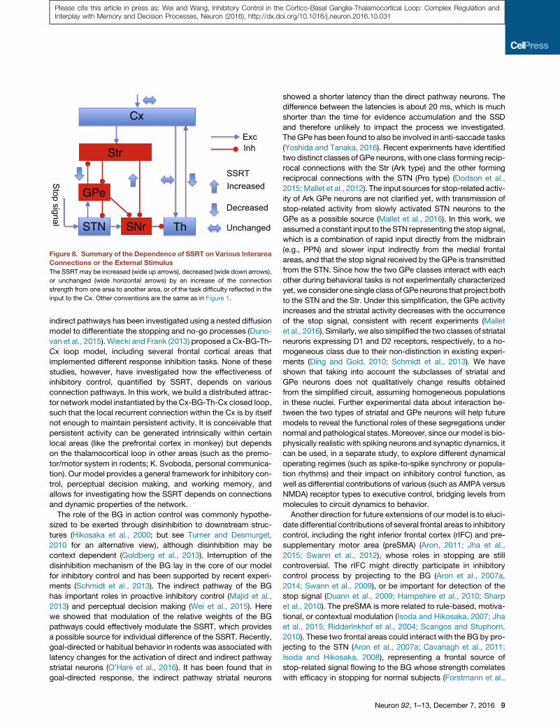
Figure 8. Summary of the Dependence of SSRT on Various Interarea
Connections or the External Stimulus
The SSRT may be increased (wide up arrows), decreased (wide down arrows),
or unchanged (wide horizontal arrows) by an increase of the connection
strength from one area to another area, or of the task difficulty reflected in the
input to the Cx. Other conventions are the same as in Figure 1.
Please cite this article in press as: Wei and Wang, Inhibitory Control in the Cortico-Basal Ganglia-Thalamocortical Loop: Complex Regulation andInterplay with Memory and Decision Processes, Neuron (2016), http://dx.doi.org/10.1016/j.neuron.2016.10.031
indirect pathways has been investigated using a nested diffusion
model to differentiate the stopping and no-go processes (Duno-
van et al., 2015). Wiecki and Frank (2013) proposed a Cx-BG-Th-
Cx loop model, including several frontal cortical areas that
implemented different response inhibition tasks. None of these
studies, however, have investigated how the effectiveness of
inhibitory control, quantified by SSRT, depends on various
connection pathways. In this work, we build a distributed attrac-
tor networkmodel instantiated by the Cx-BG-Th-Cx closed loop,
such that the local recurrent connection within the Cx is by itself
not enough to maintain persistent activity. It is conceivable that
persistent activity can be generated intrinsically within certain
local areas (like the prefrontal cortex in monkey) but depends
on the thalamocortical loop in other areas (such as the premo-
tor/motor system in rodents; K. Svoboda, personal communica-
tion). Our model provides a general framework for inhibitory con-
trol, perceptual decision making, and working memory, and
allows for investigating how the SSRT depends on connections
and dynamic properties of the network.
The role of the BG in action control was commonly hypothe-
sized to be exerted through disinhibition to downstream struc-
tures (Hikosaka et al., 2000; but see Turner and Desmurget,
2010 for an alternative view), although disinhibition may be
context dependent (Goldberg et al., 2013). Interruption of the
disinhibition mechanism of the BG lay in the core of our model
for inhibitory control and has been supported by recent experi-
ments (Schmidt et al., 2013). The indirect pathway of the BG
has important roles in proactive inhibitory control (Majid et al.,
2013) and perceptual decision making (Wei et al., 2015). Here
we showed that modulation of the relative weights of the BG
pathways could effectively modulate the SSRT, which provides
a possible source for individual difference of the SSRT. Recently,
goal-directed or habitual behavior in rodents was associated with
latency changes for the activation of direct and indirect pathway
striatal neurons (O’Hare et al., 2016). It has been found that in
goal-directed response, the indirect pathway striatal neurons
showed a shorter latency than the direct pathway neurons. The
difference between the latencies is about 20 ms, which is much
shorter than the time for evidence accumulation and the SSD
and therefore unlikely to impact the process we investigated.
TheGPe has been found to also be involved in anti-saccade tasks
(Yoshida and Tanaka, 2016). Recent experiments have identified
two distinct classes ofGPe neurons, with one class forming recip-
rocal connections with the Str (Ark type) and the other forming
reciprocal connections with the STN (Pro type) (Dodson et al.,
2015;Mallet et al., 2012). The input sources for stop-related activ-
ity of Ark GPe neurons are not clarified yet, with transmission of
stop-related activity from slowly activated STN neurons to the
GPe as a possible source (Mallet et al., 2016). In this work, we
assumed a constant input to the STN representing the stop signal,
which is a combination of rapid input directly from the midbrain
(e.g., PPN) and slower input indirectly from the medial frontal
areas, and that the stop signal received by the GPe is transmitted
from the STN. Since how the two GPe classes interact with each
other during behavioral tasks is not experimentally characterized
yet, we consider one single class ofGPe neurons that project both
to the STN and the Str. Under this simplification, the GPe activity
increases and the striatal activity decreases with the occurrence
of the stop signal, consistent with recent experiments (Mallet
et al., 2016). Similarly, we also simplified the two classes of striatal
neurons expressing D1 and D2 receptors, respectively, to a ho-
mogeneous class due to their non-distinction in existing experi-
ments (Ding and Gold, 2010; Schmidt et al., 2013). We have
shown that taking into account the subclasses of striatal and
GPe neurons does not qualitatively change results obtained
from the simplified circuit, assuming homogeneous populations
in these nuclei. Further experimental data about interaction be-
tween the two types of striatal and GPe neurons will help future
models to reveal the functional roles of these segregations under
normal and pathological states. Moreover, since our model is bio-
physically realistic with spiking neurons and synaptic dynamics, it
can be used, in a separate study, to explore different dynamical
operating regimes (such as spike-to-spike synchrony or popula-
tion rhythms) and their impact on inhibitory control function, as
well as differential contributions of various (such as AMPA versus
NMDA) receptor types to executive control, bridging levels from
molecules to circuit dynamics to behavior.
Another direction for future extensions of our model is to eluci-
date differential contributions of several frontal areas to inhibitory
control, including the right inferior frontal cortex (rIFC) and pre-
supplementary motor area (preSMA) (Aron, 2011; Jha et al.,
2015; Swann et al., 2012), whose roles in stopping are still
controversial. The rIFC might directly participate in inhibitory
control process by projecting to the BG (Aron et al., 2007a,
2014; Swann et al., 2009), or be important for detection of the
stop signal (Duann et al., 2009; Hampshire et al., 2010; Sharp
et al., 2010). The preSMA is more related to rule-based, motiva-
tional, or contextual modulation (Isoda and Hikosaka, 2007; Jha
et al., 2015; Ridderinkhof et al., 2004; Scangos and Stuphorn,
2010). These two frontal areas could interact with the BG by pro-
jecting to the STN (Aron et al., 2007a; Cavanagh et al., 2011;
Isoda and Hikosaka, 2008), representing a frontal source of
stop-related signal flowing to the BG whose strength correlates
with efficacy in stopping for normal subjects (Forstmann et al.,
Neuron 92, 1–13, December 7, 2016 9

Please cite this article in press as: Wei and Wang, Inhibitory Control in the Cortico-Basal Ganglia-Thalamocortical Loop: Complex Regulation andInterplay with Memory and Decision Processes, Neuron (2016), http://dx.doi.org/10.1016/j.neuron.2016.10.031
2012) and deficits in inhibitory control during aging (Coxon et al.,
2012). They can also selectively activate the indirect pathway
striatal neurons (Ghahremani et al., 2012; Jahfari et al., 2011).
Fixation neurons in the frontal eye field (FEF) have been found
to show activity that resembles the stop process (Boucher
et al., 2007; Hanes et al., 1998). A local network mechanism for
inhibitory control through fixation neurons in the FEF accompa-
nied by a top-down control signal has been proposed (Lo
et al., 2009). Primary motor cortex also displayed neuronal activ-
ity related to action inhibition (Stinear et al., 2009), where fixa-
tion-like neurons were observed recently (Zagha et al., 2015).
Other subcortical areas, such as the superior colliculus (SC)
(Pare and Hanes, 2003) and basal forebrain (Mayse et al.,
2015), have also been found to be involved in inhibitory control.
Whether these areas play a causal role or are downstream of a
core inhibitory control circuit remains to be better understood
in the future. Ultimately, a large-scale brain circuit model is
needed in order to systematically investigate multiple input sour-
ces for any given brain region and how interconnections be-
tween many cortical and subcortical structures actually work.
Both the thalamocortical and corticostriatal dysfunctions have
been related to cognitive deficits in mental disorders (Anticevic
et al., 2014; Gregoire et al., 2012; Parnaudeau et al., 2013; Zand-
belt et al., 2011). Our model predicts that adjusting thalamocort-
ical strength leads to a two-level modulation of the SSRT, which
could be tested experimentally by selective optogenetic inacti-
vation of neurons in corresponding thalamic nuclei (Paz et al.,
2013). Adjusting the corticostriatal strength, on the contrary,
will lead to a gradual modulation of the SSRT, since that does
not change themaximal drive from the Th to the Cx and influence
the existence of persistent activity. Impact of corticostriatal pro-
jection on inhibitory control could be exerted through its efficacy
in modulating decision threshold level (Lo and Wang, 2006): a
degraded corticostriatal projection will lead to a higher decision
threshold, which then increases the SSRT (Figures S4A–S4E).
The distinct impact on the SSRT induced by adjusting thalamo-
cortical and corticostriatal connections, i.e., two-level versus
gradual modulation, could be a differential signature for the
involvement of these two loci. Inasmuch as distinct pathways
may be impaired in different psychiatric patient groups (e.g.,
schizophrenia versus obsessive-compulsive disorder), the iden-
tification of specific connections that most effectively modulate
SSRT could suggest better loci of ‘‘circuit-based’’ treatment
for improving inhibitory control.
In conclusion, our distributed attractor network model offers a
unified framework for investigating inhibitory control, perceptual
decision making, and working memory. This model provides
experimentally testable predictions about interaction among
these functions. It can also be applied to the study of neurolog-
ical diseases such as PD and schizophrenia, and shed further in-
sights about deficits in the executive functions from a distributed
attractor network point of view.
EXPERIMENTAL PROCEDURES
Behavioral Task Simulation
We considered a classical paradigm for inhibitory control, the stop signal task.
Both the stimulus and the stop signal are constant. We simulated a two-alter-
10 Neuron 92, 1–13, December 7, 2016
native choice task and considered two types of stimuli. The first kind is a go
stimulus, indicating the position of the target (left or right) for the response
as used in the classical stop signal task (Boucher et al., 2007; Logan and
Cowan, 1984). The second one is a random-dot stimulus, as used in themotion
discrimination task (Newsome et al., 1989; Roitman and Shadlen, 2002). A cor-
rect go trial or failed (non-cancelled) stop signal trial is defined as a trial in
which the firing rate of the selective population in the Cx crosses a threshold
at 15 Hz. A successful stop signal trial is defined as a trial in which the firing
rate of the selective population in the Cx never crosses the threshold. Note
that the results in the paper do not depend on the choice of threshold value
in the Cx (Figures S4A–S4E). When there is no stop signal, model performance
is defined as the fraction of correct go trials. In stop signal trials, the non-
cancelled probability is defined as the ratio between the fraction of non-
cancelled stop signal trials and the performance when there is no stop signal.
This definition ensures that the non-cancelled probability approaches 1 for
large SSD.
Network Modeling
The full circuit includes three brain areas: the Cx, the BG, and the Th. The Cx is
the same as described in previous work (Wang, 2002), but with a lower back-
ground input to support distributed attractor state. The present model extends
an earlier one (Wei et al., 2015) by including the Th and replacing the effect of
SC in that model by a constant threshold in the Cx. We used the leaky inte-
grate-and-fire model for all the neurons.
For full details on the experimental procedures, please refer to the Supple-
mental Experimental Procedures.
SUPPLEMENTAL INFORMATION
Supplemental Information includes Supplemental Experimental Procedures
and eight figures and can be found with this article online at http://dx.doi.
org/10.1016/j.neuron.2016.10.031.
AUTHOR CONTRIBUTIONS
W.W. and X.-J.W. designed the research. W.W. performed the research and
analyzed the data. W.W. and X.-J.W. wrote the paper.
ACKNOWLEDGMENTS
This work was supported by a Swartz Foundation Fellowship (W.W.), the
NIH grant R01 MH062349, and the Naval Research grant N00014-13-1-0297
(X.-J.W.). We thank J. Rubin for insightful discussion, and F. Song and J.
Murray for comments and carefully reading an early version of the manuscript.
Received: February 9, 2016
Revised: September 12, 2016
Accepted: October 12, 2016
Published: November 17, 2016
REFERENCES
Abdi, A., Mallet, N., Mohamed, F.Y., Sharott, A., Dodson, P.D., Nakamura,
K.C., Suri, S., Avery, S.V., Larvin, J.T., Garas, F.N., et al. (2015). Prototypic
and arkypallidal neurons in the dopamine-intact external globus pallidus.
J. Neurosci. 35, 6667–6688.
Alexander, G.E., DeLong, M.R., and Strick, P.L. (1986). Parallel organization of
functionally segregated circuits linking basal ganglia and cortex. Annu. Rev.
Neurosci. 9, 357–381.
Andres, P., Guerrini, C., Phillips, L.H., and Perfect, T.J. (2008). Differential
effects of aging on executive and automatic inhibition. Dev. Neuropsychol.
33, 101–123.
Anticevic, A., Cole, M.W., Repovs, G., Murray, J.D., Brumbaugh, M.S.,
Winkler, A.M., Savic, A., Krystal, J.H., Pearlson, G.D., and Glahn, D.C.
(2014). Characterizing thalamo-cortical disturbances in schizophrenia and
bipolar illness. Cereb. Cortex 24, 3116–3130.
Please cite this article in press as: Wei and Wang, Inhibitory Control in the Cortico-Basal Ganglia-Thalamocortical Loop: Complex Regulation andInterplay with Memory and Decision Processes, Neuron (2016), http://dx.doi.org/10.1016/j.neuron.2016.10.031
Aron, A.R. (2011). From reactive to proactive and selective control: developing
a richer model for stopping inappropriate responses. Biol. Psychiatry 69,
e55–e68.
Aron, A.R., and Poldrack, R.A. (2006). Cortical and subcortical contributions to
Stop signal response inhibition: role of the subthalamic nucleus. J. Neurosci.
26, 2424–2433.
Aron, A.R., Schlaghecken, F., Fletcher, P.C., Bullmore, E.T., Eimer, M., Barker,
R., Sahakian, B.J., and Robbins, T.W. (2003). Inhibition of subliminally primed
responses is mediated by the caudate and thalamus: evidence from functional
MRI and Huntington’s disease. Brain 126, 713–723.
Aron, A.R., Behrens, T.E., Smith, S., Frank, M.J., and Poldrack, R.A.
(2007a). Triangulating a cognitive control network using diffusion-weighted
magnetic resonance imaging (MRI) and functional MRI. J. Neurosci. 27,
3743–3752.
Aron, A.R., Durston, S., Eagle, D.M., Logan, G.D., Stinear, C.M., and Stuphorn,
V. (2007b). Converging evidence for a fronto-basal-ganglia network for inhib-
itory control of action and cognition. J. Neurosci. 27, 11860–11864.
Aron, A.R., Robbins, T.W., and Poldrack, R.A. (2014). Inhibition and the right
inferior frontal cortex: one decade on. Trends Cogn. Sci. 18, 177–185.
Boucher, L., Palmeri, T.J., Logan, G.D., and Schall, J.D. (2007). Inhibitory con-
trol in mind and brain: an interactive race model of countermanding saccades.
Psychol. Rev. 114, 376–397.
Castellanos, F.X., Sonuga-Barke, E.J., Milham, M.P., and Tannock, R. (2006).
Characterizing cognition in ADHD: beyond executive dysfunction. Trends
Cogn. Sci. 10, 117–123.
Cavanagh, J.F., Wiecki, T.V., Cohen, M.X., Figueroa, C.M., Samanta, J.,
Sherman, S.J., and Frank, M.J. (2011). Subthalamic nucleus stimulation re-
verses mediofrontal influence over decision threshold. Nat. Neurosci. 14,
1462–1467.
Coxon, J.P., Van Impe, A.,Wenderoth, N., and Swinnen, S.P. (2012). Aging and
inhibitory control of action: cortico-subthalamic connection strength predicts
stopping performance. J. Neurosci. 32, 8401–8412.
Ding, L., and Gold, J.I. (2010). Caudate encodes multiple computations for
perceptual decisions. J. Neurosci. 30, 15747–15759.
Ding, L., and Gold, J.I. (2012). Separate, causal roles of the caudate in
saccadic choice and execution in a perceptual decision task. Neuron 75,
865–874.
Dodson, P.D., Larvin, J.T., Duffell, J.M., Garas, F.N., Doig, N.M., Kessaris, N.,
Duguid, I.C., Bogacz, R., Butt, S.J., and Magill, P.J. (2015). Distinct develop-
mental origins manifest in the specialized encoding of movement by adult neu-
rons of the external globus pallidus. Neuron 86, 501–513.
Duann, J.R., Ide, J.S., Luo, X., and Li, C.S. (2009). Functional connectivity de-
lineates distinct roles of the inferior frontal cortex and presupplementary motor
area in stop signal inhibition. J. Neurosci. 29, 10171–10179.
Dunovan, K., Lynch, B., Molesworth, T., and Verstynen, T. (2015). Competing
basal ganglia pathways determine the difference between stopping and
deciding not to go. eLife 4, e08723.
Floresco, S.B., Braaksma, D.N., and Phillips, A.G. (1999). Thalamic-cortical-
striatal circuitry subserves working memory during delayed responding on a
radial arm maze. J. Neurosci. 19, 11061–11071.
Forstmann, B.U., Anwander, A., Sch€afer, A., Neumann, J., Brown, S.,
Wagenmakers, E.J., Bogacz, R., and Turner, R. (2010). Cortico-striatal con-
nections predict control over speed and accuracy in perceptual decision mak-
ing. Proc. Natl. Acad. Sci. USA 107, 15916–15920.
Forstmann, B.U., Keuken, M.C., Jahfari, S., Bazin, P.L., Neumann, J., Sch€afer,
A., Anwander, A., and Turner, R. (2012). Cortico-subthalamic white matter
tract strength predicts interindividual efficacy in stopping a motor response.
Neuroimage 60, 370–375.
Gauggel,S.,Rieger,M., andFeghoff, T.A. (2004). Inhibitionofongoing responses
in patients with Parkinson’s disease. J. Neurol. Neurosurg. Psychiatry 75,
539–544.
Ghahremani, D.G., Lee, B., Robertson, C.L., Tabibnia, G., Morgan, A.T., De
Shetler, N., Brown, A.K., Monterosso, J.R., Aron, A.R., Mandelkern, M.A.,
et al. (2012). Striatal dopamine D2/D3 receptors mediate response inhibition
and related activity in frontostriatal neural circuitry in humans. J. Neurosci.
32, 7316–7324.
Goldberg, J.H., Farries, M.A., and Fee, M.S. (2013). Basal ganglia output to the
thalamus: still a paradox. Trends Neurosci. 36, 695–705.
Gregoire, S., Rivalan, M., Le Moine, C., and Dellu-Hagedorn, F. (2012).
The synergy of working memory and inhibitory control: behavioral, phar-
macological and neural functional evidences. Neurobiol. Learn. Mem. 97,
202–212.
Grinband, J., Hirsch, J., and Ferrera, V.P. (2006). A neural representation of
categorization uncertainty in the human brain. Neuron 49, 757–763.
Gulcebi, M.I., Ketenci, S., Linke, R., Hacıo�glu, H., Yanalı, H., Veliskova, J.,Moshe, S.L., Onat, F., and Cavdar, S. (2012). Topographical connections of
the substantia nigra pars reticulata to higher-order thalamic nuclei in the rat.
Brain Res. Bull. 87, 312–318.
Haber, S.N., and Calzavara, R. (2009). The cortico-basal ganglia integrative
network: the role of the thalamus. Brain Res. Bull. 78, 69–74.
Hampshire, A., Chamberlain, S.R., Monti, M.M., Duncan, J., and Owen, A.M.
(2010). The role of the right inferior frontal gyrus: inhibition and attentional con-
trol. Neuroimage 50, 1313–1319.
Hanes, D.P., and Schall, J.D. (1995). Countermanding saccades in macaque.
Vis. Neurosci. 12, 929–937.
Hanes, D.P., Patterson, W.F., 2nd, and Schall, J.D. (1998). Role of frontal eye
fields in countermanding saccades: visual, movement, and fixation activity.
J. Neurophysiol. 79, 817–834.
Harding, I.H., Harrison, B.J., Breakspear, M., Pantelis, C., and Y€ucel, M.
(2016). Cortical representations of cognitive control and working memory are
dependent yet non-interacting. Cereb. Cortex 26, 557–565.
Hegeman, D.J., Hong, E.S., Hernandez, V.M., and Chan, C.S. (2016). The
external globus pallidus: progress and perspectives. Eur. J. Neurosci. 43,
1239–1265.
Hernandez, V.M., Hegeman, D.J., Cui, Q., Kelver, D.A., Fiske, M.P., Glajch,
K.E., Pitt, J.E., Huang, T.Y., Justice, N.J., and Chan, C.S. (2015).
Parvalbumin+ neurons and Npas1+ neurons are distinct neuron classes in
the mouse external globus pallidus. J. Neurosci. 35, 11830–11847.
Hikosaka, O., Takikawa, Y., and Kawagoe, R. (2000). Role of the basal ganglia
in the control of purposive saccadic eye movements. Physiol. Rev. 80,
953–978.
Hu, S., Chao, H.H., Zhang, S., Ide, J.S., and Li, C.S. (2014). Changes in cere-
bral morphometry and amplitude of low-frequency fluctuations of BOLD sig-
nals during healthy aging: correlation with inhibitory control. Brain Struct.
Funct. 219, 983–994.
Hughes, M.E., Fulham, W.R., Johnston, P.J., and Michie, P.T. (2012). Stop-
signal response inhibition in schizophrenia: behavioural, event-related poten-
tial and functional neuroimaging data. Biol. Psychol. 89, 220–231.
Humphries, M.D., and Gurney, K.N. (2002). The role of intra-thalamic and tha-
lamocortical circuits in action selection. Network 13, 131–156.
Isoda, M., and Hikosaka, O. (2007). Switching from automatic to controlled ac-
tion by monkey medial frontal cortex. Nat. Neurosci. 10, 240–248.
Isoda, M., and Hikosaka, O. (2008). Role for subthalamic nucleus neurons in
switching from automatic to controlled eye movement. J. Neurosci. 28,
7209–7218.
Isseroff, A., Rosvold, H.E., Galkin, T.W., and Goldman-Rakic, P.S. (1982).
Spatial memory impairments following damage to the mediodorsal nucleus
of the thalamus in rhesus monkeys. Brain Res. 232, 97–113.
Jahanshahi, M., Obeso, I., Rothwell, J.C., and Obeso, J.A. (2015). A fronto-
striato-subthalamic-pallidal network for goal-directed and habitual inhibition.
Nat. Rev. Neurosci. 16, 719–732.
Jahfari, S., Waldorp, L., van den Wildenberg, W.P.M., Scholte, H.S.,
Ridderinkhof, K.R., and Forstmann, B.U. (2011). Effective connectivity reveals
important roles for both the hyperdirect (fronto-subthalamic) and the indirect
Neuron 92, 1–13, December 7, 2016 11

Please cite this article in press as: Wei and Wang, Inhibitory Control in the Cortico-Basal Ganglia-Thalamocortical Loop: Complex Regulation andInterplay with Memory and Decision Processes, Neuron (2016), http://dx.doi.org/10.1016/j.neuron.2016.10.031
(fronto-striatal-pallidal) fronto-basal ganglia pathways during response inhibi-
tion. J. Neurosci. 31, 6891–6899.
Jha, A., Nachev, P., Barnes, G., Husain, M., Brown, P., and Litvak, V. (2015).
The frontal control of stopping. Cereb. Cortex 25, 4392–4406.
Leblois, A., Boraud, T., Meissner, W., Bergman, H., and Hansel, D. (2006).
Competition between feedback loops underlies normal and pathological dy-
namics in the basal ganglia. J. Neurosci. 26, 3567–3583.
Lo, C.C., and Wang, X.J. (2006). Cortico-basal ganglia circuit mechanism for a
decision threshold in reaction time tasks. Nat. Neurosci. 9, 956–963.
Lo, C.C., Boucher, L., Pare, M., Schall, J.D., and Wang, X.J. (2009). Proactive
inhibitory control and attractor dynamics in countermanding action: a spiking
neural circuit model. J. Neurosci. 29, 9059–9071.
Logan, G.D., and Cowan, W.B. (1984). On the ability to inhibit thought and ac-
tion: a theory of an act of control. Psychol. Rev. 91, 295–327.
Logan, G.D., Van Zandt, T., Verbruggen, F., andWagenmakers, E.J. (2014). On
the ability to inhibit thought and action: general and special theories of an act of
control. Psychol. Rev. 121, 66–95.
Majid, D.S., Cai, W., Corey-Bloom, J., and Aron, A.R. (2013). Proactive selec-
tive response suppression is implemented via the basal ganglia. J. Neurosci.
33, 13259–13269.
Mallet, N., Micklem, B.R., Henny, P., Brown, M.T., Williams, C., Bolam, J.P.,
Nakamura, K.C., and Magill, P.J. (2012). Dichotomous organization of the
external globus pallidus. Neuron 74, 1075–1086.
Mallet, N., Schmidt, R., Leventhal, D., Chen, F., Amer, N., Boraud, T., and
Berke, J.D. (2016). Arkypallidal cells send a stop signal to striatum. Neuron
89, 308–316.
Mayse, J.D., Nelson, G.M., Park, P., Gallagher, M., and Lin, S.C. (2014).
Proactive and reactive inhibitory control in rats. Front. Neurosci. 8, 104.
Mayse, J.D., Nelson, G.M., Avila, I., Gallagher, M., and Lin, S.C. (2015). Basal
forebrain neuronal inhibition enables rapid behavioral stopping. Nat. Neurosci.
18, 1501–1508.
McAlonan, G.M., Cheung, V., Chua, S.E., Oosterlaan, J., Hung, S.F., Tang,
C.P., Lee, C.C., Kwong, S.L., Ho, T.P., Cheung, C., et al. (2009). Age-related
grey matter volume correlates of response inhibition and shifting in atten-
tion-deficit hyperactivity disorder. Br. J. Psychiatry 194, 123–129.
Middlebrooks, P.G., and Schall, J.D. (2014). Response inhibition during
perceptual decision making in humans and macaques. Atten. Percept.
Psychophys. 76, 353–366.
Mills, K.L., Bathula, D., Dias, T.G., Iyer, S.P., Fenesy, M.C., Musser, E.D.,
Stevens, C.A., Thurlow, B.L., Carpenter, S.D., Nagel, B.J., et al. (2012).
Altered cortico-striatal-thalamic connectivity in relation to spatial working
memory capacity in children with ADHD. Front. Psychiatry 3, 2.
Mirabella, G., Iaconelli, S., Romanelli, P., Modugno, N., Lena, F., Manfredi,
M., and Cantore, G. (2012). Deep brain stimulation of subthalamic nuclei
affects arm response inhibition in Parkinson’s patients. Cereb. Cortex 22,
1124–1132.
Newsome, W.T., Britten, K.H., and Movshon, J.A. (1989). Neuronal correlates
of a perceptual decision. Nature 341, 52–54.
O’Hare, J.K., Ade, K.K., Sukharnikova, T., Van Hooser, S.D., Palmeri, M.L., Yin,
H.H., and Calakos, N. (2016). Pathway-specific striatal substrates for habitual
behavior. Neuron 89, 472–479.
Pare, M., and Hanes, D.P. (2003). Controlled movement processing: superior
colliculus activity associated with countermanded saccades. J. Neurosci. 23,
6480–6489.
Parent, A., and Hazrati, L.N. (1995). Functional anatomy of the basal ganglia. I.
The cortico-basal ganglia-thalamo-cortical loop. Brain Res. Brain Res. Rev.
20, 91–127.
Parnaudeau, S., O’Neill, P.K., Bolkan, S.S., Ward, R.D., Abbas, A.I., Roth, B.L.,
Balsam, P.D., Gordon, J.A., and Kellendonk, C. (2013). Inhibition of mediodor-
sal thalamus disrupts thalamofrontal connectivity and cognition. Neuron 77,
1151–1162.
12 Neuron 92, 1–13, December 7, 2016
Paz, J.T., Davidson, T.J., Frechette, E.S., Delord, B., Parada, I., Peng, K.,
Deisseroth, K., and Huguenard, J.R. (2013). Closed-loop optogenetic control
of thalamus as a tool for interrupting seizures after cortical injury. Nat.
Neurosci. 16, 64–70.
Ridderinkhof, K.R., Ullsperger, M., Crone, E.A., and Nieuwenhuis, S. (2004).
The role of themedial frontal cortex in cognitive control. Science 306, 443–447.
Roitman, J.D., and Shadlen, M.N. (2002). Response of neurons in the lateral
intraparietal area during a combined visual discrimination reaction time task.
J. Neurosci. 22, 9475–9489.
Scangos, K.W., and Stuphorn, V. (2010). Medial frontal cortex motivates but
does not control movement initiation in the countermanding task. J. Neurosci.
30, 1968–1982.
Schall, J.D., and Godlove, D.C. (2012). Current advances and pressing prob-
lems in studies of stopping. Curr. Opin. Neurobiol. 22, 1012–1021.
Schmidt, R., Leventhal, D.K., Mallet, N., Chen, F., and Berke, J.D. (2013).
Canceling actions involves a race between basal ganglia pathways. Nat.
Neurosci. 16, 1118–1124.
Schroll, H., Vitay, J., and Hamker, F.H. (2012). Working memory and response
selection: a computational account of interactions among cortico-basalgan-
glio-thalamic loops. Neural Netw. 26, 59–74.
Sharp, D.J., Bonnelle, V., De Boissezon, X., Beckmann, C.F., James, S.G.,
Patel, M.C., and Mehta, M.A. (2010). Distinct frontal systems for response in-
hibition, attentional capture, and error processing. Proc. Natl. Acad. Sci. USA
107, 6106–6111.
Stinear, C.M., Coxon, J.P., and Byblow,W.D. (2009). Primarymotor cortex and
movement prevention: where Stop meets Go. Neurosci. Biobehav. Rev. 33,
662–673.
Stuphorn, V. (2015). Neural mechanisms of response inhibition. Curr. Opin.
Behav. Sci. 1, 64–71.
Swann, N., Tandon, N., Canolty, R., Ellmore, T.M., McEvoy, L.K., Dreyer,
S., DiSano, M., and Aron, A.R. (2009). Intracranial EEG reveals a time-
and frequency-specific role for the right inferior frontal gyrus and primary
motor cortex in stopping initiated responses. J. Neurosci. 29, 12675–
12685.
Swann, N.C., Cai, W., Conner, C.R., Pieters, T.A., Claffey, M.P., George, J.S.,
Aron, A.R., and Tandon, N. (2012). Roles for the pre-supplementary motor area
and the right inferior frontal gyrus in stopping action: electrophysiological re-
sponses and functional and structural connectivity. Neuroimage 59, 2860–
2870.
Tanaka, M., and Kunimatsu, J. (2011). Contribution of the central thal-
amus to the generation of volitional saccades. Eur. J. Neurosci. 33,
2046–2057.
Thakkar, K.N., Schall, J.D., Boucher, L., Logan, G.D., and Park, S. (2011).
Response inhibition and response monitoring in a saccadic countermanding
task in schizophrenia. Biol. Psychiatry 69, 55–62.
Thakkar, K.N., Schall, J.D., Logan, G.D., and Park, S. (2015). Cognitive con-
trol of gaze in bipolar disorder and schizophrenia. Psychiatry Res. 225,
254–262.
Turner, R.S., and Desmurget, M. (2010). Basal ganglia contributions to motor
control: a vigorous tutor. Curr. Opin. Neurobiol. 20, 704–716.
Verbruggen, F., and Logan, G.D. (2008). Response inhibition in the stop-signal
paradigm. Trends Cogn. Sci. 12, 418–424.
Verbruggen, F., and Logan, G.D. (2009). Models of response inhibition in the
stop-signal and stop-change paradigms. Neurosci. Biobehav. Rev. 33,
647–661.
Vitay, J., and Hamker, F.H. (2010). A computational model of Basal Ganglia
and its role in memory retrieval in rewarded visual memory tasks. Front.
Comput. Neurosci. 4, 13.
Wang, X.J. (2001). Synaptic reverberation underlying mnemonic persistent ac-
tivity. Trends Neurosci. 24, 455–463.
Wang, X.J. (2002). Probabilistic decision making by slow reverberation in
cortical circuits. Neuron 36, 955–968.

Please cite this article in press as: Wei and Wang, Inhibitory Control in the Cortico-Basal Ganglia-Thalamocortical Loop: Complex Regulation andInterplay with Memory and Decision Processes, Neuron (2016), http://dx.doi.org/10.1016/j.neuron.2016.10.031
Watanabe, Y., and Funahashi, S. (2012). Thalamic mediodorsal nucleus and
working memory. Neurosci. Biobehav. Rev. 36, 134–142.
Wei, W., Rubin, J.E., andWang, X.J. (2015). Role of the indirect pathway of the
basal ganglia in perceptual decision making. J. Neurosci. 35, 4052–4064.
Wiecki, T.V., and Frank, M.J. (2013). A computational model of inhibitory con-
trol in frontal cortex and basal ganglia. Psychol. Rev. 120, 329–355.
Wong, K.F., and Wang, X.J. (2006). A recurrent network mechanism of time
integration in perceptual decisions. J. Neurosci. 26, 1314–1328.
Yoshida, A., and Tanaka, M. (2016). Two types of neurons in the primate
globus pallidus external segment play distinct roles in antisaccade generation.
Cereb. Cortex 26, 1187–1199.
Zagha, E., Ge, X., and McCormick, D.A. (2015). Competing neural ensembles
in motor cortex gate goal-directed motor output. Neuron 88, 565–577.
Zandbelt, B.B., van Buuren, M., Kahn, R.S., and Vink, M. (2011). Reduced pro-
active inhibition in schizophrenia is related to corticostriatal dysfunction and
poor working memory. Biol. Psychiatry 70, 1151–1158.
Neuron 92, 1–13, December 7, 2016 13
Top Related