


Languages
Pages
Legal

1
1
Increased brain activation during verbal learning
in obstructive sleep apnea
Liat Ayalon et al.
Neuroimage, April 2006
University of California San Diego & Cornell University
2
Functional magnetic resonance imaging (fMRI)
• Measuring the haemodynamicresponse related to neural activity in the brain.
• Noninvasive technique.• Useing a powerful magnetic
field, radio waves and a computer to produce detailed pictures of organs, soft tissues, bone and virtually all other internal body structures.
3
H+
4
• Hemoglobin– diamagnetic when oxygenated – paramagnetic when deoxygenated
• The magnetic resonance (MR) signal of blood is therefore slightly different depending on the level of oxygenation.
• Detected by blood-oxygen-level dependent(BOLD) contrast.
• fMRI contrast is thus determined by the ratio of oxygenated to deoxygenated haemoglobin in blood
5 6
Obstructive sleep apnea (OSA) syndrome
• Repeated episodes of upper airway obstructionduring sleep that result in intermittent hypoxemia with periodic arousals (Malhotra and White, 2002)
• Patients with OSA commonly report excessive daytime sleepiness and lack of concentration.
• OSA is recognized as a significant public health problem that imposes substantial cardiovascular and neurocognitive morbidities. (Placidi et al., 1998; Engleman et al., 2000)

2
7
• A comprehensive meta-analysis in OSA concluded that – vigilance and executive functioning are impaired– general intelligence, verbal ability and short-term
verbal memory are intacted• Despite considerable data about the cognitive
correlates of OSA, less is known about the associated cerebral substrate of these changes or why some cognitive domains are impacted and others are not.
Beebe et al., 2003
8
• Untreated OSA patients – 2-back working memory task
Thomas et al., 2005
m f m
m f b
Match
not Match
9
anterior cingulate
posterior parietaldorsolateral prefrontal (PFC)
10
• From a cognitive neuroscience perspective, OSA appears to share characteristics in common with both healthy aging and acute total sleep deprivation (TSD).
• Both aging and sleep deprivation (adult) have been shown to lead to cognitive deficits.
11
• Decreased activation– both older adults and sleep deprived young
adults during arithmetic working memory tasks.(Smith et al., 2001, Drummond et al., 1999; Thomas et al., 2000)
• Increased activation– both healthy aging and sleep deprivation during
verbal learning tasks. (Cabeza et al., 1997; Morcom et al., 2003; Drummond et al., 2000, 2005)
Cerebral responses
12
• Increased activation during learning (and other tasks) is associated with better cognitive performance, leading many authors to interpret this increase activation as compensatory in nature. (Cabeza, 2002; Reuter-Lorenz and Lustig, 2005; Drummond et al., 2000, 2005)
• Given this, we postulate that the intact verbal learning typically seen in patients with OSA may also be secondary to a compensatory mechanism resulting in increased activation. (Beebe et al., 2003)

3
13
• The current study examined the cerebral substrates of intact performance in OSA patients, using fMRI
• We were interested in the mechanisms allowing OSA patients to maintain normal performance on some tasks, despite showing deficits in anumber of other areas.
• A verbal learning (VL) task was employed, because the potential parallels between OSA and both sleep deprivation and aging outlined above allowed us to make a priori hypotheses.
14
• OSA patients, relative to controls, would show intact verbal learning performance.
• Increased cerebral activation in bilateral inferior frontal and left inferior and superior parietal lobes, similar to healthy young adults following TSD. (Drummond et al., 2000)
Hypothesis
15
Lt. Inferior & superior parietal
Bilateral Inferior frontal 16
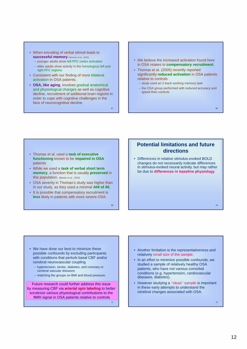
Materials and procedures
17
Participants
18
• Right-handed and free of current and past psychiatric and medical disorders as determined by history and physical exam.
• All participants obtained an average of 7.7 ± 0.9 h of sleep per night for the 3 nights preceding the study
Participants

4
19 20
21 22
Sleep questionnaires
• The Epworth Sleepiness Scale (ESS) (Johns, 1991)– is a questionnaire intended to measure daytime
sleepiness. This can be helpful in diagnosing sleep disorders.
3 = high chance of dozing
2 = moderate chance of dozing
1 = slight chance of dozing
0 = no chance of dozing
10 – 24 = sleepiness0 – 9 = average score, normal population
23In a car, while stopped for a few minutes in traffic
Sitting quietly after a lunch without alcohol
Sitting and talking to someone
Lying down to rest in the afternoon when circumstances permit
As a passenger in a car for an hour without a break
Sitting inactive in a public place (e.g a theater or a meeting)
Watching TV
Sitting and reading
Chance Of DozingSituation
24
EEG
EOG
EMG
Airflow
Thoracic and abdominal excursions
Oxymeter
ECG
Polysomnography
5
25
• Apnea was defined as any >10 s of >80% drops of respiratory amplitude.
• Hypopnea was defined as any >10 s of >30% drops of respiratory amplitude, plus >3% desaturation.
• Apnea–hypopnea index (AHI) was calculated representing the number of apnea and hypopnea events per hour of sleep.
Severe>30
Moderate15-30
Mild5-15
Normal<5
RatingAHI
26
• Records were scored for sleep stages according to the criteria of Rechtshaffen and Kales (1968).
• Number of arousals per hour of sleep (arousal index) and number of oxygen desaturation per hour of sleep (desaturation index) were calculated.
27
The verbal learning task
• During the fMRI session, participants performed a VL task.
• Stimuli, nouns, were presented visually via a video projector
28
2 4 6 8 10 12
1 3 5 7 9 11 13
Immediate free recall testStanford Sleepiness Scale, Karolinska Sleepiness Scale
a 10-point Likert scale assessing the following subjective factors: task difficulty, ability to concentrate, effort put into the task,
and motivation to perform the task well
press a button on a hand held button box to indicate whether the word was printed in all capital or all lowercase letters
Delayed free recall and recognition memory tests
30 - 40 min
2 4 6 8 10 12
1 3 5 7 9 11 131 13
Baseline blocks
Memorization blocks
29
Memorization block (6 blocks)
Cat
4 s.
Start
2.5 s.
*
1 s.
Dog *
4 s. 1 s.
Rat *
4 s. 1 s.
Ant *
4 s. 1 s.
22.5 s.
292.5 s. + 7.5 s.
300 s. = 5 min 30
The sensorimotor task
• The sensorimotor task measured basic primary sensory cortex function not dependent on cognitive processing, thus providing a measure of non-specific effects of OSA and serving as a control task.

6
31
8 cycles of 15 s. on and 15 s. off
OnOnOn
Off Off Off Off
32
• Memory (immediate free recall, delayed recall and recognition memory scores) and questionnaire data were analyzed with Student’s t tests comparing the OSA & Control groups.
• Spearman correlations between free recall performance & questionnaires were calculated.
• FMRI data were processed and analyzed with AFNI, data sets were transformed to standard atlas coordinates (Talairach & Tournoux, 1988)
Data analysis
33
• t tests were used to examine group differences (OSA vs. Control) in cerebral responses for the VL and the sensorimotor tasks.
• To assess the correlation between brain activation and performance in the OSA group
Data analysis
34
bilateral thalamus and hippocampal formation
bilateral inferior frontal gyrus
bilateral inferior and superior parietal lobes
For the VL performance analysis
35precentral & postcentral areas
(somatosensory, motor cortical)inferior, middle, and superior occipital gyri (visual cortical)
The analysis for the sensorimotor task
36
Cuneus
Longual gyrus
Fusiform gyrus

7
37
Results
38
Sleep and sleepiness• Polysomnography
– No significant differences between the groups were found in total sleep time or percentage of any sleep stage.
– OSA group had significantly higher AHI, higher arousal index and higher oxygen desaturation indexthan the control group.
• Questionnaires– OSA group reported significantly higher daytime
sleepiness than the control group as measured by the ESS
39
Behavioral data
40
Correlations between performance and subjective measures
• Subjective ratings of trait and state sleepiness were not significantly correlated with free recall performance
• Similarly, none of the other subjective factors(task difficulty, ability to concentrate, effort put into the task, and motivation to perform the task well) were related to performance
41
The verbal learning task
• Activated mainly a left hemisphere– the inferior(BA47), middle(BA4,6,9), and
superior(BA6,8) frontal gyri, middle temporal gyrus(BA21), and bilateral parahippocampalgyrus(BA28/35).
• Decreased activation– bilateral of the thalamus, right declive, right
precuneus(BA31), and right inferior parietal lobe(BA40).
In the control group
42
Lt. middle temporal gyrusbilateral parahippocampal gyrus
Lt. inferior, middle, &superior frontal gyri
8
43
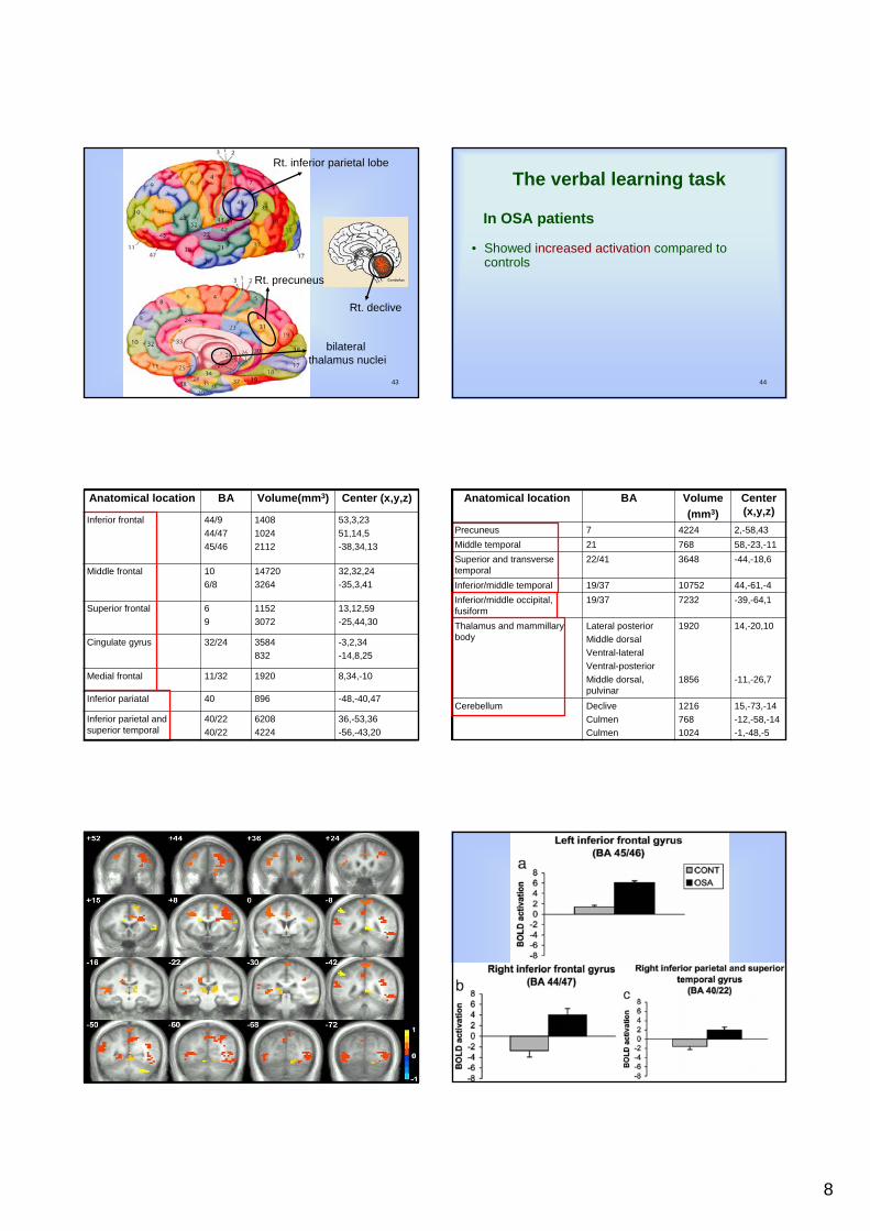
bilateral thalamus nuclei
Rt. inferior parietal lobe
Rt. declive
Rt. precuneus
44
• Showed increased activation compared to controls
The verbal learning task
In OSA patients
4536,-53,36-56,-43,20
62084224
40/2240/22
Inferior parietal and superior temporal
-48,-40,4789640Inferior pariatal
8,34,-10192011/32Medial frontal
-3,2,34-14,8,25
3584832
32/24Cingulate gyrus
13,12,59-25,44,30
11523072
69
Superior frontal
32,32,24-35,3,41
147203264
106/8
Middle frontal
53,3,2351,14,5-38,34,13
140810242112
44/944/4745/46
Inferior frontal
Center (x,y,z)Volume(mm3)BAAnatomical location
46
Center (x,y,z)
Volume(mm3)
BAAnatomical location
15,-73,-14-12,-58,-14-1,-48,-5
12167681024
DecliveCulmenCulmen
Cerebellum
14,-20,10
-11,-26,7
1920
1856
Lateral posteriorMiddle dorsalVentral-lateralVentral-posteriorMiddle dorsal, pulvinar
Thalamus and mammillarybody
-39,-64,1723219/37Inferior/middle occipital, fusiform
44,-61,-41075219/37Inferior/middle temporal
-44,-18,6364822/41Superior and transverse temporal
58,-23,-1176821Middle temporal2,-58,4342247Precuneus
47 48
9
49
Correlations between BOLD response and VL performance in the OSA group
left supramarginal arealeft inferior frontal gyrus (BA 47)
50
Correlations between BOLD response and VL performance in the OSA group
left inferior parietal lobe (BA 40)
51
The sensorimotor task
• Comparison of the BOLD response in the control & OSA groups during the sensorimotor task revealed only decreased activation in the left postcentral and precentral gyri in the OSA group.
• No differences were observed in visual areas. 52
Discussion
53
OSA patients would show intact performance on the task along with increased activation
as measured with the BOLD signal.
This study was one of the first to examine the cerebral correlates of learning and memory performance in a group of patients with OSA.
54
Increased activation in regions related to verbal encoding may reflect task-related recruitmentassociated with the concept of cognitive reserve.
Increased activation in OSA patients manifests in regions both typically and some not typically associated with verbal encoding. (Stern, 2002)
10
55
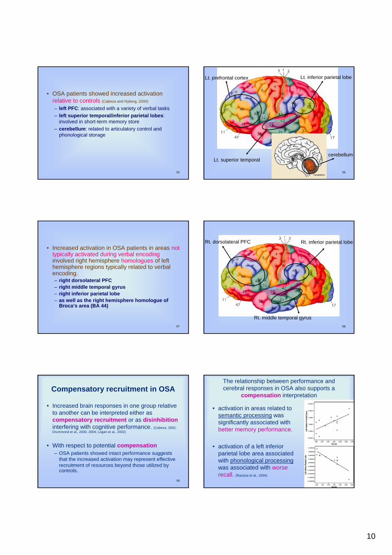
• OSA patients showed increased activation relative to controls (Cabeza and Nyberg, 2000)
– left PFC: associated with a variety of verbal tasks– left superior temporal/inferior parietal lobes:
involved in short-term memory store– cerebellum: related to articulatory control and
phonological storage
56
Lt. prefrontal cortex
Lt. superior temporal
Lt. inferior parietal lobe
cerebellum
57
• Increased activation in OSA patients in areas not typically activated during verbal encodinginvolved right hemisphere homologues of left hemisphere regions typically related to verbal encoding. – right dorsolateral PFC– right middle temporal gyrus– right inferior parietal lobe– as well as the right hemisphere homologue of
Broca’s area (BA 44)
58
Rt. dorsolateral PFC Rt. inferior parietal lobe
Rt. middle temporal gyrus
59
Compensatory recruitment in OSA
• Increased brain responses in one group relative to another can be interpreted either as compensatory recruitment or as disinhibitioninterfering with cognitive performance. (Cabeza, 2002; Drummond et al., 2000, 2004; Logan et al., 2002)
• With respect to potential compensation– OSA patients showed intact performance suggests
that the increased activation may represent effective recruitment of resources beyond those utilized by controls.
60
The relationship between performance and cerebral responses in OSA also supports a
compensation interpretation
• activation in areas related tosemantic processing was significantly associated with better memory performance.
• activation of a left inferior parietal lobe area associated with phonological processingwas associated with worse recall. (Ravizza et al., 2004)
11
61
• With respect to potential disinhibition– OSA patients showed positive activations in some
areas showing negative activations in controls: • bilateral regions in the thalamus• right hemisphere declive, precuneus and inferior
parietal lobe.
62
We suggest that recruitment of additional brain regions to participate in VL performance in OSA patients likely represents an adaptive compensatory recruitment response
63
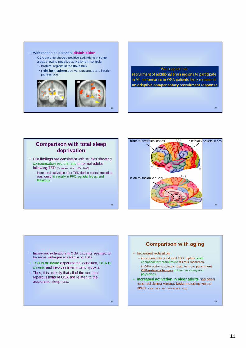
Comparison with total sleep deprivation
• Our findings are consistent with studies showing compensatory recruitment in normal adults following TSD (Drummond et al., 2000, 2005)
– increased activation after TSD during verbal encoding was found bilaterally in PFC, parietal lobes, and thalamus.
64
bilateral prefrontal cortex
bilateral thalamic nuclei
bilaterally parietal lobes
65
• Increased activation in OSA patients seemed to be more widespread relative to TSD.
• TSD is an acute experimental condition, OSA is chronic and involves intermittent hypoxia.
• Thus, it is unlikely that all of the cerebral repercussions of OSA are related to the associated sleep loss.
66
Comparison with aging• Increased activation
– in experimentally induced TSD implies acute compensatory recruitment of brain resources.
– in OSA patients actually relate to more permanent OSA-related changes in brain anatomy and physiology
• Increased activation in older adults has been reported during various tasks including verbal tasks. (Cabeza et al., 1997; Morcom et al., 2003)
12
67
• When encoding of verbal stimuli leads to successful memory (Morcom et al., 2003)
– younger adults show left PFC cortex activation– older adults show activity in the homologous left and
right PFC regions.• Consistent with our finding of more bilateral
activation in OSA patients.• OSA, like aging, involves gradual anatomical
and physiological changes as well as cognitive decline, recruitment of additional brain regions in order to cope with cognitive challenges in the face of neurocognitive decline.
68
• We believe the increased activation found here in OSA relates to compensatory recruitment.
• Thomas et al. (2005) recently reported significantly reduced activation in OSA patients relative to controls – study used an 2-back working memory task– the OSA group performed with reduced accuracy and
speed than controls
69
• Thomas et al. used a task of executive functioning known to be impaired in OSApatients
• While we used a task of verbal short term memory, a function that is usually preserved in this population. (Beebe et al., 2003)
• OSA severity in Thomas’s study was higher than in our study, as they used a minimal AHI of 40.
• It is possible that compensatory recruitment is less likely in patients with more severe OSA.
70
Potential limitations and future directions
• Differences in relative stimulus-evoked BOLD changes do not necessarily indicate differences in stimulus-evoked neural activity, but may rather be due to differences in baseline physiology.
71
• We have done our best to minimize these possible confounds by excluding participants with conditions that perturb basal CBF and/or cerebral neurovascular coupling – hypertension, stroke, diabetes, and coronary or
cerebral vascular diseases – matching the groups on BMI and blood pressure.
Future research could further address this issue by measuring CBF via arterial spin labeling to better
scrutinize various physiological contributions to the fMRI signal in OSA patients relative to controls
72
• Another limitation is the representativeness and relatively small size of the sample.
• In an effort to minimize possible confounds, we studied a sample of relatively healthy OSA patients, who have not various comorbidconditions (e.g. hypertension, cardiovascular diseases, diabetes).
• However studying a ‘‘clean’’ sample is important in these early attempts to understand the cerebral changes associated with OSA.

13
73
Future studies should employ larger samples as well as explore the role of various
comorbidities in the behavioral and cerebral abnormalities seen in OSA patients.
74
• Finally, assessing the reversibility of changes in brain activation in OSA following treatment will shed light on the nature of the compensatory processes described here and will help determine to what extent these changes resemble the anatomical/functional reorganization occurring in older adults.
75
• Given that at least some aspects of cognitive performance deficits are reversed with treatment, this may be reflected in neuroimagingmeasures of brain function.
Such outcome studies may improve ourunderstanding of OSA and its effects on the brain and assist in developing appropriate treatments
for the neurocognitive.
76
• Increased cerebral responses and intact performance during VL in OSA patients relative to well-matched controls.
• Interpreted these findings as being compensatory in nature.
• Differential patterns of compensatory vs. diminished responses may account for why OSA patients show intact performance on some tasks and deficits on others.
Summary
77
• Sleep apnea is a very common syndrome (up to 28% of the adult population), the findings of altered brain activation in this group suggest that this is a potential confound in functional neuroimaging studies
• OSA could influence the findings from studies comparing younger adults with older adults, who show a higher prevalence of OSA.
78
• Potential ways to address this would include administering the ESS along with specific questions covering the main OSA symptoms as part of subject screening procedures.
• This would allow investigators to exclude anyone at high risk for OSA.

14
79
Thank you
Top Related