


Languages
Pages
Legal


1
INSECT MOUTHPARTS
A group of appendages associated with the mouth which form collectively a feeding
apparatus of an insect commonly called the mouthparts. Mouthparts are some of the most
distinctive features of insects, and their structure tells a great deal about the feeding habits of
a species. Insect mouthparts are variously adapted for the ingestion of many types of foods.
Mouthparts vary not only among different insect groups, but also among different stages of
the same species (for example, note the mouthparts of a butterfly and a caterpillar). In some
insects (e.g., mayflies) mouthparts are not developed; this condition is termed as vestigial.
Importance of studying mouthparts
Different insects can be identified by observing their mouthparts.
The feeding mechanism of insect pests can be known by studying their mouthparts.
Nature of damage of various insect pests can be recognized by learning their
mouthparts.
Appropriate control measures can be applied for specific insect pests by
understanding their mouthparts.
Types of mouthparts
Eight major types of mouthparts based on the feeding habit of insects.
A. Chewing type (eg. Grasshopper, cockroach, beetles etc.)
B. Rasping sucking type (eg. Thrips)
C. Piercing sucking type (eg. Rice bug, bed bug, stink bug, leaf hopper, female mosquito etc.)
D. Sponging type (eg. House fly
E. Siphoning type (eg. Butterfly and moths)
F. Cutting sponging type (eg. Horse fly)
G. Chewing lapping type (eg. Honey bee)
H. Degenerate type (eg. Larvae of mosquito, fruit fly etc.)
A. CHEWING TYPE MOUTHPARTS
Insects like ground beetles and grasshoppers with chewing mouthparts have heavy crania,
adapted for muscles involved in capturing prey and biting off leaf tissue. Chewing type
mouthparts consist of a labrum (upper lip), a pair of chewing mandibles (upper jaws), a pair
of maxillae (lower/second jaws), and a labium (lower lip) [Figure 1]. These structures
surround the mouth and form the pre-oral cavity. In addition, a central tongue-like
hypopharynx drops from the membranous floor of the cranium, behind the mouth, and bears
the opening of the salivary ducts. The interior, fleshy-surface of the labrum, endowed with
numerous sensory structures, is referred to as the labrum-epipharynx (roof of the mouth).
Labrum: The labrum is a sclerite or plate that acts as the 'upper lip' in insects. It is a flap-
like structure that lies immediately in front of the mouth. There is a notch at the middle of
the labrum which hold food material during cutting or grinding. Labrum pushes food into the
mouth cavity.
Mandibles: Mandibles are strongly chitinized structures and are commonly known as the
jaws. They always articulate in a transverse plate. Mandibles are provided with strong
adductor and abductor muscles arisen from the dorsal and ventral walls of the cranium.
Teeth are blunt like molar in phytophagous insects, slightly pointed like incisors in

2
carnivorous insects. Teeth are molar near the base and incisor as blade at the tip in
omnivorous insects for cutting and grinding the food material.
Maxillae: They are also known as second pair of jaws of the insect. Each maxilla is
composed of proximal cardo, middle stipes, lateral palpifer, inner subgalea or parastipes and
the distal sclerite consisting two lobes, outer galea & inner lacinia. On the outside, the
palpifer bears the maxillary palp, which is sensory appendages and composed of 1-7
segments. The maxillae possess various types of cuticular processes, hairs and bristles etc.
Mandibles and maxillae are adapted for cutting and grinding the solid food material.
Labium: It is formed from the fusion of paired 2nd
maxillae pair. It closes the preoral cavity
and known as lower lip of insect. It is composed of distal prementum, proximal
postmentum, separated by labial suture. Postmentum is divided into proximal submentum
and distal mentum. Palpiger: lateral lobe arises from prementum bears 1-4 segmented labial
palp, which is sensory in function. Paraglosae: arise from distal margin of prementum
outward as lobe. Glossae: arise from distal margin of prementum innerward as lobe.
Hypopharynx: In some Apterygota, it is considered as composite gnathal appendages. At
embryonic development, hypopharynx is formed by a pair of lateral lobes (Superlinguae)
fused with a median lobe (Lingua) in Apterygota and some primitive Pterygota. In higher
Pterygota, it develops as a ventral median lobe from the ventral wall of the labium. At the
base it bears an orifice of salivary duct. Various muscles serve to swing the hypopharynx in
forward and backward directions.
Epipharynx and hypopharynx are membranous lobulated structures which push the food
material into the pharynx.
3
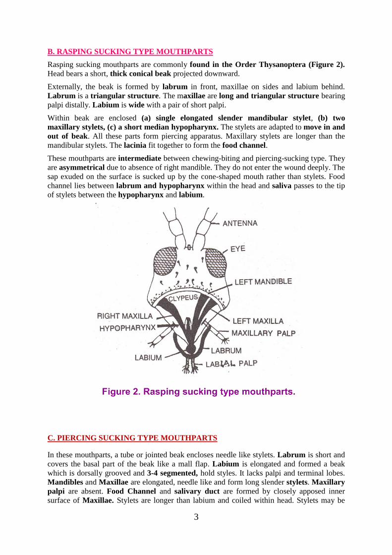
B. RASPING SUCKING TYPE MOUTHPARTS
Rasping sucking mouthparts are commonly found in the Order Thysanoptera (Figure 2).
Head bears a short, thick conical beak projected downward.
Externally, the beak is formed by labrum in front, maxillae on sides and labium behind.
Labrum is a triangular structure. The maxillae are long and triangular structure bearing
palpi distally. Labium is wide with a pair of short palpi.
Within beak are enclosed (a) single elongated slender mandibular stylet, (b) two
maxillary stylets, (c) a short median hypopharynx. The stylets are adapted to move in and
out of beak. All these parts form piercing apparatus. Maxillary stylets are longer than the
mandibular stylets. The lacinia fit together to form the food channel.
These mouthparts are intermediate between chewing-biting and piercing-sucking type. They
are asymmetrical due to absence of right mandible. They do not enter the wound deeply. The
sap exuded on the surface is sucked up by the cone-shaped mouth rather than stylets. Food
channel lies between labrum and hypopharynx within the head and saliva passes to the tip
of stylets between the hypopharynx and labium.
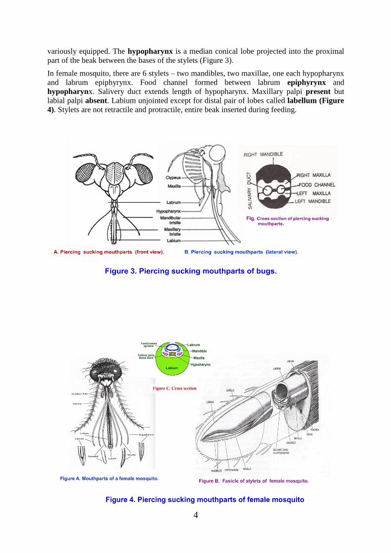
C. PIERCING SUCKING TYPE MOUTHPARTS
In these mouthparts, a tube or jointed beak encloses needle like stylets. Labrum is short and
covers the basal part of the beak like a mall flap. Labium is elongated and formed a beak
which is dorsally grooved and 3-4 segmented, hold styles. It lacks palpi and terminal lobes.
Mandibles and Maxillae are elongated, needle like and form long slender stylets. Maxillary
palpi are absent. Food Channel and salivary duct are formed by closely apposed inner
surface of Maxillae. Stylets are longer than labium and coiled within head. Stylets may be
4
variously equipped. The hypopharynx is a median conical lobe projected into the proximal
part of the beak between the bases of the stylets (Figure 3).
In female mosquito, there are 6 stylets – two mandibles, two maxillae, one each hypopharynx
and labrum epiphyrynx. Food channel formed between labrum epiphyrynx and
hypopharynx. Salivery duct extends length of hypopharynx. Maxillary palpi present but
labial palpi absent. Labium unjointed except for distal pair of lobes called labellum (Figure
4). Stylets are not retractile and protractile, entire beak inserted during feeding.

5
D. SPONGING TYPE MOUTHPARTS
They are incapable of piercing skin and found in adult house fly. The mandibles are wanting.
Feeding apparatus is proboscis which is a composite structure formed by labrum,
hypopharynx and labium. The proboscis is anteriorly associated with maxillary palpi and
distally bears a pair of labellar lobes called labellum. Labella are broad, soft pads with teeth
for rasping food.
The proboscis has 3 parts: (1) basiproboscis or rostrum which bears maxillary palpi, (2)
mediproboscis or haustellum which is cylindrical and its anterior surface is covered by
labrum, (3) distiproboscis fromed labellae or oral suckers which occur as non-piercing
spongy pads. It bears pseudotrachae. Food channel lies between labrum and hypopharynx.
The maxillae are represented by maxillary palpi (Figure 5). The hypopharynx conveys saliva
into the wound which contains anticoagulant.
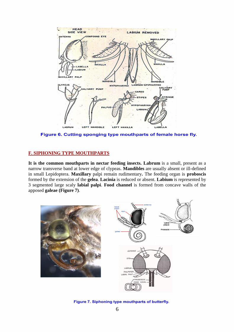
E. CUTTING SPONGING TYPE MOUTHPARTS
They are found in female horse flies (Tabanidae) only. Total number of stylets is six similar
to that of mosquitoes. Stylets are flattened blade like. Labium is similar to the sponging type
terminating into a pair of large lobes, the labellae. Mandibles are well-developed, forming
sharp blades. Maxillae are long slender tapering forming probing stylets. The mandibles cut
the tissues like scissor and the maxillae thrust and retract repeatedly. The blood vessels are
ruptured by the teeth armed at the tip of mandibles. The hypopharynx is long, narrow,
tapering stylet arising from the ventral wall of the head (Figure 6). The saliva contains
powerful anticoagulant.
6
F. SIPHONING TYPE MOUTHPARTS
It is the common mouthparts in nectar feeding insects. Labrum is a small, present as a
narrow transverse band at lower edge of clypeas. Mandibles are usually absent or ill-defined
in small Lepidoptera. Maxillary palpi remain rudimentary. The feeding organ is proboscis
formed by the extension of the gelea. Lacinia is reduced or absent. Labium is represented by
3 segmented large scaly labial palpi. Food channel is formed from concave walls of the
apposed galeae (Figure 7).
7
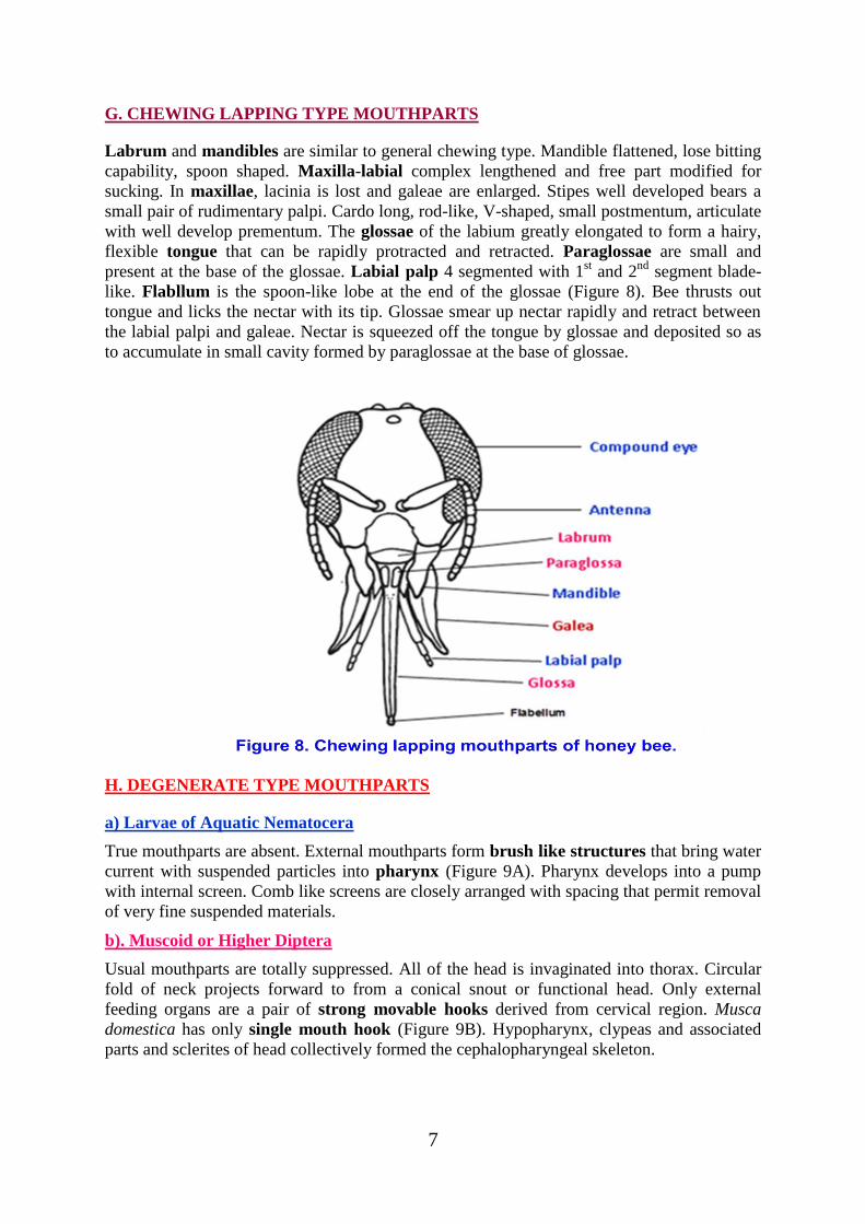
G. CHEWING LAPPING TYPE MOUTHPARTS
Labrum and mandibles are similar to general chewing type. Mandible flattened, lose bitting
capability, spoon shaped. Maxilla-labial complex lengthened and free part modified for
sucking. In maxillae, lacinia is lost and galeae are enlarged. Stipes well developed bears a
small pair of rudimentary palpi. Cardo long, rod-like, V-shaped, small postmentum, articulate
with well develop prementum. The glossae of the labium greatly elongated to form a hairy,
flexible tongue that can be rapidly protracted and retracted. Paraglossae are small and
present at the base of the glossae. Labial palp 4 segmented with 1st and 2
nd segment blade-
like. Flabllum is the spoon-like lobe at the end of the glossae (Figure 8). Bee thrusts out
tongue and licks the nectar with its tip. Glossae smear up nectar rapidly and retract between
the labial palpi and galeae. Nectar is squeezed off the tongue by glossae and deposited so as
to accumulate in small cavity formed by paraglossae at the base of glossae.
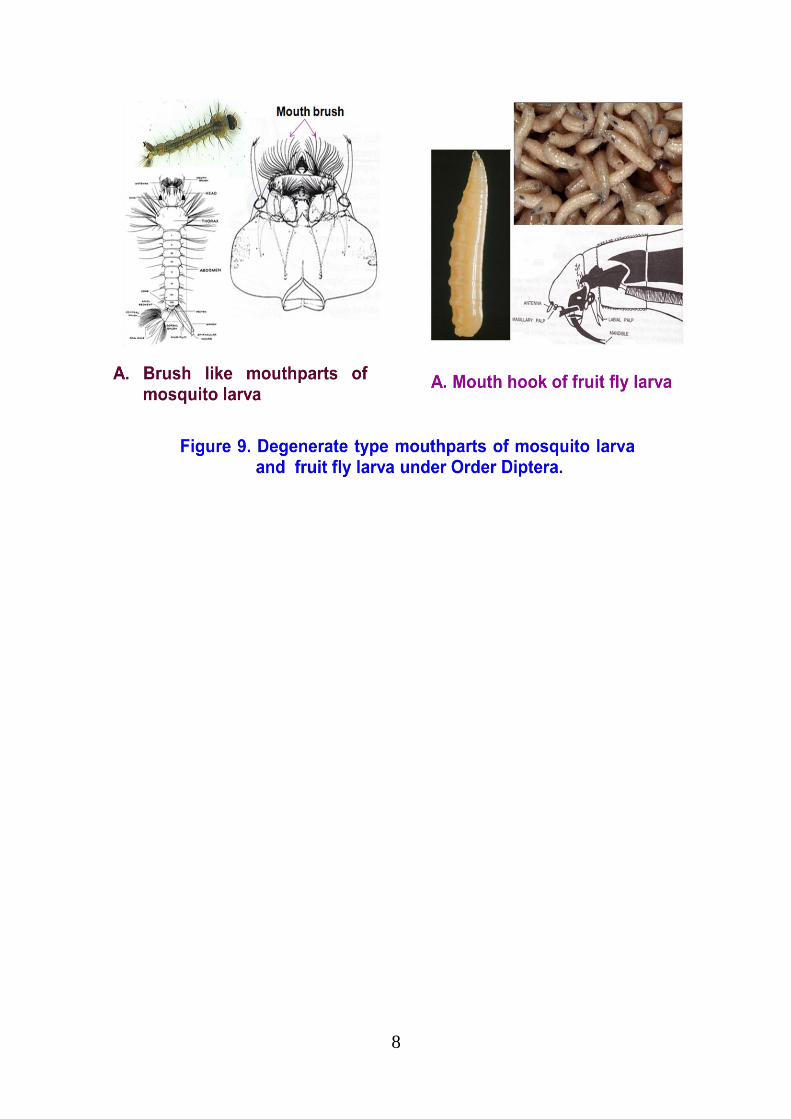
H. DEGENERATE TYPE MOUTHPARTS
a) Larvae of Aquatic Nematocera
True mouthparts are absent. External mouthparts form brush like structures that bring water
current with suspended particles into pharynx (Figure 9A). Pharynx develops into a pump
with internal screen. Comb like screens are closely arranged with spacing that permit removal
of very fine suspended materials.
b). Muscoid or Higher Diptera
Usual mouthparts are totally suppressed. All of the head is invaginated into thorax. Circular
fold of neck projects forward to from a conical snout or functional head. Only external
feeding organs are a pair of strong movable hooks derived from cervical region. Musca
domestica has only single mouth hook (Figure 9B). Hypopharynx, clypeas and associated
parts and sclerites of head collectively formed the cephalopharyngeal skeleton.
8
Top Related